A long-term study of AAV gene therapy in dogs with hemophilia A identifies clonal expansions of transduced liver cells
←
→
Page content transcription
If your browser does not render page correctly, please read the page content below
Articles
                                                                                                         https://doi.org/10.1038/s41587-020-0741-7

A long-term study of AAV gene therapy in dogs
with hemophilia A identifies clonal expansions of
transduced liver cells
Giang N. Nguyen1,8, John K. Everett2,8, Samita Kafle1, Aoife M. Roche2, Hayley E. Raymond2,
Jacob Leiby2, Christian Wood1, Charles-Antoine Assenmacher 3, Elizabeth P. Merricks4,5,
C. Tyler Long4,5, Haig H. Kazazian6, Timothy C. Nichols4,5, Frederic D. Bushman 2 and
Denise E. Sabatino 1,7 ✉

Nine dogs with hemophilia A were treated with adeno-associated viral (AAV) gene therapy and followed for up to 10 years.
Administration of AAV8 or AAV9 vectors expressing canine factor VIII (AAV-cFVIII) corrected the FVIII deficiency to 1.9–11.3%
of normal FVIII levels. In two of nine dogs, levels of FVIII activity increased gradually starting about 4 years after treatment.
None of the dogs showed evidence of tumors or altered liver function. Analysis of integration sites in liver samples from six
treated dogs identified 1,741 unique AAV integration events in genomic DNA and expanded cell clones in five dogs, with 44% of
the integrations near genes involved in cell growth. All recovered integrated vectors were partially deleted and/or rearranged.
Our data suggest that the increase in FVIII protein expression in two dogs may have been due to clonal expansion of cells har-
boring integrated vectors. These results support the clinical development of liver-directed AAV gene therapy for hemophilia A,
while emphasizing the importance of long-term monitoring for potential genotoxicity.

G
        ene therapy for hemophilia A has moved from preclinical               that integration events caused HCC seemed unlikely19–21. Another
        studies in mice and dogs to successful trials in humans1–4.           study showed that some AAV2 vectors contain a sequence in the 3′
        AAV vectors to deliver the factor VIII gene (F8) were first           untranslated region, adjacent to the AAV inverted terminal repeat
evaluated in dogs with hemophilia A in the early 2000s5–7. While              (ITR), that has enhancer-promoter activity in liver cells22. This ele-
AAV gene therapy has demonstrated promising results for hemo-                 ment has been identified in HCC in humans and may be a mecha-
philia B in clinical studies for more than 8 years8,9, studies for            nism of transformation in these tumors20. While HCC has not been
hemophilia A are at an earlier stage1–4. As recombinant (r)AAV                observed in rAAV-treated adult mice or other mammals12,17,18,23,
commonly persists in an episomal form10, rAAV may not be main-                the development of rAAV-associated HCC has been reported after
tained in tissues undergoing cell division. However, as hepatocytes           delivery to neonatal mice14,24–26 and to young adults of mouse strains
in healthy dogs are typically quiescent11, the dog model is well              that have a high incidence of spontaneous liver tumors27,28. This sug-
suited to long-term studies designed to understand the durability             gests that active cell division, such as that occurring during devel-
and safety of AAV gene therapy and to evaluate tissues that are not           opment, can be associated with genotoxic integration events14,24,25.
accessible in human patients.                                                 In neonatal mice, rAAV integration events were identified recur-
    One of the safety concerns for AAV gene therapy is the poten-             rently in specific genomic regions in HCC tissue, suggesting pos-
tial for genotoxic integration events. After introduction into cells,         sible insertional mutagenesis15. The above findings warrant further
rAAV primarily remains episomal10, but integration into the host              research to assess the safety of rAAV gene therapy.
genome has been observed in mice12–16, non-human primates17                      In this study, we treated nine dogs with hemophilia A with
and humans17,18. To date, there have been no serious adverse                  AAV-cFVIII and followed them for as long as 10 years. Levels of
events involving genotoxicity as a result of rAAV vector integra-             cFVIII activity were maintained throughout the study, represent-
tion in humans or large animals. Other studies of wild-type AAV               ing the longest sustained therapeutic levels of FVIII expression
in humans or rAAV in mouse models have detected tumors associ-                observed in a large-animal model of hemophilia A associated with
ated with AAV. Studies of wild-type AAV reported AAV integra-                 a significant reduction in spontaneous bleeding events. Two dogs
tion events in human hepatocellular carcinoma (HCC), suggesting               showed a gradual increase in FVIII expression levels. Upon termi-
a possible role of AAV integration in transformation19–21. However,           nation of the study, AAV integration analysis on the liver tissue of
the AAV copy number was much less than one per cell, thus the idea            these two dogs and four other dogs revealed clonal expansion of

The Raymond G. Perelman Center for Cellular and Molecular Therapeutics, Children’s Hospital of Philadelphia, Philadelphia, PA, USA. 2Department
1

of Microbiology, Perelman School of Medicine, University of Pennsylvania, Philadelphia, PA, USA. 3Department of Pathobiology, School of Veterinary
Medicine, University of Pennsylvania, Philadelphia, PA, USA. 4Department of Pathology and Laboratory Medicine, University of North Carolina at Chapel
Hill, Chapel Hill, NC, USA. 5UNC Blood Research Center, University of North Carolina School of Medicine, University of North Carolina at Chapel Hill,
Chapel Hill, NC, USA. 6Department of Genetic Medicine, Johns Hopkins School of Medicine, Baltimore, MD, USA. 7Division of Hematology, Department of
Pediatrics, Perelman School of Medicine, University of Pennsylvania, Philadelphia, PA, USA. 8These authors contributed equally: Giang N. Nguyen, John K.
Everett. ✉e-mail: dsabatin@pennmedicine.upenn.edu

Nature Biotechnology | www.nature.com/naturebiotechnology
Articles NATurE BIOTEchnOlOgy
a
HC 2.3 kb
TC
LC 2.1 kb
SC
SC 4.4 kb
Feature Promoter B domain F8 HC
5′ ITR Intron F8 PolyA
Spacer Signal peptide F8 LC 3′ ITR
b Linus
15 Woodstock
F24 H19
J60
10 TC
cVIII activity (%)
5
0
0 1 2 3 4 5 6 7 8 9 10
Time after vector administration (years)
c 15
M50 L51
M06 M66
cFVIII activity (%)
10
SC
5
0
0 1 2 3 4 5 6 7
Time after vector administration (years)
Fig. 1 | Long-term expression of AAV-cFVIII in dogs with hemophilia A. a, Genetic structures of the vectors used, illustrating the TC (top) and SC
(bottom) designs. The features of these constructs are indicated by the colors shown in the legend. The TC-delivery approach consisted of co-delivering
the 2.3 kb cF8 HC in one AAV vector and the 2.1 kb cF8 LC in a second AAV vector. These vectors used the thyroxine-binding globulin (TBG) promoter
and two copies of α1-microglobulin–bikunin enhancer sequences (695 bp) with an intron (175 bp) for liver-directed gene expression. The construct used a
263 bp SV40 polyA signal. The SC-delivery approach consisted of delivering the 4.4 kb B domain deleted cF8, driven by a minimal hepatic control region
(192 bp) and the human α-1-antitrypsin (HCR-hAAT) promoter (266 bp). This construct included a 134 bp SV40 polyA signal. The gray regions indicate
spacers or cloning sites. The TC vector was delivered by portal vein infusion, while the SC vector was delivered by peripheral vein infusion. b, Longitudinal
quantification of FVIII levels after the TC-delivery approach. In the TC approach, the total AAV dose was 2.5 × 10¹³ viral genomes (vg) per kg for F24
(AAV8), Woodstock (AAV9) and J60 (AAV9) (blue). Linus (AAV8) and H19 (AAV9) were treated at a low-vector dose (total vector dose of 1.2 × 10¹³ vg
per kg; green). c, FVIII levels after the SC-delivery approach. AAV8 delivery of the SC to dogs with hemophilia A was performed using a vector dose of
4 × 10¹³ vg per kg (M50, M06; blue) and a vector dose of 2 × 10¹³ vg per kg (L51, M66; green).
cells, with insertions near genes that are associated with cancer in heterodimer, which is subsequently secreted. In this approach, an
humans, but not overt nodule formation or transformation. AAV vector (AAV8 or AAV9) encoding the cF8 heavy chain (HC)
was co-delivered with an AAV vector encoding the cF8 light chain
Results (LC)6. The SC approach used an AAV8 vector to deliver cF8 with the
Sustained FVIII expression after AAV delivery. We treated dogs B domain deleted6.
with hemophilia A using two approaches, with either the two cF8 All the AAV-treated dogs with hemophilia A maintained cFVIII
chains (TC) encoded separately or a single chain (SC) encoded in expression for the duration of the entire study (2.2–10.4 years; Fig. 1b,c
one vector6 (Fig. 1a and Table 1). The TC approach takes advan- and Table 1). These dogs with hemophilia A have an intron 22
tage of the normal intracellular processing of FVIII, in which FVIII inversion that is analogous to the most common mutation in humans
undergoes proteolytic cleavage into two polypeptides that form a and have a severe bleeding phenotype (NATurE BIOTEchnOlOgy Articles
Table 1 | Summary of AAV administration, FVIII activity and DNA analysis of dogs with hemophilia A
AAV Total AAV AAV Dog with Years cFVIII activity (%) DNA analysis of liver samples
delivery vector dose serotype hemophilia A evaluatedb
Mean ± s.d. Final Number Mean VCN Copy Number of
approach (vg per kg)a
of liver per diploid number genomic
samplesc genome ± s.d. range integration
sites
recoveredd
TC 2.5 × 10¹³ AAV8 F24 2.2 5.7 ± 2.3 2.7 NDe ND ND ND
AAV9 Woodstock 8.2 3.1 ± 1.5 7.1 5 0.28 ± 0.10 0.19–0.42 ND
AAV9 J60 9.5 8.1 ± 4.0 4.5 29 3.43 ± 2.34 0.76–7.82 271
1.2 × 10¹³ AAV8 Linus 10.1 5.9 ± 4.6 11.3 15 0.28 ± 0.21 0.01–0.62 131
AAV9 H19 8.3 0.9 ± 0.7 2.5 11 0.17 ± 0.09 0.07–0.33 160
SC 4 × 10¹³ AAV8 M06 2.3 7.4 ± 2.6 9.4 8 1.44 ± 0.69 0.37–2.26 764
M50 7.3 5.2 ± 3.6 10.3 29 0.35 ± 0.34 0.00–1.14 258
2 × 10¹³ L51 6.1 0.8 ± 0.6 1.9 5 0.01 ± 0.02 0.00–0.04 ND
M66 6.0 2.6 ± 1.3 3.7 13 0.32 ± 0.18 0.00–0.60 161
a
In the TC-delivery approach, the vector dose was 1.25 × 10¹³ vg per vector per kg at the high dose and 6 × 10¹² vg per vector per kg at the low dose. bThe number of years after AAV administration.
c
The number of liver samples analyzed in the VCN analysis. dIntegration studies were performed on three liver samples per dog. eND, not determined.
After AAV administration, the mean levels of cFVIII activity for the administration. These dogs were older (4 years old) at the time of
dogs treated with the TC approach were 5.6% of normal for the dogs vector administration than the other dogs studied. L51 also had ALT
at the high-vector dose and 3.4% for the dogs at the low-vector dose levels that were >2 times the normal limit at multiple time points
(Table 1). One dog (J60) treated at the high-vector dose required a and are considered mildly elevated33. The aspartate aminotransfer-
splenectomy at the time of vector infusion and had a higher level ase (AST) levels were within normal limits for all dogs for the dura-
of cFVIII expression (8.1% ± 4.0%) than that of the other dogs tion of the study (Fig. 2c,d). These mild, asymptomatic elevations of
treated at this dose (see Methods for additional details). For the liver enzyme levels were not consistent with a specific liver disease
SC-treated dogs, the mean levels of cFVIII expression were 6.2% for pattern. The serum α-fetoprotein (AFP) is a biomarker for hepatic
the high-vector dose and 1.7% for the low-vector dose. At the final disease, including HCC in some patients34–36. The AFP values for all
time points, all dogs had levels of cFVIII activity that were close to dogs were within the normal range at all times analyzed (Fig. 2e,f).
or greater than the mean cFVIII expression (Table 1). FVIII activity Other blood chemistries were monitored throughout the study
was also demonstrated by a shortening of the whole-blood clotting without cause for clinical concern.
time (Supplementary Fig. 1). The liver pathology was evaluated at the final time point in the
Two dogs (Linus and M50) had a gradual increase in cFVIII AAV-treated dogs with hemophilia A and compared to naive (to
expression levels that began more than 4 years after vector admin- gene therapy) dogs with hemophilia A (n = 10) or hemophilia B
istration. Linus had a cFVIII activity of 4% at 4 years after vector (n = 9) and normal dogs (n = 9) of similar age from the same colony
delivery, which gradually increased to 11% at 10 years after AAV (Supplementary Tables 1 and 2). Some of the findings were con-
administration (Fig. 1b). M50 had a cFVIII activity of 4% at 4 years sistent with features commonly observed in older dogs and were
after AAV delivery, which gradually increased to 10% at 7 years seen in both AAV-treated and untreated (that is, naive) dogs. No
after vector administration (Fig. 1c). Two other dogs, Woodstock pathologies definitively related to the treatment were identified.
and M06, also had possible modest increases in FVIII activity levels. Three of nine AAV-treated dogs with hemophilia A had clinical
symptoms that led to ending the study early, but none were believed
Clinical observations after AAV gene therapy. A cohort of naive to be related to AAV gene therapy (Methods).
dogs with hemophilia A (n = 11) was followed prospectively for 4 The immune response to cFVIII (Supplementary Fig. 3) and
years to determine the frequency of bleeding events. All bleeding neutralizing antibodies (NAbs) to the AAV capsid (Supplementary
events required treatment with recombinant cFVIII30 or normal Table 3) were also followed during the study.
canine blood products. The annualized bleeding rate (ABR) was
12.7 ± 6.0 bleeds per year (ref. 31; Supplementary Fig. 2), simi- Investigation of the increase in FVIII expression. We investigated
lar to the ABR for patients with hemophilia who are treated epi- the mechanism of the increase in FVIII expression levels in Linus
sodically (18.5 bleeds per patient per year)32. In contrast, the nine and M50 (Supplementary Table 4). At 6 years after vector adminis-
AAV-treated dogs with hemophilia A had a total of seven bleeding tration, an ultrasound and liver biopsy were performed on Linus; no
episodes during the combined total of 60 years of follow-up after tumors, nodules or notable inflammation in the liver were detected.
AAV administration. The ABR per AAV-treated dog with hemo- The increase in FVIII levels was not associated with changes in lev-
philia A was between 0.00 and 0.45 with a mean of 0.13 ± 0.16, els of circulating AFP, fibrinogen or von Willebrand factor (vWF) or
lower than that reported for people with hemophilia on prophylaxis clearance receptor in the liver (Fig. 2e,f and Supplementary Fig. 4).
(ABRs of 4.8–6.0 bleeds per patient per year)32. We next assessed AAV vector copy number (VCN) (Table 1). For
Clinical blood chemistries were obtained two to four times dogs that had multiple liver samples per lobe available for analysis,
per year from AAV-treated dogs to monitor for adverse reactions. the VCNs showed that the vector was distributed throughout the
The alanine aminotransferase (ALT) level was within the normal liver (Supplementary Table 5). Higher VCN levels were not detected
limits for six of the nine dogs at most of the time points analyzed in Linus or M50. J60 had a higher VCN than the other dogs; we
(Fig. 2a,b). Woodstock and Linus had an elevation (Articles NATurE BIOTEchnOlOgy
a b
TC M06
500 SC
650 Woodstock M50
600 J60 400 L51
ALT (IU L–1)
F24
ALT (IU L–1)
400 M66
Linus 300
300 H19
200
200
100 100
0 0
0 1 2 3 4 5 6 7 8 9 10 0 1 2 3 4 5 6 7
c 250 d 250 M06
Woodstock
M50
200 J60 200 L51
AST (IU L–1)
F24
AST (IU L–1)
150 150 M66
Linus
100 H19 100
50 50
0 0
0 1 2 3 4 5 6 7 8 9 10 0 1 2 3 4 5 6 7
e 70 f 70
60 Woodstock 60 M06
J60
AFP (ng ml–1)
AFP (ng ml–1)
50 50 M50
40 F24 40 L51
30 Linus 30 M66
20 H19 20
10 10
0 0
0 1 2 3 4 5 6 7 8 9 10 0 1 2 3 4 5 6 7
Time after vector administration (years) Time after vector administration (years)
Fig. 2 | Assessment of liver function after AAV-cFVIII administration. ALT (a,b), AST (c,d) and AFP (e,f) levels were monitored after AAV administration
and throughout the study. In each case, the x axis shows time in years and the y axis shows the liver analyte measured in blood. a,c,e, Dogs treated using
the TC strategy. b,d,f, Dogs treated using the SC strategy. The upper limits of normal for ALT (120 IU L−1), AST (65 IU L−1) and AFP (70 ng ml−1) are shown
as dashed lines. Clinically, increases in liver enzyme levels in dogs are defined by veterinary clinical pathologists as mild (10 times the upper limit of normal)33.
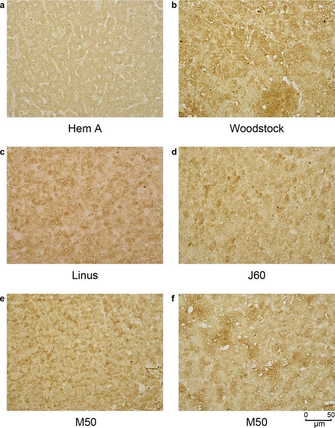
The pattern of cFVIII expression in the liver after AAV delivery containing DNA with expanded clones, there are be multiple DNA
was assessed by immunohistochemistry. Most of the tissue analyzed chains from each expanded clone that contain an identical integra-
had cFVIII expression that was dispersed throughout the tissue, tion site. Each chain is usually broken by sonication at a different
although some areas of the liver had what appeared to be clonal location in the flanking dog DNA. As a result, there are different
populations of cells expressing cFVIII (Extended Data Fig. 1). No locations of adaptor ligation in the cellular DNA flanking the inte-
major differences were noted between the dogs. gration site. These points of adaptor ligation can then be quantified
by sequencing from both ends of amplification products, providing
Analysis of integration site distributions in the livers of a measure of the number of cells sampled37.
AAV-treated dogs. AAV vector genomes are expected to be pres- We identified a total of 1,741 putative unique integration sites,
ent in multiple forms, with some integrated into the canine genome corresponding to an inference of 3,263 sampled cells (Supplementary
and some persisting as episomes. As integration can influence gene Table 6). Statistical analysis revealed modest but significant homol-
function and cell growth, we analyzed the locations of vector inte- ogy between AAV and canine sequences at vector–host junctions
gration sites in the dog genome and the behavior of cell clones that (Supplementary Fig. 6), suggesting that annealing of complemen-
harbored these integration sites. tary DNA sequences promoted integration in at least some cases.
We analyzed 20 samples of liver tissue taken at necropsy from The number of cells sampled per integration site ranged from 1 to
six AAV-treated dogs and two untreated dogs with hemophilia A 130; the high values indicate extensive clonal proliferation follow-
as controls. Liver DNA samples were selected to represent high, ing the integration event. These expanded clones were scarce; unique
medium and low VCN ranges. Integration sites were recovered from integration sites recovered from at least three cells corresponded to
genomic DNA samples by shearing DNA with sonication, ligating only 4.8% of the cell population. No integration events were identi-
DNA linkers to the broken DNA ends, then carrying out nested fied in the Rian–Dlk–Dio region (identified as canine chromosome
PCR amplification using primers that bound to the ligated linker 8, ~68,900,000–69,800,000), previously associated with AAV integra-
and the vector ITR sequences. (Supplementary Fig. 5). The resulting tion and transformation in mice14,25. The number of integration sites
DNA fragments containing junctions between dog genomic DNA recovered per sample was positively correlated with VCN (Fig. 3a;
and vector DNA were then sequenced with the Illumina platform, P = 3.0 × 10⁻⁴). Most ITRs that were associated with integration were
and reads were mapped to the dog genome (CanFam3). truncated (Supplementary Fig. 7), as seen in previous studies13,38.
From these data, we can also estimate the clonal abundance of We selected 18 integration sites for validation by targeted PCR
cells harboring each unique integrated vector. For a tissue sample and Sanger sequencing, focusing on expanded clones. We succeeded
Nature Biotechnology | www.nature.com/naturebiotechnologyNATurE BIOTEchnOlOgy Articles
a b
Chr1
Chr2
4 Chr3
Chr4
Chr5
Chr6
Chr7
3 Chr8
Integrations per sample mass
Chr9
Chr10
Chr11
Chr12
2 Chr13
Chr14
Chr15
Experiment Chr16
Dog
SC Chr17
TC Chr18 H19
1
Dog Chr19 J60
H19 Chr20 Linus
J60 Chr21
M06
Linus Chr22
Chr23 M50
0 M06
M50 Chr24 M66
M66 Chr25
Chr26
−1.0 −0.5 0 0.5
Chr27
log10 (VCN)
Chr28
Chr29
c Interval
(bp) H19 J60 Linus M06 M50 M66 Chr30
Chr31
106 CpG ROC
Chr32
105 island score
104 density Chr33
106 GC 0.6 Chr34
105 content Chr35
104
TU 106 Chr36
counts 105 Chr37
104 0.4
Chr38
In TU |
Gene width | ChrX
Distance to start site |
d e
60,000 60,000
Simulations
Simulations
40,000 40,000
Observed frequency
Observed frequency
20,000 20,000
0 0
35 40 45 50 9 10 11 12 13 14 15 16 17 18 19 20 21
Sites in TUs in simulations (%) Sites in TUs that were also in
annotated cancer genes (%)
Fig. 3 | Distributions of AAV vector integration sites in the treated dogs. a, Correlation of the VCN value and the number of integration sites recovered
from dog liver DNA specimens. Liver DNA samples (n = 3 biologically independent samples) analyzed for each dog are shown as individual points. The
gray envelope shows a 95% confidence interval. b, Distribution of AAV vector integration sites in the dog chromosomes. Chr, chromosome. c, Distribution
of AAV vector integration sites in the dog genome relative to genomic annotation. Associations were calculated using the receiver operating characteristic
(ROC) area method30. Values of the ROC area vary from 0 (negatively associated, blue) to 1 (positively associated, red). Several lengths of chromosomal
intervals were used for comparison. d, Enrichment of integration sites in dog transcription units (TUs). The dark bars show the frequency of appearance of
randomly selected sites in TUs, summarized over 106 random simulations. The blue arrow shows the observed frequency of AAV vector integration sites
in TUs. e, Enrichment of integration sites within dog TUs that are also cancer-associated genes. A list of human cancer-associated genes49 was used to
annotate the dog genome.
in validating 13 sites (Supplementary Fig. 5 and Supplementary (TUs) (Fig. 3c,d) and near CpG islands, which are associated with
Table 7). Two sites that were found in negative controls could not be active transcription (Fig. 3c).
validated, indicating probable laboratory contamination. One possible contributor to cell persistence is insertional muta-
Global analysis of integration site distributions showed that inte- genesis of genes involved in growth control. Comparison of our
gration events were distributed throughout the dog chromosomes results to catalogs of cancer-associated genes in humans, mapped
(Fig. 3b). Integration was significantly favored in transcription units to canine homologs, showed that integration events were found
Nature Biotechnology | www.nature.com/naturebiotechnologyArticles NATurE BIOTEchnOlOgy
a M06
Linus J60 M50 M66
100
Clonal abundance
50
0
Nearest feature CCND1 DLEU2, DLEU2L GLT8D2 MAN2A1 PARD3,PARD3B TBC1D5
ADO CELF4 EGR2 JAG2,PACS2,TMEM121 MET PEBP4 TPCN2
ALMS, EGR4 CP EGR3 LTO1 NRBF2 RPLP0P2 ZNF365
b Integrated vector fragments
DLEU2 Intact HC Linus chr22–1840213
EGR3 Linus chr25–34,587,602 PARD3 Linus chr37+13,443,531
EGR3 M50 chr25–34,590,970 EGR2 M50 chr4+14,757,686
Fig. 4 | Clonal expansion of cells harboring AAV vectors. a, Summary of notable expanded clones in five of the six dogs studied. No expanded clones were
detected in H19. Shown are the most abundant 15 clones per dog represented by five or more cells sampled. The genes affected are indicated by the color
code. Data for each integration site studied are presented in Supplementary Table 12. b, Genetic map of vector sequences integrated in several of the dogs.
The color code for vector segments is the same as in Fig. 1a. The schematics illustrate truncations and rearrangements that may not contain the entire
vector sequence. Sequences are provided (NIH SRA BioProject ID: PRJNA606282). The numbers indicate the chromosome location and coordinates of a
junction between AAV vector sequences and the dog genome. The gene at the site of integration is labeled. The curved lines represent genomic DNA at
the site of integration, with the color matching the gene in a.
modestly more frequently in cancer-associated genes than expected Clustering of integration sites in the canine genome. Vector
by chance (Fig. 3e), and gene ontology analysis disclosed modestly integration sites recovered in treated dogs can cluster on the tar-
higher integration frequency in genes from several pathways regu- get genome if integration at specific locations leads to increased
lating cellular growth (Supplementary Table 9). cell growth or survival, so that at later times, cells with such
To assess possible effects of insertional mutagenesis on cel- integration events are present at increased frequency. We used
lular proliferation, we studied the vector integration sites in the model-independent scan statistics42 to assess possible clustering and
most expanded clones. The most expanded clones (≥5 cells; identified five clusters (limiting cluster sizes to less than 0.5 Mb).
n = 54) are shown in Fig. 4a; all clones detected at least twice are These corresponded to apparent clusters at EGR2, EGR3, CCND1,
in Supplementary Table 12. One challenge is that vector integra- ALB and DUSP1 (Supplementary Table 10). Genetic maps of clus-
tion may mark a cell clone that expanded for a reason independent ters at EGR2, EGR3 and CCND1 are shown in Fig. 5. All three of
of insertional mutagenesis. Therefore, we looked for integrations these genes also hosted integration events in expanded clones, pro-
that occurred multiple times independently in expanded clones viding two different indications that integration at these genes was
from any of the dogs. Five genes were found at integration sites associated with preferential cell proliferation or persistence.
that expanded in multiple dogs (Fig. 4a): EGR2 (four dogs), EGR3
(two dogs), CCND1 (two dogs), LTO1 (two dogs) and ZNF365 (two Structures of integrated vector DNA. As the structures of AAV
dogs). All these genes have been associated with transformation in vector sequences in cells can be complex12,16,26, we investigated the
humans. Two clones expanded beyond 100 cells, with vectors inte- structures of integrated vectors in expanded clones. We designed
grated at the genes ‘deleted in leukemia 2’ (DLEU2) and ‘phospha- PCR primers that bound to the dog genome on either side of seven
tidylethanolamine binding protein 4’ (PEBP4). In humans, DLEU2 loci in expanded clones that were inferred to contain integrated
is a commonly deleted gene in leukemia39. The product of PEBP4 is vectors, amplified the full vector sequence and sequenced them by
implicated as a modulator of signaling pathways in multiple cancer Illumina sequencing, successfully validating five loci (Fig. 4b and
cell types40,41. Supplementary Fig. 5).
Nature Biotechnology | www.nature.com/naturebiotechnologyNATurE BIOTEchnOlOgy Articles
a 100,000 kb Discussion
After years of preclinical studies, AAV gene therapy for hemo-
philia A has yielded therapeutic levels of FVIII in human patients.
Although the durability and safety of the approach remain mostly
ADO EGR2 unstudied in humans, long-term outcomes can be readily evaluated
in dogs. We have demonstrated that expression of FVIII can persist
b 100,000 kb in dogs for as long as 10 years. Unexpectedly, two of the nine treated
dogs had gradual increases in FVIII expression, reaching levels that
were four-fold higher than those observed in the first 4 years. There
CCND1 were no clinical adverse events or evidence of malignancy related to
AAV administration in any dog. Analysis of integration site distri-
c 5 kb butions showed the expansion of clones that harbored vectors inte-
grated in genes potentially associated with growth control.
The clinical phenotype correlated with the levels of FVIII expres-
EGR3
sion. After AAV administration, there was a >97% reduction in the
ABR (mean = 0.13 bleeds per year), even in dogs that expressed very
M66 M50 M06 Linus J60 H19 low levels of FVIII. An ABR of 15 cells
human patients on prophylaxis32. Because gene therapy provides
continuous expression of FVIII, it has the advantage that the out-
Fig. 5 | Three examples of clusters of AAV vector integration sites found come does not depend on the maintenance of trough FVIII levels
in the dog genome. Clusters are shown schematically at ADO–EGR2 (a), and compliance to a treatment regimen. Our data confirm that even
CCND1 (b) and EGR3 (c). TUs are indicated by gray rectangles. Integration low levels of FVIII achieved by gene therapy substantially improve
sites are indicated by the shapes, with the inferred number of recovered the disease phenotype.
cells harboring each site indicated by the nature of the shape (circle, 1–4 We considered several hypotheses that might explain the increase
cells; square, 5–15 cells; and triangle, >15 cells). The colors of the shapes in FVIII expression in two of the dogs. The increase was indepen-
indicate the dog of origin. The scales are shown by the bar at the top. dent of the delivery approach (TC versus SC), vector dose and pro-
moter element (Supplementary Table 4). The TC and SC vectors
had different promoter elements, TBG and hAAT, respectively. The
TBG promoter has been linked to HCC in mouse models14,28 while
no studies have associated the hAAT promoter with HCC12,14,44.
The most expanded clone from Linus (integrated at DLEU2) Both promoters are in clinical development (NCT03173521)4,8.
contained an intact copy of the promoter and HC portion of the F8 The increase in FVIII expression levels was also not associated with
transgene (Fig. 4b). The ITRs were truncated at both ends, and in any pathological findings, such as inflammation or biomarkers of
addition, a portion of the polyadenylation (polyA) site was dupli- hepatic disease.
cated. After extensive attempts, four additional integrants were The most plausible explanation emerged from the analysis of
also characterized, integrated in two cases at EGR3 and one each clonal behavior based on integration site analysis. Integration was
at PARD3 and EGR2. In all cases, the vectors were greatly trun- favored in TUs, and, of the most expanded clones, 44% were found
cated, resulting in transgene deletion and significant shortening of near genes that are associated with cell growth control and cancer in
ITRs. Our reconstruction studies showed that intact ITRs inhibit humans. However, we could document only one intact transgene in
PCR amplification, and internal repeat structures and longer con- our sequence data (an intact HC at the DLEU2 site in Linus), sug-
catemers were likely also recovered inefficiently, all of which could gesting a potential role for unsampled expanded clones elsewhere in
potentially bias the types of products recovered. the liver to increase FVIII levels.
In previous studies, a 46-nucleotide liver-specific enhancer–pro-
Rearrangements of the AAV vector. Our data provided additional moter element adjacent to the wild-type AAV2 ITR was associated
evidence of extensive vector rearrangement. An average of 82% of with cancer in humans20–22. In our integration site analysis, we used
integration sites showed apparent integration of AAV into F8 itself the 46-nucleotide element as the primer-binding site for our AAV
(Supplementary Figs. 5 and 8). These could be confidently ascribed primer by chance; therefore, this sequence was inferred to be pres-
to vector rearrangements (that is, the apparent integration of the ent in all the AAV sequences reported here. The 46-nucleotide
vector into itself) and not to integration into the genomic F8 copy, region is present in early AAV vectors45 but not in many of the cur-
based on three observations. 1) No integration was seen in introns rent clinical AAV vectors22.
of the cellular F8 locus; introns were absent in the F8 cDNA in Structures of AAV sequences in dog livers were complex.
the vector (Supplementary Figs. 9 and 10). 2) The B domain Mapping of AAV sequences at five integration sites yielded only one
coding region of F8, which is absent in the vector but not in the vector with an intact transgene, and even that vector was truncated
genomic locus, lacked apparent integration sites (Supplementary and partially rearranged. Rearrangements were identified in both
Fig. 10). 3) Numerous sequence reads were seen in which the F8 TC- and SC-treated dogs, indicating that the large size of the trans-
sequence crossed exon boundaries; such junctions were present in gene in the SC construct did not make it more prone to rearrange-
the F8 cDNA but not the genomic locus (Supplementary Fig. 9). ments. Fusion of vector ITRs to internal vector sequences was seen
This documents the extensive formation of rearranged concate- in 82% of all apparent integration sites. These events were clearly
meric AAV vector forms, which persisted in the dog liver for integrations into F8 in the vector and not into the genomic F8 locus,
the full duration of the study. For most of the concatemers, it is because numerous reads spanned exon boundaries, the B domain
not known whether they were integrated or episomal or whether (deleted in the vector) was not targeted and no integration events
the rearrangements occurred during vector production or in were detected in F8 introns. One implication of these data is that
the target cells. Examples of rearranged sequences are shown in the VCN as usually measured in patient samples may greatly over-
Supplementary Fig. 11. A previous report also identified the fusion estimate the number of functional copies of the encoded therapeu-
of AAV vector sequences with each other as a prominent output tic gene. It will be of interest to investigate whether modification
from integration site analysis43 of the vector structure or the transduction procedure can increase
Nature Biotechnology | www.nature.com/naturebiotechnologyArticles NATurE BIOTEchnOlOgy
the proportion of intact transgenes ultimately present in target cells. 10. Nakai, H. et al. Extrachromosomal recombinant adeno-associated virus
Another question for future investigation is whether the structure vector genomes are primarily responsible for stable liver transduction in vivo.
J. Virol. 75, 6969–6976 (2001).
of loci containing vector DNA changes over time after AAV infu- 11. Schotanus, B. A., Penning, L. C. & Spee, B. Potential of regenerative medicine
sion, for example, through iterative recombination that yields the techniques in canine hepatology. Vet. Q. 33, 207–216 (2013).
greatly truncated vector derivatives observed at some integration 12. Li, H. et al. Assessing the potential for AAV vector genotoxicity in a murine
sites (Fig. 4b). model. Blood 117, 3311–3319 (2011).
This study has several limitations. The sample size is small, as 13. Nakai, H. et al. Large-scale molecular characterization of adeno-associated
virus vector integration in mouse liver. J. Virol. 79, 3606–3614 (2005).
is common in gene therapy studies of large vertebrates. Integration 14. Chandler, R. J. et al. Vector design influences hepatic genotoxicity after
site analysis recovers some but not all integration events; therefore, adeno-associated virus gene therapy. J. Clin. Invest. 125, 870–880 (2015).
inferences are based on incomplete data. Because of the sampling 15. Chandler, R. J., Sands, M. S. & Venditti, C. P. Recombinant adeno-associated
scheme used, we were unable to pair integration site analysis with viral integration and genotoxicity: insights from animal models. Hum. Gene
an analysis of the expression of nearby host genes. In the integra- Ther. 28, 314–322 (2017).
16. Zhong, L. et al. Recombinant adeno-associated virus integration sites in
tion site analysis, integration events associated with large deletions murine liver after ornithine transcarbamylase gene correction. Hum. Gene
of the host chromosome could have been misidentified as two inde- Ther. 24, 520–525 (2013).
pendent integration events. For the vector-into-vector integration 17. Gil-Farina, I. et al. Recombinant AAV integration is not associated with
events, it is unknown whether the sequences were integrated or hepatic genotoxicity in nonhuman primates and patients. Mol. Ther. 24,
episomal. Reconstruction studies showed that the DNA folding in 1100–1105 (2016).
18. Kaeppel, C. et al. A largely random AAV integration profile after LPLD gene
the full ITR sequence interferes with efficient PCR, creating a pos- therapy. Nat. Med. 19, 889–891 (2013).
sible recovery bias in favor of molecules with deleted ITRs. Lastly, 19. Nault, J.-C. et al. Recurrent AAV2-related insertional mutagenesis in human
hepatocytes can become polyploid during normal liver homeo- hepatocellular carcinomas. Nat. Genet. 47, 1187–1193 (2015).
stasis, which could have been scored in our assays as low-level 20. La Bella, T. et al. Adeno-associated virus in the liver: natural history and
clonal expansion46. consequences in tumour development. Gut 69, 737–747 (2020).
21. Büning, H. & Schmidt, M. Adeno-associated vector toxicity—to be or not to
Our findings pose questions for ongoing gene therapy trials for be? Mol. Ther. 23, 1673–1675 (2015).
hemophilia and other AAV gene therapy studies. To our knowledge, 22. Logan, G. J. et al. Identification of liver-specific enhancer-promoter activity in
no previous study has documented an increase in FVIII expression the 3′ untranslated region of the wild-type AAV2 genome. Nat. Genet. 49,
levels after AAV-FVIII gene therapy in small or large animals. The 1267–1273 (2017).
FVIII levels in the dogs we studied remained within the range of a 23. Bell, P. et al. No evidence for tumorigenesis of AAV vectors in a large-scale
study in mice. Mol. Ther. 12, 299–306 (2005).
mild human hemophilia A phenotype; however, supraphysiologi- 24. Donsante, A. et al. Observed incidence of tumorigenesis in long-term rodent
cal FVIII levels have been associated with thrombosis and would studies of rAAV vectors. Gene Ther. 8, 1343–1346 (2001).
be concerning47,48. Although AAV clinical studies for hemophilia A, 25. Donsante, A. et al. AAV vector integration sites in mouse hepatocellular
with 3 years of follow-up4, and for hemophilia B, with about 10 years carcinoma. Science 317, 477 (2007).
of follow-up10, have not reported increases in transgene expression 26. Walia, J. S. et al. Long-term correction of Sandhoff disease following intravenous
delivery of rAAV9 to mouse neonates. Mol. Ther. 23, 414–422 (2016).
or vector-mediated serious adverse events, our data emphasize the 27. Rosas, L. E. et al. Patterns of scAAV vector insertion associated with
importance of long-term monitoring after AAV gene therapy. oncogenic events in a mouse model for genotoxicity. Mol. Ther. 20,
2098–2110 (2012).
Online content 28. Bell, P. et al. Analysis of tumors arising in male B6C3F1 mice with and
Any methods, additional references, Nature Research report- without AAV vector delivery to liver. Mol. Ther. 14, 34–44 (2006).
29. Lozier, J. N. et al. The Chapel hill hemophilia A dog colony exhibits a factor
ing summaries, source data, extended data, supplementary infor- VIII gene inversion. Proc. Natl Acad. Sci. USA 99, 12991–12996 (2002).
mation, acknowledgements, peer review information; details of 30. Sabatino, D. E. et al. Recombinant canine B-domain-deleted FVIII exhibits
author contributions and competing interests; and statements of high specific activity and is safe in the canine hemophilia A model. Blood
data and code availability are available at https://doi.org/10.1038/ 114, 4562–4565 (2009).
s41587-020-0741-7. 31. McCormack, W. M. et al. Helper-dependent adenoviral gene therapy mediates
long-term correction of the clotting defect in the canine hemophilia A model.
J. Thromb. Haemost. 4, 1218–1225 (2006).
Received: 13 February 2020; Accepted: 15 October 2020; 32. Berntorp, E., Spotts, G., Patrone, L. & Ewenstein, B. M. Advancing
Published: xx xx xxxx personalized care in hemophilia A: ten years’ experience with an advanced
category antihemophilic factor prepared using a plasma/albumin-free
References method. Biologics 8, 115–127 (2014).
1. Rangarajan, S. et al. AAV5-factor VIII gene transfer in severe hemophilia A. 33. Center, S. A. Interpretation of liver enzymes. Vet. Clin. North Am. Small
N. Engl. J. Med. 377, 2519–2530 (2017). Anim. Pract. 37, 297–333 (2007).
2. High, K. A. et al. A phase 1/2 trial of investigational Spk-8011 in hemophilia 34. Galle, P. R. et al. Biology and significance of α-fetoprotein in hepatocellular
A demonstrates durable expression and prevention of bleeds. Blood 132, carcinoma. Liver Int. 39, 2214–2229 (2019).
487 (2018). 35. Kitao, S. et al. α-fetoprotein in serum and tumor tissues in dogs with
3. Nathwani, A. C. et al. GO-8: preliminary results of a phase I/II dose escalation hepatocellular carcinoma. J. Vet. Diagn. Invest. 18, 291–295 (2006).
trial of gene therapy for haemophilia A using a novel human factor VIII 36. Yamada, T. et al. Serum α-fetoprotein values in dogs with various hepatic
variant. Blood 132, 489 (2018). diseases. J. Vet. Med. Sci. 61, 657–659 (1999).
4. Pasi, K. J. et al. Multiyear follow-up of AAV5-hFVIII-SQ gene therapy for 37. Berry, C. C. et al. Estimating abundances of retroviral insertion sites from
hemophilia A. N. Engl. J. Med. 382, 29–40 (2020). DNA fragment length data. Bioinformatics 28, 755–762 (2012).
5. Jiang, H. et al. Multiyear therapeutic benefit of AAV serotypes 2, 6, and 38. Yang, C. C. et al. Cellular recombination pathways and viral terminal repeat
8 delivering factor VIII to hemophilia A mice and dogs. Blood 108, hairpin structures are sufficient for adeno-associated virus integration in vivo
107–115 (2006). and in vitro. J. Virol. 71, 9231–9247 (1997).
6. Sabatino, D. E. et al. Efficacy and safety of long-term prophylaxis in severe 39. Gaidano, G., Foà, R. & Dalla-Favera, R. Molecular pathogenesis of chronic
hemophilia A dogs following liver gene therapy using AAV vectors. lymphocytic leukemia. J. Clin. Invest. 122, 3432–3438 (2012).
Mol. Ther. 19, 442–449 (2011). 40. Huang, R. Q. et al. Knockdown of PEBP4 inhibits human glioma cell growth
7. Sarkar, R. et al. Long-term efficacy of adeno-associated virus serotypes 8 and and invasive potential via ERK1/2 signaling pathway. Mol. Carcinog. 58,
9 in hemophilia A dogs and mice. Hum. Gene Ther. 17, 427–439 (2006). 135–143 (2019).
8. Nathwani, A. C. et al. Long-term safety and efficacy of factor IX gene therapy 41. Zhang, D. et al. PEBP4 promoted the growth and migration of cancer cells in
in hemophilia B. N. Engl. J. Med. 371, 1994–2004 (2014). pancreatic ductal adenocarcinoma. Tumour Biol. 37, 1699–1705 (2016).
9. Nathwani, A. C. et al. Adeno-associated mediated gene transfer for hemophilia 42. Berry, C. C., Ocwieja, K. E., Malani, N. & Bushman, F. D. Comparing
B: 8 year follow up and impact of removing ‘empty viral particles’ on safety DNA integration site clusters with scan statistics. Bioinformatics 30,
and efficacy gene transfer. Blood 132, 491 (2018). 1493–1500 (2014).
Nature Biotechnology | www.nature.com/naturebiotechnologyNATurE BIOTEchnOlOgy Articles
43. Cogné, B. et al. NGS library preparation may generate artifactual integration 47. Kyrle, P. A. et al. High plasma levels of factor VIII and the risk of recurrent
sites of AAV vectors. Nat. Med. 20, 577–578 (2014). venous thromboembolism. N. Engl. J. Med. 343, 457–462 (2000).
44. Kao, C.-Y. et al. Incorporation of the factor IX Padua mutation into 48. Rietveld, I. M. et al. High levels of coagulation factors and venous thrombosis
FIX-Triple improves clotting activity in vitro and in vivo. Thromb. Haemost. risk: strongest association for factor VIII and von Willebrand factor.
110, 244–256 (2013). J. Thromb. Haemost. 17, 99–109 (2019).
45. Samulski, R. J., Chang, L. S. & Shenk, T. A recombinant plasmid 49. Sadelain, M., Papapetrou, E. P. & Bushman, F. D. Safe harbours for the
from which an infectious adeno-associated virus genome can be integration of new DNA in the human genome. Nat. Rev. Cancer 12,
excised in vitro and its use to study viral replication. J. Virol. 61, 51–58 (2011).
3096–3101 (1987).
46. Donne, R., Saroul-Aïnama, M., Cordier, P., Celton-Morizur, S. & Desdouets, Publisher’s note Springer Nature remains neutral with regard to jurisdictional claims in
C. Polyploidy in liver development, homeostasis and disease. Nat. Rev. published maps and institutional affiliations.
Gastroenterol. Hepatol. 17, 391–405 (2020). © The Author(s), under exclusive licence to Springer Nature America, Inc. 2020
Nature Biotechnology | www.nature.com/naturebiotechnologyArticles NATurE BIOTEchnOlOgy
Methods rabbit monoclonal anti-LRP1 antibodies (ab92544, Abcam) and goat anti-rabbit
AAV administration in dogs with hemophilia A. Dogs with hemophilia A were IgG (H&L) IRDye 800CW (925-32211, LI-COR). After LRP staining, the
maintained at the University of North Carolina, Chapel Hill. All procedures with membrane was stripped using LI-COR Stripping Buffer (LI-COR) and stained for
the dogs were approved by the Institutional Animal Care and Use Committee at GAPDH as a loading control (mouse recombinant anti-GAPDH, 6C5, Abcam;
the University of North Carolina. Vector administration and cFVIII transgene goat anti-mouse IgG (H&L) IRDye 680RD, LI-COR). After densitometry, the levels
constructs were previously described. The TC vector was delivered by the portal of protein for each dog were adjusted based on GAPDH and normalized to those
vein which requires exteriorization of the spleen6,50. While performing this from an untreated dog with hemophilia A. An uncropped image of the western
procedure, one dog (J60) developed a small splenic laceration that bled blot is provided in the Supplementary Data.
despite attempts to close the wound with suture material. A decision was
made to perform a splenectomy from which the dog recovered without Immunohistochemistry. Liver samples were fixed in formalin before being
sequelae. J60 had a decline in FVIII expression in the absence of any increase embedded in paraffin. Slides (5 μm thick) were boiled in 1 mM EDTA for
in levels of liver enzymes (Fig. 2a,c) or evidence of an immune response antigen retrieval in a microwave oven. Endogenous peroxidase was blocked
(Supplementary Fig. 3). The SC vector was delivered by intravenous infusion using a 2.13% sodium meta-periodate buffer, followed by further blocking in
using a peripheral vein. buffer containing 5% goat serum (Cell Signaling Technology). FVIII was
detected using a mouse monoclonal anti-cFVIII antibody or a rabbit polyclonal
cFVIII activity, antigen and antibody assays. cFVIII activity was determined anti-cFVIII antibody (Green Mountain Antibodies), followed by a biotinylated
using the Chromogenix Coatest SP4 FVIII kit (DiaPharma) using normal goat anti-rabbit IgG antibody (Vector Laboratories). Slides were incubated
canine plasma as a standard. Whole-blood clotting time assays were performed with reagents from the VECTASTAIN ABC kit (Vector Laboratories), and
as previously described6. cFVIII antigen levels were determined by ELISA as immunohistochemical reactions were carried out using the SignalStain DAB
previously described6. To confirm the observation that cFVIII activity and antigen Substrate kit (Cell Signaling Technology).
levels increased over time in Linus and M50, the activity and antigen levels were
each measured in a single assay. Anti-cFVIII antibodies were detected with Analysis of anti-FVIII and anti-AAV antibodies. Although the dogs in this study
cFVIII-specific IgG1 and IgG2 antibodies by ELISA6. In each IgG ELISA assay, the had been exposed to plasma-derived cFVIII protein to treat bleeding episodes
pretreatment baseline sample for each dog was used as a control and subtracted before vector delivery, they did not have any evidence of an immune response
as the background for each dog for the duration of the study to account for any to cFVIII before AAV administration. Eight of the nine dogs had no evidence of
variation in the background for each assay. The Bethesda assay was used to anti-cFVIII antibodies throughout the study (Supplementary Fig. 2)6.
measure anti-cFVIII NAbs as previously described6. NAbs to the AAV capsid can prevent the transduction of AAV at the time
of vector administration or interfere with readministration. Before vector
Serological studies of antibody responses to the AAV vector. Heat-inactivated administration, the anti-AAV8 NAb titers were 1:3,000 and remained between 1:1,000 and 1:3,000
samples incubated with the AAV8 vector were transferred onto human embryonic at the terminal time points. These data indicate that anti-AAV antibody titers
kidney cells stably expressing Ad-E4 (ATCC, CRL-2784) and incubated overnight persisted at high levels for years following AAV administration.
at 37 °C. Using the Renilla Luciferase Assay System (Promega) and a luminometer
(Veritas Microplate Luminometer, Turner BioSystems), luciferase expression was Liver pathology. The liver was removed within minutes after euthanasia in
assayed. The NAb titer was defined as the first dilution of the dog plasma at which eight of the nine dogs. The one exception was Woodstock, who died from a fatal
there was 50% or greater inhibition of reporter gene expression compared to the hemorrhage on the kidney. All five lobes were identified, palpated and examined
untreated HA canine plasma control sample. for macroscopic abnormalities. All lobes were then cut into ~1-cm serial sections,
and the cut surfaces were inspected for abnormalities. Samples were then taken
Annualized bleeding rate. Naive dogs with hemophilia A (n = 11) from two litters from all five lobes that were snap-frozen in liquid nitrogen, frozen in optimal
were followed prospectively for 4 years to obtain a detailed bleeding history for the cutting temperature compound (Fisher Scientific) or placed in 10% buffered
dogs. The AAV-treated dogs were monitored for bleeding events for the duration formaldehyde.
of the study. The ABR was calculated based on the total number of bleeding events The liver histopathology was assessed by analysis of hematoxylin- and
per number of years on the study. eosin-stained liver sections (Supplementary Tables 1 and 2). The veterinary
pathologist who read these slides was blinded to the treatment groups. Two of
Quantitative PCR for DNA copy number analysis. DNA from liver samples the dogs had liver biopsies taken before AAV gene therapy (Woodstock and
from different liver lobes of treated dogs was isolated using the DNeasy Blood Linus), and one had a liver biopsy 6 years after gene therapy due to asymptomatic
& Tissue kit (Qiagen). When multiple liver samples from each liver lobe were elevation of liver enzymes levels and then was followed for an additional 4 years
available, an analysis of the VCN per liver lobe was performed (Supplementary (Linus). As a control for the potential effect of AAV gene therapy, liver sections
Table 5). Gene copy numbers from each sample were determined using real-time from naive dogs with hemophilia A (n = 10) or hemophilia B (n = 9) and wild-type
quantitative PCR (qPCR) with TaqMan (Thermo Fisher Scientific). Primers dogs (n = 9) of similar age from the same colony were also analyzed. No definitive
were designed to recognize either the cF8 LC (forward, 5′-AAGTGGCACAGT treatment-related findings were observed in any of the dogs examined. Most of
TACCGAGGGAAT-3′; reverse, 5′-GCAACTGTTGAAGTCACAGCCCAA-3′; the AAV-treated and control naive dogs in this study exhibited at least one of the
probe with a 5′ fluorescein derivative (6-FAM), Iowa Black FQ and Zen quenchers, following findings that were compatible with age-related or background changes
5′-AGTACATCCGTTTGCACCCAACCCAT-3′) or the cF8 HC region (forward, seen in dogs. These changes included a slight increase in connective tissue within
5′-AAGGGAGTCTGGCCAAAGAAAGGA-3′; reverse, 5′-CATTGATGGTGTG the portal regions and around central veins, mixed inflammatory cell infiltrates
CAGCTCATGCT-3′; probe with a 5′ fluorescein derivative (6-FAM), Iowa Black in the same regions, bile duct hyperplasia, variable numbers of inflammatory cell
FQ and Zen quenchers, 5′-AAATGCGTCTTTGACACAGGCTGAGG-3′). These aggregates and lipogranulomas, subcapsular post-necrotic scars and multifocal
primers bind within the cF8 cDNA sequence, while the primers for the integration nodular hyperplasia. In some cases, the connective tissue minimally extended away
analysis bind outside of the cF8 sequence (Analysis of AAV integration sites and from the portal and centrilobular regions, without definitive evidence of bridging
Supplementary Table 11). The number of cF8 copies was standardized against a between these regions.
series of dilutions of linearized pAAV plasmid containing the cF8-BDD (B domain Acute hepatocellular necrosis was observed in one AAV-treated dog, H19,
deleted) transgene. and mainly involved the centrilobular region. While an effect of the AAV
treatment could not be entirely ruled out, it was considered less likely, given the
Assays to assess fibrinogen, AFP and vWF. Fibrinogen levels in plasma were distribution and time since the treatment administration (8.3 years). The history
assayed using the Canine Fibrinogen ELISA kit (ab205083, Abcam). Canine AFP of severe diarrhea that required the euthanasia of this dog likely played a role in
was assayed in serum using a Dog AFP ELISA kit (Kamiya Biomedical). vWF this phenomenon (that is, hypovolemia and/or endotoxin translocation from the
was detected with an ELISA assay. Canine vWF in citrated plasma samples was gastrointestinal tract).
captured using a rabbit anti-human vWF antibody (Dako, 95051) and detected Although more commonly seen in association with hyperadrenocorticism
using the same HRP-conjugated antibodies with the Lighting-Link HRP kit (that is, Cushing’s disease or exogenous steroid administration), the hepatocellular
(Novus Biologicals). swelling and clearing seen in most dogs can likely be considered as a background
finding. This change may be associated with cytoplasmic glycogen accumulation
Western blot analysis of LRP1. For western blot analysis of LRP1, whole-cell and osmotic fluid intake. Alternatively, prolonged fixation of the tissues in buffered
lysate extraction was carried out from frozen liver tissue using RIPA lysis buffer 10% formaldehyde may have contributed to these changes, which would then be
(Cell Signaling Technology) with a Complete Protease Inhibitor cocktail (Roche). artifacts due to tissue processing. These findings are likely not consequential, given
Protein concentrations were determined using the Coomassie Bradford Protein that levels of liver enzymes were not significantly elevated. Of the nine pathological
Assay kit (Thermo Scientific), and similar amounts of proteins were loaded onto a variables assessed (Supplementary Tables 1 and 2), only hepatocellular swelling and
NuPAGE 4–12% Bis-Tris gel (Thermo Fisher Scientific) under reducing conditions. clearing showed a significant difference between the AAV-treated and naive dogs
Samples were then blotted onto a nitrocellulose membrane and detected using with hemophilia A (exact Wilcoxon–Mann–Whitney test51, P = 0.004). However,
Nature Biotechnology | www.nature.com/naturebiotechnologyNATurE BIOTEchnOlOgy Articles
the AAV-treated dogs with hemophilia A were also significantly older than the internal edge of integrated AAV ITR sequences and end with flanking genomic
naive dogs at time of necropsy (8.9 versus 5.5 years), and when the AAV-treated sequences. ITR sequence remnants were recorded, after which viral and adaptor
animals were compared to older naive wild-type dogs, there was no significant sequences were removed from sequencing reads before alignment to the host
difference in hepatocellular swelling. Thus, further studies are needed to determine genome with the BLAT aligner. Aligned reads were then assembled into genomic
whether this pathology is associated with age or with treatment. fragments in which the number of unique fragment lengths associated with
Of the three AAV-treated dogs with increased levels of ALT, Woodstock and integration positions was used as a lower-end estimate for clonal abundance53.
Linus exhibited slightly more swelling and clearing than other dogs, while L51 did The canine genome was annotated using a sequence homology xenoRef human
not have any evidence of hepatocyte swelling or clearing. The latter dog presented annotation track (https://genome.ucsc.edu/cgibin/hgTrackUi?hgsid=866058135_
with more pronounced hemosiderin-laden macrophages, which likely represents C33ztc5VtUOsQYEJhmgFwr4Xh4sH&c=chr26&g=xenoRefGene). This track was
sequelae to small hemorrhages. In contrast, dog M06 presented similar levels of produced at UCSC from RNA sequence data generated by scientists worldwide
hepatocyte swelling and clearing as Woodstock and Linus, without the associated and curated by the NCBI RefSeq project54,55. The RNA sequences were aligned
increase in levels of liver enzymes. A definitive histopathologic explanation for the against the dog genome using BLAT56; those with an alignment of less than 15%
increased ALT levels in these three dogs was difficult to ascertain. were discarded. When a single RNA sequence aligned in multiple locations, the
Liver stained with hematoxylin and eosin from naive (to gene therapy) dogs alignment having the highest base identity was used. Only alignments having a
with hemophilia A or hemophilia B and wild-type dogs were also analyzed base identity level within 0.5% of the best and at least 25% base identity with the
(Supplementary Table 2). These dogs were similar in age to the AAV-treated genomic sequence were retained.
dogs with hemophilia A in this study. The mean age of the dogs at the time of The AAVenger software, raw sequencing data and analysis software supporting
necropsy was 5.5 ± 2.2 years for the dogs with hemophilia A, 5.6 ± 3.1 years for this study are available at the Zenodo data server (https://doi.org/10.5281/
those with hemophilia B and 11.7 ± 2.4 years for the wild-type dogs. The changes zenodo.3666122), while the demultiplexed sample reads that were generated during
seen in these naive dogs were very similar to the age-related changes seen in the the analysis are available at the NIH SRA (BioProject ID: PRJNA606282).
AAV-treated dogs with hemophilia A, both in the type of lesion and degree of
severity. Three naive control dogs had elevations in levels of their liver enzymes Numbers of AAV integration site junctions expected to be present in assay
(alkaline phosphatase) (P19, X13, I32). The liver capsule in P19, a naive dog with reactions. Following recovery of integration sites using Illumina sequencing,
hemophilia A, was regionally thickened and fibrotic and was elevated by a large we sought to validate several of the sites using targeted amplification and Sanger
accumulation of hemosiderin-laden macrophages and fibrous connective tissue. sequencing. As a first step, we calculated the expected numbers of target sequences
This likely represented a focus of chronic hemorrhage. Two additional naive dogs in available samples. We approximated the number of integration site junctions
(G35, hemophilia B; Diamond, hemophilia A) showed vascular changes that may from VCN and relative sonic abundance data as follows.
be related to either amyloid deposition or arteriosclerosis. Three naive dogs had
malignant neoplasms in their liver. Based on the hematoxylin and eosin staining, Abundance of integration
¼
1; 000 canine genomes
the neoplasms were diagnosed as of epithelial origin (Elton, wild type), metastatic ng DNA 5:5 ng
osteosarcoma (I34, wild type) and lymphoma (O94, hemophilia A). AAV copies abundance of integration
´ genome ´ AAV copy
Clinical reasons for early termination. While six of the dogs were followed
until the end of the study without clinical concerns, three dogs had clinical issues One thousand canine genomes per 5.5 ng DNA is the size of the canine
that led to the termination of the study. H19 had severe diarrhea and weight loss genome, AAV copies per genome is the VCN of the DNA sample, and abundance
for unknown reasons and was euthanized 8.3 years after AAV administration. of integration per AAV copy is the relative sonic abundance of the integration site
M06 had recurring rectal bleeds due to mucosal polyps and was euthanized 2.3 of interest (Supplementary Tables 7 and 8). For each AAV ITR-to-genomic PCR
years after vector administration. Woodstock was found deceased 8.8 years after amplification, we maximized the number of integration site junctions in the PCR
AAV delivery and, upon autopsy, the cause of death was determined to be a template input while never using more than 10% of the sample per PCR reaction
hemorrhage on the kidney. None of these clinical observations were related to AAV (Supplementary Tables 7 and 8). For the long amplification of the high abundance
administration. integration into DLEU2, we input approximately 400 integration site junctions per
PCR reaction, performing reactions in triplicate and using a total of 26% of the
Analysis of AAV integration sites. Six dogs were selected for AAV integration DNA sample.
analysis, the two dogs that had an increase in FVIII expression, Linus (TC approach)
and M50 (SC approach), and the dogs that received the same AAV dose of the TC Validation of AAV integration site data. In reconstruction experiments, we found
vector (H19) or the SC vector (M06) were included as controls to analyze alongside that amplification across intact AAV ITR structures was inefficient, presumably
Linus and M50. In addition, one of the high-dose TC-treated dogs and one of the due to extensive DNA secondary structure. In our sequence data, the great majority
low-dose SC-treated dogs were included for comparison (see Table 1). Integration of ITRs were found to be truncated (Supplementary Fig 6). Based on previous
site isolation and sequencing were carried out using ligation-mediated PCR52 literature14,38, we suggest that these probably provide a representative sample of the
under the following conditions. DNA for each sample (60–250 ng) was sheared authentic population of integrants, but it remains possible that the data were biased
using a Covaris M220 ultrasonicator to achieve a fragment size of roughly 1,000 bp in favor of recovering integrants with truncated ITRs.
under the following conditions: peak power, 50 W; duty factor, 2%; cycles, 200 AAV integration results in heterogeneous junctions; therefore, it can be
for 95 s. Two negative controls were added, for which molecular-grade water was challenging to distinguish between authentic AAV integration events in the dog
used instead of DNA. Bead purification of sheared DNA and linker ligation were genome and artifactual molecules containing AAV and dog sequences generated
performed as previously described52. The first PCR reaction (PCR1) was performed during the PCR steps used to make sequence libraries. For this reason, we validated
in triplicate to suppress effects of PCR jackpotting, with a total volume of 25 µl a set of the observed junctions using targeted PCR and Sanger sequencing.
per reaction, 300 nM PCR1 linker primer, 300 nM ITR primer 1, 1× Clontech Twenty integration sites were chosen for validation. These included high
Advantage 2 PCR Buffer (Takara Bio), 200 µM dNTPs and 1× Clontech Advantage abundance sites in inferred expanded clones near cancer-associated genes, sites
2 Polymerase Mix (Takara Bio). PCR1 thermocycling parameters were as follows: from less prominent clones and two sites from negative control dogs that were
initial denaturation for 1 min at 95 °C, five linear amplification cycles of 95 °C for expected to be artifacts. Primers were chosen so that one matched the expected
30 s, 80 °C for 30 s (annealing) and 72 °C for 1 min 30 s (extension), followed by sequence of the AAV ITR near the AAV–dog DNA junction, and the second
20 exponential amplification cycles of 95 °C for 30 s, 80 °C for 30 s (annealing) primer was chosen to match flanking dog DNA. Of the 18 sites from AAV-treated
and 70 °C 1 min 30 s (extension), followed by a final extension at 72 °C for 4 min dogs, 13 could be validated by target PCR (Supplementary Tables 7 and 8). Neither
and an infinite hold at 4 °C. Two microliters of PCR1 product were diluted into of the two sites from negative controls could be validated (Supplementary Tables 7
the second PCR reaction (PCR2), performed in triplicate, with a total volume of and 8). Thus, we were successful in validating integration sites in expanded clones
25 µl per reaction, 300 nM PCR2 linker primer, 300 nM ITR primer 2, 1× Clontech and partially successful in validating sites in rare clones.
Advantage 2 PCR Buffer (Takara Bio), 200 µM dNTPs and 1× Clontech Advantage Primer designs are shown in Supplementary Table 11. We designed forward
2 Polymerase Mix (Takara Bio). PCR2 thermocycling parameters were as follows: and reverse genomic primers to flank each integration site and an additional
1 min initial denaturation at 95 °C, five linear amplification cycles of 95 °C for primer to bind to both the 5′ and 3′ AAV ITR regions (Supplementary Table 11).
30 s, 80 °C for 30 s (annealing) and 72 °C for 1 min 30 s (extension), followed by Before amplifying samples from the livers of treated dogs, we verified that dog
15 exponential amplification cycles of 95 °C for 30 s, 80 °C for 30 s (annealing), at genomic primers flanking the expected integration site yielded dog genomic DNA
70 °C 1 min 30 s (extension), followed by a final extension at 72 °C for 4 min and products of the expected size, when tested in PCR reactions. We then performed
an infinite hold at 4 °C. Finally, samples were pooled, purified and quantified as 40 PCR reactions using the dog genomic forward primer or reverse primer paired
previously described and sequenced on an Illumina MiSeq with a library loading with an AAV ITR primer binding to the A region of the ITR that is common to
concentration of 10 pM, and PhiX was spiked in at 15%. both ends of the genome. Dog liver DNA samples were amplified in a total volume
of 25 µl with 2.5 U LongAmp Taq DNA Polymerase (New England Biolabs), 1×
Computational analysis of AAV integration sites. AAV integrations were LongAmp Taq Reaction Buffer (New England Biolabs), 300 µM dNTPs, 0.4 µM
identified with the AAVenger software pipeline, which identifies AAV integrations forward primer and 0.4 µM reverse primer. PCR thermocycling parameters were
from paired-end sequencing data, in which sequenced fragments begin with the as follows: 30 s initial denaturation at 94 °C, 35 cycles of 94 °C for 30 s, 60 °C for
Nature Biotechnology | www.nature.com/naturebiotechnologyYou can also read