Application of counter-selectable marker PIGA in engineering designer deletion cell lines and characterization of CRISPR deletion efficiency
←
→
Page content transcription
If your browser does not render page correctly, please read the page content below
2642–2654 Nucleic Acids Research, 2021, Vol. 49, No. 5                                                             Published online 16 February 2021
doi: 10.1093/nar/gkab035

Application of counter-selectable marker PIGA in
engineering designer deletion cell lines and
characterization of CRISPR deletion efficiency
Donghui Li 1,2 , Xiaoji Sun1 , Fangzhou Yu1 , Mary Ann Perle3 , David Araten4 and
Jef D. Boeke1,5,*
1
  Institute for Systems Genetics and Department of Biochemistry & Molecular Pharmacology, NYU Langone Health,
New York, NY 10016, USA, 2 McKusick-Nathans Institute of Genetic Medicine, Johns Hopkins University School of

                                                                                                                                                                  Downloaded from https://academic.oup.com/nar/article/49/5/2642/6138593 by guest on 11 September 2021
Medicine, Baltimore, MD 21205, USA, 3 Department of Pathology, NYU Langone Health, New York, NY 10016, USA,
4
  Division of Hematology, Laura and Isaac Perlmutter Cancer Center, NYU Langone Health, and the New York VA
Medical Center, New York, NY 10016, USA and 5 Department of Biomedical Engineering, NYU Tandon School of
Engineering, Brooklyn NY 11201, USA

Received September 08, 2020; Revised January 11, 2021; Editorial Decision January 11, 2021; Accepted February 15, 2021

ABSTRACT                                                                          INTRODUCTION
The CRISPR/Cas9 system is a technology for                                        Genome engineering has become an essential study tool
genome engineering, which has been applied to in-                                 for fundamental research and industry, including the tar-
del mutations in genes as well as targeted gene dele-                             geted engineering of genes, biosynthetic pathways, and net-
tion and replacement. Here, we describe paired gRNA                               worked systems analysis. Such designer genomic manipu-
                                                                                  lations rely on selectable markers to distinguish success-
deletions along the PIGA locus on the human X chro-
                                                                                  fully targeted cells from a much larger number of wild-type
mosome ranging from 17 kb to 2 Mb. We found no                                    cells. Since the number of useful selectable markers discov-
compelling linear correlation between deletion size                               ered thus far is limited, reusing markers that feature coun-
and the deletion efficiency, and there is no substan-                             terselection (also known as negative selection) is extremely
tial impact of topologically associating domains on                               helpful for multistep engineering. Counterselectable mark-
deletion frequency. Using this precise deletion tech-                             ers have been extensively applied in yeast genetics since the
nique, we have engineered a series of designer dele-                              exploitation of 5-fluoroorotic acid (5-FOA) counterselec-
tion cell lines, including one with deletions of two X-                           tion against the URA3 gene, which confers uracil prototro-
chromosomal counterselectable (negative selection)                                phy and thus makes URA3 both selectable and counters-
markers, PIGA and HPRT1, and additional cell lines                                electable (1). In addition to marker reuse and sequential
bearing each individual deletion. PIGA encodes a                                  delivery, counterselection contributes to removing auxiliary
component of the glycosylphosphatidylinositol (GPI)                               genes, cassettes and other engineered DNAs whenever this
                                                                                  may be desirable.
anchor biosynthetic apparatus. The PIGA gene coun-                                   Genome engineering and stem cell reprogramming may
terselectable marker has unique features, including                               require transient transfection of auxiliary genes (like
existing single cell level assays for both function and                           CRISPR) for permanent genomic modification (2) or mod-
loss of function of PIGA and the existence of a potent                            ification of cell fate (3). Sometimes, for efficient reprogram-
counterselectable agent, proaerolysin, which we use                               ming or for therapeutic purposes, these systems are also
routinely for selection against cells expressing PIGA.                            packaged on an episomal vector, allowing for sufficiently
These designer cell lines may serve as a general plat-                            long-term expression. First, selection for cells that have un-
form with multiple selection markers and may be par-                              dergone stable genome modification is valuable. Second, it
ticularly useful for large scale genome engineering                               may also be important to subsequently selectively lose the
projects such as Genome Project-Write (GP-write).                                 auxiliary genes or the whole episomal vector, to eliminate
                                                                                  potential toxicity, e.g. ultimately, during clinical treatments.

* To   whom correspondence should be addressed. Tel: 646 501 0504; Email: jef.boeke@nyulangone.org
Present addresses:
Donghui Li, Tessera Therapeutics, Cambridge, MA, USA.
Xiaoji Sun, Cellarity, Cambridge, MA, USA.
Fangzhou Yu, University of the Pacific Arthur A. Dugoni School of Dentistry, San Francisco, CA, USA.

C The Author(s) 2021. Published by Oxford University Press on behalf of Nucleic Acids Research.
This is an Open Access article distributed under the terms of the Creative Commons Attribution-NonCommercial License
(http://creativecommons.org/licenses/by-nc/4.0/), which permits non-commercial re-use, distribution, and reproduction in any medium, provided the original work
is properly cited. For commercial re-use, please contact journals.permissions@oup.com
Nucleic Acids Research, 2021, Vol. 49, No. 5 2643

For use in mammalian cells, a number of natural and fusion      homologous end joining (NHEJ). While performing gene
genes serve the purpose of counterselection, like the thymi-    targeting, PIGA cDNA can be used as a positive selection
dine kinase (Tk) gene of HSV (4), the human hypoxanthine-       marker (26). Using the paired CRISPR deletion tool, we
guanine phosphoribosyl transferase gene (HPRT1) (5), and        engineered a series of PIGA deficient human HCT116+ch3
the fusion constructs of various positive selection markers     cell lines to establish a genetic background that prepares the
with the Tk gene or the codA (cytosine deaminase) gene of       cell line for large-scale genome rearrangements. HCT116 is
Escherichia coli (6–9). The further development of counter-     a human male colon cancer cell line. Different from other
selectable markers with potent selection is needed to facili-   massively rearranged cancer genomes, HCT116 is nearly
tate the expanding needs of gene targeting and genomic re-      diploid, making it well-suited for genome rearrangement
arrangement research.                                           targeting. The extra chromosome 3 (ch3) in HCT116+ch3
   The human PIGA gene is a suitable candidate for a            was previously introduced to complement the microsatellite
counterselectable marker, which encodes phosphatidylinos-       instability and mismatch repair deficiency resulting from a
itol N-acetylglucosaminyltransferase subunit A (PIG-A),         homozygous mutation in the mismatch repair gene MLH1

                                                                                                                                 Downloaded from https://academic.oup.com/nar/article/49/5/2642/6138593 by guest on 11 September 2021
the catalytic subunit of the multiprotein complex that cat-     on chromosome 3 (27), resulting in a cell line that is pre-
alyzes the first step of glycosylphosphatidylinositol (GPI)     dicted to be much more stable genomically than the parental
anchor synthesis (10–12). The GPI anchor is attached post-      HCT116 line.
translationally to various eukaryotic proteins to direct them      In this study, we also employed a quantitative assay based
to the cell membrane where it provides a high degree of         on the counterselection associated with PIGA to measure
membrane fluidity relative to anchoring the protein directly    paired gRNA based deletion efficiency in HCT116+ch3.
into the membrane and protecting the attached proteins          This assay is distinct from single gRNA CRISPR/Cas9
from most extracellular proteases and lipases (13,14). Of       based NHEJ, often used to inactivate genes through IN-
the 22 genes encoding proteins that are involved in GPI         DELs in the coding region. In the paired gRNA assay,
anchor synthesis, only PIGA is located on the X chromo-         CRISPR deletion can be used for a wide variety of pur-
some in mammals. Thus ‘single hit’ mutations in PIGA dis-       poses, such as making nonrevertable mutations, completely
rupt GPI anchor synthesis, resulting in a general deficiency    eliminating entire native genes with their noncoding ele-
of GPI-anchored proteins, a phenotype which can be de-          ments, or eliminating noncoding RNA gene regions. Pre-
tected and quantified by flow cytometry. Flow sorting can       vious studies reported robust efficiency of paired gRNA
also be used to collect live cells based on their expression    CRISPR deletion in multiple mammalian cell lines, target-
of GPI-linked proteins (15). Flow cytometry is used to di-      ing different loci (28–30) but systematic studies are lim-
agnose the disease paroxysmal nocturnal hemoglobinuria          ited. We sought to understand whether there was a cor-
(PNH), characterized by the deficiency of GPI-linked pro-       relation of deletion efficiency with intended deletion size,
teins on the surface of blood cells caused by clonal expan-     as there was disagreement between prior studies (29,30).
sion of hematopoietic cells with acquired somatic muta-         Our assay was designed to be performed at and flanking
tions in PIGA (16). A broad spectrum of somatic mutations       the PIGA locus in the nearly diploid HCT116+ch3 cell
in the PIGA gene from PNH patients has been reported            line, with one gRNA always anchored at a consistent lo-
(17), including small a 737 bp deletion, large deletions ∼4     cation (the ‘anchor’ gRNA), and paired with various ‘part-
kb (18,19) and chromosome aberrations ∼500 kb spanning          ner’ gRNAs positioned at different distances, allowing for
PIGA (20). In addition to flow cytometry-based cell sorting,    a systematic analysis of paired gRNA CRISPR deletion
GPI-deficient cells can be selected for using proaerolysin, a   efficiency.
protoxin form of aerolysin, a cytolytic pore-forming toxin         We designed the correlation analysis of deletion efficiency
produced by Aeromonas hydrophila that causes cell death         not only by examining inter-gRNA distance but also by
by forming transmembrane pores and cell content release         considering potential 3D topological intracellular associa-
(21). Proaerolysin binds to the GPI anchor and is proteolyt-    tions between them. Recent technological advancements in
ically cleaved into active aerolysin. All these features make   Hi-C and related methods have enabled the study of the hi-
PIGA a well-suited counterselectable marker. In addition        erarchy and dynamics of chromatin domains in a genome
to the diagnosis of PNH, published applications of PIGA         manner at a high resolution. Hi-C has helped generate
include in vivo mutation assays (22,23), quantitative mea-      genome-wide DNA interaction maps for different cell types
surement of somatic mutation rates (15), and monitoring of      and organisms, defining discrete self-interacting genomic
gene targeting efficiency (24). Moreover, the mutation as-      regions in the genome, termed topologically associated do-
says of PIGA have been extended to in vitro mammalian           mains (TADs) (31). DNA sequences within a TAD phys-
cells for chemically-induced mutagenesis and safety assess-     ically interact with each other more frequently than with
ment in the pharmaceutical and consumer product indus-          sequences outside TADs, and TADs are bordered by low
tries (25).                                                     interaction regions called TAD boundaries (32). Based on
   Since PIGA is an endogenous human gene, a marker-            published Hi-C data for the closely related HCT116 cell line,
deficient background is required in order to carry out          we were able to generate a heat map of the target region to
negative selection. We conducted paired gRNA CRISPR             allow combined evaluation of paired CRISPR gRNA effi-
deletion, to remove endogenous PIGA, in which two in-           ciencies and assess the impact of local TAD boundary anal-
dependent constructs of Cas9 coupled with gRNA were             ysis. Surprisingly, we conclude from these experiments that
co-transfected to create two double strand breaks (DSBs)        TAD boundaries do not interfere significantly with deletion
around PIGA, which are subsequently repaired by non-            formation.
2644 Nucleic Acids Research, 2021, Vol. 49, No. 5
MATERIALS AND METHODS Mini-PIGA expression plasmid construction
gRNA design and plasmid construction The human PIGA expression plasmids pDL365 (TK pro-
moter) and pDL366 (native PIGA promoter) were first
The target gRNAs for deletion efficiency analysis were se-
made by the use of Gibson assembly to combine the
lected using the website http://crispor.tefor.net/crispor.py/
promoter and PIGA cDNA into the pCEP4 plasmid
by feeding 2000 bp sequence of the targeted region to the
(XhoI/BamHI) which contains the SV40 polyadenylation
website and selecting at least two gRNAs. The target gR-
signal. In order to exchange the human PIGA cDNA with
NAs for engineering cell lines were selected using the on-
other PIGA ortholog cDNAs, a BstZ17I site was intro-
line tool listed in the CRISPR/Cas9 protocol (33). Follow-
duced into pDL365 between the promoter and the CDS
ing this protocol, gRNAs were ordered as oligos and cloned
to make the parent plasmid, pDL371. Protein sequences
into the human codon optimized Cas9 plasmid. gRNAs for
of Mus musculus (mouse, AAI38759.1), Macaca mulatta
cell line engineering and deletion efficiency analysis at PIGA
(monkey, NP 001247532.1), Drosophila melanogaster (fruit
locus are summarized in Supplementary Table S1. gRNAs
Downloaded from https://academic.oup.com/nar/article/49/5/2642/6138593 by guest on 11 September 2021
fly, XP 020815746.1) and Plasmodium falciparum (malaria
for deletion efficiency analysis at HPRT1 locus are summa-
parasite, PKC43453.1) PIGA genes were acquired from
rized in Supplementary Table S2.
NCBI. Human codon-optimized cDNA sequences of each
ortholog were synthesized by a commercial provider. The
Cell culture and transfection for HCT116+ch3 cell line cDNA was then inserted into the parent plasmid by Gibson
HCT116+ch3 cells were maintained in DMEM/F12, assembly after cutting host plasmid pDL371 with BstZ17I
HEPES (ThermoFisher Scientific, catalog # 11330032) sup- and BamHI. Plasmids are summarized in Supplementary
plemented with 10% fetal bovine serum (FBS) and 400 Table S5.
g/ml active G418 (Geneticin). Cells were seeded in 6-
well plates (30,000 cells/well) the day before transfection.
The next day, paired gRNA plasmids (750g of each) were
co-transfected using 3 l Fugene HD (Promega, Madison Drug selection
WI) according to the manufacturer’s protocol into each In order to isolate the HCT116+ch3 pigaΔ cell line, cells
well. The day after transfection, DMEM/F12 medium with from paired CRISPR deletion with gRNAs 5 USS and 3
0.6 g/ml puromycin was applied for 48 hours. Three bi- DSS were trypsinized and resuspended in culture medium
ological repeats of each paired CRISPR deletion transfec- with 5nM proaerolysin 7 days post transfection. After
tions were done. Cell lines used and generated in this study 24 h of treatment, dead cells were removed and fresh
are summarized in Supplementary Table S3. medium without proaerolysin was applied. In order to iso-
late HPRT1 deletion cell lines, 72 h post CRISPR transfec-
Deletion junction analysis tion, cells were treated with 30 M 6-thioguanine (Sigma,
catalog # A4660). For both selections, after at least 7 days
Genomic DNA (gDNA) was extracted 3 d after CRISPR of outgrowth, clones were picked into 96-well plates for ge-
transfection with QIAamp DNA mini Kit (Qiagen, Valen- nomic deletion verification. Proaerolysin was obtained from
cia, CA, USA). To detect genomic deletions, gDNA was Aerohead Scientific Ltd, 1533 Cairns Avenue, Saskatoon
subjected to PCR analysis using GoTaq Green Master Mix SK, S7H 2H5, Canada. One noteworthy point is the poten-
(Promega) and appropriate primers (Supplementary Ta- tial toxicity of proaerolysin. The active form of proaerolysin
ble S4). PCR products were analyzed by agarose gel elec- is known to be quite toxic. Its LD50 in mice is 7 g/kg.
trophoresis. For sequencing analysis, PCR product bands However, proaerolysin is inactive, and requires cleavage of
corresponding to genomic deletions were cut and purified a small fragment from its carboxyl terminal to be activated.
using Zymoclean Gel DNA Recovery Kit (Zymo Research) Proaerolysin is least 250 times less active than aerolysin in
and cloned into pCR™4-TOPO® TA vector with TOPO™ mice (34). Further, proaerolysin is about 200 times less ac-
TA Cloning™ Kit for Sequencing (ThermoFisher Scien- tive on human erythrocytes than mouse erythrocytes (35),
tific). Cloned plasmids were sequenced using M13 primers. suggesting toxicity of proaerolysin to humans will be even
lower. Nonetheless, appropriate protection of laboratory
GPI staining and Flow cytometry personnel and attention to standard lab safety precautions
Ten days post-transfection, cells were detached and stained for procedures involving toxic compounds are important
in 24 l DMEM/F12, HEPES (ThermoFisher Scientific, when using proaerolysin.
catalog # 11330032) with 3l HLA-ABC PE antibody
(ThermoFisher Scientific, Catalog # 12-9983-42) and 3 l
CD59-FITC antibody (BioRad, catalog # MCA1054F) for
High throughput human genomic DNA extraction
30 min on ice, cells were washed with PBS twice. The fre-
quency of GPI (−) cells from each deletion reaction was After the clones were grown to confluence in 96-well plates,
evaluated by FACS using BD Accuri™ C6 flow cytome- medium was removed and the attached cells were washed
ter. HCT116+ch3 WT and HCT116+ch3 pigaΔ cells were with 100 l PBS twice. Then the plate was fast frozen at –
used for gating. The result of three repeated experiments 80◦ C for at least 30 min. Cells were resuspended in 50 l
was used to calculate the average frequency and standard TE buffer with 0.3 g/l proteinase K and transferred to
deviation. The fcs files of the flow are available at https: a 96-well PCR plate using the following program: 37◦ C for
//flowrepository.org/id/FR-FCM-Z3BN. 60 min→ 99◦ C for 5 min→ 4◦ C.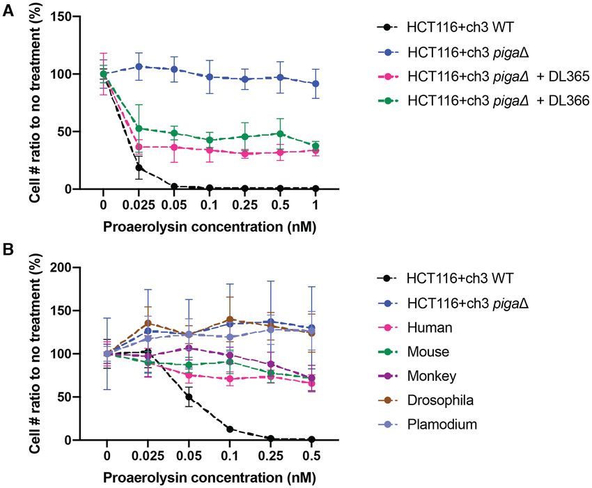
Nucleic Acids Research, 2021, Vol. 49, No. 5 2645

Hi-C data analysis and visualization                              products from deletion junction PCR. The deletion junc-
                                                                  tion fragments of six clones were gel purified and revealed
Hi-C data (.hic files) of HCT116 cells were downloaded
                                                                  by Sanger sequencing. Three were precise deletions, and
from NCBI GEO (accession number GSM2795535) (36).
                                                                  the other three were deletions with insertions (Figure 2B).
We extracted the interaction matrices from .hic format at
                                                                  After blasting the two insertion sequences, #4 showed no
50 kb resolution using the straw software (37) and Knight-
                                                                  genomic similarities, whereas #6 had a 99% identity to
Ruiz Matrix Balancing (KR) (38) normalization was ap-
                                                                  a sequence on chromosome X 15336146–15336415, which
plied to the matrices. Interaction scores within the range
                                                                  lies within the deletion region between gRNAs 3 DSS and
of interest were plotted using in-house developed scripts
                                                                  5 USS (Supplementary Table S6). In addition to the pigaΔ
in R. Heat maps in Figure 4 were generated using the 3D
                                                                  cell line, we used similar methods to obtain precise hprt1Δ
Genome Browser (39). All Hi-C analysis scripts are avail-
                                                                  cell lines in the backgrounds of HCT116+ch3 WT and
able at https://github.com/sunnysun515/hic analysis.
                                                                  HCT116+ch3 pigaΔ (Figure 2C). Sequencing revealed four
                                                                  clones of HCT116+ch3 pigaΔ hprt1Δ of which two were

                                                                                                                                   Downloaded from https://academic.oup.com/nar/article/49/5/2642/6138593 by guest on 11 September 2021
RESULTS                                                           precise deletions and the other two had INDELs. For the
                                                                  two clones of HCT116+ch3 hprt1Δ analyzed, one was a pre-
Robust selection with proaerolysin in HCT116+ch3 cell line
                                                                  cise deletion, and the other one had a total 74 bp deletion
To evaluate the robustness of the proaerolysin, we eval-          beyond the two expected Cas9 cut sites (Figure 2D). After
uated its killing curve and then examined the kinetics of         blasting the 74 bp insertion against the human genome, no
appearance of proaerolysin-resistant cells after treatment        similarities were found.
with a single exon-targeted CRISPR guide sequence. The               In addition to these modest sized designer deletion cell
proaerolysin killing curve indicates nearly 100% cell death       lines, we recovered cell lines with ∼2 Mb mega-deletions
after 24 h treatment with as little as 0.1 nM proaerolysin        centered on PIGA by using CRISPR deletion with the
(Figure 1A). Usually cell shrinkage (a consequence of             outermost pair of gRNAs 1 Mb and 1 Mb’ (Supple-
leakage of cellular contents through transmembrane pores          mentary Figure S1A). Eleven clones were recovered from
caused by aerolysin) can be observed within 1h after addi-        proaerolysin selected populations corresponding to the 2
tion of proaerolysin to the medium (Figure 1B). Dead cells        Mb deletion, and six of these were positive for deletion junc-
were floating within 24 h and were removed by changing            tion PCR. Sequencing indicated all six had INDELs which
the medium (Figure 1B). By employing a single gRNA tar-           fell into two groups, seemingly representing sibling clones
geting PIGA Exon 2, we showed that there is a clean back-         derived from two original deletion events (Supplementary
ground after proaerolysin treatment by comparing transfec-        Figure S1B, Supplementary Table S6). This could be due to
tions with and without gRNA (Figure 1C). Importantly, we          the fact that the 2 Mb deletion is a relatively rare event and
found that it was critical to incubate the cells for at least 7   the proaerolysin treatment was only applied 16 days post-
days prior to initiating counterselection with proaerolysin.      transfection, in order to recover the largest number of sur-
This time is presumably required to allow for turnover of         vivors. Remarkably, although this 2 Mb deletion removes
both preexisting PIGA enzyme as well as preexisting GPI-          15 human genes in addition to PIGA, namely GLRA2,
anchored proteins in the membrane. Figure 1D indicates            FANCB, MOSPD2, ASB9, ASB11, VEGFD, PIR, BMX,
the single gRNA targeting efficiency reflected on differ-         ACE2, TMEM27, CA5B, ZRSR2, AP1S2, GRPR and
ent days of proaerolysin treatment post-CRISPR transfec-          MAGEB17, the cells grow normally.
tion. When proaerolysin was added less than 7 days after
CRISPR, there were barely any survivors. As indicated in
Figure 1E, the correlation of CRISPR efficiency (reflected        Complementing the loss of PIGA with miniPIGA cDNA
as proaerolysin survival rate) is almost linearly correlated      In order to complement the loss of PIGA in the dele-
with the number of days between proaerolysin application          tion cell line, we cloned the cDNA of PIGA gene into
and CRISPR transfection.                                          the episomal (oriP-containing) pCEP4 plasmid under ei-
                                                                  ther the TK (pDL365) or the native promoter (pDL366)
                                                                  (Supplementary Figure S2A). After transfection and 7 days
Designer deletion cell lines with counterselectable markers
                                                                  of puromycin selection, we treated transfected cells with
In order to generate a ‘designer deletion’ cell line in which     proaerolysin and employed PrestoBlue cell viability assay
the entire PIGA transcription unit was deleted, we designed       to evaluate drug resistance. Unexpectedly, we only observed
paired gRNA 3 DSS and 5 USS in the flanking region              ∼40% complementation (Figure 3A). To further analyze
of PIGA. Even though INDELs caused by single gRNA                 whether this result was due to problems in initial trans-
CRISPR in an exon of PIGA are sufficient to disrupt its           fection or gradual loss/silencing of transfected plasmids,
function, we preferred to engineer a marker-deficient cell        previously described flow cytometry methods employing
line via elimination of the full gene, because the PIGA DNA       staining with fluorescent CD59 antibody (40) were used to
sequence in the genome might otherwise allow for subse-           sort the fully complemented population, and proaerolysin
quent recombination with incoming DNA when introduc-              treatment was initiated after outgrowth. We conducted the
ing it as a selectable marker.                                    proaerolysin treatment right after sorting, 5 and 9 days af-
   Single clones were picked after CRISPR–Cas9 trans-             ter sorting and outgrowth (Supplementary Figure S3). The
fection, followed by outgrowth and proaerolysin selection         PrestoBlue assay after outgrowth demonstrated a gradual
(Figure 2A). Of 15 clones picked, 13 gave rise to positive        loss of complementation (Supplementary Figure S3A–C),
2646 Nucleic Acids Research, 2021, Vol. 49, No. 5
120%
A Ratio to no treatment
B
100%
80%
60%
Treatment
40%
(h)
20%
1
0 1 5 5 .1 5 .5 1 2 .5 5 7 .5 1 0
0 .0 0 .02 0 .0 0 0 .2 0
Proaerolysin concentration (nM)
C
17
Downloaded from https://academic.oup.com/nar/article/49/5/2642/6138593 by guest on 11 September 2021
0 0.1 0.5 1
Proaerolysin concentration (nM)
gRNA - + E
Drug resistant colonies/10e6 cells
10e6
D Day1 Puro 48 h
Day-1 Seed Cell Day0 Transfect
10e5
Proaero Day 3 Day 5 Day 7 Day 8 Day 10 Day 23
10e4
-gRNA 10e3
10e2
+gRNA
10e1
0
~ 1-2 million cells per well ~ 1000 cells 0 2 4 6 8 10 12 14 16 18 20 22 24 26
per well Days post Transfection
Figure 1. Proaerolysin treatment in HCT116+ch3 cell line. (A) Proaerolysin killing curve conducted in HCT116+ch3 cell line using the PrestoBlue assay.
(B) Cell images for HCT116+ch3 cells after 1 and 17 h showing treatment with different concentrations of proaerolysin. (C) Crystal violet stain for CRISPR
Cas9 targeting PIGA Exon 2 with proaerolysin treatment. (D) Crystal violet stain for CRISPR Cas9 targeting PIGA Exon 2 with proaerolysin treatment
on different days post-transfection. (E) CRISPR efficiency from (D) calculated on a log10 scale, plotting proaerolysin resistant colony number per million
treated cells.
which may be due to episomal plasmid loss. pCEP4 plas- What factors affect paired gRNA deletion efficiency?
mid was used in this analysis because of its episomal ex-
Since PIGA is essential for the synthesis of GPI anchors,
pression. However, our result suggests that there is still a
measuring the loss of GPI anchors using flow cytometry in
chance to lose this episomal vector even under the proper
a cell population can help provide an estimate of deletion ef-
selection.
ficiency. In order to investigate the associated factors of the
GPI anchor is a highly conserved post-translational mod-
deletion efficiency, series of partner gRNAs were designed
ification. PIGA is essential for the first step of GPI synthe-
50 kb, 100 kb, 250 kb, 500 kb, 750 kb and 1 Mb upstream
sis and has the active site of the enzyme, but it is only one
and downstream of the PIGA gene (Figure 4C, Supplemen-
of multiple subunits involved in this step, so we planned to
tary Table S1). gRNAs were chosen to minimize the likeli-
investigate the degree of conservation of this biosynthetic
hood of off-target effects based on publicly available online
process. We cloned cDNA of PIGA orthologs from M. mus-
tools (33). At each position, at least two gRNAs were de-
culus (mouse), M. mulatta (monkey), D. melanogaster (fruit
signed. gRNA IN2-3 was used as the ‘anchor gRNA’ and
fly) and P. falciparum (malaria parasite) into pCEP4 (Sup-
paired with each of the partner gRNAs to systematically
plementary Figure S2B and C). After transfecting these
analyze deletion efficiency over a wide deletion size range
plasmids into the pigaΔ cell line, we used proaerolysin re-
from 50 kb to 1 Mb. All these deletions contain the loss
sistance analysis to evaluate the complementation efficiency
of at least one PIGA exon, and all are able to disrupt the
(Figure 3B). The orthologs of mouse and monkey comple-
synthesis of GPI anchors through deletion of at least one
mented as efficiently as the human cDNA, but those of
critical segment(s) of PIGA. Since PIGA was located at the
Drosophila and plasmodium did not demonstrate any ef-
boundary between two well-defined TADs and the anchor
fect. This result agrees that PIGA participates in a multi-
gRNA IN2–3 belongs to the TAD upstream of PIGA (Fig-
subunit step.Nucleic Acids Research, 2021, Vol. 49, No. 5 2647
Downloaded from https://academic.oup.com/nar/article/49/5/2642/6138593 by guest on 11 September 2021
Figure 2. Designer deletion cell lines. (A) gRNA designed to delete the full sequence of PIGA. The gel image indicates the deletion product amplified
with P1 and P2 for the 15 clones picked after proaerolysin treatment. Green triangles indicate the band cut and purified for sequencing. (B) Sequencing
results for the bands cut from the gel image in (A). (C) gRNA designed to delete the full sequence of HPRT1 in HCT116+ch3 WT and pigaΔ cell lines.
The gel image shows the deletion product for the numbered clones which were picked. Green triangles indicate band cut and purified for sequencing. (D)
Sequencing results for bands cut from gel image in (C). gRNA sequences are in bold uppercase letters. The PAM sequence is in red. Blue triangles indicate
predicted cleavage sites. WT refers to the wild type sequence from ensemble.org. Green italic lowercase letters represent mismatch sequences or insertions.2648 Nucleic Acids Research, 2021, Vol. 49, No. 5
Downloaded from https://academic.oup.com/nar/article/49/5/2642/6138593 by guest on 11 September 2021
Figure 3. Proaerolysin resistance analysis for mini-PIGA cDNA complementation. (A) pDL365 (TK promoter) and pDL366 (native PIGA promoter) were
transfected to pigaΔ cell line, after 7 days of puromycin selection, transfected cells were treated with proaerolysin for 24 h and then assayed with PrestoBlue.
(B) Plasmids expressing cDNA copies of the orthologs were transfected into the pigaΔ cell line, and after 7 days of puromycin selection, the transfected
cells were treated with proaerolysin for 24 h and assayed with PrestoBlue.
ure 4A), we used this design to test the effect of TAD on sulted in overall slightly higher deletion efficiencies (Fig-
deletion efficiency. Even though the mechanism of paired ure 5B and Supplementary Figure S4A), the opposite of
gRNA CRISPR deletion is not clear, it is likely that NHEJ what one would expect if TAD boundaries interfered with
is required to join the ends generated by CRISPR/Cas9 cut- deletion formation. We also tested the correlation of TAD
ting, we hypothesized that deletion efficiency might be af- boundaries with paired gRNA mediated deletion at the
fected by Topologically Associated Domains (TADs) as two HPRT1 locus. Similar to PIGA, HPRT1 is located at the
targeted regions might be more likely to be ligated when boundary of two TADs. gRNAs at 50, 100, 250 and 500 kb
they in close proximity. The gRNAs PIGA 50 kb D, 100 upstream or downstream of HPRT1 were designed to pair
kb D, 250 kb D, 500 kb D, 750 kb D and 1 Mb D, mapping with the anchor gRNA inside HPRT1(Supplementary Ta-
downstream of PIGA, all lie within the TAD downstream ble S2). The deletion efficiency measured by G418 selection
of the anchor gRNA IN2–3. On the other hand, all gRNAs and crystal violet staining and it again indicated no signif-
50 kb U, 100 kb U, 250 kb U, 500 kb U, 750 kb U and 1 icant difference between deletion efficiency and whether or
Mb U are within the same TAD in which gRNA IN2–3 is not the paired gRNAs reside in the same or the neighboring
located (Figure 4A, C). Interaction scores between the an- TAD (Supplementary Figure S4B and C). These observa-
chor guide region and the regions of the upstream partner tions are inconsistent with the hypothesis that TAD bound-
guides within the same TAD were much higher relative to aries interfere with deletion formation.
downstream partner guides that lie in the downstream TAD
(Figure 4B)
Using flow cytometry, deletion efficiency was measured Analysis of a heterozygous mutation in an autosomal gene,
as percentage of GPI (−) cells out of a total 20,000 cells, PIGL, suggests high frequency loss of heterozyogosity
which varied between 30% and 0.5%. Using this assay, we In addition to PIGA, a number of other ‘PIG’ genes are
observed no clear correlation between deletion efficiency required for the formation of GPI anchors, and the other
and distance between paired gRNAs (Figure 5A). The dele- PIG genes are all autosomal (10). We were interested in
tion efficiency of the anchor gRNA paired with the 1 Mb D using the GPI system to study the stability of heterozy-
gRNA was as high as 20%. (Figure 5B). Moreover, by com- gous CRISPR induced mutations in autosomal ‘PIG’ genes.
paring deletion efficiency of paired gRNAs within the same Because homozygous missense mutations of PIGU result
TAD or crossing the TAD boundary, we noticed that the in reduced cell-surface expression of fluorescent aerolysin
paired gRNAs that cross the TAD boundary actually re- (FLAER) flow cytometry profile (41), we anticipated thatNucleic Acids Research, 2021, Vol. 49, No. 5 2649
Downloaded from https://academic.oup.com/nar/article/49/5/2642/6138593 by guest on 11 September 2021
Figure 4. Paired gRNA deletion design. (A) Interaction frequencies from HI-C data were shown as a heat map at the PIGA region. Positions of gRNAs
located 50 kb–1 Mb from the anchored gRNA IN2–3 were indicated by gray dashed lines. Anchor icon indicates position of gRNA IN2–3. (B) Hi-C
interaction scores of partner gRNAs regions and anchor gRNA (IN2–3) region were plotted according to the partner gRNA positions around PIGA. (C)
gRNA target sites. Red dots and triangles represent gRNAs 50 kb U, 100 kb U, 250 kb U, 500 kb U, 750 kb U and 1 Mb U. Green dots and triangles
represent gRNAs 50 kb D, 100 kb D, 250 kb D, 500 kb D, 750 kb D and 1 Mb D.2650 Nucleic Acids Research, 2021, Vol. 49, No. 5
Downloaded from https://academic.oup.com/nar/article/49/5/2642/6138593 by guest on 11 September 2021
Figure 5. Paired gRNA deletion efficiency is not significantly affected by deletion size nor TAD boundaries. (A) Deletion efficiency versus deletion size.
Red dots represent gRNAs in the TAD upstream of PIGA, green dots represent gRNAs in the TAD downstream of PIGA. (B) Deletion efficiencies versus
partner gRNA position. Red dots represent gRNAs in the TAD upstream of PIGA, green dots represent gRNAs in the TAD downstream of PIGA. The
shading from dark to light represent gRNAs with deletion efficiency from high to low at the same position. Error bars are standard deviations calculated
from three biological repeats.Nucleic Acids Research, 2021, Vol. 49, No. 5 2651
homozygous mutations in all other autosomal PIG genes based on studies of asynchronous cells, and the highly com-
would also confer proaerolysin resistance. Thus, we used partmentalized organization is restricted to interphase (42),
CRISPR–Cas9 to generate a heterozygous NHEJ-induced the consideration of the TAD effect is practically relevant to
mutation at a single site in the PIGL gene (Supplementary those wishing to perform paired gRNA CRISPR deletion.
Figure S5A). These studies revealed that a homozygous mu- It is reasonable to ask whether the TAD boundaries are
tation in PIGL indeed led to proaerolysin resistance, as ex- active in cells actually undergoing CRISPR-mediated dele-
pected, and also that cells with a heterozygous mutation in tion. Based on single cell Hi-C studies, the locations of TAD
PIGL remained sensitive to proaerolysin. borders are generally unchanged in G1, early-S and late-
We then studied the heterozygous cell line, HCT116+ch3 S/G2 cells and are absent during mitosis (43). The duration
PIGL (+/−) by selecting derivatives that were proaerolysin of mitosis is short, constant and independent of varied cell
resistant, expecting to find new spontaneous mutations in cycle length, and lasts about 1 hour in rapidly proliferating
the wild-type PIGL allele at a high frequency. However, human cells (44). Thus, more than 90% of the cells assayed
when using PCR amplification of the gRNA target site in this study were in interphase and presumably the bound-
Downloaded from https://academic.oup.com/nar/article/49/5/2642/6138593 by guest on 11 September 2021
to verify heterozygosity in single clones from this cell line aries at the PIGA and HPRT1 loci were intact. Further-
after proaerolysin treatment and outgrowth, we unexpect- more, the NHEJ repair pathway utilized by paired gRNA
edly observed that the majority of the survivors (21/24) CRISPR deletion is active throughout interphase and pre-
had lost heterozygosity for the original CRISPR/NHEJ- dominates in the G1 phase when sister chromatids are avail-
induced mutation (Supplementary Figure S5B, primers in able for homologous recombination based repair (45). Since
Supplementary Table S4). The wild type allele was main- G1 phase is the longest phase of the cell cycle, the popula-
tained in only three of the lines. Further, cytogenetic anal- tion engineered by paired gRNA CRISPR deletion is pre-
yses showed that the loss of heterozygosity (LoH) was not sumed to have active TAD boundaries. The lack of impact
accompanied by loss of chromosome 17p, on which PIGL of nearby TAD boundaries on paired gRNA CRISPR dele-
resides, in four of seven cases evaluated. It may be that the tion provides confidence when using paired gRNA CRISPR
observed LoH occurred through gene conversion, although to engineer large deletions.
the experiments done thus far cannot be considered defini- As a genome engineering technology, CRISPR is a
tive on this point. promising tool and has been applied extensively in re-
search and clinical fields due to its high efficiency. INDELs
associated with NHEJ repair of CRISPR/Cas9-mediated
DISCUSSION
DSBs that occur within an exon can disrupt protein-coding
Canver et al. reported an inverse relationship between open reading frames. However, INDELS placed in non-
deletion efficiency and deletion size based on their coding DNA are usually insufficient to disrupt the func-
CRISPR/Cas9-mediated genomic deletions in mouse ery- tion of noncoding transcripts, gene clusters or regulatory se-
throleukemia (MEL) cells (29). However, another analy- quences, where large genomic deletions would be preferable.
sis conducted along the HPRT1 locus in HEK293FT cell Also, CRISPR deletions could play roles in genetic diseases
line did not report any apparent correlation between inter- driven by genome amplification or copy number abnormali-
gRNA distance and deletion efficiency (30). Both groups ties for animal model generation and therapeutics with large
achieved deletions up to 1 Mb. For the paired gRNA genomic rearrangements. During our study of cell line engi-
CRISPR deletion study conducted in MEL cells, a range neering via CRISPR, we also observed the value of paired
of deletions with different inter-gRNA distances were de- gRNA CRISPR deletion in the generation of heterozygous
signed and constructed at different loci throughout the mutations. The loss of heterozygosity which occurred in the
genome. This design introduced many confounding vari- HCT116+ch3 PIGL (+/−) cell line indicates the high fre-
ables beyond inter-gRNA distance, and thus is not an op- quency of LoH of point alterations like the small INDELs
timal experimental design for directly evaluating the effect produced by single gRNA targeting. It might be possible
of length on deletion efficiency. For the analysis conducted to harness such LoH, particularly if it is produced by gene
by He et al. (30) at the HPRT1 locus, the cell line chosen conversion, to make homozygous designer alterations to
was HEK 293FT, which is hypotriploid and contains at least diploid mammalian chromosomes. Since gene-conversion
three X chromosomes. Such a complex genome confounds tracts are usually short in mammalian cells, and rarely ex-
direct genotyping to detect deletions, but did benefit from ceed 1 kb in length (46), it is interesting to consider ways
being a single locus study, with an anchored guide sequence to limit the impact of such gene conversion, e.g. in order
to make results potentially more comparable. to develop an assay for mutation in a large autosomal gene
By employing the nearly diploid HCT116+ch3 cell line, like PIGL. By making a heterozygous deletions very large,
the PIGA locus on the X chromosome and the quantita- or even extending them into an adjacent essential gene, it
tive measurement of GPI(–) cells, we performed a system- might be possible to minimize or entirely eliminate the re-
atic analysis for the efficiency of paired gRNA CRISPR covery of this type of LoH event.
deletion. Our results reveal no significant impact of gRNA The era of editing and writing mammalian genomes using
positioning on deletion efficiency associated with the dis- synthetic genomics techniques is well underway. Launched
tance between the two gRNAs nor their positions relative in 2016, the GP-write project aims to reduce genomic engi-
to nearby TAD boundaries. This finding was confirmed by neering and testing costs to 1/1000th of the previous within
the same design in the HPRT1 locus, providing a valuable 10 years and address a number of human health challenges
extension to the observations of Canver et al. (29) and He (47). A recently announced plan by GP-write, making virus-
et al. (30). Even though TAD boundaries are mostly defined resistant human cells, would require >400 000 changes to2652 Nucleic Acids Research, 2021, Vol. 49, No. 5
the genome (48). All this editing will be associated with mation by length variation and inclusion/exclusion TAD
extensive recoding, DNA fragment delivery, and sequen- boundary deletions. Since PIGA has the potential to be-
tial delivery with marker swapping. Paired gRNA CRISPR come a widely used counterselection marker, its endogenous
deletion may be important in endogenous genomic content locus may well serve as a future target of DNA delivery or
removal when introducing synthetic fragments. Designer other types of analysis, more extensive studies of TADs in
deletion cell lines described in this study may be useful for our cell lines (Supplementary Table S3) may be of interest.
DNA delivery, and cell lines in which both HPRT1 and In summary, we have engineered a series of designer dele-
PIGA are deleted could be used as a platform for deliver- tion cell lines featureing counterselectable markers of PIGA
ing DNAs independently or perhaps sequentially. The wide and HPRT1. These cell lines can facilitate large DNA frag-
use and adoption of the 5-Foa selection in Saccharomyces ment delivery and other types of study involved with PIGA
cerevisiae (1), and similarly, the wide use of TK negative se- and HPRT1 biology. In the process of building this re-
lection using gancyclovir in mammalian cells (4) suggests source, we acquired systematic data on a controlled set
that the proaerolysin selection, in conjunction with deploy- of deletions with a common anchor point, and used this
Downloaded from https://academic.oup.com/nar/article/49/5/2642/6138593 by guest on 11 September 2021
ment of CRISPR represents a highly versatile mammalian to provide evidence supporting the hypothesis that TAD
genome engineering tool that will enjoy wide usage. boundaries do not interfere in a practical way with deletion
One potential limitation of our study is that it was per- formation using paired CRISPR guides.
formed in male cell lines, and thus it remains to be prac-
tically tested how well these counterselections work in fe- DATA AVAILABILITY
male cells. The 6-thioguanine selection for hprt1 mutants
works equally well in male and female cells (49) and fur- All the Hi-C analysis scripts are available at https://
thermore, the gene lies well outside the pseudoautosomal github.com/sunnysun515/hic analysis. Flow cytometry ex-
region, and is not a gene known to ‘escape’ X inactivation periments were deposited in FlowRepository under Repos-
(50). Nonetheless some caution might be exercised in female itory ID FR-FCM-Z3BN.
embryonic stem cells, as these are known to lose X inactiva-
tion. The availability of lines with deletions in both PIGA SUPPLEMENTARY DATA
and HPRT1, located on the left and right arms of the X
chromosome, respectively opens up some interesting oppor- Supplementary Data are available at NAR Online.
tunities to look at aspects of X inactivation and beyond.
Counterselection markers might be useful in selective ACKNOWLEDGEMENTS
loss of DNA templates delivered on episomal vectors af-
ter proper genome editing has occurred. The instability of We thank Drs Jeff Corden, Maria Jasin, Todd Macfar-
episomal vectors discovered in this study indicates that the lan and David Truong, and anonymous reviewers for valu-
selected loss of auxiliary gene on an episomal vector via able discussions and suggestions. We thank Dr Thomas A.
proaerolysin treatment seems more efficient than expected. Kunkel for sharing the HCT116+ch3 cell line. Portions of
It is worth noting that an intronless PIGA processed pseu- this work were previously published in a PhD thesis by D.L.
dogene (PIGA) resides on chromosome 12 (51). PIGA and supported in part by NIH grants P50-GM107632 and
should not cause issues when using the designer deletion RM1-HG009491 to JDB. JDB is a founder and Director
cells in the study for DNA delivery or other applications of CDI Labs, Inc., a founder of Neochromosome, Inc., a
via proaerolysin selection or other GPI anchor based assays, Founder of and Consultant to the ReOpen Diagnostics, and
for it is not functionally expressed as a consequence of sev- serves or served on the Scientific Advisory Board of the fol-
eral nonsense mutations. However, PIGA should be taken lowing: Sangamo, Inc., Modern Meadow, Inc. and Sam-
into account due to its homology to PIGA cDNA, which ple6, Inc.
might in theory lead to recombination with PIGA cDNA
constructs. FUNDING
In addition to the use of designer deletion cell lines in
DNA delivery, it would be interesting to investigate the National Human Genome Research Institute [RM1-
TAD architecture of the cell line HCT116+ch3 piga 2Mb HG009491]; National Institute of General Medical Sci-
deletion, since the deleted region harbors extensive CTCF ences [P50-GM107632]. Funding for open access charge:
(a master regulator of TAD structure) binding sites and in- National Human Genome Research Institute [RM1-
cludes a TAD boundary (Figure 4A). It has been reported HG009491].
that TAD boundary disruption can occur after deletions of Conflict of interest statement. Jef Boeke is a Founder and
minimal or very large genomic regions with CTCF bind- Director of CDI Labs, Inc., a Founder of Neochromosome,
ing sites (52–54). However, a recent study focusing on the Inc., a Founder of and Consultant to the ReOpen Diagnos-
CTCF-rich Firre locus on X chromosome revealed that tics, and serves or served on the Scientific Advisory Board
its deletion preserved the surrounding TAD even though of the following: Sangamo, Inc., Modern Meadow, Inc. and
CTCF binding was depleted (55). It would be interesting Sample6, Inc.
to evaluate another X chromosome locus like PIGA to in-
vestigate whether such TAD preservation is a sex chromo- REFERENCES
some associated phenomena. The series of paired gRNAs 1. Boeke,J.D., Trueheart,J., Natsoulis,G. and Fink,G.R. (1987)
designed for deletion efficiency measurements in this study 5-Fluoroorotic acid as a selective agent in yeast molecular genetics.
can also be utilized to screen the effects on TAD refor- Methods Enzymol., 154, 164–175.Nucleic Acids Research, 2021, Vol. 49, No. 5 2653
2. Steyer,B., Bu,Q., Cory,E., Jiang,K., Duong,S., Sinha,D., Steltzer,S., using the endogenous Pig-A gene: I. Flow cytometric detection of
Gamm,D., Chang,Q. and Saha,K. (2018) Scarless genome editing of CD59-negative peripheral red blood cells and CD48-negative spleen
human pluripotent stem cells via transient puromycin selection. Stem T-cells from the rat. Environ. Mol. Mutagen., 49, 614–621.
Cell Rep., 10, 642–654. 23. Bryce,S.M., Bemis,J.C. and Dertinger,S.D. (2008) In vivo mutation
3. Yu,J., Vodyanik,M.A., Smuga-Otto,K., Antosiewicz-Bourget,J., assay based on the endogenous Pig-a locus. Environ. Mol. Mutagen.,
Frane,J.L., Tian,S., Nie,J., Jonsdottir,G.A., Ruotti,V., Stewart,R. 49, 256–264.
et al. (2007) Induced pluripotent stem cell lines derived from human 24. Karnan,S., Konishi,Y., Ota,A., Takahashi,M., Damdindorj,L.,
somatic cells. Science, 318, 1917–1920. Hosokawa,Y. and Konishi,H. (2012) Simple monitoring of gene
4. Wigler,M., Silverstein,S., Lee,L.S., Pellicer,A., Cheng,Y. and Axel,R. targeting efficiency in human somatic cell lines using the PIGA gene.
(1977) Transfer of purified herpes virus thymidine kinase gene to PLoS One, 7, e47389.
cultured mouse cells. Cell, 11, 223–232. 25. Bemis,J.C. and Heflich,R.H. (2019) In vitro mammalian cell mutation
5. Lester,S.C., LeVan,S.K., Steglich,C. and DeMars,R. (1980) assays based on the Pig-a gene: a report of the 7th International
Expression of human genes for adenine phosphoribosyltransferase Workshop on Genotoxicity Testing (IWGT) Workgroup. Mutat. Res.,
and hypoxanthine-guanine phosphoribosyltransferase after genetic 847, 403028.
transformation of mouse cells with purified human DNA. Somatic 26. Zou,J., Maeder,M.L., Mali,P., Pruett-Miller,S.M.,
Cell Genet., 6, 241–259. Thibodeau-Beganny,S., Chou,B.K., Chen,G., Ye,Z., Park,I.H.,
Downloaded from https://academic.oup.com/nar/article/49/5/2642/6138593 by guest on 11 September 2021
6. Lupton,S.D., Brunton,L.L., Kalberg,V.A. and Overell,R.W. (1991) Daley,G.Q. et al. (2009) Gene targeting of a disease-related gene in
Dominant positive and negative selection using a hygromycin human induced pluripotent stem and embryonic stem cells. Cell Stem
phosphotransferase-thymidine kinase fusion gene. Mol. Cell. Biol., Cell, 5, 97–110.
11, 3374–3378. 27. Koi,M., Umar,A., Chauhan,D.P., Cherian,S.P., Carethers,J.M.,
7. Schwartz,F., Maeda,N., Smithies,O., Hickey,R., Edelmann,W., Kunkel,T.A. and Boland,C.R. (1994) Human chromosome 3 corrects
Skoultchi,A. and Kucherlapati,R. (1991) A dominant positive and mismatch repair deficiency and microsatellite instability and reduces
negative selectable gene for use in mammalian cells. Proc. Natl. Acad. N-methyl-N’-nitro-N-nitrosoguanidine tolerance in colon tumor cells
Sci. U.S.A., 88, 10416–10420. with homozygous hMLH1 mutation. Cancer Res., 54, 4308–4312.
8. Karreman,C. (1998) New positive/negative selectable markers for 28. Zheng,Q., Cai,X., Tan,M.H., Schaffert,S., Arnold,C.P., Gong,X.,
mammalian cells on the basis of Blasticidin deaminase-thymidine Chen,C.Z. and Huang,S. (2014) Precise gene deletion and
kinase fusions. Nucleic Acids Res., 26, 2508–2510. replacement using the CRISPR/Cas9 system in human cells.
9. Karreman,C. (1998) A new set of positive/negative selectable markers BioTechniques, 57, 115–124.
for mammalian cells. Gene, 218, 57–61. 29. Canver,M.C., Bauer,D.E., Dass,A., Yien,Y.Y., Chung,J., Masuda,T.,
10. Kawagoe,K., Takeda,J., Endo,Y. and Kinoshita,T. (1994) Molecular Maeda,T., Paw,B.H. and Orkin,S.H. (2014) Characterization of
cloning of murine pig-a, a gene for GPI-anchor biosynthesis, and genomic deletion efficiency mediated by clustered regularly
demonstration of interspecies conservation of its structure, function, interspaced short palindromic repeats (CRISPR)/Cas9 nuclease
and genetic locus. Genomics, 23, 566–574. system in mammalian cells. J. Biol. Chem., 289, 21312–21324.
11. Watanabe,R., Kinoshita,T., Masaki,R., Yamamoto,A., Takeda,J. and 30. He,Z., Proudfoot,C., Mileham,A.J., McLaren,D.G., Whitelaw,C.B.
Inoue,N. (1996) PIG-A and PIG-H, which participate in and Lillico,S.G. (2015) Highly efficient targeted chromosome
glycosylphosphatidylinositol anchor biosynthesis, form a protein deletions using CRISPR/Cas9. Biotechnol. Bioeng., 112, 1060–1064.
complex in the endoplasmic reticulum. J. Biol. Chem., 271, 31. Nora,E.P., Lajoie,B.R., Schulz,E.G., Giorgetti,L., Okamoto,I.,
26868–26875. Servant,N., Piolot,T., van Berkum,N.L., Meisig,J., Sedat,J. et al.
12. Watanabe,R., Inoue,N., Westfall,B., Taron,C.H., Orlean,P., Takeda,J. (2012) Spatial partitioning of the regulatory landscape of the
and Kinoshita,T. (1998) The first step of glycosylphosphatidylinositol X-inactivation centre. Nature, 485, 381–385.
biosynthesis is mediated by a complex of PIG-A, PIG-H, PIG-C and 32. Pombo,A. and Dillon,N. (2015) Three-dimensional genome
GPI1. EMBO J., 17, 877–885. architecture: players and mechanisms. Nat. Rev. Mol. Cell Biol., 16,
13. Low,M.G. and Saltiel,A.R. (1988) Structural and functional roles of 245–257.
glycosyl-phosphatidylinositol in membranes. Science, 239, 268–275. 33. Ran,F.A., Hsu,P.D., Wright,J., Agarwala,V., Scott,D.A. and Zhang,F.
14. Paulick,M.G. and Bertozzi,C.R. (2008) The (2013) Genome engineering using the CRISPR-Cas9 system. Nat.
glycosylphosphatidylinositol anchor: a complex Protoc., 8, 2281–2308.
membrane-anchoring structure for proteins. Biochemistry, 47, 34. Howard,S.P. and Buckley,J.T. (1985) Activation of the hole-forming
6991–7000. toxin aerolysin by extracellular processing. J. Bacteriol., 163, 336–340.
15. Araten,D.J., Golde,D.W., Zhang,R.H., Thaler,H.T., Gargiulo,L., 35. Bernheimer,A.W., Avigad,L.S. and Avigad,G. (1975) Interactions
Notaro,R. and Luzzatto,L. (2005) A quantitative measurement of the between aerolysin, erythrocytes, and erythrocyte membranes. Infect.
human somatic mutation rate. Cancer Res., 65, 8111–8117. Immun., 11, 1312–1319.
16. Takeda,J., Miyata,T., Kawagoe,K., Iida,Y., Endo,Y., Fujita,T., 36. Rao,S.S.P., Huang,S.C., Glenn St Hilaire,B., Engreitz,J.M.,
Takahashi,M., Kitani,T. and Kinoshita,T. (1993) Deficiency of the Perez,E.M., Kieffer-Kwon,K.R., Sanborn,A.L., Johnstone,S.E.,
GPI anchor caused by a somatic mutation of the PIG-A gene in Bascom,G.D., Bochkov,I.D. et al. (2017) Cohesin loss eliminates all
paroxysmal nocturnal hemoglobinuria. Cell, 73, 703–711. loop domains. Cell, 171, 305–320.
17. Young,N.S. and Moss,J. (2000) In: Paroxysmal Nocturnal 37. Durand,N.C., Robinson,J.T., Shamim,M.S., Machol,I., Mesirov,J.P.,
Hemoglobinuria and the Glycosylphosphatidylinositol-linked Proteins. Lander,E.S. and Aiden,E.L. (2016) Juicebox provides a visualization
Academic Press, San Diego. system for Hi-C contact maps with unlimited zoom. Cell Syst., 3,
18. Bessler,M., Mason,P., Hillmen,P. and Luzzatto,L. (1994) Somatic 99–101.
mutations and cellular selection in paroxysmal nocturnal 38. Philip,A. and Knight,D.R. (2013) A fast algorithm for matrix
haemoglobinuria. Lancet, 343, 951–953. balancing. IMA J. Numer. Anal., 33, 1029–1047.
19. Endo,M., Ware,R.E., Vreeke,T.M., Singh,S.P., Howard,T.A., 39. Wang,Y., Song,F., Zhang,B., Zhang,L., Xu,J., Kuang,D., Li,D.,
Tomita,A., Holguin,M.H. and Parker,C.J. (1996) Molecular basis of Choudhary,M.N.K., Li,Y., Hu,M. et al. (2018) The 3D Genome
the heterogeneity of expression of glycosyl phosphatidylinositol Browser: a web-based browser for visualizing 3D genome
anchored proteins in paroxysmal nocturnal hemoglobinuria. Blood, organization and long-range chromatin interactions. Genome Biol.,
87, 2546–2557. 19, 151.
20. O’Keefe,C.L., Sugimori,C., Afable,M., Clemente,M., Shain,K., 40. Araten,D.J., Martinez-Climent,J.A., Perle,M.A., Holm,E.,
Araten,D.J., List,A., Epling-Burnette,P.K. and Maciejewski,J.P. Zamechek,L., DiTata,K. and Sanders,K.J. (2010) A quantitative
(2011) Deletions of Xp22.2 including PIG-A locus lead to analysis of genomic instability in lymphoid and plasma cell
paroxysmal nocturnal hemoglobinuria. Leukemia, 25, 379–382. neoplasms based on the PIG-A gene. Mutat. Res., 686, 1–8.
21. Buckley,J.T. and Howard,S.P. (1988) Aerolysin from Aeromonas 41. Knaus,A., Kortum,F., Kleefstra,T., Stray-Pedersen,A., Dukic,D.,
hydrophila. Methods Enzymol., 165, 193–199. Murakami,Y., Gerstner,T., van Bokhoven,H., Iqbal,Z., Horn,D. et al.
22. Miura,D., Dobrovolsky,V.N., Kasahara,Y., Katsuura,Y. and (2019) Mutations in PIGU impair the function of the GPI
Heflich,R.H. (2008) Development of an in vivo gene mutation assay2654 Nucleic Acids Research, 2021, Vol. 49, No. 5
transamidase complex, causing severe intellectual disability, epilepsy, 50. Carrel,L. and Willard,H.F. (2005) X-inactivation profile reveals
and brain anomalies. Am. J. Hum. Genet., 105, 395–402. extensive variability in X-linked gene expression in females. Nature,
42. Naumova,N., Imakaev,M., Fudenberg,G., Zhan,Y., Lajoie,B.R., 434, 400–404.
Mirny,L.A. and Dekker,J. (2013) Organization of the mitotic 51. Bessler,M., Hillmen,P., Longo,L., Luzzatto,L. and Mason,P.J. (1994)
chromosome. Science, 342, 948–953. Genomic organization of the X-linked gene (PIG-A) that is mutated
43. Nagano,T., Lubling,Y., Varnai,C., Dudley,C., Leung,W., Baran,Y., in paroxysmal nocturnal haemoglobinuria and of a related autosomal
Mendelson Cohen,N., Wingett,S., Fraser,P. and Tanay,A. (2017) pseudogene mapped to 12q21. Hum. Mol. Genet., 3, 751–757.
Cell-cycle dynamics of chromosomal organization at single-cell 52. Lupianez,D.G., Kraft,K., Heinrich,V., Krawitz,P., Brancati,F.,
resolution. Nature, 547, 61–67. Klopocki,E., Horn,D., Kayserili,H., Opitz,J.M., Laxova,R. et al.
44. Araujo,A.R., Gelens,L., Sheriff,R.S. and Santos,S.D. (2016) Positive (2015) Disruptions of topological chromatin domains cause
feedback keeps duration of mitosis temporally insulated from pathogenic rewiring of gene-enhancer interactions. Cell, 161,
upstream cell-cycle events. Mol. Cell, 64, 362–375. 1012–1025.
45. Aylon,Y., Liefshitz,B. and Kupiec,M. (2004) The CDK regulates 53. Franke,M., Ibrahim,D.M., Andrey,G., Schwarzer,W., Heinrich,V.,
repair of double-strand breaks by homologous recombination during Schopflin,R., Kraft,K., Kempfer,R., Jerkovic,I., Chan,W.L. et al.
the cell cycle. EMBO J., 23, 4868–4875. (2016) Formation of new chromatin domains determines
46. Chen,J.M., Cooper,D.N., Chuzhanova,N., Ferec,C. and Patrinos,G.P. pathogenicity of genomic duplications. Nature, 538, 265–269.
Downloaded from https://academic.oup.com/nar/article/49/5/2642/6138593 by guest on 11 September 2021
(2007) Gene conversion: mechanisms, evolution and human disease. 54. Rodriguez-Carballo,E., Lopez-Delisle,L., Zhan,Y., Fabre,P.J.,
Nat. Rev. Genet., 8, 762–775. Beccari,L., El-Idrissi,I., Huynh,T.H.N., Ozadam,H., Dekker,J. and
47. Boeke,J.D., Church,G., Hessel,A., Kelley,N.J., Arkin,A., Cai,Y., Duboule,D. (2017) The HoxD cluster is a dynamic and resilient TAD
Carlson,R., Chakravarti,A., Cornish,V.W., Holt,L. et al. (2016) boundary controlling the segregation of antagonistic regulatory
GENOME ENGINEERING. The Genome Project-Write. Science, landscapes. Genes Dev., 31, 2264–2281.
353, 126–127. 55. Barutcu,A.R., Maass,P.G., Lewandowski,J.P., Weiner,C.L. and
48. Dolgin,E. (2018) Scientists downsize bold plan to make human Rinn,J.L. (2018) A TAD boundary is preserved upon deletion of the
genome from scratch. Nature, 557, 16–17. CTCF-rich Firre locus. Nat. Commun., 9, 1444.
49. Fukuchi,K., Martin,G.M. and Monnat,R.J. Jr. (1989) Mutator
phenotype of Werner syndrome is characterized by extensive
deletions. Proc. Natl. Acad. Sci. U.S.A., 86, 5893–5897.You can also read