Review Article Exploring the Pivotal Immunomodulatory and Anti-Inflammatory Potentials of Glycyrrhizic and Glycyrrhetinic Acids - Hindawi.com
←
→
Page content transcription
If your browser does not render page correctly, please read the page content below
Hindawi
Mediators of Inflammation
Volume 2021, Article ID 6699560, 15 pages
https://doi.org/10.1155/2021/6699560

Review Article
Exploring the Pivotal Immunomodulatory and Anti-
Inflammatory Potentials of Glycyrrhizic and Glycyrrhetinic Acids

 Seidu A. Richard
 Department of Medicine, Princefield University, P. O. Box MA 128, Ho, Ghana

 Correspondence should be addressed to Seidu A. Richard; gbepoo@gmail.com

 Received 5 November 2020; Revised 9 December 2020; Accepted 19 December 2020; Published 7 January 2021

 Academic Editor: Rômulo Dias Novaes

 Copyright © 2021 Seidu A. Richard. This is an open access article distributed under the Creative Commons Attribution License,
 which permits unrestricted use, distribution, and reproduction in any medium, provided the original work is properly cited.

 Licorice extract is a Chinese herbal medication most often used as a demulcent or elixir. The extract usually consists of many
 components but the key ingredients are glycyrrhizic (GL) and glycyrrhetinic acid (GA). GL and GA function as potent
 antioxidants, anti-inflammatory, antiviral, antitumor agents, and immuneregulators. GL and GA have potent activities against
 hepatitis A, B, and C viruses, human immunodeficiency virus type 1, vesicular stomatitis virus, herpes simplex virus, influenza
 A, severe acute respiratory syndrome-related coronavirus, respiratory syncytial virus, vaccinia virus, and arboviruses. Also, GA
 was observed to be of therapeutic valve in human enterovirus 71, which was recognized as the utmost regular virus responsible
 for hand, foot, and mouth disease. The anti-inflammatory mechanism of GL and GA is realized via cytokines like interferon-γ,
 tumor necrotizing factor-α, interleukin- (IL-) 1β, IL-4, IL-5, IL-6, IL-8, IL-10, IL-12, and IL-17. They also modulate anti-
 inflammatory mechanisms like intercellular cell adhesion molecule 1 and P-selectin, enzymes like inducible nitric oxide synthase
 (iNOS), and transcription factors such as nuclear factor-kappa B, signal transducer and activator of transcription- (STAT-) 3,
 and STAT-6. Furthermore, DCs treated with GL were capable of influencing T-cell differentiation toward Th1 subset. Moreover,
 GA is capable of blocking prostaglandin-E2 synthesis via blockade of cyclooxygenase- (COX-) 2 resulting in concurrent
 augmentation nitric oxide production through the enhancement of iNOS2 mRNA secretion in Leishmania-infected
 macrophages. GA is capable of inhibiting toll-like receptors as well as high-mobility group box 1.

1. Introduction production of interferons (IFNs), accelerated the activities
 of natural killer (NK) cells as well as regulated the growth
Licorice extract is a Chinese herbal medication most often response of lymphocytes via the acceleration of interleukins-
used as a demulcent or elixir [1]. Glycyrrhizin (GL) is one (IL-) 2 production [1, 8, 9]. Furthermore, GL has the ability
of the principally effective and efficient ingredients of licorice to modulate the immune response at the initial stage of the
extract [1–3]. GL is a triterpene saponin which has aglycone disease process via the dendritic cells (DCs) [10]. GA inhib-
component known as glycyrrhetinic acid (GA) [1]. GA is a ited anti-FAS antibody-triggered mouse liver injury but did
pentacyclic triterpenoid of oleanene type with a hydroxyl not facilitate the upregulation of tumor necrotizing factor-α
group at C-3, a carboxyl moiety at C-30 as well as a ketone (TNF-α) messenger RNA (mRNA) secretion in the liver [11].
functional group at C-11 [2]. GL and GA have been demon- This review explores the fundamental immune and
strated to possess antioxidant properties as well as robust inflammatory players regulated by GL and GA. The “boolean
anti-inflammatory, antiviral, antitumor, and immuneregula- logic” was utilized to search for the article on the subject mat-
tory properties [4–6]. GL was capable of triggering the ter. Most of the articles were indexed in PubMed with strict
blockade of receptor-mediated endocytosis resulting in the inclusion criteria being in vitro and in vivo up or downregu-
inhibition of viral infiltration into the cells [5, 7]. lation of these immune and inflammatory biomarkers in
 GL triggers biological activities at the cellular level via diverse disease conditions. Inflammation, DCs, cyclooxygen-
novel gbPs, which are responsible for anti-inflammatory ase, and prostaglandins, cytokines like ILs, IFNs, TNF-α,
and antiviral effects [5, 8]. GL was capable of triggering the nuclear factor-κB (NF-κB), mitogen-activated protein kinase
2 Mediators of Inflammation

(MAPK), Toll-like receptors (TLRs), high-mobility group hydrolyzing GL into GA possess a specific β-glucuronidase,
box 1 (HMGB1), and chemokines like CCL11 as known as because common β-glucuronidases like Escherichia coli
eotaxin 1 as well as enzymes like nitric oxide were explored. were unable to hydrolyze GL [12, 28]. After hydrolysis of
 GL into GA, intestinal bacteria convert GA partially into
2. Uses 3-α-18β-GA, through a metabolic intermediary 3-oxo-18β-
 GA [12, 27].
Glycyrrhizin (GL) obtained from the dried roots of the lico- Also, the plasma clearance of GL after an intravenous
rice shrub is very sweet tasting and has been utilized as fla- bolus dose to rats exhibited a biphasic pattern, in which the
vors in diverse food products and treatment of diseases for distribution phase was preceded by a slower elimination
over 4000 years [12]. Currently, GL is used to flavor consum- phase [30, 31]. However, realistic plasma levels of GA were
able products like chocolate, chewing gum, some alcoholic observed to be approximately 100 mg/ml after intravenous
beverages, and cigarettes [12, 13]. Carbenoxolone (GC), the administration [30, 31]. Also, the distribution of GA to the
derivative of glycyrrhetinic acid (3β-11-oxoolean-12-en-30- body tissues was negligible because tissue-to-blood partition
oic acid 3-hemisuccinate), was used to treat peptic ulcer coefficients were observed to
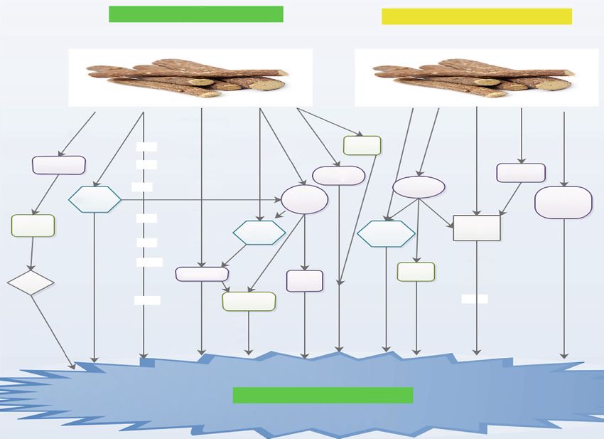
Mediators of Inflammation 3

 Downregulatory Upregulatory
 GL/GA pathway
 pathway

 VEGF, ICAM–1
 GM-CSF, GRO/KC
 PLA2/ARA, PGE2 SOD
 TXA2, LTB4 GSH-Px
 LC-1, PLA2 p-Akt
 MMP-9, STAT-3 p-ERK
 pADR, NTS
 PMN, IL-8
 Eotaxin 1, Pselectin
 LPS-TLR-4/MD-2

 Anti-inflammation

 Figure 1: Shows a comprehensive down and upregulatory pathways via which GL and GA elicits anti-inflammation.

pathway (Figure 1) [36]. GA ominously decreased the con- maturation status [48–50]. Their prime function is to bridge
centration of ICAM-1 as well as matrix metalloproteinase-9 the innate as well as adaptive immune systems [48–50]. DCs
(MMP-9) (Figure 1) [41, 43]. Furthermore, it augmented were able to accelerate allogeneic T-cell proliferation in vitro
the actions of Superoxide dismutase (SOD) and glutathione [4]. A study revealed that only a minute quantity of DCs was
peroxidase (GSH-Px), as well as the secretion of p-Akt and enough to trigger an allogeneic mixed lymphocyte reaction
p-ERK (Figure 1) [41, 44]. (MLR) [4, 48]. Studies have demonstrated that DCs are the
 GL and GA efficiently blocked the generation of free rad- most crucial antigen-presenting cells (APCs) associated with
icals in LPS-treated Raw264.7 macrophage models [41]. They the uptake, processing, transport, and presentation of
also decreased the configuration of the LPS-TLR-4/MD-2 antigens to CD4+ and CD8+ T-cells [4, 49, 51].
complexes, leading to the blockade of homodimerization of Also, DC subsets are capable of triggering or inhibiting
TLR-4 (Figure 1) [45, 46]. Thus, GA was able to regulate immune responses via the secretion of different costimula-
the TLR-4/MD-2 complex at the receptor level, resulting in tory molecules and cytokines [4, 52]. DCs were able to trigger
the inhibition of LPS-induced triggering of signaling cascades as well as target naive T-cells to differentiate into T-helper
as well as cytokine generation [45]. This signifies that GA (Th)1 or T-helper (Th)2 cells [4, 53]. Thus, DCs have
blocked inflammatory responses as well as regulated innate potential immunomodulatory therapeutic targets for some
immune responses [45, 47]. pharmacological compounds [4, 10]. Bordbar et al. demon-
 Furthermore, GA inhibited the stimulation of signal strated that DCs treated with GL were capable of influencing
transducers and activators of transcription-3 (STAT-3), T-cell differentiation toward Th1 subset (Figure 2) (Table 1)
decreased the upregulation of ICAM-1 as well as P- [4]. Abe et al. also observed the upregulation of IL-10 expres-
selectin secretion, decreased the configuration of poly- sion by liver DCs [54]. Hua et al. established that GL was
adenosine diphosphate-ribose (pADR) and nitrotyrosine capable of augmenting IL-10 production in DC2.4 cell line
(NTS), and decreased polymorphonuclear neutrophil infil- (Figure 2) [55]. A current study demonstrated that GL was
tration (PMN) (Figure 1 and Table 1) [45–47]. Moreover, capable of augmenting IL-10 production along with IFN-γ
GA elicited broad anti-inflammatory actions via its interac- in MLR [4]. On the other hand, Bhattacharjee et al. exhibited
tion with the lipid bilayer resulting in the decrease of that GA was capable of blocking the expression of the Th2,
receptor-mediated signaling [45, 46]. GA was capable of IL-10, and TGF-β from the splenocytes of infected mice
blocking the lytic pathway of the complement system as well (Figure 2) [25].
as averted tissue injury triggered by membrane attack
complexes [45]. 6. Nuclear Factor-κB
5. Dendritic Cells The nuclear factor-κB (NF-κB) family is made up of five
 groups such as NF-κB1, which comprise of p50/p105 with
Dendritic cells (DCs) are a group of bone-marrow-derived p50 as the precursor, NF-κB2 which comprise of p52/p100
cells found in blood, tissues, and lymphoid organs [48–50]. with p52 as the precursor, Rel A with p65 as the precursor,
These cells initiate and control immune responses that are Rel B with p68 as the precursor, and c-Rel with p75 as the
affected by numerous factors like origin, phenotype, and precursor [56, 57]. Almost all the groupings are capable of
4 Mediators of Inflammation

 Table 1: Shows the explicit effect of GL or GA on various immune/inflammatory factors.

Immune/inflammatory factors Type Effect of GL/GA Citations
Inflammation VEGF Inhibitory [40]
 ICAM-1 Inhibitory [40]
 GM-CSF Inhibitory [40]
 GRO/KC Inhibitory [40]
 PLA2/ARA Inhibitory [36, 41, 42]
 MMP-9 Inhibitory [41, 43]
 STAT-3 Inhibitory [45–47]
 STAT-6 Inhibitory [45–47]
 pADR Inhibitory [45–47]
 NTS Inhibitory [45–47]
 PMN Inhibitory [45–47]
 SOD Facilitatory [41, 44]
 GSH-Px Facilitatory [41, 44]
 TGF-β Facilitatory [25]
Dendritic cells (DCs) T-cell Facilitatory [4]
 Th1 Facilitatory [4]
 Th2 Facilitatory [25]
Nuclear factor-κB — Inhibitory [6, 20, 62–65]
 IKK Inhibitory [56]
Chemokines CXCL10 Inhibitory [20, 30, 70, 71]
 CCL5 Inhibitory [20, 30, 70, 71]
 CCL11 Inhibitory [39, 76–78]
Interferons IFN-γ Facilitatory [85–89]
Cyclooxygenase COX-1 — —
 COX-2 Inhibitory [25, 65, 98]
Interleukins IL-1 Inhibitory [8, 104–106]
 IL-2 Facilitatory [4, 9, 106]
 IL-3 Inhibitory [8, 104–106]
 IL-4 Inhibitory [8, 104–106]
 IL-5 Inhibitory [8, 104–106]
 IL-6 Inhibitory [8, 104–106]
 IL-10 Inhibitory [8, 104–106]
 IL-12 Inhibitory [4, 8, 104–106]
 IL-13 Inhibitory [8, 104–106]
 IL-18 Inhibitory [8, 104–106, 111]
Mitogen-activated
 — Inhibitory [6, 115, 116, 141]
protein kinase
 p38MAPK Inhibitory [6, 115, 116]
 ERK Inhibitory [45]
 JNK Inhibitory
Nitric oxide iNOS Inhibitory [45]
 eNOS — —
 nNOS — —
Toll-like receptors TLR-3 Inhibitory [131–134]
 TLR-4 Inhibitory [131–134, 141]
 TLR-7 Inhibitory [131–134]
 TLR-9 Inhibitory [131–134]
 TLR-10 Inhibitory [131–134]
High-mobility group box 1 — Inhibitory [141, 142]
Mediators of Inflammation 5

 Inhibitory pathways Facilitatory pathways

 GL/GA GL/GA

 ICAM-1
 LPS
 VCAM-1 AGE
 IL-1 Th2
 TNF-a IKKb
 COX-2 IL-2 IKKy TCR
 IL-8 TGF-b Con A
 IL-3
 IL-1 DCs
 CCL11 NF- B TLR-4
 IL-5 IL-6
 PGE2 T-cell
 IL-4 IFN- TLRs IFN-
 IL-6

 IL-10 IL-2
 TNF-a Th1
 HMGB1
 NO PI3K IL-2R
 IL-13 MAPK IL-12
 MIP-1
 STAT-6
 iNOS2 p38
 mRNA IL-18
 JNK
 ERK

 Ameliorates disease

 Figure 2: Shows the inhibitory and facilitatory pathways via which GL and GA ameliorate disease.

preserving homodimeric as well as heterodimeric complexes NF-κB-inducing kinase, PI3K, or MAPK in the signaling
[56]. Nevertheless, the most predominant-stimulated form of pathway (Figure 2) [63].
NF-κB is the heterodimer p50-p65, which has the transactiv- GL was capable of treating coxsackievirus B3- (CVB3-)
ity territory obligatory for gene modification [58–60]. In triggered myocarditis via the blockade of CVB3-triggered
most cells, NF-κB exists as a latent, inactive, IκB bound com- NF-κB activity via the inhibition of NF-κB inhibitor IκB
plex in the cytoplasm [56]. Nevertheless, upon stimulation by (Table 1) [20, 64]. Wang and Du revealed that pretreatment
extracellular stimuli, NF-κB promptly translocates to the with GL substantially inhibited the facilitation of NF-κB p65
nucleus and triggers gene release [56, 61]. protein secretion, in methotrexate-stimulated enteritis
 IκB kinase (IKK) is a large multisubunit protein kinase (Table 1) [6]. Cherng et al. showed that GL blocked NF-κB
active via numerous signal pathways [56]. The IKK complex secretion, averted DNA damage, and accelerated DNA
when triggered results in the phosphorylation or degradation repair (Table 1) [65]. Feng et al. demonstrated that GA safe-
of IκBα leading to the expression of NF-κB [56]. NF-κB then guards advanced glycation end-product- (AGE-) stimulated
translocates to the nucleus and triggers the transcription of endothelial dysfunction via blockade of the receptor for
numerous κB-dependent genes, such as iNOS as well as AGE/NF-κB signaling pathway (Figure 2 and Table 1) [66].
Th1 cytokines [56]. Thus, some pathogens are capable of
blocking the action of NF-κB via the inhibition of the degra-
dation of IκB during infection [56]. Also, in macrophages, the 7. Chemokines
MAPK cascade and the NF-κB pathway are the key pathways
via which modulation of inflammation as well as host defense Chemokines are a family of molecules associated with the
occurs [56]. trafficking of leukocytes in normal immune surveillance and
 Ukil et al. demonstrated that the kinase properties of recruitment of inflammatory cells in host defense [67–69].
IKK were triggered in cells that were stimulated with GA They are made up of over 40 groups, which are classified into
via a mechanism that most probably involves upregulatory four classes founded on the sites of essential cysteine residues
signaling pathways [56]. They however did not observe any like C, CC, CXC, and CX3C [67]. GL was capable of subduing
influence of GA on IKK activity when GA was added the H5N1-triggered generation of CXCL10, and CCL5 resulting
directly to the assay mixture containing IKK immunopreci- in the blockade of H5N1-triggered apoptosis [20, 70]. Michaelis
pitated from normal macrophages (Table 1) [56]. An earlier et al. demonstrated that 100 mg/ml of GA drastically blocked
study revealed that GA influenced the inhibitory interaction secretion of CXCL10, and CCL5 at the mRNA and the protein
between NF-κB, which is a fundamental modifier to IKKβ levels (Table 1) [30]. Augmented CXCL10 levels were observed
and IKKγ (Figure 2) [62]. Another study indicated that in patients with H5N1, and the elevated levels of CXCL10 were
GA inhibited one of the essential upregulatory kinases like associated with poor prognosis (Table 1) [30, 71].
6 Mediators of Inflammation

 CCL11 known as eotaxin 1 was primarily detected as the thromboxane, and levuloglandins [90]. Prostaglandins are
prime eosinophil chemoattractant in the lung lavage fluid autocoid facilitators that influence practically all recognized
after allergic exposure in guinea pigs [39]. Subsequently, it physiological as well as pathological activities via their
was cloned for further studies [39, 72, 73]. Several studies reversible communication with G-protein attached mem-
have demonstrated that numerous types of cells, such as lung brane receptors [90]. Amongst the COX isoenzymes, COX-
or dermal fibroblasts, as well as lung or bronchial epithelial 2 was more inducible with low secretory levels in most
cells are capable of producing eotaxin 1 [39, 74, 75]. Studies tissues under normal circumstances [6, 91]. It was estab-
further revealed that the production of eotaxin 1 was lished that numerous cell types such as vascular smooth
triggered by IL-4 and inhibited by IFN-γ [39, 74, 75]. It was muscle cells, endothelial cells, mononuclear macrophages,
also observed that eotaxin 1 facilitated the infiltration of and fibroblasts were capable of secreting COX-2 up to about
eosinophils into allergic inflammatory sites [39, 74, 75]. 8-10-fold the normal level when stimulated by proinflam-
 Matsui et al. indicated that GL may be capable of mod- matory cytokines [6, 92].
ulating chemokine generation via the posttranscriptional It was further observed that augmentation of COX-2
level such as protein expression or mortification [39]. They levels resulted in the generation as well as buildup of prosta-
demonstrated that GL derivatives had inhibitory effects on glandin inflammatory factors, facilitating inflammatory
eotaxin 1 generation via TNF-α as well as IL-4 induction responses as well as tissue damage [6, 91]. Studies have
in lung fibroblasts (Figure 2) [39]. Studies have shown that shown that oversecretion of COX-2 facilitated cell prolifera-
induction of IL-4 and TNF-α in combination synergisti- tion, blocked apoptosis, and blocked immune responses,
cally accelerated the generation of eotaxin 1 via the trigger- resulting in abnormal modulation of the balance between
ing of transcriptional factors like STAT-6 and NF-κB proliferation and apoptosis [6, 91, 92]. Bhattacharjee et al.
(Figure 2) [76–78]. GL and its derivatives thus blocked demonstrated that a robust antileishmanial protection was
eotaxin 1 production at protein or mRNA secretary levels observed via the modulation of macrophage-secreted COX-
(Table 1) [39, 76]. 2-determined PGE2 levels [25]. Also, Leishmania organisms
 were capable of using immune modulators like TGF-β, IL-
8. Interferons 4, and arachidonic acid metabolites to inhibit macrophage
 functions and facilitated the organism’s survival within the
Interferons (IFNs) are a family of broad-spectrum antiviral host [93].
glycoproteins expressed by cells upon attack by viruses. They PGE2 biosynthesis comprises two successive enzymatic
are often involved in numerous immune responses as trig- reactions [25]. The first one is a rate-limiting step involving
gers, modulators, and effectors of both innate as well as adap- the COX enzyme, while the second is a precise PGE synthesis
tive immune systems during viral infections [79, 80]. They step [25]. In pathophysiological processes, the inducible iso-
have the ability of blocking viral replication and are often form of COX-2 was capable of modulating PGE2 production
the most prominent cytokines produced during viral infec- while COX-1 was principally copied [25, 94, 95]. Studies have
tions [79, 80]. IFN-γ, which is expressed by lymphocytes, shown that augmented level PGE2 was capable of modulat-
has been implicated in the secretion of histocompatibility ing several immune responses via mechanisms involving
antigen as well as immune modifications [6, 81]. Studies have the blockade of Th1 cytokines like IL-2, IL-12, and IFN-γ,
demonstrated that IFN-γ was capable of facilitating the as well as inhibition of phagocytosis and lymphocyte prolifer-
endotoxin-stimulated generation of NO in murine macro- ation [25, 96, 97]. Thus, PGE2 ability to modulated immune
phages [79, 80]. response is champion by Th1- or Th2-associated lympho-
 Studies have shown that IFN with or without adenine kines [25, 96].
arabinoside was capable of curing hepatitis B patients [1, It was established that GA was capable of blocking PGE2
82, 83]. IFNs were capable of reducing the level of either synthesis via blockade of COX-2 resulting in concurrent aug-
DNA polymerase or hepatitis B surface antigen in hepatitis mentation NO production through enhancement of iNOS2
patients [1, 84]. Furthermore, GL was capable of facilitating mRNA secretion in Leishmania-infected macrophages
IFN-γ production in human T-lymphocytes [85, 86]. Also, (Figure 2 and (Table 1) [25]. Wang and Du demonstrated
GL was capable of inducing the production of IFN in mice, that pretreatment with GL significantly blocked the facilita-
which was preceded by stimulation of macrophages as well tion of COX-2 activity in methotrexate-triggered enteritis
as the increase of NK activity [87, 88]. Bhattacharjee et al. [6]. Cherng et al. also demonstrated that GL was able to block
demonstrated that splenic expression of IFN-γ, TNF-α, and COX-2 secretion, inhibited DNA damage, and promoted
IL-12 elevated after GA treatment. Wu et al. also demon- DNA repair (Table 1) [65]. Ni et al. observed an upsurge in
strated that GL drastically decreased inflammatory via IFN- COX-2 secretion in lung tissues after introducing LPS in their
γ (Figure 2) [89]. They concluded that blockade of the IFN- experiment, which was subsequently decreased in a dose-
γ signaling pathway may be linked to anti-inflammatory dependent manner after GL pretreatment (Figure 2 and
effects of GL in enteritis [89]. Table 1) [98].

9. Cyclooxygenase and Prostaglandins 10. Interleukins
COX-1 and COX-2 are the main cyclooxygenase (COX) iso- Interleukin belongs to a group of cytokines, which are
enzymes, which catalyze the formation of prostaglandins, perhaps the most essential messenger molecules generated
Mediators of Inflammation 7

by leukocytes to modulate the biological activities of target introduction of GL [111]. Also, GL was capable of blocking
cells via autocrine or paracrine means [99]. Several groupings the infiltration of neutrophils and macrophages in liver
of ILs have been identified [99]. Notable amongst them are injury [111]. Furthermore, GL-stimulated decrease in immu-
IL-1, IL-2, IL-3, IL-5, IL-6, IL-10, IL-12, IL-13, and so many noreactive IL-18 was probably due to blockade of cell infiltra-
others [3, 8, 99–101]. Although most of the ILs are influenced tion in the liver [111]. GL was able to inhibit an upsurge in
by GL and GA, IL-12 is the most influential. IL-12 is a hetero- alanine aminotransferase activity when exogenous IL-18
dimeric cytokine produced primarily by macrophages and was administered in mice treated with LPS/D-galactosamine
monocytes [8]. Its key function is the modulation of cyto- [111]. Thus, GL blocked IL-18-mediated inflammatory
kines as well as T-cell subsets [8]. A study revealed that a response in the pathogenesis of liver injury [111]. Nakanishi
deficiency in endogenous IL-12 production influenced the et al. demonstrated that IL-18 was capable of triggering gene
progression of immunodeficiency in HIV-infected patients secretion as well as the synthesis of TNF-α, IL-1, FAS ligand,
[8, 102]. Studies have proven that IL-12 salvaged numerous and many chemokines [112].
activities of cells infected with HIV [8, 103].
 Several studies have demonstrated that IL-12 was capable 11. Mitogen-Activated Protein Kinase
of influencing T-cells and natural NK cells resulting in cell
proliferation, cytolytic activities, and triggering of IFN-γ [8, Mitogen-activated protein kinase (MAPK) signal transduc-
104]. Studies further revealed that the polarization of the T tion pathways are linked with cell proliferation, differentia-
helper response to a Th1-dominant form via IL-12 was accel- tion, apoptosis, and angiogenesis [6]. Specifically, the p38
erated by IFN-γ resulting in the blockade of IL-4 production mitogen-activated protein kinase (p38MAPK) signal trans-
[8, 100, 101]. GA was capable of blocking IL-1β, IL-3, IL-5, duction pathway modulates stress responses, like inflamma-
IL-6, IL-10, IL-12 subtypes, IL-13 (Figure 2), eotaxin, and tion as well as apoptosis [6, 113]. Studies have shown that
TNF-α expression (Table 1) [8, 104–106]. GL was also capa- LPS as well as other factors is capable of triggering the MAPK
ble of accelerating the proliferation of lymphocytes and acted pathways resulting in the secretion of many inflammatory
as a facilitator of the late signal transduction of T lympho- mediators via complex signal conduction pathways, which
cytes for IL-2 generation (Table 1) [4, 106]. facilitates inflammation [6]. Furthermore, the modulation
 Zhang et al. also indicated that GL facilitated TCR- of p38MAPK was observed in various transduction path-
mediated T-cell proliferation by selectively influencing the ways, which in turn stimulated many transcription factors
late signal transduction for IL-2 generation as well as IL-2R as well as mediated a variety of biological activities [6, 114].
secretion [9]. They further indicated that GL exhibited two Wang and Du demonstrated that pretreatment with GA
separate activities on immature thymocytes resulting in the remarkably inhibited the facilitation of p38MAPK in
facilitation of IL-2 generation on one hand and blocked methotrexate-stimulated enteritis (Table 1) [6]. They
growth response on the other [9, 107]. A hepatitis study concluded that the anti-inflammatory actions of GA were
revealed that IL-4 was capable of stimulating STAT6, which probably linked to p38MAPK signaling (Figure 2) [6]. Also,
in turn stimulated eotaxin secretion as well as triggered IL- studies have shown that GA lessens glycative stress in the
5 secretion [40, 108]. Wang and Du established that GA kidneys of diabetic mice via the blockade of p-p38MAPK
was capable of relieving methotrexate-stimulated upsurge of [115, 116]. It was further established that GA was capable
TNF-α, IL-1β, and IL-6 levels, as well as elevated IL-10 levels, of blocking the modulation of JNK, p38 protein, and ERK
in rats with enteritis (Table 1) [6]. GL was able to facilitate the (Figure 2 and Table 1) in bone marrow-derived macrophages
IL-10 production by hepatic dendritic cells in mice with (BMMs) [45].
hepatitis (Table 1) [76].
 Studies have proven that IL-10 is a well-known anti- 12. Nitric Oxide
inflammatory cytokine [40, 109, 110]. It was capable of mod-
ulating STAT3 in hepatocytes as well as macrophages/Kupf- Nitric oxide (NO) is a radical messenger molecule generated
fer cells [40, 109, 110]. A study revealed that GA was capable by the enzyme nitric oxide synthase (NOS) [117–119]. So far,
of accelerating LPS-triggered IL-12 generation by peritoneal only three isoforms of NOS have been identified. Amongst
macrophages (Table 1) [4, 8]. Its optimal effect on IL-12 gene the three, only two of them, NOS in neurons (nNOS) and
secretion was linked to an upsurge in NF-κB modulation [4, in the endothelial cells of blood vessels (eNOS), are intensely
8]. Dai et al. demonstrated that GL accelerated both IL-12 secreted [117–120]. These two are capable of producing only
mRNA buildup as well as protein expression by peritoneal minute quantities of NO, which is sufficient to trigger cellular
macrophages in response to LPS [8]. They indicated that signaling in stress conditions.
the priming influence of GL on IL-12 generation did not Studies have shown that NO in an inflammatory media-
depend on IFN-γ or GM-CSF [8]. Thus, they also affirmed tor is capable of modulating innate immunity as well as path-
that the facilitation of IL-12 p40 mRNA secretion by GL ophysiology of many infectious diseases [117, 121, 122]. The
may be via the modulation of NF-κB [8]. third kind of NOS is the inducible nitric oxide synthase
 Yoshida et al. demonstrated that GL was able to block the (iNOS) [117, 119].
upsurge in serum levels of IL-18 in LPS/D-galactosamine- Studies have further proven that iNOS generates NO in
induced liver injury (Figure 2 and Table 1) [111]. Thus, GL hepatocytes as well as macrophages [117, 119, 121, 122].
blocked the generation of IL-18 in this model [111]. They The stimulation of iNOS is modulated via a posttranscrip-
also observed fewer IL-18-positive infiltrating cells after the tional mechanism that is mediated by antisense transcripts
8 Mediators of Inflammation

(asRNAs) [117, 122]. Several studies have shown that the way (Figure 2) [129, 139, 140]. Studies further revealed that
asRNAs are transcribed from the iNOS gene and interact the inactivation of NF-κB led to a reduced expression of
with iNOS mRNA to stabilize the same iNOS mRNA [122, different proinflammatory cytokines like IL-1β, IL-6, TNF-
123]. Studies have demonstrated that the iNOS is triggered α, and HMGB1 (Figure 2) [129, 139, 140]. GL was capable
by cytokines like IFN-γ and TNF-α, which in turn produce of blocking porcine epidemic diarrhea virus infection, as
large quantities of NO [117, 119, 121, 123]. It is well proven well as reduced proinflammatory cytokine expression via
that NO generated by iNOS was capable of triggering an the HMGB1/TLR4-p38MAPK pathway (Figure 2 and
inflammatory liver damage [117, 124]. Table 1) [141].
 Studies have demonstrated that concanavalin A (Con A)
was capable of triggering the stimulating the T-cells in mice
and induced the secretion of proinflammatory cytokines 14. High-Mobility Group Box 1
associated with the progression of hepatitis (Figure 2) [119,
125]. Furthermore, GL was capable of inhibiting Con A- High-mobility group box 1 (HMGB1) protein is a nuclear
stimulated mouse liver damage without influencing the protein that functions as an architectural chromatin-
generation of IFN-γ and TNF-α [119, 126]. Tsuruoka et al. binding factor [142, 143]. HMGB1 is the prime signal during
demonstrated that GL blockade of liver damage was via the tissue damage usually involving necrotic and apoptotic cells
inhibition of iNOS mRNA as well as its protein secretion [142]. Furthermore, HMGB1 performs dual functions in
(Table 1) [119]. Thus, GL inhibited iNOS mRNA and protein the nucleus and the cytoplasm [142]. Also, extracellular
in Con A-stimulated hepatitis [119]. Also, GL was capable of HMGB1 facilitates both local as well as systemic responses
blocking the secretion of iNOS mRNA stimulated by carbon in the organism [142]. These responses often include inflam-
tetrachloride in hepatic tissue (Table 1) [119, 127]. mation, modulation of innate as well as adaptive immunity
 [142, 143]. Several studies have demonstrated that HMGB1
13. Toll-Like Receptors is secreted by monocytes, macrophages, neutrophils, plate-
 lets, and dendritic and NK cells [142, 144].
Toll-like receptors (TLRs) are sensors for pathogen-associated Several studies have shown that HMGB1 induces macro-
molecular patterns (PAMPs) [128]. TLRs are capable of mod- phages, monocytes, and neutrophils to secrete proinflamma-
ulating several immune responses, especially during the infec- tory cytokines like TNF-α, IL-1, IL-6, IL-8, and MIP-1 via
tious process [128]. Several studies have shown that the p38- and JNK MAPK-dependent pathways (Figure 2) [145,
secretion of TLR-3, TLR-4, TLR-7, TLR-9, and TLR-10 genes 146]. It was established that HMGB1 was passively secreted
from hepatic tissue was upregulated in some viral infection by damage alveolar endothelial cells or macrophages during
models [129, 130], and GA or GL is capable of inhibiting these virus-mediated cytolysis [145]. Once expressed, extracellular
receptors (Table 1) [131–134]. It was established that the TLR- HMGB1 was capable of mediating injurious pulmonary
4 pathway comprises of two dissimilar signaling pathways inflammatory response like neutrophil infiltration, derange-
such as the myeloid differentiating primary response gene ment of epithelial barrier, lung edema, and lung injury [145,
88- (MyD88-) dependent as well as the MyD88-independent 147]. These injurious pulmonary inflammatory responses sub-
pathway [135, 136]. It was further revealed that stimulation sequently result in respiratory failure as well as death [147].
of the MyD88-dependent pathway led to the generation of Also, human microvascular endothelial cells are capable of
proinflammatory cytokines via triggering of NF-κB, while secreting ICAM-1, vascular adhesion molecule–1 (VCAM-1),
the stimulation MyD88-independent pathway led to the gen- proinflammatory cytokines like TNFα, IL-8, and chemokines
eration of type 1 IFNs [135, 136]. in response to HMGB1 activation (Figure 2) [145, 148]. This
 A study revealed that TLR-4 was the fundamental recep- means that HMGB1 was capable of disseminating inflamma-
tor of the innate immune signaling responses to influenza tory response in the endothelium during infection or injury
virus as well as other respiratory viruses [137]. Several studies [145]. Chemotactic as well as mitogenic actions of HMGB1
have shown that the TLR-4 was more associated with respira- depends on its association with the receptor of advanced gly-
tory syncytial virus and human papillomavirus infections cation end products (RAGE) [142, 149]. GL was capable of
[129, 138, 139]. Shi et al. revealed that TLR-4 gene deficiency blocking the chemoattractant as well as mitogenic activities
was not associated with the downregulation of virus titer in of HMGB1 (Table 1) [142].
the liver during MHV-A59 infection [129]. They observed GL was capable of binding to both HMG boxes of
that in MHV-A59 infection, the HMGB1-TLR-4 axis utilizes HMGB1 in both NMR and fluorescence studies without
proinflammatory activities without directly influencing virus altering their secondary structure, which was observed as
replication [129]. an absence of changes in CD spectra [142]. It was further
 A study demonstrated that GA was not capable of established that amino acids interacting with GL clusters at
influencing TLR-4 gene secretion during viral infection the junction of both arms of the classical L-shape fold of both
[129]. Nevertheless, the secretion of the TLR-4 gene facili- HMG boxes in chemical-shift perturbation experiments
tated MHV-stimulated hepatic inflammation injury as well [142]. Furthermore, the binding sites for GL on the HMG
as determined HMGB1 secretory levels in the serum boxes partly overlap with the DNA binding sites, shielding
(Figure 2) [129]. Several studies have proven that pretreat- residues like R23, which is recognized to be crucial for
ment with a TLR-4 inhibition agent reduced the HMGB1 DNA binding [142, 150]. Nevertheless, the RAGE-binding
levels from virus-infected cells via the TLR4-NF-κB path- surface on HMGB1 was characterized with the stretch of
Mediators of Inflammation 9

basic amino acids between box B and the acidic tail and did inflammation via IFN-γ, which means that GL and GA have
not match with the binding surfaces of GL [142, 149]. very crucial antiviral properties. Also, GA decreased the secre-
 Influenza type A, B, and C viruses are responsible for tion of VEGF, MCP-1, GM-CSF, and GRO/KC in alcoholic
influenza infection (“flu”) [145]. This infection is often hepatitis rats’ models. GA was capable of blocking the modu-
depicted with massive virus replication as well as excessive lation of JNK, p38 protein, and ERK in BMMs. Further studies
inflammation [145]. Studies have shown that influenza on GL/GA-HMGB1 axis are needed to elucidate their poten-
viruses are capable of infecting monocytes and macrophages tial role in the treatment for patients with coronavirus
resulting in the stimulation of proinflammatory cytokines disease-19 in the current SARS-coronavirus pandemic.
like TNF-α, IL-1, IL-6, IL-8, IFN-α, and chemokines in
infected areas (Figure 2) [145, 151]. Moisy et al. demon-
strated that HMGB1 binds to the nucleoprotein section of Abbreviations
influenza ribonucleoproteins (vRNPs) freely in the company
of viral RNA in vitro and interacts with the viral nucleopro- asRNAs: Antisense transcripts
tein VCAM-1 in infected cells [152]. They revealed that APCs: Antigen-presenting cells
HMGB1 was capable of facilitating viral growth as well as BMMs: Bone marrow-derived macrophages
augmented the transcription or replication activity of the COX: Cyclooxygenase
viral polymerase in HMGB1-depleted cells [152]. Thus, GC: Carbenoxolone
HMGB1 binding to DNA was a prerequisite for the augmen- CVB3: Coxsackievirus B3
tation of influenza virus replication [152]. Therefore, GA and Con A: Concanavalin A
GL may be capable of treating influenza viral infection via the DCs: Dendritic cells
HMGB1-TNF-α pathway (Figure 2). Further studies should GM-CSF: Granulocyte-macrophage colony-stimulating
focus on this pathway. factor
 HMGB1 was able to trigger necrotic cell death resulting in GL: Glycyrrhizin
abundant budding of West Nile (WN) progeny virus particles GA: Glycyrrhetinic acid
at higher infectious doses [145, 153]. Furthermore, HMGB1 HMGB1: High-mobility group box 1
mediated in injurious inflammatory response resulting in the GRO/KC: Human growth-regulated oncogene/keratino-
pathogenesis of WN encephalitis [145, 153, 154]. Besides cyte chemoattractant
WN viruses, other viruses like the salmon anemia virus were HIV: Human immunodeficiency virus
capable of triggering necrotic cell death of infected cells, ICAM-1: Intercellular cell adhesion molecule 1
leading to simultaneous HMGB1 expression [145, 154]. GL IKK: IκB kinase
and GA may be potential treatment options for WN viral via IFNs: Interferons
HMGB1. Further studies are warranted in this direction. IL: Interleukin
 Studies have shown that an increase in proinflammatory iNOS: Inducible nitric oxide synthase
cytokines like IL-1, IL-6, TNF-α, and IFN-γ may trigger the MAPK: Mitogen-activated protein kinase
expression of HMGB1 from innate immune cells in SARS p38MAPK: p38 mitogen-activated protein kinase
patients (Figure 2) [145, 155]. Thus, further studies on MLR: Mixed lymphocyte reaction
GL/GA-HMGB1 axis are needed to elucidate their potential mRNA: Messenger RNA
role in the treatment for patients with coronavirus disease- NK: Natural killer
19 in the current SARS-coronavirus pandemic. Acute viral NO: Nitric oxide
hepatitis is caused by hepatitis A, B, C, and D viruses. Their NF-κB: Nuclear factor-κB
pathogenesis is often depicted with acute necrosis of hepato- NTS: Nitrotyrosine
cytes, inflammation, and followed by fibrosis as well as cir- PLA2: Phospholipase A2
rhosis [145, 156]. HMGB1, passively secreted by necrotic pADR: Poly-adenosine diphosphate-ribose
hepatocytes, may stimulate tissue macrophages especially PMN: Polymorphonuclear neutrophil infiltration
Kupffer cells to express proinflammatory cytokines during TNF-α: Tumor necrotizing factor-α
an acute infection [145]. Thus, HMGB1 alone or in combi- PGE2: Prostaglandin-E 2
nation with other proinflammatory cytokines may cause vRNPs: Ribonucleoproteins
chronic liver damage in hepatitis patients [145]. GL and TLRs: Toll-like receptors
GA are potential treatment options for chronic viral hepati- Th: T-helper
tis. Further studies are warranted on HMGB1 and/or SARS: Severe acute respiratory syndrome
GL/GA axis. STAT: Signal transducer and activator of transcription
 VCAM-1: Vascular adhesion molecule–1
15. Conclusion VEGF: Vascular endothelial growth factor
 WN: West Nile.
GL and GA are able to block the secretion of IL-1β, IL-3, IL-
4, IL-5, IL-6, IL-10, IL-12, IL-13, eotaxin, and TNF-α expres-
sion. This means that GL and GA are capable of inhibiting Data Availability
cytokine storms elicited during various infectious diseases
most especially viral diseases. GL and GA drastically decreased No data was used in this paper.
10 Mediators of Inflammation

Conflicts of Interest C,” Alimentary Pharmacology & Therapeutics, vol. 12, no. 3,
 pp. 199–205, 1998.
The authors declare that they have no conflicts of interest. [16] U. A. Ashfaq, M. S. Masoud, Z. Nawaz, and S. Riazuddin,
 “Glycyrrhizin as antiviral agent against hepatitis C virus,”
 Journal of Translational Medicine, vol. 9, no. 1, article 800,
References pp. 1–7, 2011.
 [17] L. Baltina, R. Kondratenko, L. A. Baltina Jr., O. A. Plyasunova,
 [1] N. Abe, T. Ebina, and N. Ishida, “Interferon induction by gly-
 A. G. Pokrovskii, and G. A. Tolstikov, “Prospects for the crea-
 cyrrhizin and glycyrrhetinic acid in mice,” Microbiology and
 tion of new antiviral drugs based on glycyrrhizic acid and its
 Immunology, vol. 26, no. 6, pp. 535–539, 1982.
 derivatives (a review),” Pharmaceutical Chemistry Journal,
 [2] D. Langer, B. Czarczynska-Goslinska, and T. Goslinski, “Gly- vol. 43, no. 10, article 348, pp. 539–548, 2009.
 cyrrhetinic acid and its derivatives in infectious diseases,”
 [18] C. Fiore, M. Eisenhut, R. Krausse et al., “Antiviral effects
 Current Issues in Pharmacy and Medical Sciences, vol. 29,
 ofGlycyrrhiza species,” Phytotherapy Research, vol. 22,
 no. 3, pp. 118–123, 2016.
 no. 2, pp. 141–148, 2008.
 [3] F. Maione, P. Minosi, A. di Giannuario et al., “Long-lasting
 anti-inflammatory and antinociceptive effects of acute [19] S. Harada, T. Maekawa, E. Haneda, Y. Morikawa, N. Nagata,
 ammonium glycyrrhizinate administration: pharmacological, and K. Ohtsuki, “Biochemical characterization of recombinant
 biochemical, and docking studies,” Molecules, vol. 24, no. 13, HIV-1 reverse transcriptase (rRT) as a glycyrrhizin-binding
 p. 2453, 2019. protein and the CK-II-mediated stimulation of rRT activity
 potently inhibited by glycyrrhetinic acid derivative,” Biological
 [4] N. Bordbar, M. H. Karimi, and Z. Amirghofran, “The effect of
 and Pharmaceutical Bulletin, vol. 21, no. 12, pp. 1282–1285,
 glycyrrhizin on maturation and T cell stimulating activity of
 1998.
 dendritic cells,” Cellular Immunology, vol. 280, no. 1,
 pp. 44–49, 2012. [20] L. Wang, R. Yang, B. Yuan, Y. Liu, and C. Liu, “The antiviral
 and antimicrobial activities of licorice, a widely-used Chinese
 [5] F. Shamsa, K. Ohtsuki, E. Hasanzadeh, and S. Rezazadeh,
 herb,” Acta Pharmaceutica Sinica B, vol. 5, no. 4, pp. 310–
 “The anti-inflammatory and anti-viral effects of an ethnic
 315, 2011.
 medicine: glycyrrhizin,” Journal of Medicinal Plants, vol. 9,
 no. 33, pp. 1–28, 2010. [21] M. H. Salari, S. Eshraghi, and M. Noroozi, “Antibacterial
 [6] Y. M. Wang and G. Q. Du, “Glycyrrhizic acid prevents enter- effect of glycyrrhetinic acid on 55 hospital strains of staphylo-
 itis through reduction of NF-κB p65 and p38MAPK expres- coccus aureus and 32 actinobacillus actinomycetemcomi-
 sion in rat,” Molecular Medicine Reports, vol. 13, no. 4, tans,” DARU Journal of Pharmaceutical Sciences, vol. 9,
 pp. 3639–3646, 2016. no. 3-4, pp. 37–39, 2001.
 [7] J.-M. Crance, F. Lévêque, E. Biziagos, H. van Cuyck-Gandré, [22] M. M. Nitalikar, K. C. Munde, B. V. Dhore, and S. N. Shikalgar,
 A. Jouan, and R. Deloince, “Studies on mechanism of action “Studies of antibacterial activities of Glycyrrhiza glabra root
 of glycyrrhizin against hepatitis A virus replication in vitro,” extract,” International Journal of PharmTech Research, vol. 2,
 Antiviral Research, vol. 23, no. 1, pp. 63–76, 1994. no. 1, pp. 899–901, 2010.
 [8] J. H. Dai, Y. Iwatani, T. Ishida et al., “Glycyrrhizin enhances [23] M. Salari and Z. Kadkhoda, “In vitro antibacterial effects of
 interleukin-12 production in peritoneal macrophages,” glycyrrhetinic acid on periodontopathogenic and capnophilic
 Immunology, vol. 103, no. 2, pp. 235–243, 2001. bacteria isolated from adult periodontitis,” Clinical Microbi-
 [9] Y. Zhang, K. Isobe, F. Nagase et al., “Glycyrrhizin as a pro- ology and Infection, vol. 9, no. 9, pp. 987-988, 2003.
 moter of the late signal transduction for interleukin-2 pro- [24] I. A. Rodrigues, A. M. Mazotto, V. Cardoso et al., “Natural
 duction by splenic lymphocytes,” Immunology, vol. 79, products: insights into leishmaniasis inflammatory response,”
 no. 4, p. 528, 1993. Mediators of Inflammation, vol. 2015, Article ID 835910, 12
 [10] X. Chen, L. Yang, O. Howard, and J. J. Oppenheim, “Den- pages, 2015.
 dritic cells as a pharmacological target of traditional Chinese [25] S. Bhattacharjee, A. Bhattacharjee, S. Majumder, S. B.
 medicine,” Cellular & Molecular Immunology, vol. 3, no. 6, Majumdar, and S. Majumdar, “Glycyrrhizic acid suppresses
 pp. 401–410, 2006. Cox-2-mediated anti-inflammatory responses during Leish-
 [11] T. Okamoto, “The protective effect of glycyrrhizin on anti- mania donovani infection,” Journal of Antimicrobial Chemo-
 Fas antibody-induced hepatitis in mice,” European Journal therapy, vol. 67, no. 8, pp. 1905–1914, 2012.
 of Pharmacology, vol. 387, no. 2, pp. 229–232, 2000. [26] S. Krähenbühl, F. Hasler, B. M. Frey, F. J. Frey, R. Brenneisen,
 [12] B. Ploeger, T. Mensinga, A. Sips, W. Seinen, J. Meulenbelt, and R. Krapf, “Kinetics and dynamics of orally administered
 and J. DeJongh, “The pharmacokinetics of glycyrrhizic acid 18 beta-glycyrrhetinic acid in humans,” The Journal of Clinical
 evaluated by physiologically based pharmacokinetic model- Endocrinology & Metabolism, vol. 78, no. 3, pp. 581–585, 1994.
 ing,” Drug Metabolism Reviews, vol. 33, no. 2, pp. 125–147, [27] T. Akao, “Localization of enzymes involved in metabolism of
 2001. glycyrrhizin in contents of rat gastrointestinal tract,” Biological
 [13] G. Fenwick, J. Lutomski, and C. Nieman, “Liquorice, Glycyr- and Pharmaceutical Bulletin, vol. 20, no. 2, pp. 122–126, 1997.
 rhiza glabra L. –Composition, uses and analysis,” Food [28] T. Akao, T. Akao, and K. Kobashi, “Glycyrrhizin β-D-glucu-
 Chemistry, vol. 38, no. 2, pp. 119–143, 1990. ronidase of Eubacterium sp. from human intestinal flora,”
 [14] L. J. Ming and A. C. Y. Yin, “Therapeutic effects of glycyr- Chemical and Pharmaceutical Bulletin, vol. 35, no. 2,
 rhizic acid,” Natural Product Communications, vol. 8, no. 3, pp. 705–710, 1987.
 article 1934578X1300800335, 2013. [29] M. Hattori, T. Sakamoto, K. Kobashi, and T. Namba, “Metab-
 [15] T. V. Rossum, Vulto, R. A. D. Man, Brouwer, and Schalm, olism of glycyrrhizin by human intestinal flora,” Planta Med-
 “Glycyrrhizin as a potential treatment for chronic hepatitis ica, vol. 48, no. 5, pp. 38–42, 1983.
Mediators of Inflammation 11

[30] M. Michaelis, J. Geiler, P. Naczk et al., “Glycyrrhizin inhibits injury,” International Journal of Clinical and Experimental
 highly pathogenic H5N1 influenza A virus-induced pro- Pathology, vol. 7, no. 8, p. 4755, 2014.
 inflammatory cytokine and chemokine expression in human [45] J. Y. Li, H. Y. Cao, P. Liu, G. H. Cheng, and M. Y. Sun, “Gly-
 macrophages,” Medical Microbiology and Immunology, cyrrhizic acid in the treatment of liver diseases: literature
 vol. 199, no. 4, pp. 291–297, 2010. review,” BioMed Research International, vol. 2014, Article
[31] T. G. van Rossum, A. G. Vulto, W. C. Hop, and S. W. Schalm, ID 872139, 15 pages, 2014.
 “Pharmacokinetics of intravenous glycyrrhizin after single [46] B. Schröfelbauer, J. Raffetseder, M. Hauner, A. Wolkerstorfer,
 and multiple doses in patients with chronic hepatitis C infec- W. Ernst, and O. H. Szolar, “Glycyrrhizin, the main active
 tion,” Clinical Therapeutics, vol. 21, no. 12, pp. 2080–2090, compound in liquorice, attenuates pro-inflammatory
 1999. responses by interfering with membrane-dependent receptor
[32] S. Ishida, Y. Sakiya, T. Ichikawa, and S. AwAzu, “Pharmaco- signalling,” Biochemical Journal, vol. 421, no. 3, pp. 473–482,
 kinetics of glycyrrhetic acid, a major metabolite of glycyrrhi- 2009.
 zin, in rats,” Chemical and Pharmaceutical Bulletin, vol. 37, [47] H. Honda, Y. Nagai, T. Matsunaga et al., “Glycyrrhizin and
 no. 9, pp. 2509–2513, 1989. isoliquiritigenin suppress the LPS sensor toll-like receptor
[33] F. Størmer, R. Reistad, and J. Alexander, “Glycyrrhizic acid in 4/MD-2 complex signaling in a different manner,” Journal
 liquorice—evaluation of health hazard,” Food and Chemical of Leukocyte Biology, vol. 91, no. 6, pp. 967–976, 2012.
 Toxicology, vol. 31, no. 4, pp. 303–312, 1993. [48] R. M. Steinman, B. Gutchinov, M. D. Witmer, and M. C. Nus-
[34] B. R. Walker and C. R. Edwards, “Licorice-induced hyperten- senzweig, “Dendritic cells are the principal stimulators of the
 sion and syndromes of apparent mineralocorticoid excess,” primary mixed leukocyte reaction in mice,” The Journal of
 Endocrinology and Metabolism Clinics of North America, Experimental Medicine, vol. 157, no. 2, pp. 613–627, 1983.
 vol. 23, no. 2, pp. 359–377, 1994. [49] K. Liu and M. C. Nussenzweig, “Development and homeosta-
[35] K. T. Feehan and D. W. Gilroy, “Is resolution the end of sis of dendritic cells,” European Journal of Immunology,
 inflammation?,” Trends in Molecular Medicine, vol. 25, vol. 40, no. 8, pp. 2099–2102, 2010.
 no. 3, pp. 198–214, 2019. [50] M. Collin and V. Bigley, “Human dendritic cell subsets: an
[36] M. P. Manns, H. Wedemeyer, A. Singer et al., “Glycyrrhizin update,” Immunology, vol. 154, no. 1, pp. 3–20, 2018.
 in patients who failed previous interferon alpha-based thera- [51] R. Tisch, “Immunogenic versus tolerogenic dendritic cells: a
 pies: biochemical and histological effects after 52 weeks,” matter of maturation,” International Reviews of Immunology,
 Journal of Viral Hepatitis, vol. 19, no. 8, pp. 537–546, 2012. vol. 29, no. 2, pp. 111–118, 2010.
[37] K. Ohtsuki, Y. Abe, Y. Shimoyama, T. Furuya, H. Munakata, [52] T. Miloud, G. J. Hämmerling, and N. Garbi, “Review of
 and C. Takasaki, “Separation of phospholipase A2 in Habu murine dendritic cells: types, location, and development,” in
 snake venom by glycyrrhizin (GL)-affinity column chroma- Dendritic Cell Protocols, pp. 21–42, Springer, 2010.
 tography and identification of a GL-sensitive enzyme,” Bio- [53] A. Azadmehr, A. A. Pourfathollah, Z. Amirghofran, Z. M.
 logical and Pharmaceutical Bulletin, vol. 21, no. 6, pp. 574– Hassan, and S. M. Moazzeni, “Enhancement of Th1 immune
 578, 1998. response by CD8α+ dendritic cells loaded with heat shock
[38] Y. Shimoyama, H. Ohtaka, N. Nagata, H. Munakata, proteins enriched tumor extract in tumor-bearing mice,” Cel-
 N. Hayashi, and K. Ohtsuki, “Physiological correlation lular Immunology, vol. 260, no. 1, pp. 28–32, 2009.
 between glycyrrhizin, glycyrrhizin-binding lipoxygenase and [54] M. Abe, F. Akbar, A. Hasebe, N. Horiike, and M. Onji,
 casein kinase II,” FEBS Letters, vol. 391, no. 3, pp. 238–242, “Glycyrrhizin enhances interleukin-10 production by liver
 1996. dendritic cells in mice with hepatitis,” Journal of Gastroen-
[39] S. Matsui, H. Matsumoto, Y. Sonoda et al., “Glycyrrhizin and terology, vol. 38, no. 10, pp. 962–967, 2003.
 related compounds down-regulate production of inflamma- [55] H. Hua, Z. Liang, W. Li et al., “Phenotypic and functional
 tory chemokines IL-8 and eotaxin 1 in a human lung fibro- maturation of murine dendritic cells (DCs) induced by puri-
 blast cell line,” International Immunopharmacology, vol. 4, fied Glycyrrhizin (GL),” International Immunopharmacol-
 no. 13, pp. 1633–1644, 2004. ogy, vol. 12, no. 3, pp. 518–525, 2012.
[40] X. Huo, X. Sun, Z. Cao et al., “Optimal ratio of 18α-and 18β- [56] A. Ukil, A. Biswas, T. Das, and P. K. Das, “18β-glycyrrhetinic
 glycyrrhizic acid for preventing alcoholic hepatitis in rats,” acid triggers curative Th1 response and nitric oxide up-
 Experimental and Therapeutic Medicine, vol. 18, no. 1, regulation in experimental visceral leishmaniasis associated
 pp. 172–178, 2019. with the activation of NF-κB,” The Journal of Immunology,
[41] R. Yang, B.-C. Yuan, Y.-S. Ma, S. Zhou, and Y. Liu, “The anti- vol. 175, no. 2, pp. 1161–1169, 2005.
 inflammatory activity of licorice, a widely used Chinese [57] P. Takk and G. Firestein, “NF-κB: a key role in inflammatory
 herb,” Pharmaceutical Biology, vol. 55, no. 1, pp. 5–18, 2017. disease,” Journal of Clinical Investigation, vol. 107, pp. 7–11,
[42] C. Xie, X. Li, J. Wu et al., “Anti-inflammatory activity of mag- 2001.
 nesium isoglycyrrhizinate through inhibition of phospholi- [58] A. S. Baldwin Jr., “The NF-κB and IκB proteins: new discov-
 pase A2/arachidonic acid pathway,” Inflammation, vol. 38, eries and insights,” Annual review of immunology, vol. 14,
 no. 4, pp. 1639–1648, 2015. no. 1, pp. 649–681, 1996.
[43] Z. Xiao, W. Zhang, L. Ma, and Z. Qiu, “Therapeutic effect of [59] S. S. Makarov, “NF-κB as a therapeutic target in chronic
 magnesium isoglycyrrhizinate in rats on lung injury induced inflammation: recent advances,” Molecular medicine today,
 by paraquat poisoning,” European Review for Medical and vol. 6, no. 11, pp. 441–448, 2000.
 Pharmacological Sciences, vol. 18, no. 3, pp. 311–320, 2014. [60] A. S. Baldwin, “Series introduction: the transcription factor
[44] X. Huang, J. Qin, and S. Lu, “Magnesium isoglycyrrhizinate NF-κB and human disease,” The Journal of clinical investiga-
 protects hepatic L02 cells from ischemia/reperfusion induced tion, vol. 107, no. 1, pp. 3–6, 2001.
12 Mediators of Inflammation

[61] M. J. May and S. Ghosh, “Signal transduction through NF- duction via STAT6 in human lung fibroblasts,” International
 κB,” Immunology Today, vol. 19, no. 2, pp. 80–88, 1998. immunopharmacology, vol. 6, no. 3, pp. 369–375, 2006.
[62] S. Ghosh and M. Karin, “Missing pieces in the NF-κB puzzle,” [77] S. Matsukura, C. Stellato, J. R. Plitt et al., “Activation of
 Cell, vol. 109, no. 2, pp. S81–S96, 2002. eotaxin gene transcription by NF-kappa B and STAT6 in
[63] F. Mercurio and A. M. Manning, “Multiple signals converg- human airway epithelial cells,” The Journal of Immunology,
 ing on NF-κB,” Current Opinion in Cell Biology, vol. 11, vol. 163, no. 12, pp. 6876–6883, 1999.
 no. 2, pp. 226–232, 1999. [78] J. Hoeck and M. Woisetschläger, “STAT6 mediates eotaxin-1
[64] H. Zhang, Y. Song, and Z. Zhang, “Glycyrrhizin administra- expression in IL-4 or TNF-α-induced fibroblasts,” The Jour-
 tion ameliorates coxsackievirus B3-induced myocarditis in nal of Immunology, vol. 166, no. 7, pp. 4507–4515, 2001.
 mice,” The American journal of the medical sciences, [79] L. Malmgaard, “Induction and regulation of IFNs during viral
 vol. 344, no. 3, pp. 206–210, 2012. infections,” Journal of interferon & cytokine research, vol. 24,
[65] J.-M. Cherng, K.-D. Tsai, Y.-W. Yu, and J.-C. Lin, “Molecular no. 8, pp. 439–454, 2004.
 mechanisms underlying chemopreventive activities of glycyr- [80] G. C. Sen, “Viruses and interferons,” Annual Reviews in
 rhizic acid against UVB-radiation-induced carcinogenesis in Microbiology, vol. 55, no. 1, pp. 255–281, 2001.
 SKH-1 hairless mouse epidermis,” Radiation Research, [81] C.-F. Huang, T.-C. Wu, C.-C. Wu et al., “Sublingual vaccina-
 vol. 176, no. 2, pp. 177–186, 2011. tion with sonicated Salmonella proteins and mucosal adjuvant
[66] L. Feng, M.-m. Zhu, M.-h. Zhang et al., “Protection of glycyr- induces mucosal and systemic immunity and protects mice
 rhizic acid against AGEs-induced endothelial dysfunction from lethal enteritis,” Apmis, vol. 119, no. 7, pp. 468–478, 2011.
 through inhibiting RAGE/NF-κB pathway activation in [82] J. K. Dunnick and G. J. Galasso, “Update on clinical trials
 human umbilical vein endothelial cells,” Journal of ethno- with exogenous interferon,” The Journal of infectious diseases,
 pharmacology, vol. 148, no. 1, pp. 27–36, 2013. vol. 142, no. 2, pp. 293–299, 1980.
[67] J. Park, K. Choi, E. Jeong, D. Kwon, E. N. Benveniste, and [83] H. B. Greenberg, R. B. Pollard, L. I. Lutwick, P. B. Gregory,
 C. Choi, “Reactive oxygen species mediate chloroquine- W. S. Robinson, and T. C. Merigan, “Effect of human leuko-
 induced expression of chemokines by human astroglial cells,” cyte interferon on hepatitis B virus infection in patients with
 Glia, vol. 47, no. 1, pp. 9–20, 2004. chronic active hepatitis,” New England Journal of Medicine,
[68] W. J. Streit, J. R. Conde, and J. K. Harrison, “Chemokines and vol. 295, no. 10, pp. 517–522, 1976.
 Alzheimer’s disease,” Neurobiology of aging, vol. 22, no. 6, [84] S. Matsubara, M. Suzuki, F. Suzuki, and N. Ishida, “The
 pp. 909–913, 2001. induction of viral inhibitor (s) in mice treated with biological
[69] A. Bajetto, R. Bonavia, S. Barbero, T. Florio, and G. Schettini, and synthetic immunopotentiators,” Microbiology and
 “Chemokines and their receptors in the central nervous sys- immunology, vol. 24, no. 1, pp. 87–90, 1980.
 tem,” Frontiers in neuroendocrinology, vol. 22, no. 3, [85] Y. Kondo and F. Takano, “Nitric oxide production in mouse
 pp. 147–184, 2001. peritoneal macrophages enhanced with glycyrrhizin,” Biolog-
[70] H. Soufy, S. Yassein, A. R. Ahmed et al., “Antiviral and ical and Pharmaceutical Bulletin, vol. 17, no. 5, pp. 759–761,
 immune stimulant activities of glycyrrhizin against duck hep- 1994.
 atitis virus,” African Journal of Traditional, Complementary [86] M. Shinada, M. Azuma, H. Kawai et al., “Enhancement of
 and Alternative Medicines, vol. 9, no. 3, pp. 389–395, 2012. interferon-γ production in glycyrrhizin-treated human
[71] M. D. de Jong, C. P. Simmons, T. T. Thanh et al., “Fatal out- peripheral lymphocytes in response to concanavalin A and
 come of human influenza A (H5N1) is associated with high to surface antigen of hepatitis B virus,” Proceedings of the soci-
 viral load and hypercytokinemia,” Nature medicine, vol. 12, ety for experimental biology and medicine, vol. 181, no. 2,
 no. 10, pp. 1203–1207, 2006. pp. 205–210, 1986.
[72] P. Jose, D. Griffiths-Johnson, P. Collins et al., “Eotaxin: a [87] J. Y. Djeu, J. A. Heinbaugh, H. T. Holden, and R. B. Herber-
 potent eosinophil chemoattractant cytokine detected in a man, “Augmentation of mouse natural killer cell activity by
 guinea pig model of allergic airways inflammation,” The Jour- interferon and interferon inducers,” The Journal of Immunol-
 nal of experimental medicine, vol. 179, no. 3, pp. 881–887, ogy, vol. 122, no. 1, pp. 175–181, 1979.
 1994. [88] R. M. Schultz, J. D. Papamatheakis, and M. A. Chirigos,
[73] M. Kitaura, T. Nakajima, T. Imai et al., “Molecular cloning of “Interferon: an inducer of macrophage activation by polya-
 human eotaxin, an eosinophil-selective CC chemokine, and nions,” Science, vol. 197, no. 4304, pp. 674–676, 1977.
 identification of a specific eosinophil eotaxin receptor, CC [89] Q. Wu, Y. Tang, J. Zhang, X. Hu, Q. Wang, and J. Huang,
 chemokine receptor 3,” Journal of Biological Chemistry, “Therapeutic effects of glycyrrhizic acid on asthma airway
 vol. 271, no. 13, pp. 7725–7730, 1996. inflammation in mice and its mechanism,” Zhonghua yi xue
[74] L. M. Teran, M. Mochizuki, J. Bartels et al., “Th1-and Th2- za zhi, vol. 94, no. 42, pp. 3338–3344, 2014.
 type cytokines regulate the expression and production of [90] F. Fitzpatrick, “Cyclooxygenase enzymes: regulation and
 eotaxin and RANTES by human lung fibroblasts,” American function,” Current pharmaceutical design, vol. 10, no. 6,
 journal of respiratory cell and molecular biology, vol. 20, pp. 577–588, 2004.
 no. 4, pp. 777–786, 1999. [91] J. Li, G. Feng, J. Liu et al., “Renal cell carcinoma may evade
[75] M. Miyamasu, T. Nakajima, Y. Misaki et al., “Dermal fibro- the immune system by converting CD4+ Foxp3-T cells into
 blasts represent a potent major source of human eotaxin: CD4+ CD25+ Foxp3+ regulatory T cells: Role of tumor
 in vitro production and cytokine-mediated regulation,” Cyto- COX-2-derived PGE2,” Molecular medicine reports, vol. 3,
 kine, vol. 11, no. 10, pp. 751–758, 1999. no. 6, pp. 959–963, 2010.
[76] S. Matsui, Y. Sonoda, T. Sekiya, E. Aizu-Yokota, and [92] L. Sun, J. Liu, D. Cui et al., “Anti-inflammatory function of
 T. Kasahara, “Glycyrrhizin derivative inhibits eotaxin 1 pro- Withangulatin A by targeted inhibiting COX-2 expression
You can also read