Monoubiquitination by the human Fanconi anemia core complex clamps FANCI: FANCD2 on DNA in filamentous arrays - eLife
←
→
Page content transcription
If your browser does not render page correctly, please read the page content below
RESEARCH ARTICLE

                                    Monoubiquitination by the human Fanconi
                                    anemia core complex clamps FANCI:
                                    FANCD2 on DNA in filamentous arrays
                                    Winnie Tan1,2, Sylvie van Twest1, Andrew Leis3, Rohan Bythell-Douglas1,
                                    Vincent J Murphy1, Michael Sharp1, Michael W Parker3,4, Wayne Crismani1,2,
                                    Andrew J Deans1,2*
                                    1
                                     Genome Stability Unit, St. Vincent’s Institute of Medical Research, Fitzroy,
                                    Australia; 2Department of Medicine (St. Vincent’s Health), The University of
                                    Melbourne, Melbourne, Australia; 3Bio21 Institute, University of Melbourne,
                                    Parkville, Australia; 4Structural Biology Unit, St. Vincent’s Institute of Medical
                                    Research, Fitzroy, Australia

                                    Abstract FANCI:FANCD2 monoubiquitination is a critical event for replication fork stabilization
                                    by the Fanconi anemia (FA) DNA repair pathway. It has been proposed that at stalled replication
                                    forks, monoubiquitinated-FANCD2 serves to recruit DNA repair proteins that contain ubiquitin-
                                    binding motifs. Here, we have reconstituted the FA pathway in vitro to study functional
                                    consequences of FANCI:FANCD2 monoubiquitination. We report that monoubiquitination does not
                                    promote any specific exogenous protein:protein interactions, but instead stabilizes FANCI:
                                    FANCD2 heterodimers on dsDNA. This clamping requires monoubiquitination of only the FANCD2
                                    subunit. We further show using electron microscopy that purified monoubiquitinated FANCI:
                                    FANCD2 forms filament-like arrays on long dsDNA. Our results reveal how monoubiquitinated
                                    FANCI:FANCD2, defective in many cancer types and all cases of FA, is activated upon DNA
                                    binding.

*For correspondence:
adeans@svi.edu.au
                                    Introduction
Competing interests: The
                                    Fanconi anemia (FA) is a devastating childhood syndrome that results in bone marrow failure, leuke-
authors declare that no
                                    mia and head and neck cancers (Gillio et al., 1997; Butturini et al., 1994). FA is caused by inheri-
competing interests exist.
                                    tance of one of 22 dysfunctional FA genes (FANCA-FANCW) (Tan and Deans, 2017). Absence of
Funding: See page 16                any one member of the pathway causes genome instability during DNA replication, which results in
Received: 03 December 2019          mutagenic (cancer-causing) DNA damage and hypersensitivity to chemotherapeutic (normal and can-
Accepted: 12 March 2020             cer-killing) DNA damage (Deans and West, 2011). Central to the FA pathway is the conjugation of
Published: 13 March 2020            ubiquitin to FANCI:FANCD2 (ID2) complexes (Walden and Deans, 2014). ID2 monoubiquitination is
                                    critical to prevention of bone marrow failure in FA, but it is currently unknown how ID2-ub differs in
Reviewing editor: Wolf-Dietrich
Heyer, University of California,
                                    its function to ID2. Several proteins have been proposed to specifically bind FANCIUb or FANCD2Ub
Davis, United States                but not the un-ubiquitinated proteins (Smogorzewska et al., 2010; Lachaud et al., 2014). For
                                    example, FAN1 nuclease was proposed to interact with FANCD2Ub via its ubiquitin-binding domain
   Copyright Tan et al. This
                                    (UBZ) (Smogorzewska et al., 2010), whereas recruitment of SLX4 endonuclease to the interstrand
article is distributed under the
                                    crosslink site was shown to be dependent on FANCD2 ubiquitination (Klein Douwel et al., 2014).
terms of the Creative Commons
Attribution License, which          However, support for these interactions is limited to analysis of ubiquitination-deficient (K > R)
permits unrestricted use and        mutants, rather than evidence for direct ubiquitin-mediated protein interactions.
redistribution provided that the        The retention of FANCD2 in chromatin foci is dependent on its monoubiquitination by a ‘core
original author and source are      complex’ of Fanconi anemia proteins (Walden and Deans, 2014). FANCI and the FA core complex
credited.                           are required to generate FANCD2-foci that mark the location of double strand breaks, stalled

Tan et al. eLife 2020;9:e54128. DOI: https://doi.org/10.7554/eLife.54128                                                           1 of 20
Research article                                                                             Chromosomes and Gene Expression

                                    eLife digest Bone marrow is the spongy tissue inside bones that produces blood cells. Fanconi
                                    anemia is the most common form of inherited bone marrow death and affects children and young
                                    adults. In this disease, bone marrow cells cannot attach a protein tag called ubiquitin to another
                                    protein called FANCD2. When DNA becomes damaged, FANCD2 helps cells to respond and repair
                                    the damage but without ubiquitin it cannot do this correctly. Without ubiquitin linked to FANCD2
                                    bone marrow cells die from damaged DNA. Another protein, called FANCI, works in partnership
                                    with FANCD2 and also gets linked to ubiquitin.
                                       Tan et al. studied purified proteins in the laboratory to understand how linking ubiquitin changes
                                    the behavior of FANCD2 and FANCI. When the proteins have ubiquitin attached, they can form
                                    stable attachments to DNA. Without ubiquitin, however, the proteins only attach to DNA for short
                                    periods of time. Using electron microscopy, Tan et al. discovered that large numbers of the
                                    modified proteins become tightly attached to damaged DNA, helping to protect it and triggering
                                    DNA repair processes.
                                       Understanding the role of FANCD2 in Fanconi anemia could lead to new treatments. FANCD2
                                    and FANCI have similar roles in other cells too. Stopping them from protecting damaged DNA in
                                    cancer cells could be used to enhance the success of chemotherapies and radiotherapies.

                                    replication forks and R-loops (Taniguchi et al., 2002; Schwab et al., 2015; Wienert et al., 2019) in
                                    the nucleus, and protect nascent DNA at these sites from degradation by cellular nucleases
                                    (Schlacher et al., 2012). The ubiquitinated form of FANCD2, and also its ubiquitinated partner pro-
                                    tein FANCI, become resistant to detergent and high-salt extraction from these foci
                                    (Smogorzewska et al., 2007; Montes de Oca et al., 2005), leading to speculation about the exis-
                                    tence of a chromatin anchor or altered DNA binding specificity post-monoubiquitination
                                    (Longerich et al., 2014).
                                       A recent electron microscopy study revealed a DNA interacting domain that is required for
                                    FANCI:FANCD2 binding to DNA (Liang et al., 2016). The crystal structure of the non-ubiquitinated
                                    FANCI:FANCD2 shows that the monoubiquitination sites of FANCI:FANCD2 are buried and there-
                                    fore inaccessible in the dimer interface of the complex (Joo et al., 2011), suggesting that DNA bind-
                                    ing might be required to expose the ubiquitin binding sites. Based on biochemical analyses, non-
                                    ubiquitinated FANCI and FANCD2 preferentially bind to branched DNA molecules which mimic
                                    DNA replication and repair intermediates (Longerich et al., 2014; Longerich et al., 2009;
                                    Niraj et al., 2017); however, how that activates monoubiquitination of FANCI:FANCD2 remains
                                    poorly understood. DNA is a cofactor for maximal ubiquitination (Longerich et al., 2014; van Twest
                                    et al., 2017), as is phosphorylation by the ATR kinase (Tan et al., 2020b; Ishiai et al., 2008).
                                       Here, we have reconstituted the FA pathway using recombinant FA core complex and fluores-
                                    cently labeled DNA oligomer substrates. We show that once monoubiquitinated, FANCI:FANCD2
                                    forms a tight interaction with double-stranded containing DNA. We report the successful purification
                                    of monoubiquitinated FANCI:FANCD2 complex bound to DNA using an Avi-ubiquitin construct, and
                                    show that the monoubiquitination does not promote any new protein:protein interactions with other
                                    factors in vitro. Instead, we reveal a new role of monoubiquitinated FANCI:FANCD2 in forming
                                    higher order structures and demonstrate how monoubiquitinated FANCI:FANCD2 interacts with
                                    DNA to initiate DNA repair. Our work uncovers the molecular function of the pathogenetic defect in
                                    most cases of FA.

                                    Results
                                    Monoubiquitination does not promote association of FANCI:FANCD2
                                    with a panel of proteins previously hypothesized to bind the
                                    ubiquitinated form
                                    Mono-ubiquitinated FANCI:FANCD2 (henceforth IUbD2Ub) is the active form of the complex in repair
                                    of DNA damage. Many previous studies have speculated about the existence of DNA repair proteins
                                    that specifically associate with IUbD2Ub. A summary of these proteins is presented in Table 1. Using

Tan et al. eLife 2020;9:e54128. DOI: https://doi.org/10.7554/eLife.54128                                                          2 of 20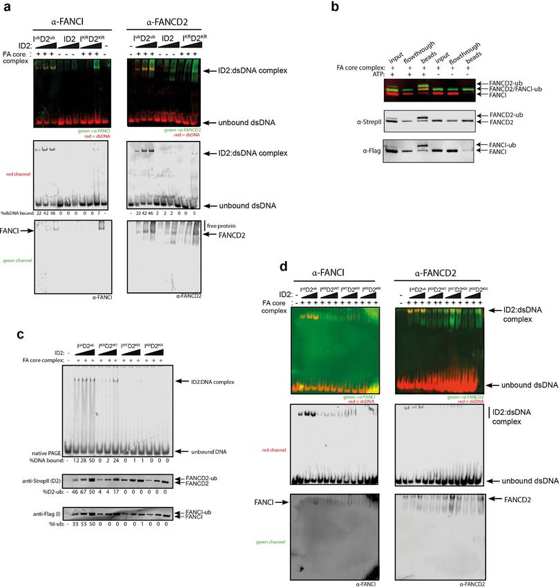
Research article                                                                                     Chromosomes and Gene Expression

                                    Table 1. List of proteins containing ubiquitin binding domain that are described or predicted to bind
                                    to ubiquitinated FANCD2.
                                    Protein             Function                        Domain                   Reference
                                    FAN1                Nuclease                        UBZ4                     (Kratz et al., 2010)
                                    SLX4                Nuclease                        UBZ1                     (Lachaud et al., 2014)
                                    FAAP20              FANCA partner                   UBZ                      (Hein et al., 2015)
                                    RAP80               BRCA1 partner                   UIM                      (Castillo et al., 2014)
                                    SMARCAD1            Chromatin remodeler             CUE                      (Densham et al., 2016)
                                    FANCJ               Helicase                        -                        (Raghunandan et al., 2015)
                                    PSMD4               Protease                        UIM                      (Jacquemont and Taniguchi, 2007)
                                    SF3B1               RNA binding protein             UBZ                      (Moriel-Carretero et al., 2017)
                                    TRIM25              E3 ligase                       RING finger              (Lossaint et al., 2013)
                                    MCM5                CMG component                   -                        (Lossaint et al., 2013)
                                    BRE                 BRCA1 partner                   -                        (Wang, 2007)
                                    BRCC                BRCA1 partner                   -                        (Wang, 2007)
                                    SNM1A               Nuclease                        UBZ                      (Yang et al., 2010)
                                    CtIP*               Nuclease activator              C2H2 zinc finger         (Murina et al., 2014)
                                    Rev1*               Translesion polymerase          UBZ3                     (Moldovan et al., 2010)
                                    *
                                    Indicates not tested in our experiments, because protein not produced in TnT system.

                                    recombinant ID2 or IUbD2Ub prepared by Avi-ubiquitin purification method (Tan et al., 2020a), we
                                    sought to directly compare the binding of this panel of ID2-associated proteins. Each of the partner
                                    proteins was expressed using reticulocyte extracts (Figure 1a), and the majority bound to the ID2
                                    complex as predicted based on previously identified associations (Figure 1b). The strongest binding
                                    proteins in terms of fraction of protein recovered were SLX4, FAAP20, SMARCAD, FANCJ, PSMD4,
                                    SF3B1, MCM5 and BRE. Although SMARCAD and SF3B1 appeared ‘sticky’ in control experiments,
                                    recovery of these proteins was still enriched by FANCD2:FANCI beads compared to background.
                                    Luciferase protein was used as a control 35S-labeled prey-protein. Surprisingly, our main observation
                                    was that none of the proteins showed any increased affinity for IUbD2Ub over ID2 (Figure 1d).

                                    Monoubiquitination clamps FANCI:FANCD2 on DNA
                                    An alternative explanation for the observed increase in association between ID2 and its associated
                                    proteins after DNA damage is that IUbD2Ub has an increased affinity for DNA, which brings the pro-
                                    tein into closer proximity to these partners. The majority of ID2 associated proteins are chromatin
                                    localized. In order to explore the stability of IUbD2Ub on DNA, we performed in vitro monoubiquiti-
                                    nation reactions in the presence of IR-dye700 labeled 60 bp double-stranded DNA (dsDNA). As pre-
                                    viously characterized (van Twest et al., 2017), we observed DNA-dependent appearance of
                                    monoubiquitinated forms of FANCD2 and FANCI when using recombinant FA core complex compo-
                                    nents (Figure 2a–b). ID2 monoubiquitination readily lead to DNA mobility shifts using EMSA (elec-
                                    tromobility shift assay) even at low concentrations, but this was not observed for the unmodified
                                    (apo)-ID2 complex in the absence of the enzymatically active FA core complex, or when monoubiqui-
                                    tination-defective K-to-R mutants of ID2 were used in the reaction (Figure 2c, lanes 2–4).
                                       Previously, we and others showed that various different dsDNA-containing structures could
                                    robustly stimulate ID2 monoubiquitination (Longerich et al., 2014; van Twest et al., 2017;
                                    Sareen et al., 2012), but that single-stranded DNA (ssDNA) does not. To determine if monoubiquiti-
                                    nated ID2 had increased affinity for other dsDNA containing structures, we repeated the monoubi-
                                    quitination reactions in the presence of different IR-dye700 labeled DNA structures. Interestingly,
                                    non-ubiquitinated ID2 also exhibited high affinity toward 3’ flap DNA structure (similar to a replica-
                                    tion fork stalled on the lagging strand), which has been previously observed (Liang et al., 2016).
                                    The 3’-Flap structure, and each of the other dsDNA containing structures, led to increased ID2
                                    monoubiquitination and increased retention of an EMSA shifted band (Figure 3). Conversely,

Tan et al. eLife 2020;9:e54128. DOI: https://doi.org/10.7554/eLife.54128                                                                      3 of 20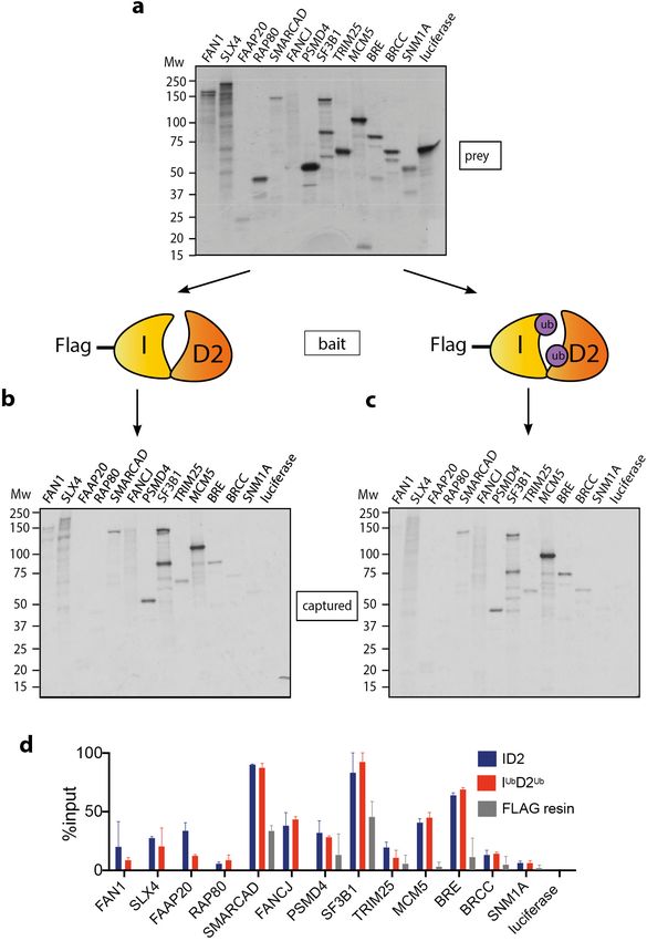
Research article                                                                                      Chromosomes and Gene Expression

                                    Figure 1. Mono-ubiquitination does not alter interaction of FANCI:FANCD2 with DNA repair proteins. (a) 35S-
                                    labelled FAN1, SLX4, FAAP20, RAP80, SMARCAD, FANCJ, PSMD4, SF3B1, TRIM25, MCM5, BRE, BRCC, SNM1A
                                    or luciferase (control) inputs were expressed using reticulocyte extracts. (b–c) The inputs prepared from (a) were
                                    incubated with the indicated FLAG-ID2 (b) or FLAG-IubD2ub (c) followed by FLAG pull-down and elution. The
                                    complexes were subjected to SDS-PAGE, and radiolabelled proteins were detected by autoradiography
                                    (representative experiment of n = 2). (d) Quantification showing percentage of ID2, IubD2ub or FLAG resin binding
                                    to inputs.

Tan et al. eLife 2020;9:e54128. DOI: https://doi.org/10.7554/eLife.54128                                                                        4 of 20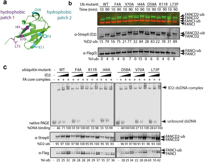
Research article                                                                                   Chromosomes and Gene Expression

                                    Figure 2. Monoubiquitination locks FANCI:FANCD2 on DNA. (a) Schematic of the electrophoretic mobility shift
                                    assay (EMSA) using IRDye-700 labeled dsDNA. (b) Coomassie stained SDS-PAGE gel showing monoubiquitination
                                    of FANCI:FANCD2 using recombinant FA core complex and IR-dye700 labeled dsDNA. 25, 50 and 100 nM of ID2
                                    or IKRD2KR were incubated with 25 nM of the IR-dye700 dsDNA for 90 min. The respective percentage of FANCI or
                                    FANCD2 monoubiquitination were calculated and shown under SDS-PAGE gel. (c) Monoubiquitination reactions
                                    from (b) were resolved on 6% native PAGE gel for EMSA analysis. The percentage of ID2 binding to DNA was
                                    calculated and shown under native PAGE gel.

                                    ssDNA, which stimulates monoubiquitination more slowly (van Twest et al., 2017) but to similar lev-
                                    els at the long time point of this assay, did not cause an EMSA shift.

                                    Figure 3. Monoubiquitinated FANCI:FANCD2 binds to any type of dsDNA. EMSA gels showing binding of
                                    monoubiquitinated or unmodified ID2 complex to different oligo-based DNA substrates. Above each panel, a
                                    schematic representing the tested DNA substrate is shown. 25, 50 and 100 nM of ID2 or IKRD2KR were incubated
                                    with 25 nM of the indicated DNA substrate and the protein:DNA complexes were resolved on 6% PAGE gels (top).
                                    The percentage of DNA binding was calculated and shown under each EMSA gel. Coomassie stained SDS-PAGE
                                    gel (bottom) showing the ubiquitination reactions used in the EMSA. The percentage of FANCD2
                                    monoubiquitination was calculated and shown under each SDS-PAGE gel.

Tan et al. eLife 2020;9:e54128. DOI: https://doi.org/10.7554/eLife.54128                                                                  5 of 20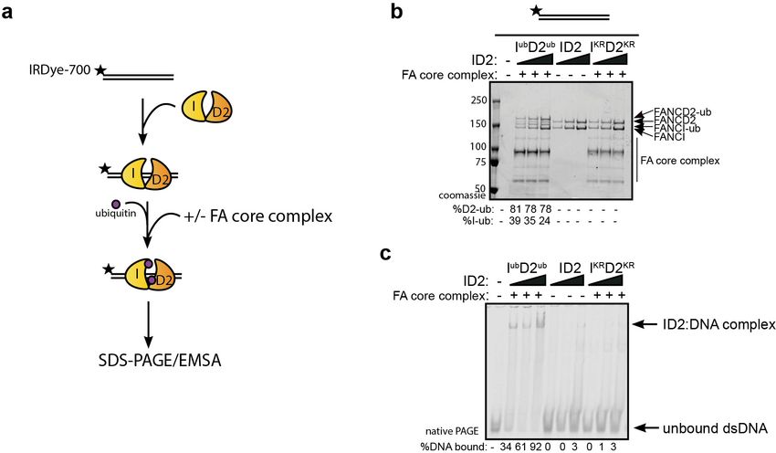
Research article                                                                                    Chromosomes and Gene Expression

                                    Both FANCIub and FANCD2ub are associated with a ‘clamped’ protein:
                                    DNA complex
                                    Previous studies reported that monoubiquitination of ID2 complex may lead to dissociation of the
                                    heterodimer to its individual subunits, as measured by loss of co-immunoprecipitation of FANCI with
                                    FANCD2 (40, 41). In contrast, we did not observe any Ub-mediated dissociation of ID2 in vitro. First,
                                    western blotting of the EMSA gels confirmed that the gel shifted DNA band contains both FANCI
                                    and FANCD2 proteins (Figure 4a). Second, FANCIUb still co-immunoprecipitated with FANCD2Ub at
                                    the plateau of the in vitro ubiquitination reaction (Figure 4b).

                                    Figure 4. FANCD2 monoubiquitination is sufficient for FANCI:FANCD2 locking to DNA, but stimulated by FANCI
                                    monoubiquitination. (a) Western blots of the EMSA gels containing 50, 100 and 200 nM of IubD2ub, ID2 or IKRD2KR
                                    in the presence of 25 nM IRDye-700 labeled dsDNA (red). Left panels correspond to anti-FANCI antibody (green)
                                    and right panels correspond to anti-FANCD2 antibody (green) (b) StrepII affinity purification of mono-
                                    ubiquitinated (+ATP) and non-ubiquitinated ID2 (-ATP). (c) EMSA gels (top) and western blots (bottom) showing
                                    the monoubiquitination of 25, 50 and 100 nM IWTD2WT, IKRD2WT, IWTD2KR or IKRD2KR in the presence of 25 nM
                                    IRDye-700 labeled dsDNA. (d) Western blots of the EMSA gels showing monoubiquitination of 50, 100 and 200 nM
                                    IWTD2WT, IKRD2WT, IWTD2KR or IKRD2KR in the presence of 25 nM IRDye-700 labeled dsDNA. FANCI (left, green)
                                    and FANCD2 (right, green) remained bound to IRDye-700 labeled DNA (red) after mono-ubiquitination.

Tan et al. eLife 2020;9:e54128. DOI: https://doi.org/10.7554/eLife.54128                                                                    6 of 20
Research article Chromosomes and Gene Expression
To determine the contribution of each of FANCD2Ub and FANCIUb to the clamping of IUbD2 Ub
complex to DNA, we used ubiquitination-deficient (KR) mutants in the ubiquitination reaction. FAN-
CIKR:FANCD2WT or FANCIWT:FANCD2KR mutant results in decrease in EMSA shift, and FANCIKR:
FANCD2KR did not bind to DNA (Figure 4c). However, this retention on DNA correlated with the
extent of FANCD2 monoubiquitination retained by these mutant complexes. Western blotting the
EMSA gels confirmed that both FANCD2 and FANCI are found in the EMSA shifted product,
although in higher amounts when both proteins are capable of being monoubiquitinated
(Figure 4d).
Mutant forms of ubiquitin can still clamp ID2 onto DNA
We postulated that the altered affinity for DNA induced by monoubiquitination must result from
either a conformational change in the ID2 heterodimer after monoubiquitination, or participation of
the conjugated ubiquitin directly in protein:DNA or protein:protein binding. To help distinguish
these possibilities, we utilized mutants of ubiquitin that have previously been shown to mediate the
known protein:ubiquitin or protein:DNA interactions in other ubiquitinated protein interactions
(Figure 5a; Husnjak and Dikic, 2012). Each of these Ub mutants were conjugated to ID2 by the FA
core complex with similar efficiency (Figure 5b) and their clamping onto DNA was then measured.
Mutations in surface patch 1 (F4A, D58A), surface patch 2 (I44A, V70A), a DNA binding residue
(K11R) or a tail mutant (L73P) had no apparent effect on DNA clamping (Figure 5c). This result
Figure 5. Mutations in different ubiquitin patches do not affect ID2 mono-ubiquitination or DNA binding. (a)
Crystal structure of ubiquitin with ubiquitin mutant sites depicted (PDB: 1UBQ). Hydrophobic binding pockets are
indicated in blue and pink. (b) Western blots showing the time course ubiquitination assays of ID2 using wild-type
ubiquitin or ubiquitin F4A, V70A, I44A, D58A, K11R and L73P mutants. (c) EMSA gels showing 25, 50 and 100 nM
monoubiquitinated ID2 binding to 25 nM IRDye-700 dsDNA using various ubiquitin mutants (top). Western blots
of ID2 ubiquitination products were shown at the bottom and the percentage of FANCI and FANCD2
ubiquitination were shown at the bottom of each western blot panel.
Tan et al. eLife 2020;9:e54128. DOI: https://doi.org/10.7554/eLife.54128 7 of 20Research article Chromosomes and Gene Expression
suggests that no canonical surface or region of ubiquitin is critical for DNA clamping of ID2, and
instead ubiquitin conjugation to ID2 probably induces a conformational rearrangement of the
heterodimer.
Purification of monoubiquitinated FANCI:FANCD2 complex bound to
dsDNA reveals a filamentous architecture
In order to examine the architecture of purified recombinant IUbD2Ub complex in the presence of
dsDNA plasmid, we utilized a recombinant Avi-tag ubiquitin construct containing a 3C protease site
between the biotinylated Avi-tag and the N-terminus of ubiquitin (Tan et al., 2020a; Figure 6a).
This tagged ubiquitin is incorporated onto FANCI:FANCD2 by the FA core complex, allowing Avi-
din-Sepharose purification of monoubiquitinated ID2 that is then eluted by 3C protease cleavage.
We recovered monoubiquitinated FANCI:FANCD2 complex only when FANCI is monoubiquitinated,
suggesting that the N-terminus of D2-attached ubiquitin may be buried within the di-ubiquitinated
Figure 6. Mono-ubiquitinated FANCI:FANCD2 complex assemble into filament-like arrays. (a) Schematic of
purification of monoubiquitinated FANCI:FANCD2 using Avi-ubiquitin. (b) Coomassie stained SDS-PAGE gel (top)
and western blots (bottom) showing the purification of monoubiquitinated FANCI:FANCD2 complex eluted using
PreScission protease (lanes 1–7) compared to without PreScission protease (lanes 8–14). (c–e) Representative
negative-stained EM image of purified FANCIub:FANCD2ub complex bound to 2.7 kb plasmid, unmodified FANCI:
FANCD2 incubated with 2.7 kb plasmid and unmodified FANCI:FANCD2 complex. Pseudo-colored regions are
shown to highlight particular filament-like arrays in FANCIub:FANCD2ub but not other samples.
Tan et al. eLife 2020;9:e54128. DOI: https://doi.org/10.7554/eLife.54128 8 of 20Research article Chromosomes and Gene Expression
complex, but the N-terminus of ubiquitin attached to FANCI is accessible for avidin binding
(Figure 6b).
Using this purified protein, we compared FANCIub:FANCD2ub to unmodified FANCI:FANCD2
using electron microscopy (EM). Surprisingly, we observed that FANCIub:FANCD2ub forms filament-
like arrays when bound to dsDNA plasmid (Figure 6c). Such arrays were not observed in the unmod-
ified FANCI:FANCD2 protein preparation in the absence of presence of plasmid DNA, nor in previ-
ous investigations of human or Xenopus FANCI:FANCD2 complexes studied by EM (Figure 6d–e
and Swuec et al., 2017; Liang et al., 2016; Lopez-Martinez et al., 2019).
When smaller DNA molecules were used as the substrate for ID2 binding, we either observed no
filament-like structures (60 bp, Figure 7a) or shorter filament-like structures (150 bp, Figure 7b)
compared to structures that were on average 7-8x longer than the characteristic double saxophone
structure of ID2 heterodimer in the non-ubiquitinated state (Figure 7c).
Figure 7. Monoubiquitinated FANCI:FANCD2 assembles into filamentous arrays along the length of dsDNA. (a–
d) Representative EM image of monoubiquitinated FANCI:FANCD2 bound to (a) 60 bp dsDNA, (b) 150 bp
dsDNA, (c) 2.7 kb dsDNA, and (d) 2.7 kb dsDNA and Benzonase-treated. Scale bar, 100 nm. Arrows indicate
formation of 1–2 ID2 array; boxes indicate multiple ID2 arrays. (e) Representative 2D class average of ID2. Views of
the side and top of ID2 are shown, framed in blue for comparison. (f) Representative 2D class average of IubD2ub
bound to 60 bp DNA. Views of the side and top of IubD2ub are shown, framed in red for comparison. (g–h)
Example comparison of the length (y) and width (x) of class average images ID2 and IubD2ub (likely an
overestimate, because uranyl formate staining increases apparent particle size).
Tan et al. eLife 2020;9:e54128. DOI: https://doi.org/10.7554/eLife.54128 9 of 20Research article Chromosomes and Gene Expression
The observation that array length correlated with the size of DNA available for ID2 binding
strongly suggested that the association between heterodimer subunits in the array was DNA-medi-
ated. To test whether the array of IubD2ub is also dependent upon binding to the same DNA mole-
cule, we examined the plasmid-stimulated ubiquitination reaction products after treatment with the
non-specific endonuclease, Benzonase. It is apparent from EM images that addition of Benzonase
breaks the long arrays formed by IubD2ub complex into very short or heterodimer-sized units
(Figure 7d). This finding is consistent with Benzonase cleaving exposed DNA between IubD2ub units,
leading to destabilization of the filamentous arrays. Together our results show that, in vitro, ubiquiti-
nation of ID2 leads to a ubiquitin- and DNA- stabilized filament-like structure.
Single IubD2ub heterodimers on short 60 bp DNA have an altered
architecture
Due to variability in the length and shape of filament-like IubD2ub structures on longer DNA mole-
cules we have not been able to uncover the shape or subunit rearrangement of the individual units
of the arrays because attempts to class average arrays failed due to a lack of order. However, exami-
nation of IubD2ub purified together with short 60 bp DNA allowed us to collect sufficient images of
individual particles for analysis. These particles were similar in size to non-ubiquitinated ID2, but it is
clear from individual molecule and class average views that the IubD2ub complex forms a distinct
architecture from that of ID2 (Figure 7e–f). In particular, the overall shape of individual particles and
their class averages reveal a twisting that repositions the solenoid arms of one or both of the subu-
nits bringing them into closer proximity. The conformational change induced appears to reduce the
size of ID2 in one direction (x vs y) but not the other (Figure 7g–h), similar to that predicted in a pre-
viously proposed model that placed DNA in a channel between FANCI and FANCD2 post DNA
binding (Longerich et al., 2014). These images support the view that monoubiquitination induces a
conformational change in the ID2 complex that clamps it upon DNA.
Discussion
The protection of stalled forks by DNA repair factors is essential for proper DNA replication and the
maintenance of genome stability. The primary mechanism of replication fork stabilization at inter-
strand crosslinks, and other replication blocking damage, utilizes the proteins of the FA-BRCA DNA
repair pathway. Monoubiquitination of FANCI:FANCD2 by the FA core complex is the central event
in this pathway. Here, we showed that monoubiquitination directly clamps the ID2 complex onto
double-stranded DNA, and promotes a filament-like coating of long DNA molecules. This finding
answers a long-standing question about the nature of the biochemical reaction that is absent in the
majority of patients with FA (Wang and Smogorzewska, 2015; Gregory et al., 2003).
‘Fork protection’ involves (i) exclusion of cellular nucleases such as MRE11 and DNA2 from the
stalled DNA replication fork, and (ii) specific recruitment of other factors that are able to restart
DNA replication (Schlacher et al., 2012; Tian et al., 2017; Madireddy et al., 2016). The role of
nuclease exclusion seems most important because inhibition of either of two nucleases, MRE11 or
DNA2, can significantly alleviate the sensitivity of FA-BRCA mutant cells to replication stalling agents
(Schlacher et al., 2012; Tian et al., 2017). But in many studies, specific association of various repair
factors with IUbD2Ub compared to ID2 have been described. These proteins mostly contain ubiquitin-
binding domains, providing an impetus for the recruitment-based hypothesis (summary and referen-
ces in Table 1). However, because these previous studies focused on use of ubiquitination-deficient
mutants, they could not address the underlying question of whether ubiquitin on either FANCI,
FANCD2 or both proteins directly mediated the interaction. Now, we have shown that none of these
proteins specifically bind recombinant purified IUbD2Ub compared to ID2. This finding was unex-
pected, and included proteins such as SLX4 (FANCP), MCM5, and FAN1, which have a demon-
strated and essential role in the FA-pathway (Walden and Deans, 2014; Lossaint et al., 2013;
MacKay et al., 2010).
Instead, our data provide direct evidence that ID2 undergoes a conformational change after
monoubiquitination, that leads to it becoming clamped on double-stranded DNA. It is likely that in
previous cell-based experiments specific interaction between SLX4 and FAN1 with FANCD2 but not
FANCD2K561R was observed because FANCD2K561R does not become clamped on DNA, even in the
presence of active FA core complex. This finding is supported by the observation that FANCD2K561R
Tan et al. eLife 2020;9:e54128. DOI: https://doi.org/10.7554/eLife.54128 10 of 20Research article Chromosomes and Gene Expression
also does not becomes chromatin localized at damage sites even after extensive DNA damage
(Matsushita et al., 2005). We propose that IUbD2Ub does not demonstrate restricted interaction
with any specific protein partner. None-the-less, its retention in chromatin after ubiquitination would
be much more likely to bring the complex into proximity of these other DNA repair factors, where it
could still influence their activity.
Clamping onto DNA occurs through a ubiquitin-mediated conformational change in the ID2 com-
plex. A ubiquitin binding-domain (UBD) in FANCD2 has previously been shown necessary for the
retention of the protein in the chromatin faction, and for strong binding to FANCI (Rego et al.,
2012). This UBD domain sits in the FANCD2 structure opposite to where ubiquitin is likely to reside
after its conjugation on to FANCI by the FA core complex, and most likely mediates the clamping
function and conformational rearrangement. A high-resolution cryo-EM structure of chicken ID2Ub
published by Alcón et al alongside our study (Alcón et al., 2020), also uncovered a conformational
rearrangement of ID2 that is then stapled in place by ubiquitin:UBD association. Other DNA-binding
proteins such as histone H2A show an increased association with DNA after monoubiquitination
(Zhang, 2003) and monoubiquitination also increases the DNA occupancy of transcription factors
such as FOXO4 and CIITA (Greer et al., 2003). It is possible that ubiquitin to UBD mediated clamp-
ing is a general mechanism of protein:DNA target stabilization.
IUbD2Ub clamped in nucleoprotein arrays
In addition to a conformational change in ID2 induced by monoubiquitination (that has also been
concurrently discovered and reported by the Pavletich, Walden and Passmore labs Alcón et al.,
2020; Wang et al., 2020; Rennie et al., 2020) we found that monoubiquitinated IUbD2Ub formed
large filament-like arrays when it was purified together with plasmid DNA, but not short 60 bp DNA
fragments. Fourier Transformation of the EM images did not reveal clear evidence for layer lines,
that are expected for fiber diffraction, so we expect that the IUbD2Ub is not a true filament. On aver-
age, the length of plasmid-associated structures is 7-8x (but up to 40x) that of that associated with
60 bp DNA. Larger or longer arrays may potentially be obscured from view because the purification
strategy makes elution exponentially more difficult with increasing numbers of conjugated ubiquitin-
molecules. Steps to remove ‘aggregates’ may have also inadvertently removed larger arrays. How-
ever, as the number of potential plasmid DNA binding sites for ID2 was in large excess the concen-
tration of ID2 used to stimulate reaction, there appears to be some purpose to creation of these
filamentous arrays. The modular nature of IUbD2Ub arrays suggests that IUbD2Ub binding to DNA is
flexible and can adopt multiple conformations, akin to RPA binding and protecting ssDNA
(Yates et al., 2018).
There is evidence that IUbD2Ub clamped in nucleoprotein filaments exist in cells. Antibodies
against FANCD2 have long been used as a marker of double strand breaks, stalled replication forks
and R-loops because the protein forms large, intensely stained foci during S-phase that are
increased after treatment with DNA damaging agents (Taniguchi et al., 2002; Schwab et al., 2015;
Deans and West, 2009). We suspect that these intense foci are due to coating of DNA around dam-
aged forks, potentially in filamentous arrays similar to those we observed by EM. Support for the
large size and extent of DNA binding reflective of filamentous arrays also comes from chromatin
immunoprecipitation and sequencing (ChIP-Seq) using anti-FANCD2 (13). FANCD2, and two other
damage markers MRE11 and gH2AX, showed no specific localization in a bulk population of cells,
but strongly localized adjacent to a Cas9-induced site-specific DNA break. Both gH2AX and
FANCD2 produced a broad peak centered at the target site kilobases (kb) to megabases (mb) in
length. In contrast, MRE11 is located within a very tight peak within ~100 bp of the break. Chromatin
within 1–2 kb of the DSB showed reduced occupancy by gH2AX, consistent with dechromatinization
around break sites (Iacovoni et al., 2010), but FANCD2 was present right up to the DSB. Accumula-
tion of FANCD2 increases at the DSB early after cleavage, and accumulates more distant from the
DSB progressively with time post-cleavage. This is suggestive of a polymerization of the FANCD2
signal away from the break site, as we hypothesize would occur for a protein that forms a growing
array of molecules at broken DNA (Wienert et al., 2019). The conserved function of FANCD2 as a
histone chaperone (Sato et al., 2012; Higgs et al., 2018) may even be directly linked to displace-
ment of nucleosomes as filamentous arrays extend into break-adjacent chromatin.
In this study, we also observed direct association of two ID2 heterodimers by co-immunopurifica-
tion only after the protein becomes monoubiquitinated. This approach, if performed in cells, could
Tan et al. eLife 2020;9:e54128. DOI: https://doi.org/10.7554/eLife.54128 11 of 20Research article Chromosomes and Gene Expression
be used to further delineate the mechanism and cellular factors required for the extension of IUb-
D2Ub arrays during fork protection. Of particular interest will be determining the role of BRCA1 in
clamping and/or array extension. BRCA1:BARD1 was initially thought to be the E3 for FANCD2
monoubiquitination, because it co-immunoprecipitates FANCD2, and FANCD2 does not form
nuclear foci after damage in BRCA1-deficient cells (Raghunandan et al., 2015). However, in various
assays it was later shown that FANCD2 monoubiquitination does occur in BRCA1-deficient cells, but
it is uncoupled from FANCD2 foci formation (Jacquemont and Taniguchi, 2007; Moriel-
Carretero et al., 2017).
How would a clamped IUbD2Ub array mediate fork protection?
Filamentous structures on DNA play a genome protective role in prokaryotes: eg DAN protein forms
a rigid collaborative filament that reduces accessibility during anoxia (Lim et al., 2013), while the
Vibrio cholera protein ParA2 forms protective filamentous structures on DNA during segregation
(Hui et al., 2010). Structural characterization has demonstrated how these filaments function and, in
the case of ParA2, can be targeted therapeutically (Misra et al., 2018). The coating of ssDNA by
RPA in eukaryotes, also protect DNA from the activity of nucleases, and directs the specific activity
of others (de Laat et al., 1998; Chen et al., 2013; Nguyen et al., 2017). We propose that a FAN-
CIub:D2ub arrays may have a similar stabilizing role on newly synthesized dsDNA at a stalled replica-
tion fork. This property would explain why stalled forks are prone to degradation in FA and BRCA
patient cells (Schlacher et al., 2012; Tian et al., 2017). In particular, we hypothesize that filamentous
DNA-clamped Iub:D2ub could prevent access to DNA by MRE11 and DNA2 nucleases and prevent
aberrant ligation of broken DNA to other parts of the genome by non-homologous end-joining.
Second, the tight binding of FANCIub:D2ub to dsDNA, when localized to stalled replication forks,
may also prevent the branch migration of replication forks and inhibit their spontaneous or helicase-
mediated reversal (Neelsen and Lopes, 2015). Reversed forks are the substrate for degradation by
DNA2 and WRN nuclease activities, providing a hypothetical link between the activities of FANCD2-
monoubiquitination and the nuclease activity of DNA2 and WRN (Thangavel et al., 2015;
Sidorova et al., 2013).
Third, Iub:D2ub arrays may also locally suppress non-homologous end-joining (NHEJ) factors, and/
or delineate the newly synthesized chromatin from unreplicated regions during the promotion of
templated repair processes such as homologous recombination. FANCD2, FANCI, and components
of the FA core complex were identified amongst relatively few other factors, in a genome-wide
screen for genes that promote templated repair over NHEJ (Richardson et al., 2017). Stabilization
of RAD51 filaments, required for HR, is also an in vitro property of ID2 (Sato et al., 2016), suggest-
ing Iub:D2ub filamentous arrays may exist adjacent to or coincident with RAD51 filaments in cells, in
order to provide a polarity to the homologous recombination reaction without loss or gain of geno-
mic sequences.
Role of FANCI-monoubiquitination
Fanci and Fancd2 have common and distinct functions in mouse models of Fanconi anemia
(Dubois et al., 2019), while the double knockout of FANCI and FANCD2 has an unexpectedly dis-
tinct phenotype compared to single knockouts in human cells (Thompson et al., 2017). But FAN-
CIK523R expressing cells are less sensitive to DNA damage than FANCI knockout in human
cells (Smogorzewska et al., 2007), so what is the role of FANCI monoubiquitination? Previous stud-
ies demonstrated that FANCI monoubiquitination is always subsequent to FANCD2 monoubiquitina-
tion, both in cells (Sareen et al., 2012) and in biochemical assays (van Twest et al., 2017). FANCI
also likely plays a role in recruiting the FA core complex to the substrate (Castella et al., 2015). In
this study, we show that FANCI-monoubiquitination is not necessary for clamping of the ID2 com-
plex onto DNA (Figure 4). However, in vivo it is likely that FANCI monoubiquitination plays a critical
role in regulating deubiquitination of the ID2 complex. FANCI recruits the deubiquitinating enzyme
USP1:UAF1 (Yang et al., 2011), which prevents trapping of monoubiquitinated FANCD2 at non-pro-
ductive DNA damage sites, but only ID2Uband not IUbD2Ub is a substrate (van Twest et al., 2017). It
is also clear from our EM investigations that FANCI must play an important role in the structural
integrity of IUbD2Ub filamentous arrays on DNA, possibly creating an asymmetry necessary for a spe-
cific polarity to array assembly.
Tan et al. eLife 2020;9:e54128. DOI: https://doi.org/10.7554/eLife.54128 12 of 20Research article Chromosomes and Gene Expression
Implications for understanding the deficiency of Fanconi anemia
Onset of progressive bone marrow failure occurs at a median age of 7 in children with FA
(Butturini et al., 1994). Almost all these patients lack FANCD2 and FANCI monoubiquitination, due
to mutation in either FANCD2 or FANCI or one of the nine other FANC proteins required for their
monoubiquitination (Walden and Deans, 2014). The importance of the monoubiquitin signal is
highlighted by the observation that up to 20% of patients acquire somatic reversion of the inherited
mutation in a fraction of blood cells (Soulier et al., 2005). These mutations restore monoubiquitina-
tion and prevent bone marrow failure. Our work suggests two potential strategies for treatment of
FA: restoration of gene function, such as that which occurs in somatic revertants or, identification of
novel mechanisms to stabilize an ID2:DNA-clamped complex for fork protection by ubiquitin-medi-
ated or innovative means. New small molecule activators or inhibitors of ID2:DNA clamping could
be therapeutics in FA or cancer-treatment. In vitro biochemistry has proven to be the most powerful
tool in uncovering new functions of FANCD2-monoubiquination that had gone undiscovered for
nearly 20 years. The approach is likely to be formidable in drugging the FA pathway in future
studies.
Materials and methods
Key resources table
Reagent type
(species) or resource Designation Source or reference Identifiers Additional information
Recombinant pFastbac1- (Klein Douwel et al., 2014) Gift from Puck
DNA reagent FLAG-xFANCI Knipscheer
(X. laevis)
Recombinant pFastbac1- (Klein Douwel et al., 2014) Gift from Puck
DNA reagent StrepII-xFANCD2 Knipscheer
(X. laevis)
Recombinant pFL-EGFP- (Tan et al., 2020a)
DNA reagent His-hFANCI
(H. sapiens)
Recombinant pFastbac1- (Tan et al., 2020a) RRID:Addgene_ 134904 Gift from Angelos
DNA reagent FLAG-hFANCD2opt Constantinou
(H. sapiens)
Recombinant pFL/pSPL-EGFP- (van Twest et al., 2017) Codon optimized
DNA reagent FLAG-B-L-100 FANCB
(H. sapiens)
Recombinant pFL-MBP-C-E-F (van Twest et al., 2017)
DNA reagent
(H. sapiens)
Recombinant pGEX-KG-GST- (van Twest et al., 2017) Codon optimized
DNA reagent UBE2T
(H. sapiens)
Recombinant pet16b-Avi- (Tan et al., 2020a) RRID:Addgene_134897
DNA reagent ubiquitin_
(E. coli) rbs_BirA
Recombinant pSRK2706-GST- (Raran-Kurussi and Waugh, 2016) RRID:Addgene_78571 A gift from
DNA reagent HRV-3Cprotease David Waugh
(E. coli)
Recombinant pUC19 plasmid New N3041S
DNA reagent England BioLabs
(E. coli)
Strain, strain BL21 (DE3) Agilent Technologies 200131
background
(E. coli)
Cell line Sf9 Thermo RRID:CVCL_0549 Maintained in
(Spodoptera Fisher Scientific Sf-900 II SFM
frugiperda)
Continued on next page
Tan et al. eLife 2020;9:e54128. DOI: https://doi.org/10.7554/eLife.54128 13 of 20Research article Chromosomes and Gene Expression
Continued
Reagent type
(species) or resource Designation Source or reference Identifiers Additional information
Cell line High Five Thermo RRID:CVCL_C190 Maintained in
(Trichoplusia ni) Fisher Scientific Sf-900 II SFM
Antibody Rabbit polyclonal Abcam RRID:AB_76949 one in 3000
antibodies against dilution
StrepII
Antibody Rabbit polyclonal Abcam RRID:AB_74332 one in 3000
antibodies against dilution
FANCI
Antibody Rabbit polyclonal Abcam RRID:AB_10862535 one in 3000
antibodies against dilution
FANCD2
Antibody Mouse monoclonal Aviva Biosciences RRID:AB_10884242 one in 3000
antibodies against dilution
FLAG
Peptide, FLAG peptide Sigma-Aldrich F3290
recombinant protein
Peptide, Recombinant Human Boston Biochem E-304–050
recombinant protein His6-Ubiquitin E1
Enzyme carrier free
Peptide, Ubiquitin and Boston Biochem U-110H, UM-I44A,
recombinant protein associated mutant UM-D58A, UM-F4A,
variants UM-L73P, UM-K11R
Commercial TNT T7 Quick Promega Corporation L1170
assay or kit Coupled
Transcription/
Translation
System
Commercial Anti-FLAG-M2 Sigma Aldrich RRID:AB_10063035
assay or kit affinity gel
Chemical EasyTagL-[35S]- PerkinElmer NEG709A500UC
compound, drug Methionine Life Sciences
Software, XMIPP (de la Rosa-Trevı́n et al., 2013)
algorithm
Protein purification
Flag-FANCI and StrepII-FANCD2 were expressed using the pFastBac1 vector (Life Technologies).
For FANCI:FANCD2 complex, Hi5 cell pellets were resuspended in lysis buffer (50 mM Tris-HCl pH
8.0, 0.1 M NaCl, 1 mM EDTA, 10% glycerol and 1X mammalian protease inhibitor), and sonicated.
Lysates were clarified by centrifugation at 20,000 g and the supernatants were incubated with M2
anti-FLAG agarose resin for 2 hr. The resin was washed 5 5 min incubation with wash buffer (20
mM Tris-HCl pH 8.0, 0.1 M NaCl, 10% glycerol), and the protein was eluted in the same buffer con-
taining 100 mg/mL FLAG peptide. GST-UBE2T, Flag-BL100, MBP-CEF were purified as described
(van Twest et al., 2017). Ubiquitin and His-UBE1 were purchased from Boston Biochem.
Biotinylated-Avi-ubiquitin purification
His-Avi-ubiquitin was purified as described in Tan et al. (2020a).
In vitro ubiquitination assay
Standard ubiquitination reactions contained 10 mM recombinant human avidin-biotin-ubiquitin, 50
nM human recombinant UBE1, 100 nM UBE2T, 100 nM PUC19 plasmid, 2 mM ATP, 100 nM FANCI:
FANCD2 complex wild type (WT) or ubiquitination-deficient (KR), in reaction buffer (50 mM Tris-HCl
pH 7.4, 2.5 mM MgCl2, 150 mM NaCl, 0.01% Triton X-100). 20 mL reactions were set up on ice and
incubated at 25˚C for 90 min. Reactions were stopped by adding 10 mL NuPage LDS sample buffer
and heated at 80˚C for 5 min. Reactions were loaded onto 4–12% SDS PAGE and run using NuPAGE
Tan et al. eLife 2020;9:e54128. DOI: https://doi.org/10.7554/eLife.54128 14 of 20Research article Chromosomes and Gene Expression
MOPS buffer and assessed by western blot analysis using Flag (Aviva Biosciences) or StrepII (Abcam)
antibody.
In vitro transcription/translation pull down of 35S-labeled proteins
Flag-tagged FANCI:FANCD2 and monoubiquitinated FANCI:FANCD2 was prepared by incubating
purified FANCI:FANCD2 or monoubiquitinated FANCI:FANCD2 on Flag beads for 2 hr followed by
extensive washes in buffer A (20 mM TEA pH 8.0, 150 mM NaCl, 10% glycerol). 35S-labeled proteins
containing UBZ or other ubiquitin domains (Table 1) were generated using the TNT Quick Coupled
T7 Transcription/Translation System (Promega) and 35S-labeled methionine (Perkin Elmer). 10 mL of
TNT product was incubated for 4 hr at 4˚C in buffer A with 100 ng Flag-tagged FANCI:FANCD2 or
monoubiquitinated FANCI:FANCD2, 20 mL of Flag-beads (Sigma-Aldrich) in a 100 mL reaction. Beads
were washed five times with buffer A and resuspended in LDS loading buffer. Proteins were sepa-
rated by SDS-PAGE and visualized by autoradiography.
Electrophoretic mobility shift assay
Oligonucleotides used to create fluorescently labeled DNA were IRDYE-700-labeled X0m1 (IDTDNA)
and other oligos with the sequences shown in Supplementary file 1. Assembly of the different DNA
structures was performed exactly as previously described (Supplementary file 1; van Twest et al.,
2017). 25 nM DNA substrates were incubated in 20 mL ubiquitination buffer containing 100 nM
FANCI:FANCD2, 100 nM BL100, 100 nM CEF, 10 uM HA-ubiquitin (Boston Biochem), 50 nM UBE1
(Boston Biochem) and 100 nM UBE2T at room temperature for 90 min to initiate ubiquitination. The
reaction was resolved by electrophoresis through a 6% non-denaturing polyacrylamide gel in TBE
(100 mM Tris, 90 mM boric acid, 1 mM EDTA) buffer and visualized by Licor Odyssey system.
Purification of monoubiquitinated FANCI:FANCD2 complex
Di-monoubiquitinated FANCI:FANCD2 complex was purified as described (Tan et al., 2020a). DNA
molecules of 60 bp or 150 bp (dsDNA from oligonucleotides) or 2.6 kb (circular plasmid DNA) were
used to stimulate the reaction for different experiments, as indicated.
Mass spectrometry analysis of monoubiquitinated FANCI:FANCD2
complex
Gels containing monoubiquitinated FANCI and FANCD2 bands were excised and in-gel digested
with trypsin and subjected to LC/MS analysis on ESI-FTICR mass spectrometer at Bio21, The Univer-
sity of Melbourne. The analysis program MASCOT was used to identify ubiquitination sites on FANCI
and FANCD2.
Negative stain electron microscopy
Freshly purified monoubiquitinated or non-ubiquitinated FANCI:FANCD2 complex was applied to
glow-discharged, carbon/formvar grids and allowed to adsorb for 60 s. Specimen was then stained
with 2% uranyl formate for 60 s. Specimen were imaged at a magnification of 73,000 x on a Ceta
camera (corresponding to a pixel size of 1.9 Å) in Talos 120 kV. For FANCI:FANCD2 complex, 20
micrographs were analyzed and 4553 particles were picked for 2D classification. For monoubiquiti-
nated FANCI:FANCD2 complex, 20 micrographs were analyzed and 4698 particles were picked for
2D classification. The length and width of 2D class average were measured using ImageJ.
Single-particle image processing
Monoubiquitinated or non-ubiquitinated FANCI:FANCD2 particles were semi-automatically picked
using XMIPP3 (83). The parameters of the contrast transfer function (CTF) for negative stained data
was estimated on each micrograph using CTFFIND3 (Mindell and Grigorieff, 2003). Finally, refer-
ence free 2D alignment and averaging were executed using XMIPP3.
Acknowledgements
We are grateful to Alessandro Costa and Paolo Swuec for discussions and suggestions for analysis of
the EM data. We thank Puck Knipscheer, Steve West, Johan de Winter, KJ Patel, Paul Hasty, Dario
Tan et al. eLife 2020;9:e54128. DOI: https://doi.org/10.7554/eLife.54128 15 of 20Research article Chromosomes and Gene Expression
Alessi, Timothy Richmond, Beverlee Buzon, Andrew Blackford, Steve Jackson and Stephen Elledge
for reagents. We thank Eric Hanssen from the Electron Microscopy facility, and Nick Shuai from the
Mass Spectrometry facility, at Bio21 Institute, University of Melbourne. WT was supported by an
Australian Government Research Training Program postgraduate scholarship. AJD is a Victorian Can-
cer Agency fellow. WC is an NHMRC career development fellow and Maddie Riewoldt’s vision fellow
(WC-MRV2016). MWP is an NHMRC Australia Senior Research Fellow. This work was funded by
grants from the Fanconi Anemia Research Fund (to AJD and WC), Maddie Riewoldt’s Vision (SVI-
MRV2017G to WC), the National Health and Medical Research Council (GNT1123100 and
GNT1181110 to AJD and GNT1156343 to WC), and the Victorian Government’s OIS Program.
Additional information
Funding
Funder Grant reference number Author
Fanconi Anemia Research Wayne Crismani
Fund Andrew J Deans
Maddie Riewoldt’s Vision SVI-MRV2017G Wayne Crismani
Victorian Government OIS Program Winnie Tan
Sylvie van Twest
Rohan Bythell-Douglas
Vincent J Murphy
Michael Sharp
Michael W Parker
Wayne Crismani
Andrew J Deans
Victorian Cancer Agency Andrew J Deans
National Health and Medical GNT1123100 Andrew J Deans
Research Council
National Health and Medical GNT1156343 Wayne Crismani
Research Council
National Health and Medical GNT1117183 Michael W Parker
Research Council
National Breast Cancer Foun- IIRS-19-017 Rohan Bythell-Douglas
dation Andrew J Deans
Australian Government Re- Winnie Tan
search Training Program
National Health and Medical GNT1129757 Wayne Crismani
Research Council
National Health and Medical GNT1181110 Andrew J Deans
Research Council
Maddie Riewoldt’s Vision MRV2016 Wayne Crismani
The funders had no role in study design, data collection and interpretation, or the
decision to submit the work for publication.
Author contributions
Winnie Tan, Conceptualization, Formal analysis, Validation, Investigation, Visualization, Methodol-
ogy, Writing - original draft, Writing - review and editing; Sylvie van Twest, Investigation, Methodol-
ogy; Andrew Leis, Resources, Software, Supervision, Methodology; Rohan Bythell-Douglas, Formal
analysis, Supervision, Investigation, Methodology; Vincent J Murphy, Michael Sharp, Resources,
Methodology; Michael W Parker, Conceptualization, Resources, Supervision, Writing - review and
editing; Wayne Crismani, Conceptualization, Resources, Formal analysis, Supervision, Funding acqui-
sition, Project administration, Writing - review and editing; Andrew J Deans, Conceptualization,
Resources, Formal analysis, Supervision, Funding acquisition, Investigation, Visualization, Methodol-
ogy, Writing - original draft, Project administration, Writing - review and editing
Tan et al. eLife 2020;9:e54128. DOI: https://doi.org/10.7554/eLife.54128 16 of 20Research article Chromosomes and Gene Expression
Author ORCIDs
Winnie Tan https://orcid.org/0000-0003-3229-4157
Sylvie van Twest https://orcid.org/0000-0001-5602-0906
Andrew Leis https://orcid.org/0000-0003-4905-9401
Rohan Bythell-Douglas https://orcid.org/0000-0002-3823-8749
Michael Sharp http://orcid.org/0000-0002-1019-3729
Michael W Parker http://orcid.org/0000-0002-3101-1138
Wayne Crismani https://orcid.org/0000-0003-0143-8293
Andrew J Deans https://orcid.org/0000-0002-5271-4422
Decision letter and Author response
Decision letter https://doi.org/10.7554/eLife.54128.sa1
Author response https://doi.org/10.7554/eLife.54128.sa2
Additional files
Supplementary files
. Supplementary file 1. (A) DNA oligonucleotides used in this study. The following oligonucleotides
were ordered from IDTDNA. (B) Combination of oligonucleotides annealed to generate DNA sub-
strates used in this study.
. Transparent reporting form
Data availability
All data generated or analysed during this study are included in the manuscript and supporting files.
References
Alcón P, Shakeel S, Chen ZA, Rappsilber J, Patel KJ, Passmore LA. 2020. FANCD2-FANCI is a clamp stabilized
on DNA by monoubiquitination of FANCD2 during DNA repair. Nature Structural & Molecular Biology 27:240–
248. DOI: https://doi.org/10.1038/s41594-020-0380-1, PMID: 32066963
Butturini A, Gale RP, Verlander PC, Adler-Brecher B, Gillio AP, Auerbach AD. 1994. Hematologic abnormalities
in fanconi Anemia: an international fanconi Anemia registry study. Blood 84:1650–1655. DOI: https://doi.org/
10.1182/blood.V84.5.1650.1650, PMID: 8068955
Castella M, Jacquemont C, Thompson EL, Yeo JE, Cheung RS, Huang JW, Sobeck A, Hendrickson EA, Taniguchi
T. 2015. FANCI regulates recruitment of the FA core complex at sites of DNA damage independently of
FANCD2. PLOS Genetics 11:e1005563. DOI: https://doi.org/10.1371/journal.pgen.1005563, PMID: 26430909
Castillo A, Paul A, Sun B, Huang TH, Wang Y, Yazinski SA, Tyler J, Li L, You MJ, Zou L, Yao J, Wang B. 2014. The
BRCA1-interacting protein Abraxas is required for genomic stability and tumor suppression. Cell Reports 8:
807–817. DOI: https://doi.org/10.1016/j.celrep.2014.06.050, PMID: 25066119
Chen H, Lisby M, Symington LS. 2013. RPA coordinates DNA end resection and prevents formation of DNA
hairpins. Molecular Cell 50:589–600. DOI: https://doi.org/10.1016/j.molcel.2013.04.032, PMID: 23706822
de la Rosa-Trevı́n JM, Otón J, Marabini R, Zaldı́var A, Vargas J, Carazo JM, Sorzano CO. 2013. Xmipp 3.0: an
improved software suite for image processing in electron microscopy. Journal of Structural Biology 184:321–
328. DOI: https://doi.org/10.1016/j.jsb.2013.09.015, PMID: 24075951
de Laat WL, Appeldoorn E, Sugasawa K, Weterings E, Jaspers NG, Hoeijmakers JH. 1998. DNA-binding polarity
of human replication protein A positions nucleases in nucleotide excision repair. Genes & Development 12:
2598–2609. DOI: https://doi.org/10.1101/gad.12.16.2598, PMID: 9716411
Deans AJ, West SC. 2009. FANCM connects the genome instability disorders bloom’s Syndrome and Fanconi
Anemia. Molecular Cell 36:943–953. DOI: https://doi.org/10.1016/j.molcel.2009.12.006, PMID: 20064461
Deans AJ, West SC. 2011. DNA interstrand crosslink repair and Cancer. Nature Reviews Cancer 11:467–480.
DOI: https://doi.org/10.1038/nrc3088, PMID: 21701511
Densham RM, Garvin AJ, Stone HR, Strachan J, Baldock RA, Daza-Martin M, Fletcher A, Blair-Reid S, Beesley J,
Johal B, Pearl LH, Neely R, Keep NH, Watts FZ, Morris JR. 2016. Human BRCA1-BARD1 ubiquitin ligase activity
counteracts chromatin barriers to DNA resection. Nature Structural & Molecular Biology 23:647–655.
DOI: https://doi.org/10.1038/nsmb.3236, PMID: 27239795
Dubois EL, Guitton-Sert L, Béliveau M, Parmar K, Chagraoui J, Vignard J, Pauty J, Caron MC, Coulombe Y,
Buisson R, Jacquet K, Gamblin C, Gao Y, Laprise P, Lebel M, Sauvageau G, D d’Andrea A, Masson JY. 2019. A
Tan et al. eLife 2020;9:e54128. DOI: https://doi.org/10.7554/eLife.54128 17 of 20Research article Chromosomes and Gene Expression
fanci knockout mouse model reveals common and distinct functions for FANCI and FANCD2. Nucleic Acids
Research 47:7532–7547. DOI: https://doi.org/10.1093/nar/gkz514, PMID: 31219578
Gillio AP, Verlander PC, Batish SD, Giampietro PF, Auerbach AD. 1997. Phenotypic consequences of mutations
in the fanconi Anemia FAC gene: an international fanconi Anemia registry study. Blood 90:105–110.
DOI: https://doi.org/10.1182/blood.V90.1.105, PMID: 9207444
Greer SF, Zika E, Conti B, Zhu XS, Ting JP. 2003. Enhancement of CIITA transcriptional function by ubiquitin.
Nature Immunology 4:1074–1082. DOI: https://doi.org/10.1038/ni985, PMID: 14528304
Gregory RC, Taniguchi T, D’Andrea AD. 2003. Regulation of the fanconi Anemia pathway by
monoubiquitination. Seminars in Cancer Biology 13:77–82. DOI: https://doi.org/10.1016/S1044-579X(02)00102-
5, PMID: 12507559
Hein MY, Hubner NC, Poser I, Cox J, Nagaraj N, Toyoda Y, Gak IA, Weisswange I, Mansfeld J, Buchholz F,
Hyman AA, Mann M. 2015. A human interactome in three quantitative dimensions organized by stoichiometries
and abundances. Cell 163:712–723. DOI: https://doi.org/10.1016/j.cell.2015.09.053, PMID: 26496610
Higgs MR, Sato K, Reynolds JJ, Begum S, Bayley R, Goula A, Vernet A, Paquin KL, Skalnik DG, Kobayashi W,
Takata M, Howlett NG, Kurumizaka H, Kimura H, Stewart GS. 2018. Histone methylation by SETD1A protects
nascent DNA through the nucleosome chaperone activity of FANCD2. Molecular Cell 71:25–41. DOI: https://
doi.org/10.1016/j.molcel.2018.05.018, PMID: 29937342
Hui MP, Galkin VE, Yu X, Stasiak AZ, Stasiak A, Waldor MK, Egelman EH. 2010. ParA2, a Vibrio cholerae
chromosome partitioning protein, forms left-handed helical filaments on DNA. PNAS 107:4590–4595.
DOI: https://doi.org/10.1073/pnas.0913060107, PMID: 20176965
Husnjak K, Dikic I. 2012. Ubiquitin-binding proteins: decoders of ubiquitin-mediated cellular functions. Annual
Review of Biochemistry 81:291–322. DOI: https://doi.org/10.1146/annurev-biochem-051810-094654,
PMID: 22482907
Iacovoni JS, Caron P, Lassadi I, Nicolas E, Massip L, Trouche D, Legube G. 2010. High-resolution profiling of
gammaH2AX around DNA double strand breaks in the mammalian genome. The EMBO Journal 29:1446–1457.
DOI: https://doi.org/10.1038/emboj.2010.38, PMID: 20360682
Ishiai M, Kitao H, Smogorzewska A, Tomida J, Kinomura A, Uchida E, Saberi A, Kinoshita E, Kinoshita-Kikuta E,
Koike T, Tashiro S, Elledge SJ, Takata M. 2008. FANCI phosphorylation functions as a molecular switch to turn
on the fanconi Anemia pathway. Nature Structural & Molecular Biology 15:1138–1146. DOI: https://doi.org/10.
1038/nsmb.1504, PMID: 18931676
Jacquemont C, Taniguchi T. 2007. Proteasome function is required for DNA damage response and fanconi
Anemia pathway activation. Cancer Research 67:7395–7405. DOI: https://doi.org/10.1158/0008-5472.CAN-07-
1015, PMID: 17671210
Joo W, Xu G, Persky NS, Smogorzewska A, Rudge DG, Buzovetsky O, Elledge SJ, Pavletich NP. 2011. Structure
of the FANCI-FANCD2 complex: insights into the fanconi Anemia DNA repair pathway. Science 333:312–316.
DOI: https://doi.org/10.1126/science.1205805, PMID: 21764741
Klein Douwel D, Boonen RA, Long DT, Szypowska AA, Räschle M, Walter JC, Knipscheer P. 2014. XPF-ERCC1
acts in unhooking DNA interstrand crosslinks in cooperation with FANCD2 and FANCP/SLX4. Molecular Cell
54:460–471. DOI: https://doi.org/10.1016/j.molcel.2014.03.015, PMID: 24726325
Kratz K, Schöpf B, Kaden S, Sendoel A, Eberhard R, Lademann C, Cannavó E, Sartori AA, Hengartner MO,
Jiricny J. 2010. Deficiency of FANCD2-associated nuclease KIAA1018/FAN1 sensitizes cells to interstrand
crosslinking agents. Cell 142:77–88. DOI: https://doi.org/10.1016/j.cell.2010.06.022, PMID: 20603016
Lachaud C, Castor D, Hain K, Muñoz I, Wilson J, MacArtney TJ, Schindler D, Rouse J. 2014. Distinct functional
roles for the two SLX4 ubiquitin-binding UBZ domains mutated in fanconi Anemia. Journal of Cell Science 127:
2811–2817. DOI: https://doi.org/10.1242/jcs.146167, PMID: 24794496
Liang CC, Li Z, Lopez-Martinez D, Nicholson WV, Vénien-Bryan C, Cohn MA. 2016. The FANCD2-FANCI complex
is recruited to DNA interstrand crosslinks before monoubiquitination of FANCD2. Nature Communications 7:
12124. DOI: https://doi.org/10.1038/ncomms12124, PMID: 27405460
Lim CJ, Lee SY, Teramoto J, Ishihama A, Yan J. 2013. The nucleoid-associated protein dan organizes
chromosomal DNA through rigid nucleoprotein filament formation in E. coli during Anoxia. Nucleic Acids
Research 41:746–753. DOI: https://doi.org/10.1093/nar/gks1126, PMID: 23180762
Longerich S, San Filippo J, Liu D, Sung P. 2009. FANCI binds branched DNA and is monoubiquitinated by
UBE2T-FANCL. Journal of Biological Chemistry 284:23182–23186. DOI: https://doi.org/10.1074/jbc.C109.
038075, PMID: 19589784
Longerich S, Kwon Y, Tsai MS, Hlaing AS, Kupfer GM, Sung P. 2014. Regulation of FANCD2 and FANCI
monoubiquitination by their interaction and by DNA. Nucleic Acids Research 42:5657–5670. DOI: https://doi.
org/10.1093/nar/gku198, PMID: 24623813
Lopez-Martinez D, Kupculak M, Yang D, Yoshikawa Y, Liang CC, Wu R, Gygi SP, Cohn MA. 2019.
Phosphorylation of FANCD2 inhibits the FANCD2/FANCI complex and suppresses the fanconi Anemia pathway
in the absence of DNA damage. Cell Reports 27:2990–3005. DOI: https://doi.org/10.1016/j.celrep.2019.05.
003, PMID: 31167143
Lossaint G, Larroque M, Ribeyre C, Bec N, Larroque C, Décaillet C, Gari K, Constantinou A. 2013. FANCD2
binds MCM proteins and controls replisome function upon activation of s phase checkpoint signaling.
Molecular Cell 51:678–690. DOI: https://doi.org/10.1016/j.molcel.2013.07.023, PMID: 23993743
MacKay C, Déclais AC, Lundin C, Agostinho A, Deans AJ, MacArtney TJ, Hofmann K, Gartner A, West SC,
Helleday T, Lilley DM, Rouse J. 2010. Identification of KIAA1018/FAN1, a DNA repair nuclease recruited to
Tan et al. eLife 2020;9:e54128. DOI: https://doi.org/10.7554/eLife.54128 18 of 20You can also read