Opposing p53 and mTOR/AKT promote an in vivo switch from apoptosis to senescence upon telomere shortening in zebrafish - eLife
←
→
Page content transcription
If your browser does not render page correctly, please read the page content below
RESEARCH ARTICLE

                                    Opposing p53 and mTOR/AKT promote
                                    an in vivo switch from apoptosis to
                                    senescence upon telomere shortening in
                                    zebrafish
                                    Mounir El Maı̈1,2†, Marta Marzullo2†, Inês Pimenta de Castro2†,
                                    Miguel Godinho Ferreira1,2*
                                    1
                                     Institute for Research on Cancer and Aging of Nice (IRCAN), Université Côte
                                    d’Azur, Nice, France; 2Instituto Gulbenkian de Ciência, Oeiras, Portugal

                                    Abstract Progressive telomere shortening during lifespan is associated with restriction of cell
                                    proliferation, genome instability and aging. Apoptosis and senescence are the two major outcomes
                                    upon irreversible cellular damage. Here, we show a transition of these two cell fates during aging
                                    of telomerase deficient zebrafish. In young telomerase mutants, proliferative tissues exhibit DNA
                                    damage and p53-dependent apoptosis, but no senescence. However, these tissues in older animals
                                    display loss of cellularity and senescence becomes predominant. Tissue alterations are
                                    accompanied by a pro-proliferative stimulus mediated by AKT signaling. Upon AKT activation,
                                    FoxO transcription factors are phosphorylated and translocated out of the nucleus. This results in
                                    reduced SOD2 expression causing an increase of ROS and mitochondrial dysfunction. These
                                    alterations induce p15/16 growth arrest and senescence. We propose that, upon telomere
                                    shortening, early apoptosis leads to cell depletion and insufficient compensatory proliferation.
*For correspondence:
                                    Following tissue damage, the mTOR/AKT is activated causing mitochondrial dysfunction and p15/
Miguel-Godinho.FERREIRA@
unice.fr
                                    16-dependent senescence.
†
 These authors contributed
equally to this work

Competing interests: The            Introduction
authors declare that no
                                    Accumulation of DNA damage impairs cellular function giving rise to defects in tissue function, dis-
competing interests exist.
                                    eases and aging (Jackson and Bartek, 2009). Cells evolved DNA repair mechanisms to counteract
Funding: See page 21                the accumulation of DNA damage. However, when the damage persists, cells undergo cell-cycle
Received: 07 January 2020           arrest, resulting in either apoptosis or senescence (Childs et al., 2014).
Accepted: 05 May 2020                  Apoptosis constitutes a highly regulated pathway that results in cell death, in a p53-dependent
Published: 19 May 2020              manner. In order to maintain organ homeostasis, apoptotic cells are eliminated from the tissue and
                                    replaced by dividing cells in a compensatory mechanism (Fogarty and Bergmann, 2017). Senes-
Reviewing editor: Dario
                                    cence is a permanent cell-cycle arrest, generally associated with pro-inflammatory events, and with a
Riccardo Valenzano, Max Planck
Institute for Biology of Ageing,
                                    secretome known as Senescence Associated Secretory Phenotype (SASP) (Coppé et al., 2008). In
Germany                             contrast to apoptosis, senescent cells are eliminated by the immune system. Inefficient removal of
                                    senescent cells results in their accumulation and persistent SASP production. This phenomenon was
   Copyright El Maı̈ et al. This
                                    previously associated with aging and age-related phenotypes (Krishnamurthy et al., 2004).
article is distributed under the
                                       The CDK inhibitor (CKI) p16 encoded by the INK4a/ARF locus (CDKN2A), is a tumor suppressor
terms of the Creative Commons
Attribution License, which          gene that limits cell proliferation and is associated with cell senescence (Liao and Hung, 2003). It
permits unrestricted use and        belongs to the INK family of CKIs that includes p15INK4b, p18INK4c and p19INK4d (Kamb, 1995;
redistribution provided that the    Vidal and Koff, 2000). The comparisons of the INK4a/ARF gene structure between human, mouse,
original author and source are      chicken and the fugu fish revealed a dynamic evolutionary history of this locus (Gilley and Fried,
credited.                           2001; Kim et al., 2003). p15INK4b (p15), a close relative of p16, is encoded by the INK4b

El Maı̈ et al. eLife 2020;9:e54935. DOI: https://doi.org/10.7554/eLife.54935                                                      1 of 26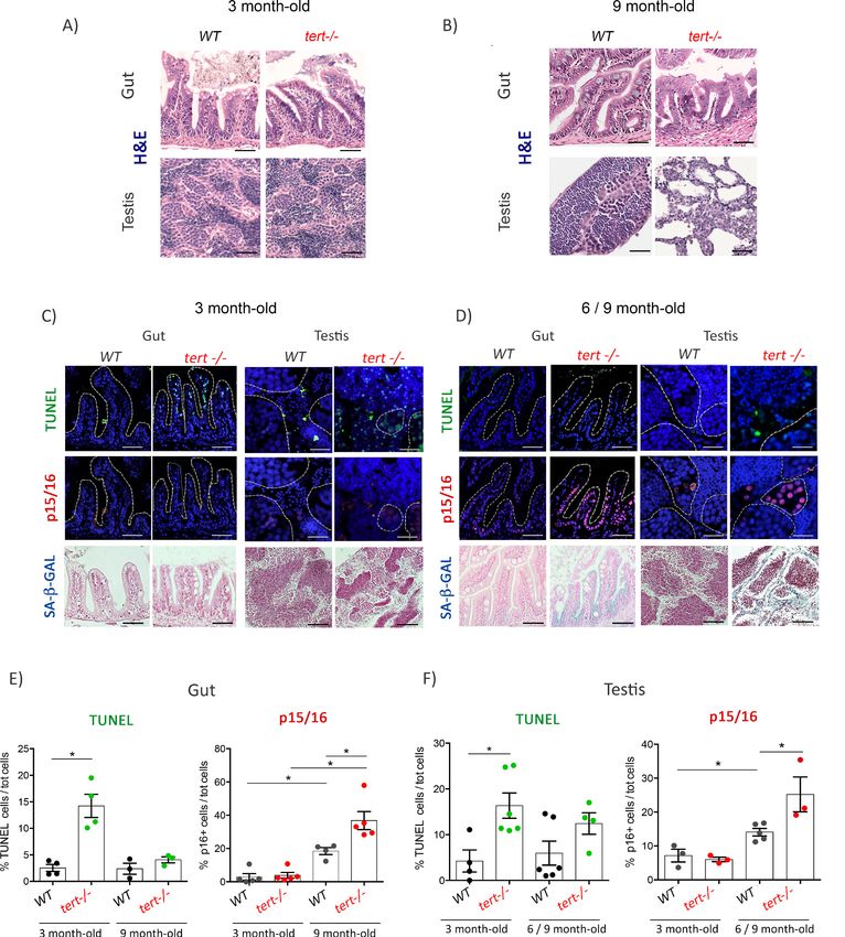
Research article                                                                Cell Biology Stem Cells and Regenerative Medicine

                                    gene (CDKN2B), located immediately upstream of the INK4a/ARF locus. Similar to p16, p15 is
                                    involved in cell senescence (Fuxe et al., 2000; Hitomi et al., 2007; Senturk et al., 2010). In fugu
                                    fish, as in chicken, p16INK4a is mutated and does not encode a functional protein. Thus, fugu and
                                    chicken INK4a loci express Arf but not p16 (Liao and Hung, 2003). Consequently, the authors pro-
                                    posed that, in these animals, the function of p16 was evolutionarily replaced by p15.
                                        Under regular physiological conditions, cells with a high turn-over rate (including epithelial and
                                    germinal cells) preserve tissue homeostasis by undergoing continuous apoptosis and compensatory
                                    cell proliferation. However, in some animals including humans, cells can only go through a finite
                                    number of divisions before entering irreversible cell-cycle arrest, a process known as replicative
                                    senescence. Telomere erosion was proposed to constitute the ‘molecular clock’ that determines the
                                    number of divisions a cell can undergo before reaching senescence, a phenomenon known as Hay-
                                    flick limit (Bodnar et al., 1998; Hayflick, 1965). In most eukaryotes, telomere shortening is counter-
                                    acted by telomerase, though its expression is restricted in most human somatic cells (Forsyth et al.,
                                    2002). Consequently, telomeres shorten significantly during human aging (Aubert and Lansdorp,
                                    2008).
                                        Vertebrate telomerase mutant animal models have been used to investigate the direct association
                                    between telomere shortening and tissue dysfunction. Even though the common lab mouse has 5-10x
                                    longer telomeres than humans, upon several generations of in-crossing (G4-6), telomerase-deficient
                                    mice demonstrated premature features of aging, including reduced cell proliferation and increased
                                    apoptosis in several tissues (Lee et al., 1998; Rudolph et al., 1999). As observed in humans, telo-
                                    mere length in zebrafish ranges between 5–15 Kb, and telomerase is restricted in somatic tissues
                                    (Anchelin et al., 2011; Anchelin et al., 2013; Carneiro et al., 2016b; Henriques et al., 2013). As a
                                    consequence, we showed that telomeres shorten throughout zebrafish natural aging
                                    (Carneiro et al., 2016b). First-generation adult zebrafish telomerase mutants (tert-/-) exhibit short
                                    telomeres and die as young adults. They display premature aging phenotypes including reduced cell
                                    proliferation, loss of tissue homeostasis and functional organ decline (Anchelin et al., 2013;
                                    Carneiro et al., 2016b; Henriques et al., 2013). Strikingly, we observed that telomeres in both
                                    aged wild-type and tert-/- zebrafish reach a similar length as they exhibit aging phenotypes
                                    (Carneiro et al., 2016b). Accumulation of DNA damage, decline in cell division and organ dysfunc-
                                    tion are associated with tissue-dependent telomere shortening (Anchelin et al., 2013;
                                    Carneiro et al., 2016b; Henriques et al., 2013). Likewise, old age afflictions including infertility,
                                    infections, cachexia and cancer are accelerated in young telomerase mutant zebrafish
                                    (Carneiro et al., 2016b). Similar to humans affected by telomeropathies (Opresko and Shay, 2017),
                                    young zebrafish telomerase mutants display phenotypes of old age, including ‘genetic anticipation’,
                                    in which second generation telomerase deficient animals have aggravated phenotypes and die as
                                    larva (Henriques et al., 2013; Anchelin et al., 2013). Overall, telomeres of naturally aged zebrafish
                                    shorten to critical lengths and this phenomenon is related with age-associated dysfunction and dis-
                                    eases. Because, like humans, telomere shortening is part of physiologic aging, zebrafish constitutes
                                    an appropriate vertebrate model to study the consequences of short telomeres in aging
                                    (Carneiro et al., 2016a).
                                        As telomeres become critically short, they accumulate gH2A.X and activate the DNA Damage
                                    Responses (DDRs) (d’Adda di Fagagna et al., 2003). One of the mediators of DDR is the onco-sup-
                                    pressor p53, which accumulates upon telomere shortening and may result in either cell senescence
                                    or apoptosis (Li et al., 2016). The signals leading to each cell fate in response to p53 accumulation
                                    are unclear to date. Previous studies suggested that cellular senescence is associated with increased
                                    levels of mTOR/AKT signaling (Miyauchi et al., 2004; Moral et al., 2009; Leontieva and Blagos-
                                    klonny, 2013). AKT is a serine/threonine protein kinase that is activated upon pro-proliferative extra-
                                    cellular signals. mTOR/AKT pathway is triggered by growth factor receptors, including the Insulin
                                    Growth Factor Receptor (IGFR) (Liao and Hung, 2010). Activation of AKT- and mTORC2-mediated
                                    phosphorylation results in the phosphorylation of the forkhead transcription factors, FoxO1 and
                                    FoxO4 (Tuteja and Kaestner, 2007). Once phosphorylated, the FoxO family proteins translocate
                                    outside the nucleus, followed by repression of their main target genes, including
                                    mitochondrial superoxide dismutase SOD2. Prolonged and uncontrolled activation of this pathway
                                    results in mitochondrial dysfunction and increased ROS levels (Nogueira et al., 2008).
                                        In our study, we investigated the in vivo switch between apoptosis and cell senescence as a con-
                                    sequence of telomere shortening in tert-/- zebrafish. We describe that early in life, telomerase

El Maı̈ et al. eLife 2020;9:e54935. DOI: https://doi.org/10.7554/eLife.54935                                                         2 of 26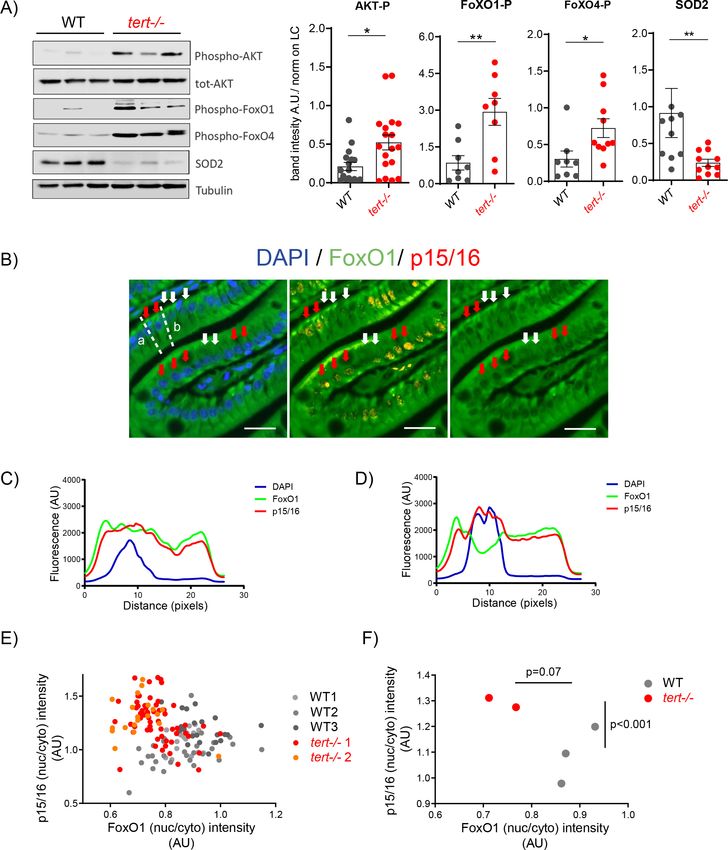
Research article                                                                 Cell Biology Stem Cells and Regenerative Medicine

                                    deficiency results in p53 mediated apoptosis and loss of tissue homeostasis, causing pro-proliferative
                                    AKT pathway activation in older individuals. AKT/FoxO signal cascade then triggers a switch that
                                    results in mitochondrial dysfunction, increased levels of ROS and p15/16, leading to cell senescence.

                                    Results
                                    tert-/- zebrafish proliferative tissues undergo a time-dependent switch
                                    from apoptosis to senescence
                                    Apoptosis is a process in which programmed cell death allows for clearance of damaged cells
                                    (Hawkins and Devitt, 2013). In contrast, replicative senescence is a state of terminal proliferation
                                    arrest associated with irreparable DNA damage, such as critically short telomeres resulting from cell
                                    division (Olovnikov, 1973; Shay and Wright, 2000). To explore the molecular mechanisms underly-
                                    ing the cell-fate decision between apoptosis and senescence, we used telomere attrition as a trigger
                                    of these two possible outcomes.
                                        First-generation tert-/- zebrafish have shorter telomeres than their wild-type (WT) siblings,
                                    develop several degenerative conditions affecting mainly highly proliferative tissues, such as the tes-
                                    tis and gut, and die prematurely (Anchelin et al., 2013; Carneiro et al., 2016b; Henriques et al.,
                                    2013). At 3 months of age, tert-/- fish are macroscopically similar to their WT siblings, with gut and
                                    testis being histologically indistinguishable from WT (Figure 1A). However, at this early age, we pre-
                                    viously observed that average telomere length in tert-/- mutants is shorter than WT and triggers the
                                    onset of DDR and increased apoptosis (Carneiro et al., 2016b).
                                        We analyzed the presence of apoptotic cells in 3-month-old tert-/- gut and testis using the TUNEL
                                    assay. We confirmed that, even in the absence of macroscopic defects, tert-/- gut and testis exhibit
                                    a higher number of apoptotic TUNEL-positive cells when compared to their WT siblings (Figure 1C,
                                    E and F; WT N = 3–6, tert-/- N = 3–6, p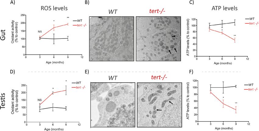
Research article                                                                        Cell Biology Stem Cells and Regenerative Medicine

Figure 1. Proliferative tissues of tert-/- zebrafish undergo an in vivo switch from apoptosis to senescence with age. (A-B) Representative haematoxylin
and eosin-stained sections of gut (scale bar: 40 mm) and testis (scale bar: 25 mm) from 3-month-old (A) or 9-month-old (B) of WT and tert-/- siblings.
While no macroscopic tissue defects are distinguishable at 3 months (N = 3), 9 month tert-/- (N = 3) exhibit altered gut and testis structures. (C-D)
Representative immunofluorescence images of apoptosis (TUNEL) or senescence (p15/16 and SA-b-GAL) of gut and testis from 3 month (C) or 6–9
month-old (D) WT and tert-/- siblings (N = 3–6 each)(scale bar: 25 mm). Dashed outlines locate cysts of spermogonia cells or spermatocytes (testis) or
Figure 1 continued on next page

El Maı̈ et al. eLife 2020;9:e54935. DOI: https://doi.org/10.7554/eLife.54935                                                                      4 of 26
Research article                                                                        Cell Biology Stem Cells and Regenerative Medicine

Figure 1 continued
villi (gut). At 3 months, both tissues show an increased number of apoptotic cells in tert-/- compared to WT. At that age, no signs of senescence are
visible in these tissues. However, senescent cells appear in the gut and testis of 6–9 month-old tert-/- fish depicting a switch between apoptosis and
senescence. (E-F) Quantification of the percentage of TUNEL and p15/16 positive cells in 3 month and 6–9 month-old tert-/- or WT. Data are
represented as mean ± SEM. * p-value
Research article Cell Biology Stem Cells and Regenerative Medicine
mitochondrial dysfunction through a p53-dependent suppression of PGC1a expression, the master
regulator of mitochondrial biogenesis (Sahin et al., 2011). G4 mTERT deficient mice exhibit signifi-
cant alterations in mitochondrial morphology, accumulation of ROS and reduced ATP generation
(Sahin et al., 2011).
We investigated if mitochondrial dysfunction could play a part in the apoptosis-to-senescence
switch observed in tert-/- zebrafish. First, we started by examining if p53 activation triggers the
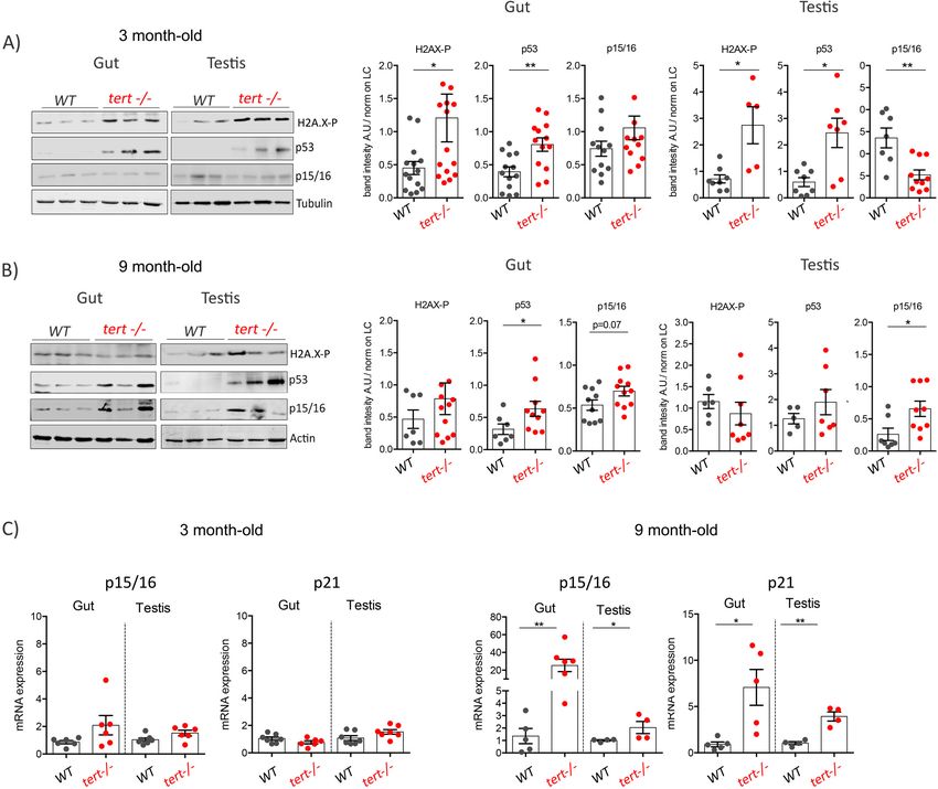
Figure 2. Quantification of senescence markers in gut and testis of older tert-/- zebrafish. (A-B) Western blot analysis of DNA damage and senescence-
associated proteins in gut and testis of 3 month (A) or 9-month-old (B) of WT and tert-/- siblings (N >= 5 fish). Representative western blot (left panel)
and corresponding quantification (right panel) showing induction of DNA Damage Response (H2A.X-P and p53) in 3-month-old and senescence (p15/
16) in 9-month-old tert-/- zebrafish. (C) RT-qPCR analysis of senescence associated genes p15/16 and p21. RT-qPCR graphs are representing
mean ± SEM mRNA fold increase after normalisation by rpl13a gene expression levels (* p-value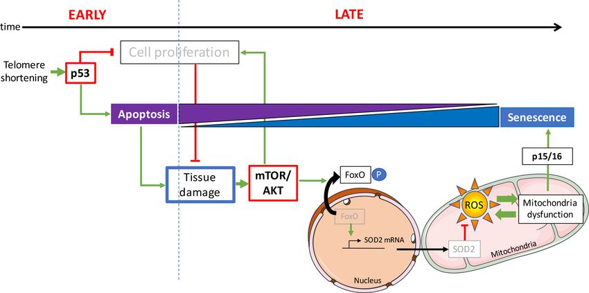
Research article                                                                       Cell Biology Stem Cells and Regenerative Medicine

                                     repression of PGC1a in zebrafish. Despite significant accumulation of p15/16 and p53 (Figures 1F,
                                     2A, B and C), we did not observe differences in neither mRNA nor protein levels of PGC1a in older
                                     tert-/- gut extracts (Figure 3—figure supplement 1; WT N = 3, tert-/- N = 3). However, we detected
                                     a robust increase in oxidative damage with age. By 3 months of age, the levels of ROS in tert-/- gut
                                     and testis did not differ significantly from their WT siblings (Figure 3A–B; WT N = 3, tert-/- N = 3).
                                     From 6 months onward, we observed a gradual and significant accumulation of ROS in both tissues
                                     in tert-/- relative to WT controls (Figure 3A–B; WT N = 3, tert-/- N = 3; p
Research article Cell Biology Stem Cells and Regenerative Medicine
AKT activation leads to ROS production by blocking the FoxO1/FoxO4-
SOD2 molecular axis
Excessive ROS formation gives rise to oxidative stress, leading to cellular damage and, eventually,
senescence (Velarde et al., 2012). Mitochondrial manganese superoxide dismutase (SOD2) is one of
the major ROS scavengers. Notably, SOD2 expression decreases with age (Tatone et al., 2006;
Velarde et al., 2012). SOD2 KO mice and connective tissue-specific SOD2 KOs have reduced life-
span and exhibit premature aging phenotypes associated with senescence but no onset of apoptosis
(Treiber et al., 2011; Velarde et al., 2012).
To gain mechanistic insights into the nature of the oxidative damage observed in the tert-/-
zebrafish, we decided to analyze the expression levels of this important antioxidant defense enzyme.
Western blot analysis of 9-month-old gut and testis extracts showed a significant reduction of SOD2
protein levels in tert-/- mutants compared to WT (Figure 4A and Figure 4—figure supplement 1;
WT N = 9–15, tert-/- N = 9–15, pResearch article Cell Biology Stem Cells and Regenerative Medicine Figure 4. Activation of AKT in older tert-/- mutants results in FoxO1/4 translocation to the cytoplasm and reduction of mitochondria OxPhos defenses. (A) Activation of Akt leads to the inhibitory phosphorylation of FoxO1 and FoxO4 and corresponding reduction of SOD2 expression in 9-month-old tert-/- mutants. Western blot analysis for AKT-P, total AKT, FoxO1-P, FoxO4-P and SOD2 from gut extracts of 9-month-old tert-/- mutant and WT siblings (N >= 9). Representative western blot (left panel) and corresponding normalised quantification (right panel). Data are represented as Figure 4 continued on next page El Maı̈ et al. eLife 2020;9:e54935. DOI: https://doi.org/10.7554/eLife.54935 9 of 26
Research article                                                                        Cell Biology Stem Cells and Regenerative Medicine

Figure 4 continued
mean ± SEM. * p-value
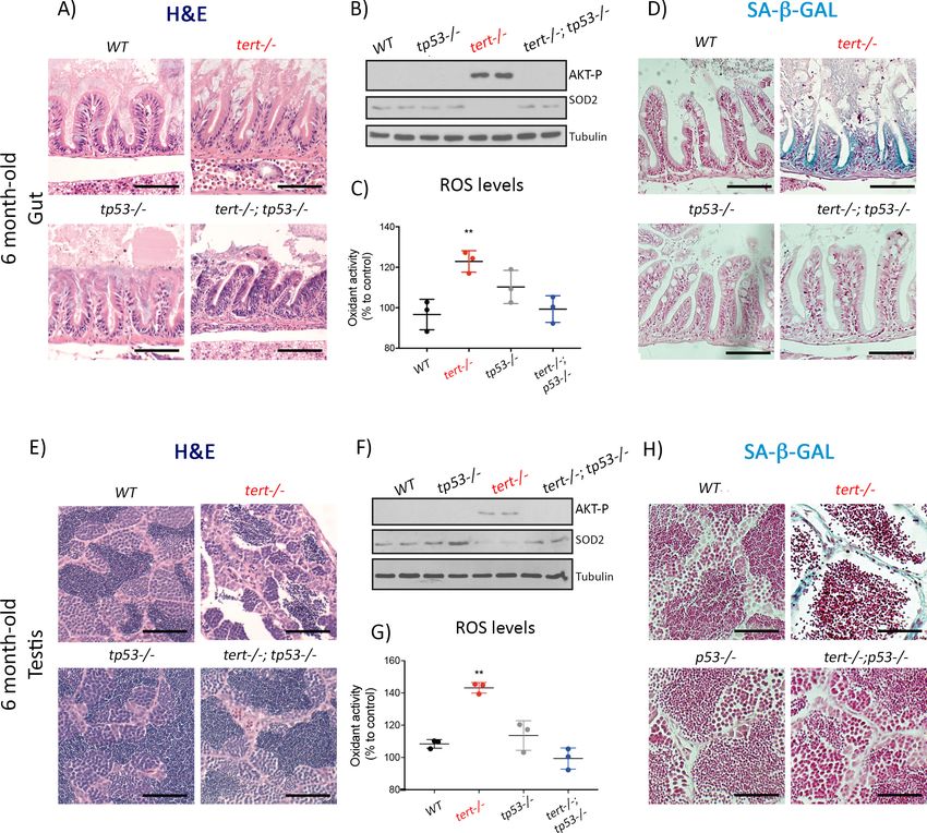
Research article                                                                          Cell Biology Stem Cells and Regenerative Medicine

Figure 5. Mutation of p53 prevents short telomeres-induced tissue degeneration, Akt activation, ROS accumulation and induction of senescence. (A
and E) Representative haematoxylin and eosin-stained sections of gut (A) (scale bar: 40 mm) and testis (E) (scale bar: 25 mm) from 6-month-old WT,
tert-/-, tp53-/- and tert-/- tp53-/- siblings (N = 3 fish each);. Mutation of tp53 in tert-/- fish rescues short-telomere induced tissue defects. (B and F)
Representative western blot analysis of AKT-p and SOD2 in gut (B) and testis (F) (N = 2 fish each). Mutation of tp53 in tert-/- fish prevents
phosphorylation of AKT and downstream downregulation of SOD2 leading to a rescue of increased ROS levels (C and G; N = 3 fish per genotype). (D
and H) Representative images of SA-b-GAL staining of gut (scale bar: 40 mm) (D) and testis (scale bar: 25 mm) (H) from 6 month-old WT, tert-/-, p53-/-
and tert-/- p53-/- siblings (N = 3 fish). Data are represented as mean ± SEM (** p-value
Research article Cell Biology Stem Cells and Regenerative Medicine
mediated cell-cycle arrest, we tested whether AKT activation was necessary, and therefore causal, to
cell senescence. Our hypothesis would predict that inhibition of AKT phosphorylation would prevent
p15/16 expression and preserve tissue homeostasis.
AKT phosphorylation is mediated by the mTORC2 complex, whose main component is the mTOR
(mammalian Target Of Rapamycin) protein (Laplante and Sabatini, 2009). To analyze the role of
AKT activation in inducing senescence upon telomere shortening, we created a double mutant bear-
ing a mutation in the tert gene combined with a mutation in the mTOR zebrafish homologue (ztor).
Previous work showed that ztor is essential for development and ztor-/- zebrafish are larval lethal
(Ding et al., 2011). However, ztor+/- mutants are haploinsufficient, with the lack of one functional
copy being sufficient to reduce AKT phosphorylation (Ding et al., 2011). Thus, we tested our
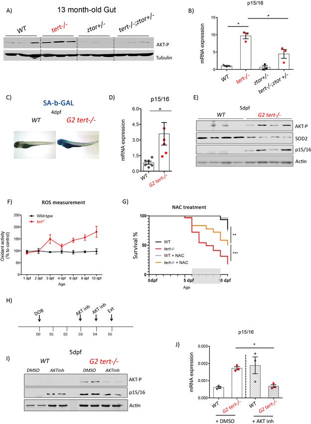
hypothesis in tert-/-ztor+/- mutant zebrafish. As expected, 13-month-old tert-/-ztor+/- present
reduction in AKT phosphorylation compared to tert-/- single mutants (Figure 6A, all genotypes
N = 3). Consistent with our hypothesis, we observed that lower AKT activation is accompanied by
a ~ 2 fold reduction of p15/16 expression (Figure 6B; all genotypes N = 3; pResearch article Cell Biology Stem Cells and Regenerative Medicine Figure 6. Genetic and pharmacological inhibition of AKT prevents short telomere-induced senescence. (A) Heterozygous mutation of zTOR counteracts telomere-shortening-induced Akt activation, leading to inhibition of p15/16 expression. Western blot analysis of AKT-P and (B) RT-qPCR analysis of p15/ 16 mRNA levels in 13-month-old gut of WT, tert-/-, ztor+/-and tert-/- ztor+/- fish (N = 3 fish). (C-F) Second generation (G2) tert-/- mutant larvae with extremely short telomeres show phenotypes associated with premature aging, as described in Figures 1, 2 and 3. (C) Representative images of SA-b- Figure 6 continued on next page El Maı̈ et al. eLife 2020;9:e54935. DOI: https://doi.org/10.7554/eLife.54935 13 of 26
Research article                                                                     Cell Biology Stem Cells and Regenerative Medicine

Figure 6 continued
GAL staining of WT and second generation (G2) tert-/- mutant four dpf larvae. (D) RT-qPCR analysis of p15/16 mRNA levels (N = 6), E) Western blot
analysis of AKT-P, SOD2, p15/16 (N = 4) and (F) ROS levels measurements determined by DCFDA assay (N = 3). G) Survival curve of G2 tert-/-upon
NAC (40 mM from day 6 to 10) treatment (WT N = 31; WT+NAC N = 27; G2 tert-/- N = 61; G2 tert-/- +NAC N = 36 fish; ** p-value
Research article                                                                Cell Biology Stem Cells and Regenerative Medicine

                                       Our study reveals that mitochondrial defects are associated with a reduction in mitochondrial
                                    OxPhos defenses. We observe that, with age, SOD2 expression is reduced in response to AKT-
                                    dependent activation and FoxO1 and FoxO4 phosphorylation. The anti-proliferative p53 and pro-
                                    survival mTOR/AKT pathways interact in a complex manner. Depending on the context, the interac-
                                    tion of these pathways modulates cell fate into either cell-cycle arrest, apoptosis or senescence
                                    (Erol, 2011; Hasty et al., 2013). Cell line studies show that p53 itself can inhibit mTOR/AKT path-
                                    way through several mechanisms including AMPK and PTEN activation (Hasty et al., 2013). p53 acti-
                                    vation of cell senescence relies on mTOR/AKT pathway activity (Davaadelger et al., 2016;
                                    Jung et al., 2019; Kim et al., 2017; Miyauchi et al., 2004; Vétillard et al., 2015). Conversely, AKT
                                    inhibition reduces p53-dependent senescence (Davaadelger et al., 2016; Duan and Maki, 2016;
                                    Kim et al., 2017). In addition, AKT mediates the inhibition of pro-apoptotic factors
                                    (Davaadelger et al., 2016) and leads to increased levels of anti-apoptotic Bcl-XL (Jones et al.,
                                    2000; Li et al., 2017b). Thus, activation of mTOR/AKT pathway can act as a negative regulator of
                                    apoptosis (Davaadelger et al., 2016; Duan and Maki, 2016). AKT was also shown to induce senes-
                                    cence and cell-cycle arrest by elevating the intracellular levels of ROS or activating p16 transcription
                                    through direct phosphorylation of the Bmi repressor (Imai et al., 2014; Li et al., 2017a; Liu et al.,
                                    2012; Miyauchi et al., 2004; Nogueira et al., 2008). Consistent with these in vitro data, we showed
                                    that AKT activation in aged tert-/- mutant zebrafish is concomitant with increased anti-apoptotic Bcl-
                                    XL and pro-senescence p15/16 and p21 levels.
                                       What constitutes the mechanistic nature of the switch from apoptosis to senescence? Even
                                    though young tert-/- mutants present no observable tissue defects, they exhibit higher levels of apo-
                                    ptosis and a reduction in proliferative capacity (Carneiro et al., 2016b; Henriques et al., 2013).
                                    High apoptosis increases the demand for cell proliferation from surrounding cells in a process
                                    termed apoptosis-induced compensatory proliferation (Fan and Bergmann, 2008). In face of cell
                                    proliferation restrictions, tissue degeneration becomes apparent in aging tert-/- zebrafish. In tissues
                                    where stem cells are not readily available or where tissue-intrinsic genetic programs constrain cell
                                    division, cellular hypertrophy represents an alternative strategy for tissue homeostasis (Losick et al.,
                                    2013; Tamori and Deng, 2013).
                                       We propose that, upon telomere shortening and p53 activation, loss of tissue integrity triggers
                                    the AKT-dependent pro-proliferative pathway (Figure 7). The combination of these antagonistic
                                    forces in the cell would result in cellular senescence. We tested this hypothesis on both pathways. By
                                    genetically ablating tp53 function, we were able to rescue tissue degeneration and avoid AKT activa-
                                    tion, increased ROS levels and induction of senescence. On a second level, we inhibited mTOR/AKT
                                    genetically by dampening the ztor pathway and, chemically, by directly inhibiting AKT in G2 tert-/-
                                    larvae. In both cases, we were able to reduce the effects of telomere shortening. Collectively, our
                                    results show that the crosstalk between the two pathways, telomere shortening/p53 and AKT/FoxO
                                    signalling, regulates the apoptosis-to-senescence switch and contributes to tissue homeostasis in
                                    vivo.
                                       Our previous studies describe a similar transition from apoptosis to senescence in WT zebrafish.
                                    While apoptosis is triggered at early stages of WT aging, loss of tissue homeostasis appears from 18
                                    to 24 month of age and is associated with predominant senescence (Carneiro et al., 2016b).
                                    We therefore anticipate that the mechanisms occurring in normal zebrafish aging and leading to
                                    degenerative phenotypes are analogous to tert-/- premature aging. However, we were unable to
                                    observe critical telomere shortening in some WT tissues, such as testis or kidney marrow (the fish
                                    hematopoietic organ), owing to continuous expression of telomerase throughout life
                                    (Carneiro et al., 2016b). In this regard, tert-/- zebrafish are better models of human telomeropa-
                                    thies, a disease spectrum that affects primarily organs that rely on telomerase expression, such as
                                    the hematopoietic tissues (Opresko and Shay, 2017). Nevertheless, these organs do exhibit degen-
                                    erative phenotypes and accumulation of senescent cells with age. Thus, other factors could trigger
                                    age-dependent dysfunctions rather than telomere shortening in WT aging. Alternatively, other
                                    organs in an aging animal, such as the immune system, may become limiting and give rise to sys-
                                    temic deficiencies that lead to a concerted demise.

El Maı̈ et al. eLife 2020;9:e54935. DOI: https://doi.org/10.7554/eLife.54935                                                        15 of 26
Research article Cell Biology Stem Cells and Regenerative Medicine
Figure 7. Opposing activities of p53 and mTOR/AKT upon telomere shortening promote a switch from apoptosis to senescence. Early telomere
shortening triggers p53-dependent apoptosis and inhibition of cell proliferation. At early age, apoptosis is the predominant cell fate and it mostly
counterbalanced by compensatory proliferation of neighboring cells. However, inhibition of cell proliferation results in a progressive loss of tissue
cellularity, eventually leading to tissue damage. As age progresses, loss of tissue homeostasis triggers the pro-proliferative mTOR/AKT pathway. Akt
phosphorylates FoxO, inducing its translocation from the nucleus to the cytoplasm. Loss of FoxO transcriptional activity reduces mitochondrial SOD2
expression generating mitochondria oxidative stress through increased ROS levels. Mitochondrial dysfunction eventually triggers p15/16 expression and
senescence becomes the predominant cell fate.
Materials and methods
Key resources table
Reagent type Additional
(species) or resource Designation Source or reference Identifiers information
Genetic reagent tert hu3430/+ Hubrecht Institute, RRID:ZFIN_ZDB-
(Danio rerio) Utrecht, Netherland GENO-100412-50
Genetic reagent tp53 M214K/+ Berghmans et al., 2005 RRID:ZDB-ALT-050428-2
(Danio rerio)
Genetic reagent ztor xu015Gt/+ Ding et al., 2011 RRID:ZDB-ALT-120412-1
(Danio rerio)
Gene tert ZDB-GENE-080405–1
(Danio rerio)
Gene tp53 ZDB-GENE-990415–270
(Danio rerio)
Gene ztor ZDB-GENE-030131–2974
(Danio rerio)
Gene cdkn2a/b (p15/16) ZDB-GENE-081104–306
(Danio rerio)
Gene cdkn1a (p21) ZDB-GENE-070705–7
(Danio rerio)
Gene bcl2l1 (Bcl-XL) ZDB-GENE-010730–1
(Danio rerio)
Continued on next page
El Maı̈ et al. eLife 2020;9:e54935. DOI: https://doi.org/10.7554/eLife.54935 16 of 26Research article Cell Biology Stem Cells and Regenerative Medicine
Continued
Reagent type Additional
(species) or resource Designation Source or reference Identifiers information
Gene ppargc1a (PGC1a) ZDB-GENE-080505–1
(Danio rerio)
Gene rpl13a ZDB-GENE-030131–168
(Danio rerio)
Antibody anti-p16 (mouse Santa Cruz #Sc-1661; IF(1:50), WB (1:700)
monoclonal; F-12) Biotechnology RRID:AB_628067
Antibody anti-FoxO1 (rabbit Cell Signaling #2880; IF(1:50)
monoclonal; C29H4) Technology RRID:AB_2106495
Antibody anti-zebrafish p53 Anaspec #55342; WB (1:1000)
(rabbit polyclonal) RRID:AB_2287635
Antibody anti-zebrafish gH2AX GeneTex #GTX127342; WB (1:1000)
(rabbit polyclonal) RRID:AB_2833105
Antibody anti-SOD2 Sigma-Aldrich #SAB2701618; WB (1:1000)
(rabbit polyclonal) RRID:AB_2833106
Antibody anti-phospho-Akt, Ser473 Cell Signaling #4060; WB (1:1000)
(rabbit monoclonal; D9E) Technology RRID:AB_2315049
Antibody anti-total-Akt Cell Signaling #9272, WB (1:1000)
(rabbit polyclonal) Technology RRID:AB_329827
Antibody anti-phospho-FoxO1, Cell Signaling #9461; WB (1:100)
Ser256 (rabbit polyclonal) Technology RRID:AB_329831
Antibody anti-tubulin (mouse Sigma #T6074; WB (1:5000)
monoclonal; B-5-1-2) RRID:AB_477582
Antibody Alexa Fluor 568 goat Invitrogen #A11004; IF (1:500)
anti-mouse RRID:AB_2534072
(goat polyclonal)
Antibody Alexa Fluor 488 goat Invitrogen #A11008; IF (1:500)
anti-rabbit RRID:AB_143165
(goat polyclonal)
Antibody HRP- anti-rabbit Santa Cruz #Sc2054; WB (1:5000)
(goat polyclonal) RRID:AB_631748
Antibody HRP- anti-mouse Santa Cruz #Sc2005; WB (1:5000)
(goat polyclonal) RRID:AB_631736
Sequence- cdkn2a/b (p15/16) Fw This paper PCR primers GAGGATGAACT
based reagent GACCACAGCA
Sequence- cdkn2a/b (p15/16) Rv This paper PCR primers CAAGAGCCAAA
based reagent GGTGCGTTAC
Sequence- bcl2l1 (Bcl-XL) Fw This paper PCR primers GGGCTTGTTT
based reagent GCTTGGTTGA
Sequence- bcl2l1 (Bcl-XL) Rv This paper PCR primers AGAACACAGTG
based reagent CACACCCTT
Sequence- cdkn1a (p21) Fw This paper PCR primers CAGCGGGTTTA
based reagent CAGTTTCAGC
Sequence- cdkn1a (p21) Rv This paper PCR primers TGAACGTAGGA
based reagent TCCGCTTGT
Sequence- ppargc1a (PGC1a) Fw This paper PCR primers CTGTGGAACCC
based reagent CAGGTCTGAC
Sequence- ppargc1a (PGC1a) Rv This paper PCR primers ACTCAGCCTGG
based reagent GCCTTTTGCT
Sequence- rpl13a Fw Henriques et al., 2013 PCR primers TCTGGAGGACTG
based reagent TAAGAGGTATG
Sequence- rpl13a Rv Henriques et al., 2013 PCR primers AGACGCACAATC
based reagent TTGAGAGCAG
Continued on next page
El Maı̈ et al. eLife 2020;9:e54935. DOI: https://doi.org/10.7554/eLife.54935 17 of 26Research article Cell Biology Stem Cells and Regenerative Medicine
Continued
Reagent type Additional
(species) or resource Designation Source or reference Identifiers information
Sequence- cdkn2a/b (p15/16) This paper morpholino TCAGTTCATCCT
based reagent Morpholino CGACGTTCATCAT
Sequence- Control Morpholino GeneTools morpholino CCTCTTACCTCAG
based reagent TTACAATTTATA
Commercial In Situ Cell Death Roche 11684795910
assay or kit Detection Kit, Fluorescein
Commercial CellTiter-Glo Luminescent Promega G7570
assay or kit Cell Viability Assay
Chemical 20 ,70 -Dichlorofluorescin Sigma Aldrich D6883
compound, drug diacetate (DCFDA)
Chemical AKT1/2 kinase inhibitor Santa Cruz sc-300173
compound, drug
Chemical N-Acetyl-L- Sigma Aldrich A7250
compound, drug Cysteine (NAC)
Chemical MitoTEMPO Sigma Aldrich SML0737
compound, drug
Other DAPI stain Sigma D9542 (0.5 mg/mL)
Ethics statement
All Zebrafish work was conducted according to Portuguese (Decreto-Lei 113/2013) and European
(Directive 2010/63/EU) legislations and approved by the Ethical Committee of the IGC (Instituto Gul-
benkian de Ciência; approval number: A001.2012) and the DGAV (Direcção Geral de Alimentação e
Veterinária, Portuguese Veterinary Authority; approval number: 010294).
Zebrafish lines and maintenance
Zebrafish were maintained in accordance with Institutional and National animal care protocols. The
telomerase mutant line tert AB/hu3430 was generated by N-Ethyl-N-nitrosourea (ENU) mutagenesis
(Utrecht University, Netherlands; Wienholds and Plasterk, 2004). tert AB/hu3430 line is available at
the ZFIN repository, ZFIN ID: ZDB-GENO-100412–50, from the Zebrafish International Re-source
Center—ZIRC. The protocols used for outcrossing mutagenized male zebrafish were previously
described (Carneiro et al., 2016a; Henriques et al., 2013). All stocks were kept in heterozygous
form and maintained strictly by outcrossing to AB strains to avoid haploinsufficiency effects in the
progeny. The tert hu3430/hu3430 homozygous mutant (referred in this work as tert-/-) was obtained
by incrossing our tert AB/hu3430 (tert+/-) strain. WT siblings were used as controls. Overall charac-
terization of tert-/- and WT zebrafish was performed in F1 animals produced by tert+/- incross. Due
to a male sex bias in our crosses, that affected mostly tert-/- progeny, we were unable to obtain sig-
nificant numbers of females for analysis and so all of our data is restricted to males.
p53 mutant line zdf1 (tp53 M214K, Berghmans et al., 2005) was kindly provided by Dr António
Jacinto (CEDOC, chronic diseases, Nova medical school, Lisbon Portugal). tp53 mutant line zdf1
(P53 M214K) is available at the ZFIN repository, ZFIN ID: ZDB-ALT-050428–2 from the Zebrafish
International Re-source Center—ZIRC. The tp53 M214K/M214K homozygous mutant (referred to as
tp53-/-) was obtained by incrossing our tp53 AB/M214K strain. ztor line was obtained from the ZFIN
repository, ZFIN ID: ZDB-ALT-120412–1 from the Zebrafish International Re-source Center—ZIRC.
The line was previously described (Ding et al., 2011) as homozygous larval lethal and it was main-
tained through outcrossing. All animals showing signs of morbidity that persisted for up to 5 days,
such as inability to eat or swim, or macroscopic lesions/tumors were sacrificed using 200 mg/L of
MS-222 (Sigma,MO, USA).
Histological analysis
Zebrafish were sacrificed by anaesthetic overdose in 200 mg/L of MS-222 (Sigma, MO, USA), fixed
for 72 hr in 10% neutral buffered formalin and decalcified in 0.5M EDTA for 48 hr at RT. Whole fish
were then paraffin-embedded and three micrometer midline sagittal sections were stained with
El Maı̈ et al. eLife 2020;9:e54935. DOI: https://doi.org/10.7554/eLife.54935 18 of 26Research article Cell Biology Stem Cells and Regenerative Medicine
haematoxylin and eosin for histopathological analysis. Sections were examined by a histopathologist
(TC), blinded to experimental groups and microphotographs were acquired in a Leica DM2500
microscope coupled to a Leica MC170 HD microscope camera. At least four animals from each age
group/genotype were analysed.
Immunofluorescence (IF) assays
Apoptosis and Senescence was detected using the In Situ Cell Death Detection Kit (Roche, SW)
according to manufacturer’s instructions combined with immunofluorescence against p15/16 senes-
cence-associated marker. Briefly, deparaffinized slides were incubated with 40 mg/mL Proteinase K
in 10 mM Tris-HCl pH 7.4, 45 min at 37˚C. Slides were left to cool down for 30 min at RT, washed
three times in dH20 for 5 min each and blocked for 1 hr at RT in 1% BSA, 0,5% Tween 20 in PBS-T
(Triton 0.5%). Subsequently, the slides were incubated overnight with anti-p16 (F-12) (1:50, Santa
Cruz Biotechnology, sc-1661), followed by 3 10 min PBS washes. Incubation with the secondary
antibody Alexa Fluor 568 goat anti-mouse (Invitrogen, UK, 1:500 dilution) overnight at 4˚C was fol-
lowed by 3 10 min PBS washes. The day after, slides were washed 2 5 min in PBS and then incu-
bated with TUNEL labelling mix (protocol indicated by the supplier). Washing and mounting were
performed by DAPI staining (Sigma, MO, USA) and mounting with DAKO Fluorescence Mounting
Medium (Sigma, MO, USA).
For FoxO-1 and p15/16 co-staining, deparaffinized slides were incubated in Citrate Buffer (2.95
g/L sodium citrate; pH 6) for 20 min at 105˚C. Samples were processed as above except for the
block solution of 0.5% Triton/5% Normal Goat Serum/PBS and an overnight staining with anti-p16
(F-12) (1:50, Santa Cruz Biotechnology, sc-1661) and anti-FoxO-1 (C29H4) (1:50, Cell Signalling Tech-
nology, 2880). A second incubation was then performed using secondary antibodies Alexa Fluor 568
goat anti-mouse (Invitrogen, UK, 1:500 dilution) and Alexa Fluor 488 goat ant-rabbit (Invitrogen, UK,
1:500 dilution).
Images were acquired on a commercial Nikon High Content Screening microscope, based on
Nikon Ti equipped with a Andor Zyla 4.2 sCMOS camera, using the a 20 1.45 NA objective, DAPI
+ GFP fluorescence filter sets and controlled with the Nikon Elements software.
For quantitative and comparative imaging, equivalent image acquisition parameters were used.
The percentage of positive nuclei was determined by counting a total of 500–1000 cells per slide,
63x amplification (N >= 3 zebrafish per time point/genotype).
Senescence-associated b-galactosidase assay
b-galactosidase assay was performed as previously described (Kishi et al., 2008). Briefly, sacrificed
zebrafish adults were fixed for 72 hr in 4% paraformaldehyde in PBS at 4˚C and then washed three
times for 1 hr in PBS-pH 7.4 and for a further 1 hr in PBS-pH 6.0 at 4˚C. b-galactosidase staining was
performed for 24 hr at 37˚C in 5 mM potassium ferrocyanide, 5 mM potassium ferricyanide, 2 mM
MgCl2 and 1 mg/mL X-gal, in PBS adjusted to pH 6.0. After staining, fish were washed three times
for 5 min in PBS pH seven and processed for de-calcification and paraffin embedding as before. Sec-
tions were stained with nuclear fast red for nuclear detection and images were acquired in a bright
field scan (Leica, APERIO).
Immunoblot analysis
Age- and sex-matched adult zebrafish fish were sacrificed in 200 mg/L of MS-222 (Sigma, MO, USA)
and portions of each tissue (gonads and gut) were retrieved and immediately snap-frozen in dry ice.
4dpf larvae were sacrificed in ice and collected in 1,5 mL Eppendorf tube, minimum 10 larvae/tube.
Gonads tissues and larvae were then homogenized in RIPA buffer (sodium chloride 150 mM; Triton-
X-100 1%; sodium deoxycholate 0,5%; SDS 0,1%; Tris 50 mM, pH = 8.0), including complete prote-
ase and phosphatase inhibitor cocktails (Roche diagnostics), with the help of a motor pestle. Protein
extracts were incubated on ice for 30 min and centrifuged at 4˚C, 13.000 rpm, for 10 min. The super-
natant was collected and added to 100 mL of protein sample buffer containing DTT.
Gut samples were homogenized in TRIzol (Invitrogen, UK) by mashing each individual tissue with
a pestle in a 1.5 mL Eppendorf tube. After incubation at RT for 10 min in TRIzol, chloroform extrac-
tions were performed. The organic phase was collected, and proteins were precipitated according
to the manufacture protocol. The protein pellet was resuspended in 100 mL of Lysis Buffer (150 mM
El Maı̈ et al. eLife 2020;9:e54935. DOI: https://doi.org/10.7554/eLife.54935 19 of 26Research article Cell Biology Stem Cells and Regenerative Medicine
NaCl, 4% SDS, 50 mM Tris-HCl pH 8.0, 10 mM EDTA, complete protease and phosphatase inhibitor
cocktails-Roche diagnostics).
For each sample, a fraction of proteins was separated on 12% SDS-PAGE gels and transferred to
Immobilon PVDF membranes (Millipore). The membranes were blocked in 5% milk or 5% BSA
(depending on the primary antibody), then incubated with the indicated primary antibody prior to
incubation with the appropriate HRP-conjugated secondary antibody. Antibody complexes were
visualised by enhanced chemiluminescence (ECL). Antibodies concentration: anti-p53 (1:1000, Anas-
pec, 55342); anti-g-H2AX (1:1000, GeneTex, GTX127342); anti-p16 (F-12) (1:700, Santa Cruz Biotech-
nology, sc-1661); anti-SOD-2 (1:1000, Sigma, SAB2701618); anti-phospho-AKT, Ser473 (1:1000, Cell
Signaling, #4060); anti-total-AKT (1:1000 Cell Signaling, #9272, gift of Adrien Colin), anti-phospho-
FoxO1, Ser256 (1:100, Cell Signaling, #9461); anti-Tubulin (1:5000, Sigma, T 6074).
Real-time quantitative PCR
Age- and sex-matched fish were sacrificed in 200 mg/L of MS-222 (Sigma, MO, USA) and portions of
each tissue (gonads, gut and muscle) were retrieved and immediately snap-frozen in liquid nitrogen.
Similarly, larvae were sacrificed and collected in Eppendorf tubes, minimum 10 larvae each. RNA
extraction was performed in TRIzol (Invitrogen, UK) by mashing each individual tissue with a pestle in
a 1.5 mL eppendorf tube. After incubation at room temperature (RT) for 10 min in TRIzol, chloroform
extractions were performed. Quality of RNA samples was assessed through BioAnalyzer (Agilent
2100, CA, USA). Retro-transcription into cDNA was performed using a RT-PCR kit NZY First-Strand
cDNA Synthesis Kit # MB12501 (NZYtech).
Quantitative PCR (qPCR) was performed using iTaq Universal SYBR Green Supermix # 1725125
(Bio-Rad) and an ABI-QuantStudio 384 Sequence Detection System (Applied Biosystems, CA, USA).
qPCRs were carried out in triplicate for each cDNA sample. Relative mRNA expression was normal-
ized against rpl13a mRNA expression using the DCT method. Primer sequences are listed in
Supplementary file 1.
Detection of intracellular oxidant activity
Reactive oxygen species (ROS) accumulation was assessed by measuring the levels of the oxidized
form of the cell-permeant 5-chloromethyl-2’,7’-dichlorodihydrofluorescein diacetate (DCFDA,
Sigma). Briefly, zebrafish were euthanized with 200 mg/L of MS-222 (Sigma, MO, USA) and tissues
such as the testis, gut and muscle were dissected. Each tissue was homogenized in 100 mL of ROS
buffer (0.32 mM sucrose, 20 mM hepes, 1 mM MgCl2 and 0.5 mM phenylmethanesulfonyl fluoride-
PMSF). Homogenates were centrifuged and 20 mL of the supernatant was transferred to a 96-well
plate and incubated in 1 mg/mL of DCFDA for 30 min. Fluorescence values were measured with a
Victor three plate reader (Perkin Elmer) and normalized to total protein content, which was deter-
mined by the Bradford method. N = 3 per time point.
ATP measurement
Age- and sex-matched adult zebrafish fish were sacrificed in 200 mg/L of MS-222 (Sigma, MO, USA)
and portions of each tissue (gonads, gut and muscle) were retrieved and immediately snap-frozen in
dry ice. Each tissue was homogenised in 100 mL of 6M guanidine-HCl in extraction buffer (100 mM
Tris and 4 mM EDTA, pH 7.8) to inhibit ATPases. Homogenised samples were subjected to rapid
freezing in liquid nitrogen followed by boiling for 3 min. Samples were then cleared by centrifuga-
tion and the supernatant was diluted (1/50) with extraction buffer and mixed with luminescent solu-
tion (CellTiter-Glo Luminescent Cell Viability Assay, Promega). The luminescence was measured on a
Victor three plate reader (Perkin Elmer). The relative ATP levels were calculated by dividing the lumi-
nescence by the total protein concentration, which was determined by the Bradford method. For
Bradford assays, samples were diluted (1/50) with extraction buffer.
Electron microscopy
For electron microscopy analysis, zebrafish tissues were processed according to Schieber et al.,
2010. Briefly, zebrafish were fixed in 2% Paraformaldehyde, 2.5% Glutaraldehyde in 0.1M PHEM
buffer for 72 hr at 4˚C. Dissected tissues were then washed 3 times in 0.1 M PHEM. Tissues were
transferred in 1% Osmium Tetroxide in 0.1 M PHEM for 1 hr fixation on ice. Samples were then
El Maı̈ et al. eLife 2020;9:e54935. DOI: https://doi.org/10.7554/eLife.54935 20 of 26Research article Cell Biology Stem Cells and Regenerative Medicine
dehydrated before being processed for embedding using Epon (Schieber et al., 2010). 70 nm ultra-
thin sections were cut using Reichert Ultramicrotome. After being counterstained with uranyl acetate
and lead, samples were analyzed using a transmission electron microscope (Hitachi H-7650).
Larva chemical treatments
AKT1/2 kinase inhibitor (AKT inh) was purchased from Santa-Cruz (sc-300173). Stock solutions were
prepared in DMSO. AKT inh was applied, after a titration, at 2 mM concentration in E3 embryo
medium between 3-5dpf. Larvae were grown at 28˚C and over the incubation periods, replacing the
medium with an inhibitor every day. Since the inhibitor was dissolved in DMSO, controls were
treated with the correspondent dilution of the solvent. The inhibitor was tested in two independent
trials. Finally, 5dpf larvae were sacrificed and collected to perform protein and RNA analysis.
Second generation (G2) tert-/- survival experiments were performed as follows. Larvae were
raised in petri dishes containing E3 embryo medium at 28˚C. N-Acetyl-L-Cysteine (NAC) and Mito-
TEMPO were purchased from Sigma-Aldrich. NAC was applied at 40 mM between days 5 and 10
post fertilization. MitoTEMPO was applied at 10 mM between days 3 and 5. Medium was replaced
every day. Each drug experiment was performed in two independent replicates.
Knock-down experiments using p15/16 Morpholino injection
One-cell stage WT embryos were injected with 2.4 ng or 3.6 ng of p15/16 mRNA specific translation
blocking antisense morpholino oligonucleotides (MO, Gene Tools, USA) sequence (5’ TCAGTTCA
TCCTCGACGTTCATCAT 3’) or 3.6 ng of standard control MO (5’ CCTCTTACCTCAGTTACAATTTA
TA 3’). After four dpf, larvae were collected for further p15/16 protein expression analysis.
Statistical and image analysis
Image edition was performed using Adobe Photoshop CS5.1 Statistical analysis was performed in
GraphPad Prism5, using one-way ANOVA test with Bonferroni post-correction for all experiments
comparing WT and tert-/- over time. For real-time quantitative PCR, statistical analysis was per-
formed in GraphPad Prism5, one-way ANOVA with Bonferroni post-correction. A critical value for
significance of pResearch article Cell Biology Stem Cells and Regenerative Medicine
Fondation ARC pour la Re- PJA 20161205137 Miguel Godinho Ferreira
cherche sur le Cancer
Université Côte d’Azur - Aca- Installation Grant: Action 2 - Miguel Godinho Ferreira
démie 4 2019
Howard Hughes Medical Insti- IECS Miguel Godinho Ferreira
tute
Ville de Nice Postdoctoral fellowship Mounir El Maı̈
The funders had no role in study design, data collection and interpretation, or the
decision to submit the work for publication.
Author contributions
Mounir El Maı̈, Marta Marzullo, Inês Pimenta de Castro, Conceptualization, Data curation, Formal
analysis, Validation, Investigation, Visualization, Methodology, Writing - original draft, Writing -
review and editing; Miguel Godinho Ferreira, Conceptualization, Data curation, Formal analysis,
Supervision, Funding acquisition, Validation, Visualization, Writing - original draft, Project administra-
tion, Writing - review and editing
Author ORCIDs
Mounir El Maı̈ https://orcid.org/0000-0002-7528-2474
Marta Marzullo https://orcid.org/0000-0001-7229-1693
Inês Pimenta de Castro https://orcid.org/0000-0002-0898-6834
Miguel Godinho Ferreira https://orcid.org/0000-0002-8363-7183
Ethics
Animal experimentation: All Zebrafish work was conducted according to National Guidelines and
approved by the Ethical Committee of the Instituto Gulbenkian de Ciência and the DGAV (Direcção
Geral de Alimentação e Veterinária, Portuguese Veterinary Authority). Approval number: 010294.
Decision letter and Author response
Decision letter https://doi.org/10.7554/eLife.54935.sa1
Author response https://doi.org/10.7554/eLife.54935.sa2
Additional files
Supplementary files
. Supplementary file 1. List of primers used in RT-qPCR expression analysis. Table listing the oligo-
nucleotides used as primers for the RT-qPCR performed in this study.
. Transparent reporting form
Data availability
All data generated or analysed during this study are included in the manuscript and supporting files.
References
Anchelin M, Murcia L, Alcaraz-Pérez F, Garcı́a-Navarro EM, Cayuela ML. 2011. Behaviour of telomere and
telomerase during aging and regeneration in zebrafish. PLOS ONE 6:e16955. DOI: https://doi.org/10.1371/
journal.pone.0016955, PMID: 21347393
Anchelin M, Alcaraz-Pérez F, Martı́nez CM, Bernabé-Garcı́a M, Mulero V, Cayuela ML. 2013. Premature aging in
telomerase-deficient zebrafish. Disease Models & Mechanisms 6:1101–1112. DOI: https://doi.org/10.1242/
dmm.011635, PMID: 23744274
Aubert G, Lansdorp PM. 2008. Telomeres and aging. Physiological Reviews 88:557–579. DOI: https://doi.org/10.
1152/physrev.00026.2007, PMID: 18391173
Balaban RS, Nemoto S, Finkel T. 2005. Mitochondria, oxidants, and aging. Cell 120:483–495. DOI: https://doi.
org/10.1016/j.cell.2005.02.001, PMID: 15734681
El Maı̈ et al. eLife 2020;9:e54935. DOI: https://doi.org/10.7554/eLife.54935 22 of 26Research article Cell Biology Stem Cells and Regenerative Medicine
Berghmans S, Murphey RD, Wienholds E, Neuberg D, Kutok JL, Fletcher CD, Morris JP, Liu TX, Schulte-Merker
S, Kanki JP, Plasterk R, Zon LI, Look AT. 2005. tp53 mutant zebrafish develop malignant peripheral nerve
sheath tumors. PNAS 102:407–412. DOI: https://doi.org/10.1073/pnas.0406252102, PMID: 15630097
Bodnar AG, Ouellette M, Frolkis M, Holt SE, Chiu CP, Morin GB, Harley CB, Shay JW, Lichtsteiner S, Wright WE.
1998. Extension of life-span by introduction of telomerase into normal human cells. Science 279:349–352.
DOI: https://doi.org/10.1126/science.279.5349.349, PMID: 9454332
Brunet A, Bonni A, Zigmond MJ, Lin MZ, Juo P, Hu LS, Anderson MJ, Arden KC, Blenis J, Greenberg ME. 1999.
Akt promotes cell survival by phosphorylating and inhibiting a forkhead transcription factor. Cell 96:857–868.
DOI: https://doi.org/10.1016/S0092-8674(00)80595-4, PMID: 10102273
Campisi J, d’Adda di Fagagna F. 2007. Cellular senescence: when bad things happen to good cells. Nature
Reviews Molecular Cell Biology 8:729–740. DOI: https://doi.org/10.1038/nrm2233, PMID: 17667954
Carneiro MC, de Castro IP, Ferreira MG. 2016a. Telomeres in aging and disease: lessons from zebrafish. Disease
Models & Mechanisms 9:737–748. DOI: https://doi.org/10.1242/dmm.025130, PMID: 27482813
Carneiro MC, Henriques CM, Nabais J, Ferreira T, Carvalho T, Ferreira MG. 2016b. Short telomeres in key tissues
initiate local and systemic aging in zebrafish. PLOS Genetics 12:e1005798. DOI: https://doi.org/10.1371/
journal.pgen.1005798
Childs BG, Baker DJ, Kirkland JL, Campisi J, van Deursen JM. 2014. Senescence and apoptosis: dueling or
complementary cell fates? EMBO Reports 15:1139–1153. DOI: https://doi.org/10.15252/embr.201439245,
PMID: 25312810
Coppé J-P, Patil CK, Rodier F, Sun Y, Muñoz DP, Goldstein J, Nelson PS, Desprez P-Y, Campisi J. 2008.
Senescence-Associated secretory phenotypes reveal Cell-Nonautonomous functions of oncogenic RAS and the
p53 tumor suppressor. PLOS Biology 6:e301. DOI: https://doi.org/10.1371/journal.pbio.0060301
d’Adda di Fagagna F, Reaper PM, Clay-Farrace L, Fiegler H, Carr P, Von Zglinicki T, Saretzki G, Carter NP,
Jackson SP. 2003. A DNA damage checkpoint response in telomere-initiated senescence. Nature 426:194–198.
DOI: https://doi.org/10.1038/nature02118, PMID: 14608368
Davaadelger B, Duan L, Perez RE, Gitelis S, Maki CG. 2016. Crosstalk between the IGF-1R/AKT/mTORC1
pathway and the tumor suppressors p53 and p27 determines cisplatin sensitivity and limits the effectiveness of
an IGF-1R pathway inhibitor. Oncotarget 7:27511-26. DOI: https://doi.org/10.18632/oncotarget.8484,
PMID: 27050276
Ding Y, Sun X, Huang W, Hoage T, Redfield M, Kushwaha S, Sivasubbu S, Lin X, Ekker S, Xu X. 2011.
Haploinsufficiency of target of rapamycin attenuates cardiomyopathies in adult zebrafish. Circulation Research
109:658–669. DOI: https://doi.org/10.1161/CIRCRESAHA.111.248260, PMID: 21757652
Duan L, Maki CG. 2016. The IGF-1R/AKT pathway determines cell fate in response to p53. Translational Cancer
Research 5:664–675. DOI: https://doi.org/10.21037/tcr.2016.09.16, PMID: 28966916
Erol A. 2011. Deciphering the intricate regulatory mechanisms for the cellular choice between cell repair,
apoptosis or senescence in response to damaging signals. Cellular Signalling 23:1076–1081. DOI: https://doi.
org/10.1016/j.cellsig.2010.11.023, PMID: 21144894
Fan Y, Bergmann A. 2008. Apoptosis-induced compensatory proliferation the cell is dead long live the cell!.
Trends in Cell Biology 18:467–473. DOI: https://doi.org/10.1016/j.tcb.2008.08.001, PMID: 18774295
Farnebo M, Bykov VJ, Wiman KG. 2010. The p53 tumor suppressor: a master regulator of diverse cellular
processes and therapeutic target in Cancer. Biochemical and Biophysical Research Communications 396:85–89.
DOI: https://doi.org/10.1016/j.bbrc.2010.02.152, PMID: 20494116
Fogarty CE, Bergmann A. 2017. Killers creating new life: caspases drive apoptosis-induced proliferation in tissue
repair and disease. Cell Death & Differentiation 24:1390–1400. DOI: https://doi.org/10.1038/cdd.2017.47,
PMID: 28362431
Forsyth NR, Wright WE, Shay JW. 2002. Telomerase and differentiation in multicellular organisms: turn it off,
turn it on, and turn it off again. Differentiation 69:188–197. DOI: https://doi.org/10.1046/j.1432-0436.2002.
690412.x, PMID: 11841477
Francisco S, Blackburn EH. 2001. Telomeres. In: Encyclopedia of Life Sciences. Wiley. p. 1–7.
Freund A, Patil CK, Campisi J. 2011. p38MAPK is a novel DNA damage response-independent regulator of the
senescence-associated secretory phenotype. The EMBO Journal 30:1536–1548. DOI: https://doi.org/10.1038/
emboj.2011.69, PMID: 21399611
Fuxe J, Akusjärvi G, Goike HM, Roos G, Collins VP, Pettersson RF. 2000. Adenovirus-mediated overexpression of
p15INK4B inhibits human glioma cell growth, induces replicative senescence, and inhibits telomerase activity
similarly to p16INK4A. Cell Growth & Differentiation : The Molecular Biology Journal of the American
Association for Cancer Research 11:373–384. PMID: 10939591
Gilley J, Fried M. 2001. One INK4 gene and no ARF at the Fugu equivalent of the human INK4A/ARF/INK4B
tumour suppressor locus. Oncogene 20:7447–7452. DOI: https://doi.org/10.1038/sj.onc.1204933, PMID: 11704
876
Greer EL, Brunet A. 2005. FOXO transcription factors at the interface between longevity and tumor suppression.
Oncogene 24:7410–7425. DOI: https://doi.org/10.1038/sj.onc.1209086, PMID: 16288288
Guertin DA, Stevens DM, Thoreen CC, Burds AA, Kalaany NY, Moffat J, Brown M, Fitzgerald KJ, Sabatini DM.
2006. Ablation in mice of the mTORC components raptor, rictor, or mLST8 reveals that mTORC2 is required for
signaling to Akt-FOXO and PKCalpha, but not S6K1. Developmental Cell 11:859–871. DOI: https://doi.org/10.
1016/j.devcel.2006.10.007, PMID: 17141160
Harley CB, Futcher AB, Greider CW. 1990. Telomeres shorten during ageing of human fibroblasts. Nature 345:
458–460. DOI: https://doi.org/10.1038/345458a0, PMID: 2342578
El Maı̈ et al. eLife 2020;9:e54935. DOI: https://doi.org/10.7554/eLife.54935 23 of 26Research article Cell Biology Stem Cells and Regenerative Medicine
Hasty P, Sharp ZD, Curiel TJ, Campisi J. 2013. mTORC1 and p53: clash of the gods? Cell Cycle 12:20–25.
DOI: https://doi.org/10.4161/cc.22912, PMID: 23255104
Hawkins LA, Devitt A. 2013. Current understanding of the mechanisms for clearance of apoptotic Cells—A Fine
Balance. Journal of Cell Death 6:11037–11068. DOI: https://doi.org/10.4137/JCD.S11037
Hayflick L. 1965. The limited in vitro lifetime of human diploid cell strains. Experimental Cell Research 37:614–
636. DOI: https://doi.org/10.1016/0014-4827(65)90211-9, PMID: 14315085
Henriques CM, Carneiro MC, Tenente IM, Jacinto A, Ferreira MG. 2013. Telomerase is required for zebrafish
lifespan. PLOS Genetics 9:e1003214. DOI: https://doi.org/10.1371/journal.pgen.1003214
Hitomi T, Matsuzaki Y, Yasuda S, Kawanaka M, Yogosawa S, Koyama M, Tantin D, Sakai T. 2007. Oct-1 is
involved in the transcriptional repression of the p15(INK4b) gene. FEBS Letters 581:1087–1092. DOI: https://
doi.org/10.1016/j.febslet.2007.01.092, PMID: 17316622
Imai Y, Takahashi A, Hanyu A, Hori S, Sato S, Naka K, Hirao A, Ohtani N, Hara E. 2014. Crosstalk between the rb
pathway and AKT signaling forms a quiescence-senescence switch. Cell Reports 7:194–207. DOI: https://doi.
org/10.1016/j.celrep.2014.03.006, PMID: 24703840
Jackson SP, Bartek J. 2009. The DNA-damage response in human biology and disease. Nature 461:1071–1078.
DOI: https://doi.org/10.1038/nature08467, PMID: 19847258
Jones RG, Parsons M, Bonnard M, Chan VS, Yeh WC, Woodgett JR, Ohashi PS. 2000. Protein kinase B regulates
T lymphocyte survival, nuclear factor kappaB activation, and Bcl-X(L) levels in vivo. Journal of Experimental
Medicine 191:1721–1734. DOI: https://doi.org/10.1084/jem.191.10.1721, PMID: 10811865
Jung SH, Hwang HJ, Kang D, Park HA, Lee HC, Jeong D, Lee K, Park HJ, Ko YG, Lee JS. 2019. mTOR kinase
leads to PTEN-loss-induced cellular senescence by phosphorylating p53. Oncogene 38:1639–1650.
DOI: https://doi.org/10.1038/s41388-018-0521-8, PMID: 30337688
Kamb A. 1995. Cell-cycle regulators and cancer. Trends in Genetics 11:136–140. DOI: https://doi.org/10.1016/
S0168-9525(00)89027-7, PMID: 7732591
Kim SH, Mitchell M, Fujii H, Llanos S, Peters G. 2003. Absence of p16INK4a and truncation of ARF tumor
suppressors in chickens. PNAS 100:211–216. DOI: https://doi.org/10.1073/pnas.0135557100, PMID: 12506196
Kim YY, Jee HJ, Um JH, Kim YM, Bae SS, Yun J. 2017. Cooperation between p21 and akt is required for p53-
dependent cellular senescence. Aging Cell 16:1094–1103. DOI: https://doi.org/10.1111/acel.12639, PMID: 286
91365
Kishi S, Bayliss PE, Uchiyama J, Koshimizu E, Qi J, Nanjappa P, Imamura S, Islam A, Neuberg D, Amsterdam A,
Roberts TM. 2008. The identification of zebrafish mutants showing alterations in senescence-associated
biomarkers. PLOS Genetics 4:e1000152. DOI: https://doi.org/10.1371/journal.pgen.1000152, PMID: 18704191
Kops GJ, Dansen TB, Polderman PE, Saarloos I, Wirtz KW, Coffer PJ, Huang TT, Bos JL, Medema RH, Burgering
BM. 2002. Forkhead transcription factor FOXO3a protects quiescent cells from oxidative stress. Nature 419:
316–321. DOI: https://doi.org/10.1038/nature01036, PMID: 12239572
Krishnamurthy J, Torrice C, Ramsey MR, Kovalev GI, Al-Regaiey K, Su L, Sharpless NE. 2004. Ink4a/Arf
expression is a biomarker of aging. Journal of Clinical Investigation 114:1299–1307. DOI: https://doi.org/10.
1172/JCI22475, PMID: 15520862
Laplante M, Sabatini DM. 2009. mTOR signaling at a glance. Journal of Cell Science 122:3589–3594.
DOI: https://doi.org/10.1242/jcs.051011, PMID: 19812304
Lee HW, Blasco MA, Gottlieb GJ, Horner JW, Greider CW, DePinho RA. 1998. Essential role of mouse
telomerase in highly proliferative organs. Nature 392:569–574. DOI: https://doi.org/10.1038/33345, PMID:
9560153
Leontieva OV, Blagosklonny MV. 2013. CDK4/6-inhibiting drug substitutes for p21 and p16 in senescence:
duration of cell cycle arrest and MTOR activity determine geroconversion. Cell Cycle 12:3063–3069.
DOI: https://doi.org/10.4161/cc.26130, PMID: 23974099
Li T, Liu X, Jiang L, Manfredi J, Zha S, Gu W. 2016. Loss of p53-mediated cell-cycle arrest, senescence and
apoptosis promotes genomic instability and premature aging. Oncotarget 7:11838–11849. DOI: https://doi.
org/10.18632/oncotarget.7864, PMID: 26943586
Li LU, Zhao Y, Zhang H. 2017a. P16INK4a upregulation mediated by TBK1 induces retinal ganglion cell
senescence in ischemic injury. Cell Death & Disease 8:e2752. DOI: https://doi.org/10.1038/cddis.2017.169,
PMID: 28425986
Li X, Li B, Ni Z, Zhou P, Wang B, He J, Xiong H, Yang F, Wu Y, Lyu X, Zhang Y, Zeng Y, Lian J, He F. 2017b.
Metformin synergizes with BCL-XL/BCL-2 inhibitor ABT-263 to induce apoptosis specifically in p53-Defective
Cancer cells. Molecular Cancer Therapeutics 16:1806–1818. DOI: https://doi.org/10.1158/1535-7163.MCT-16-
0763, PMID: 28533436
Liao Y, Hung MC. 2003. Regulation of the activity of p38 mitogen-activated protein kinase by akt in Cancer and
adenoviral protein E1A-mediated sensitization to apoptosis. Molecular and Cellular Biology 23:6836–6848.
DOI: https://doi.org/10.1128/MCB.23.19.6836-6848.2003, PMID: 12972603
Liao Y, Hung MC. 2010. Physiological regulation of akt activity and stability. American Journal of Translational
Research 2:19–42. PMID: 20182580
Liu Y, Liu F, Yu H, Zhao X, Sashida G, Deblasio A, Harr M, She QB, Chen Z, Lin HK, Di Giandomenico S, Elf SE,
Yang Y, Miyata Y, Huang G, Menendez S, Mellinghoff IK, Rosen N, Pandolfi PP, Hedvat CV, et al. 2012. Akt
phosphorylates the transcriptional repressor bmi1 to block its effects on the tumor-suppressing ink4a-arf locus.
Science Signaling 5:ra77. DOI: https://doi.org/10.1126/scisignal.2003199, PMID: 23092893
López-Otı́n C, Blasco MA, Partridge L, Serrano M, Kroemer G. 2013. The hallmarks of aging. Cell 153:1194–
1217. DOI: https://doi.org/10.1016/j.cell.2013.05.039, PMID: 23746838
El Maı̈ et al. eLife 2020;9:e54935. DOI: https://doi.org/10.7554/eLife.54935 24 of 26You can also read