Alternative splicing at neuroligin site A regulates glycan interaction and synaptogenic activity - eLife
←
→
Page content transcription
If your browser does not render page correctly, please read the page content below

RESEARCH ARTICLE

                                   Alternative splicing at neuroligin site A
                                   regulates glycan interaction and
                                   synaptogenic activity
                                   Shinichiro Oku1, Huijuan Feng2, Steven Connor1,3, Andrea Toledo4, Peng Zhang1†,
                                   Yue Zhang1, Olivier Thoumine4, Chaolin Zhang2, Ann Marie Craig1*
                                   1
                                    Djavad Mowafaghian Centre for Brain Health and Department of Psychiatry,
                                   University of British Columbia, Vancouver, Canada; 2Departments of Systems
                                   Biology and Biochemistry and Molecular Biophysics, Center for Motor Neuron
                                   Biology and Disease, Columbia University, New York, United States; 3Department
                                   of Biology, York University, Toronto, Canada; 4Interdisciplinary Institute for
                                   Neuroscience UMR 5297, CNRS and University of Bordeaux, Bordeaux, France

                                   Abstract Post-transcriptional mechanisms regulating cell surface synaptic organizing complexes
                                   that control the properties of connections in brain circuits are poorly understood. Alternative
                                   splicing regulates the prototypical synaptic organizing complex, neuroligin-neurexin. In contrast to
                                   the well-studied neuroligin splice site B, little is known about splice site A. We discovered that
                                   inclusion of the positively charged A1 insert in mouse neuroligin-1 increases its binding to heparan
                                   sulphate, a modification on neurexin. The A1 insert increases neurexin recruitment, presynaptic
                                   differentiation, and synaptic transmission mediated by neuroligin-1. We propose that the A1 insert
                                   could be a target for alleviating the consequences of deleterious NLGN1/3 mutations, supported
*For correspondence:               by assays with the autism-linked neuroligin-1-P89L mutant. An enrichment of neuroligin-1 A1 in
acraig@mail.ubc.ca                 GABAergic neuron types suggests a role in synchrony of cortical circuits. Altogether, these data
                                   reveal an unusual mode by which neuroligin splicing controls synapse development through
Present address: †Department
                                   protein-glycan interaction and identify it as a potential therapeutic target.
of Neurosciences, Case Western
Reserve University, Cleveland,
United States
Competing interests: The
authors declare that no
competing interests exist.
                                   Introduction
                                   Synapses are specialized sites of intercellular communication where chemical transmission occurs
Funding: See page 20               between neurons in brain circuits. Cell surface trans-synaptic organizing complexes recruit presynap-
Received: 07 May 2020              tic vesicles and the neurotransmitter release machinery and apposing postsynaptic receptors and
Accepted: 31 August 2020           scaffold proteins. Dendritic neuroligins (NLGNs) and their axonal neurexin (NRXN) binding partners
Published: 11 September 2020       form a prototypical synaptic organizing complex (Südhof, 2017). NLGNs bind to two interfaces on
                                   NRXN, to the laminin-neurexin-sex hormone (LNS) protein domain and to the heparan sulphate (HS)
Reviewing editor: Mary B
Kennedy, California Institute of
                                   glycan chain (Zhang et al., 2018). Mutations in all NLGN and NRXN genes are associated with neu-
Technology, United States          ropsychiatric disorders. NLGNs and NRXNs are evolutionarily conserved and essential for mouse sur-
                                   vival (Missler et al., 2003; Varoqueaux et al., 2006). NLGNs have additional extracellular domain
   Copyright Oku et al. This
                                   binding partners, cell surface MDGAs, also linked to neuropsychiatric disorders (Connor et al.,
article is distributed under the
                                   2019). MDGA binding to NLGNs occludes the NRXN binding site, thus preventing NLGN-NRXN
terms of the Creative Commons
Attribution License, which         interaction and suppressing synapse development. Intracellularly, NLGNs can bind to the excitatory
permits unrestricted use and       postsynaptic scaffold PSD-95 and the inhibitory postsynaptic scaffold gephyrin, interactions that are
redistribution provided that the   regulated by phosphorylation (Jeong et al., 2019; Jeong et al., 2017; Letellier et al., 2020;
original author and source are     Letellier et al., 2018). NLGN1 functions at excitatory glutamatergic synapses, NLGN2 at inhibitory
credited.                          GABAergic synapses, and NLGN3 at both.

Oku et al. eLife 2020;9:e58668. DOI: https://doi.org/10.7554/eLife.58668                                                          1 of 26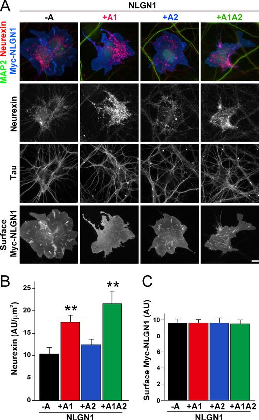
Research article                                                                           Developmental Biology Neuroscience

                                      NLGN1 is essential for normal NMDA receptor recruitment, long-term potentiation, and spatial
                                   learning and memory (Blundell et al., 2010; Budreck et al., 2013; Jiang et al., 2017; Shipman and
                                   Nicoll, 2012). Additional competitive effects of NLGN1 on synapse number were revealed with
                                   sparse knockdown (Kwon et al., 2012) and roles in long-term depression revealed in Nlgn1 hetero-
                                   zygous mice (Dang et al., 2018). Copy number, truncating, and missense variants in NLGN1 are
                                   associated with autism spectrum disorders (ASD) (Glessner et al., 2009; Nakanishi et al., 2017;
                                   O’Roak et al., 2012; Tejada et al., 2019) and obsessive-compulsive disorder (Gazzellone et al.,
                                   2016) and NLGN1 polymorphisms are linked to schizophrenia (Chen et al., 2018; Zhang et al.,
                                   2015).
                                      NLGNs are regulated by alternative splicing (see Figure 1—figure supplement 1 for gene struc-
                                   tures of NLGN1-3). The function of splice site B present only in NLGN1 is well understood. The B
                                   insert blocks the interaction of NLGN1 with a-NRXNs, reduces binding to b-NRXNs, and reduces
                                   binding to MDGAs (Boucard et al., 2005; Elegheert et al., 2017; Koehnke et al., 2010). The major
                                   form of NLGN1 contains this B insert but lacks an insert at splice site A (Chih et al., 2006). Addi-
                                   tional forms of NLGN1 and NLGN3 can have conserved A1, A2 or A1A2 splice inserts whereas
                                   NLGN2, NLGN4, and NLGN5 can have a single A insert similar to A2 (Bolliger et al., 2008). How-
                                   ever, the NLGN A inserts have little effect on interaction with NRXN or MDGA protein domains
                                   (Comoletti et al., 2006; Elegheert et al., 2017; Koehnke et al., 2010). At the cellular level, the A2
                                   insert promotes association with GABAergic synapses only in the absence of the B insert
                                   (Chih et al., 2006). To the best of our knowledge, a function for the NLGN1 A1 insert has not been
                                   reported. Here, we show that the A1 insert increases the binding of NLGN1 to HS, a modification on
                                   NRXN, increasing NLGN1 synaptogenic activity. We further report cell type regulation of A1 inser-
                                   tion, identify Rbfox1-3 as potential regulators, and show that A1 insertion can partially restore func-
                                   tion of an ASD-linked missense variant of NLGN1.

                                   Results
                                   The NLGN1 A1 insert increases heparin binding
                                   The A1 insert of NLGN1 and NLGN3 forms a positively charged surface, with 9 of the 20 amino acids
                                   being arginine or lysine (Figure 1A–C). The two internal cysteine residues form a disulfide bond
                                   (Hoffman et al., 2004), contributing to the extended surface. Another constitutive positively
                                   charged surface on NLGNs near the dimer interface binds the HS chain of NRXN, an interaction that
                                   is necessary along with protein-protein interactions for full NLGN-NRXN complex formation and
                                   function (Zhang et al., 2018). We wondered whether the A1 insert with its high density of positively
                                   charged surface residues might participate in binding of NLGN to the HS chain of NRXN. To test
                                   this idea, we compared relative binding affinity for different splice variants of recombinant purified
                                   NLGN1 ectodomain to a heparin column. Indeed, NLGN1 variants containing the A1 insert required
                                   higher concentrations of salt than variants lacking A1 for elution from heparin (Figure 1D; all NLGN1
                                   variants expressed in this study contain the B insert). This indicates that A1 insertion increases the
                                   affinity of NLGN1 for HS. We further wondered whether the A2 insert with its high acidic composi-
                                   tion might counteract the effect of A1 but we observed only a small shift in the peak of elution
                                   between NLGN1 +A1 and NLGN1 +A1A2.

                                   The NLGN1 A1 insert enhances neurexin recruitment in coculture
                                   To assess the relative interaction of NLGN1 A splice variants with NRXN, we used a cell-based
                                   recruitment assay. NLGN1-expressing non-neuronal cells cocultured with neurons recruit axonal
                                   NRXN to contact sites (Zhang et al., 2018). If the observed differences in heparin binding among
                                   NLGN splice variants reflect differences in binding to native HS modified NRXN, we expect NLGN1
                                   A1-containing splice variants to recruit more NRXN than NLGN1 variants lacking A1. Indeed, we
                                   found that Myc-tagged NLGN1 +A1 and +A1A2 splice forms expressed on COS7 cells cocultured
                                   with rat hippocampal neurons recruited more native neuronal NRXN to contact sites than NLGN1 -A
                                   or +A2 (Figure 2A–C). We used this cell-based assay to assess NRXN-NLGN1 interaction because
                                   recombinant NRXN produced in HEK293 cells is poorly HS modified (Zhang et al., 2018), making it
                                   difficult to do direct binding assays with appropriately glycosylated NRXN. Three studies used sur-
                                   face plasmon resonance to assess the effect of NLGN splicing on binding affinity to a truncated

Oku et al. eLife 2020;9:e58668. DOI: https://doi.org/10.7554/eLife.58668                                                           2 of 26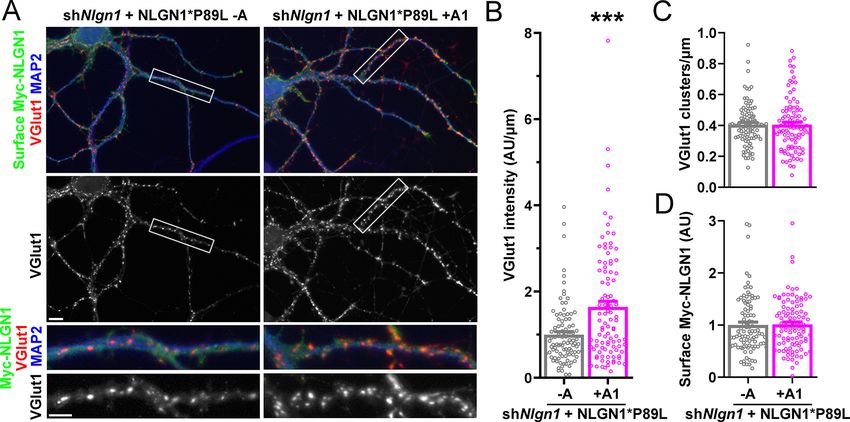
Research article                                                                                    Developmental Biology Neuroscience

                                   Figure 1. NLGN1 alternative splicing at site A regulates heparin/HS binding. (A) The amino acid sequences of
                                   each human NLGN A splice insert highlighting positively charged (blue) and negatively charged (red) residues.
                                   Splice insert sequences in mouse NLGN1-3 are identical to those in human NLGN1-3 except for one residue in
                                   NLGN1 A2 (7th residue H in mouse and Q in human). (B) Structure of the NLGN1-NRXN1b LNS domain complex
                                   (PDB: 3VKF) (Tanaka et al., 2012) showing the position of the A1 insert relative to the constitutive HS binding site
                                   (yellow asterisk) (Zhang et al., 2018). The NLGN1 surface is colored according to the electrostatic potential from
                                   blue (+8 kbT/ec) to red ( 8 kbT/ec), and the NRXN LNS domain is in cyan. (C) Structure of the NLGN1 A1 splice
                                   insert (PDB: 3VKF) highlighting the positively charged surface residues proposed to participate in HS interaction.
                                   Residues are numbered from the beginning of the A1 insert. (D) Elution profile of purified recombinant NLGN1
                                   isoform ectodomain proteins from a heparin column. Elution at a higher concentration of salt indicates stronger
                                   binding.
                                   The online version of this article includes the following figure supplement(s) for figure 1:
                                   Figure supplement 1. NLGN gene structures.

Oku et al. eLife 2020;9:e58668. DOI: https://doi.org/10.7554/eLife.58668                                                                        3 of 26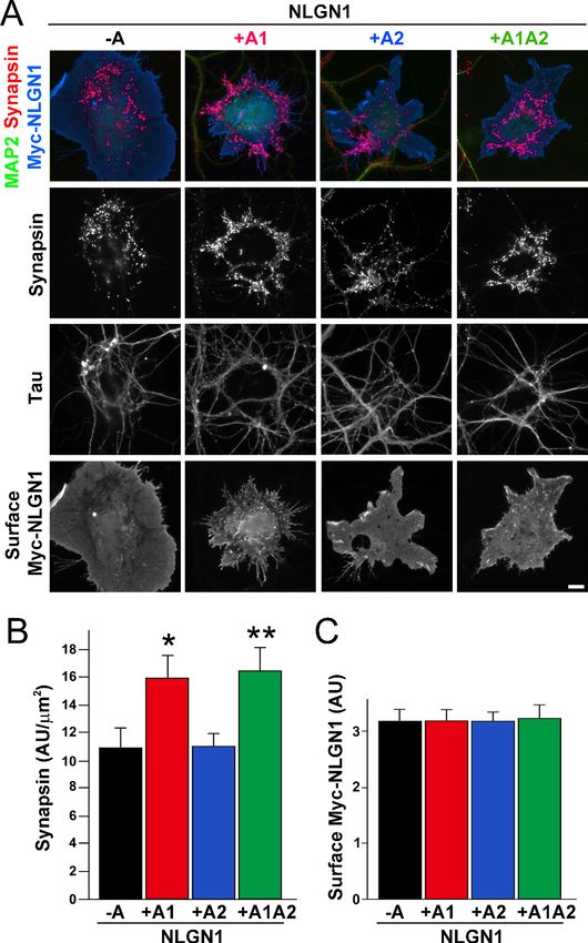
Research article                                                                                  Developmental Biology Neuroscience

                                   Figure 2. The NLGN1 A1 splice insert enhances neurexin recruitment in coculture. (A–C) Each NLGN1 isoform
                                   with an extracellular Myc tag was expressed in COS7 cells which were cocultured with hippocampal neurons.
                                   NLGN1 induced recruitment of neurexin along tau-positive axons. Axon regions contacting expressing COS7 cells
                                   and lacking contact with MAP2-positive dendrites were assessed to exclude native synapses. The total intensity of
                                   native neurexin (B) per contact area was normalized to a baseline value of 1 measured from sister cocultures
                                   performed with the negative control protein Myc-tagged Amigo. Neurexin recruitment differed among NLGN1
                                   splice variants, p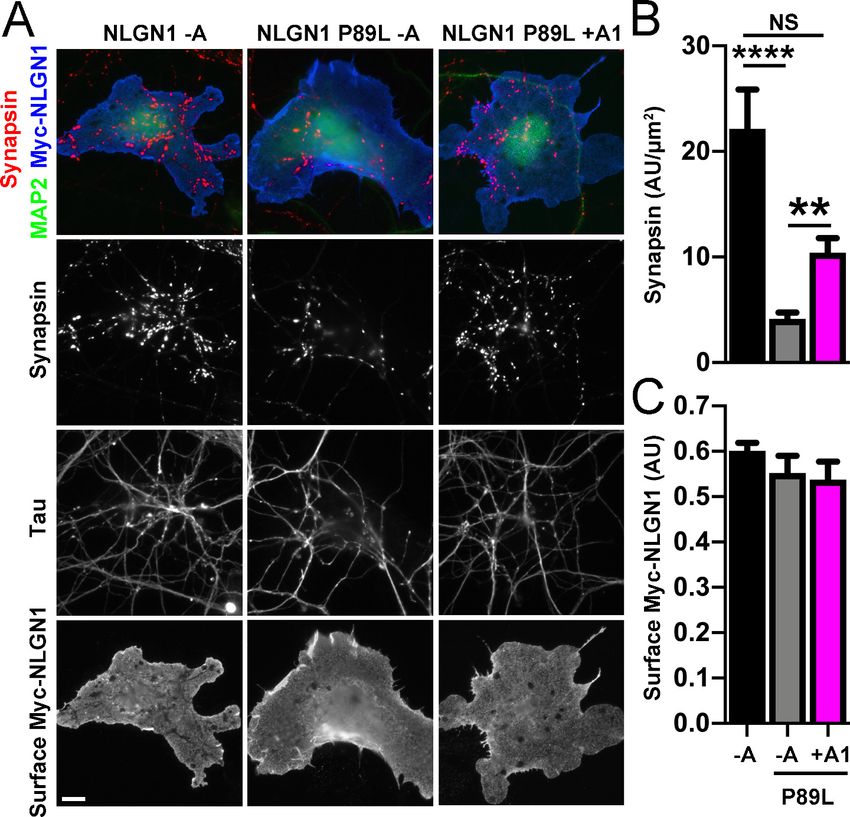
Research article                                                                           Developmental Biology Neuroscience

                                   NRXN1b ectodomain containing the full LNS domain but lacking the HS modification site. One of
                                   these studies using bacterially expressed NRXN1b reported a positive effect of the A1 insert
                                   (Comoletti et al., 2006) although two studies using NRXN1b expressed in mammalian cells reported
                                   no difference in affinity between NLGN1 -A and NLGN1 +A1 (Elegheert et al., 2017;
                                   Koehnke et al., 2010). Altogether, these previous results and our data suggest that the NLGN1 A1
                                   insert does not affect binding to the NRXN LNS domain but increases binding to the NRXN HS
                                   modification.

                                   NLGN1 A1 inclusion is high in GABAergic cell types
                                   Our data above show that alternative splicing at the NLGN1 A site affects molecular interactions.
                                   Yet, although NLGN1 transcripts containing the A1, A2 and A1A2 inserts have been detected, cell
                                   type specific splicing patterns have not been well explored. Thus, we analysed three deep RNA-seq
                                   datasets for cell type splicing patterns of the NLGN1 A site. Supplementary file 1 summarizes all
                                   RNA-seq datasets analysed in this study. Two datasets for adult mouse cortex from the Allen Insti-
                                   tute for Brain Science covered 23,822 single-cell transcriptomes with >100,000 reads per cell detect-
                                   ing approximately 9500 genes per cell (Tasic et al., 2018). Cells were isolated by FACS or manual
                                   picking following layer-enriched dissections from many Cre driver lines crossed with reporters for
                                   access to select rare cell types, and the resulting 133 transcriptomic cell types were identified by
                                   clustering analysis (Tasic et al., 2018). We analysed the primary data (Gene Expression Omnibus
                                   (GEO) GSE115746) to assess cell-type splicing at NLGN1 splice site A. In mouse primary visual cor-
                                   tex, NLGN1 A1 inserts were most prevalent in GABAergic cell types, including the well-studied VIP
                                   and parvalbumin classes (Figure 3A). Oligodendrocytes also showed high A1 inclusion whereas A1
                                   was essentially absent from NLGN1 in astrocytes and oligodendrocyte precursor cells. Cell type spe-
                                   cific splicing patterns were generally similar in anterior lateral motor cortex (Figure 3B) except here
                                   the Meis2-expressing divergent GABAergic neuron class showed more NLGN1 A1 inclusion. We fur-
                                   ther analysed an independent dataset of ribosome-engaged transcripts from genetically defined
                                   neuron types in mouse cortex and hippocampus (Furlanis et al., 2019). Their deep RNA-seq
                                   designed to study splice isoforms covered >100 million reads per biological replicate, detect-
                                   ing >12,000 genes per sample with full-length coverage across transcripts. In this dataset of actively
                                   translated transcripts (GEO GSE133291), we again found that NLGN1 A1 inclusion was highest in
                                   GABAergic neurons of the cortex, including VIP and parvalbumin classes (Figure 3C).
                                       To determine whether NLGN1 A1 inclusion changes with development, we assessed two deep
                                   RNA-seq datasets from mouse cortex spanning from embryonic day (E)14.5 to 2 years (Lister et al.,
                                   2013; Yan et al., 2015; Supplementary file 1). Previous analysis demonstrated dynamic splicing
                                   regulation of cassette exons in embryonic and postnatal cortex up to one month old (Weyn-
                                   Vanhentenryck et al., 2018). We estimated the abundance of NLGN1 isoforms by a regression anal-
                                   ysis using reads mapped to exon junctions of the alternatively spliced region. Developmentally,
                                   NLGN1 A1 inclusion in mouse cortex peaked during the first to second postnatal weeks (Figure 4A,
                                   B).

                                   NLGN1 A1 inclusion is regulated by Rbfox
                                   Cell-type specific alternative splicing is controlled by RNA-binding proteins that recognize specific
                                   regulatory sequences embedded in the pre-mRNA transcripts. Splicing factors specifically expressed
                                   or enriched in neurons include Rbfox, Mbnl, and Ptbp2 which have all been demonstrated to regu-
                                   late alternative splicing of numerous neuronal transcripts (Raj and Blencowe, 2015; Vuong et al.,
                                   2016). Thus, we determined whether genetic knockout (KO) of these families of splicing factors
                                   altered splicing of NLGN1 A1.
                                       The three Rbfox family members are thought to have considerable functional redundancy as they
                                   all bind to the same RNA sequence motif and have overlapping expression patterns in neurons
                                   (Vuong et al., 2016). To assess their combined role in NLGN1 A site splicing, due to limitations with
                                   lethality in vivo, we studied triple KO of Rbfox1, Rbfox2 and Rbfox3 in spinal neurons differentiated
                                   from mouse embryonic stem cells (Jacko et al., 2018; Supplementary file 1). At 5 days in culture,
                                   the embryonic stem cell-derived neurons elaborate branched processes and fire trains of action
                                   potentials upon current injection. Over the next 5 days of maturation, they show splicing changes
                                   paralleling those of early postnatal mouse cortex (Jacko et al., 2018). Our analyses of this dataset

Oku et al. eLife 2020;9:e58668. DOI: https://doi.org/10.7554/eLife.58668                                                           5 of 26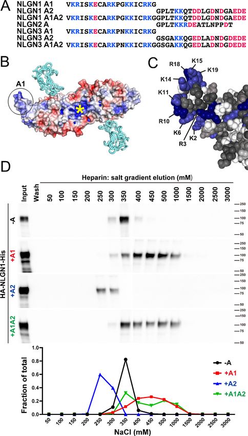
Research article                                                                                      Developmental Biology Neuroscience

Figure 3. The NLGN1 A1 splice insert is high in GABAergic neuron cell types. The fraction of each mouse NLGN1 site A splice isoform transcript is
plotted in each upper graph and the fraction of NLGN1 transcript that contains the A1 insert, that is (+A1 plus +A1A2)/total, is plotted in each lower
graph. Datasets are from Tasic et al., 2018 (A, B) and Furlanis et al., 2019 (C).

                                     revealed that Rbfox triple KO effectively eliminated NLGN1 A1 inclusion at both stages of neuron
                                     maturation (Figure 4C).
                                         To identify additional RNA binding proteins which might regulate NLGN1 splicing, we similarly
                                     examined published RNA-seq data derived upon depletion of individual RNA binding proteins
                                     (Supplementary file 1). The data were derived from: adult hippocampus of Mbnl2 KO mice
                                     (Charizanis et al., 2012); adult frontal cortex of Mbnl1 / Mbnl2loxP/loxP Nestin-Cre nervous system-
                                     specific double KO mice (Weyn-Vanhentenryck et al., 2018); embryonic day 18 brains of Ptbp2loxP/
                                     loxP
                                          Nestin-Cre nervous system-specific KO mice, and postnatal day 1 cortex of Ptbp2loxP/loxP Emx1-
                                     Cre forebrain glutamatergic neuron-selective KO mice (Li et al., 2014). These datasets were ana-
                                     lysed for alternative splicing in our previous study (Weyn-Vanhentenryck et al., 2018), from which
                                     we obtained reads mapped to the alternatively spliced region of NLGN1 to estimate abundance of
                                     different splice variants. Our analyses of these datasets essentially rules out a role for Mbnl in
                                     NLGN1 A site splicing (Figure 4D). For Ptbp2, the Emx1-Cre KO datasets did not show any differ-
                                     ence in NLGN1 A1 splicing, but NLGN1 A1 inclusion was reduced in the Nestin-Cre KO dataset
                                     (Figure 4E). These findings suggest Ptbp2 may regulate NLGN1 -A1 splicing in neurons outside the
                                     forebrain. Therefore, our transcriptomic analyses indicated a major role for Rbfox and lesser roles, if
                                     any, for Mbnl or Ptbp2 in NLGN1 A1 splicing.

Oku et al. eLife 2020;9:e58668. DOI: https://doi.org/10.7554/eLife.58668                                                                          6 of 26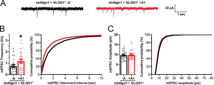
Research article                                                                                   Developmental Biology Neuroscience

                                   Figure 4. The NLGN1 A1 splice insert is regulated developmentally and by Rbfox splicing factors. The fraction of
                                   each mouse NLGN1 site A splice isoform transcript is plotted in each upper graph and the fraction of NLGN1
                                   transcript that contains the A1 insert, that is (+A1 plus +A1A2)/total, is plotted in each lower graph. In the
                                   developmental studies (A, B), ages are indicated in embryonic (E) or postnatal (P) days, or in postnatal weeks (W)
                                   or months (M). Panel (C) data are from spinal neuron cultures differentiated from Rbfox1,2,3 triple KO (TKO)
                                   embryonic stem cells and grown for the indicated number of days. Panel (D) data are from hippocampi from 2 to 3
                                   month old Mbnl2 KO mice or frontal cortex from adult Mbnl1 / Mbnl2loxP/loxP Nestin-Cre conditional double KO
                                   (cDKO) mice. Panel (E) data are from embryonic day 18 brain of Ptbp2loxP/loxP Nestin-Cre cKO mice or postnatal
                                   day 1 cortex of Ptbp2loxP/loxP Emx1-Cre cKO mice. Development datasets are from (A) (Yan et al., 2015) and (B)
                                   (Lister et al., 2013). Datasets from KO cells or mice lacking splice factors are from (C) (Jacko et al., 2018), (D)
                                   (Charizanis et al., 2012; Weyn-Vanhentenryck et al., 2018) and (E) (Li et al., 2014).

Oku et al. eLife 2020;9:e58668. DOI: https://doi.org/10.7554/eLife.58668                                                                       7 of 26
Research article Developmental Biology Neuroscience
Figure 5. The NLGN1 A1 splice insert enhances presynaptic differentiation in coculture. (A–C) Each NLGN1
isoform with an extracellular Myc tag was expressed in COS7 cells which were cocultured with hippocampal
neurons. NLGN1 induced clustering of synapsin along tau-positive axons. Axon regions contacting expressing
COS7 cells and lacking contact with MAP2-positive dendrites were assessed to exclude native synapses. The total
intensity of synapsin (B) per contact area was normalized to a baseline value of 1 measured from sister cocultures
performed with the negative control protein Myc-tagged Amigo. Synapsin clustering differed among NLGN1
splice variants, pResearch article Developmental Biology Neuroscience
The NLGN1 A1 insert enhances presynaptic differentiation in coculture
To test the functional impact of the NLGN1 A1 splice site, we first used the neuron - COS7 cell
coculture assay to assess synaptogenic activity of the NLGN1 splice variants. Via NRXN recruitment
as seen in Figure 2, NLGNs expressed on the surface of non-neuronal cells induce full presynaptic
differentiation in contacting axons, reflected by clustering of multiple presynaptic components
(Scheiffele et al., 2000). NLGN binding to axonal NRXN is required for this synaptogenic activity in
coculture, based on loss of activity upon NRXN triple knockdown (Gokce and Südhof, 2013;
Zhang et al., 2018) or by NLGN point mutations that disrupt binding to the NRXN LNS domain
(Ko et al., 2009) or to the NRXN HS modification (Zhang et al., 2018). Here we found that NLGN1
+A1 and +A1A2 induced greater presynaptic differentiation than other splice variants, assessed by
synapsin clustering at axon - COS7 cell contact sites lacking dendrite contact to exclude native syn-
apses (Figure 5). Thus, the NLGN1 -A1 insert promotes presynaptic differentiation in coculture
assays.
The NLGN1 A1 insert promotes structural and functional synapse
development
To assess the role of the NLGN1 A1 insert at native synapses, we used a molecular replacement
strategy knocking down native NLGN1 with shNlgn1 (Zhang et al., 2018) and re-expressing RNAi-
resistant Myc-NLGN1* lacking or containing the A1 insert. For these assays, we focused on NLGN1 -
A, the major neuronal form (Figures 3 and 4; Chih et al., 2006), and NLGN1 +A1. We found that
NLGN1* +A1 induced greater presynaptic differentiation than NLGN1* -A, assessed by total inten-
sity of vesicular glutamate transport VGlut1 clustering onto expressing neurons (Figure 6A–D). There
was no difference between the NLGN1 isoforms in the density of VGlut1 clusters per dendrite area,
suggesting no difference in synapse numbers, but a difference in total intensity of VGlut1 per
cluster.
We observed no difference in the localization of Myc-NLGN1* -A and +A1 in these assays. How-
ever, as in previous assays expressing YFP-NLGN1 in the absence of recombinant postsynaptic scaf-
folds (Graf et al., 2004), both isoforms of Myc-NLGN1* showed high diffuse levels with poor
postsynaptic clustering. Thus, to determine whether A1 alternative splicing mediates differential syn-
aptic recruitment, we used a more sensitive tag on NLGN1* and slightly different culture conditions
(Chamma et al., 2016; Letellier et al., 2018). NLGN1* was tagged with an extracellular 15-amino-
acid acceptor peptide (AP) that is biotinylated upon co-expression of a biotinylating enzyme BirAER
and then surface labelled with fluorescent streptavidin. The longer culture time and co-expression of
Homer1c-dsRed to mark postsynaptic sites may also enhance synapse maturation (Zeng et al.,
2018), altogether resulting in postsynaptic clustering of AP-NLGN1*. However, there was no differ-
ence in the degree of synaptic enrichment of AP-NLGN1* -A versus +A1, nor in Homer1c-dsRed syn-
aptic enrichment, cluster density, or cluster area between groups (Figure 6E–I). Collectively, our
data indicate that the NLGN1 A1 splice insert mediates differential recruitment of NRXN and presyn-
aptic differentiation but not differential postsynaptic targeting.
To assess effects of NLGN1 A1 splicing directly on synaptic function, we used a similar molecular
replacement strategy to knockdown native NLGN1 and re-express NLGN1* -A or +A1 and recorded
miniature excitatory synaptic currents (mEPSCs) in cultured hippocampal neurons. Neurons express-
ing shNlgn1 and rescued with NLGN1* +A1 had significantly greater mEPSC frequency than those
rescued with NLGN1* -A, with a corresponding reduction in mEPSC interevent interval (Figure 7).
There was no difference between groups in mEPSC amplitude. Thus, the NLGN1 A1 splice insert
enhances functional transmission as well as structural presynaptic differentiation.
A1 insertion partially restores autism-linked NLGN1 P89L synaptogenic
function
We propose that the A1 insert might be a potential target for alleviating deficits of ASD NLGN1 mis-
sense mutations by enhancing their function. To test this idea, we chose the NLGN1 P89L missense
variant found in two ASD siblings but not in controls (Nakanishi et al., 2017). Modelling in Nlgn1
P89L heterozygous knock-in mice resulted in reduced NLGN1 protein levels and deficits in social
interaction, altered social dominance, and impaired spatial memory, supporting a causative role of
this mutation in autism (Nakanishi et al., 2017). We confirmed an expected reduction in surface
Oku et al. eLife 2020;9:e58668. DOI: https://doi.org/10.7554/eLife.58668 9 of 26Research article Developmental Biology Neuroscience Figure 6. The NLGN1 A1 splice insert promotes structural synapse development. (A–D) Cultured hippocampal neurons were transfected with U6- shNlgn1-hSyn-CFP and hSyn-Myc-NLGN1* -A or +A1 at DIV 5 and neurons were fixed at DIV 12. The density of VGlut1 clusters did not differ but the total fluorescence intensity of VGlut1 inputs was higher for neurons expressing the NLGN1* +A1 than the -A isoform, ***p=0.0006 by Mann Whitney test, n = 59–66 neurons from three independent experiments. Cells were chosen for equal intensity of surface Myc-NLGN1. Scale bar, top 10 mm, bottom 5 mm. (E–I) Cultured hippocampal neurons were electroporated at plating with shNlgn1-GFP, Homer1c-dsRed, BirAER, and hSyn-AP-NLGN1* -A or +A1 and neurons imaged at DIV 14 following live cell labeling with streptavidin-Alexa647. The synaptic enrichment of AP-NLGN1*, defined as the intensity of AP-NLGN1* in Homer1c-dsRed-positive clusters relative to the intensity in the local dendrite shaft, did not differ between the splice variants, nor was there any difference in the synaptic enrichment or density or mean area of Homer1c-dsRed clusters (all p>0.1 by Mann Whitney test, n > 14 neurons from two independent experiments). Scale bar, 10 mm. Oku et al. eLife 2020;9:e58668. DOI: https://doi.org/10.7554/eLife.58668 10 of 26
Research article                                                                                     Developmental Biology Neuroscience

Figure 7. The NLGN1 A1 splice insert promotes functional synapse development. Cultured hippocampal neurons were transfected with U6-shNlgn1-
hSyn-YFP, hSyn-DIO-YFP-P2A-HA-NLGN1* -A or +A1, and CAG-Cre at DIV 5. YFP positive neurons were selected for mEPSC recording at DIV 13 and
14. (A) Representative mEPSC traces from neurons expressing NLGN1* lacking (black trace) or containing (red trace) the A1 splice variant. (B) mEPSC
frequency was significantly increased in NLGN1* +A1-expressing neurons (n = 26) relative to cells expressing NLGN1* -A (n = 26; *p=0.040 by Welch’s t
test), with a corresponding change in interevent interval. (C) mEPSC amplitude did not significantly differ between groups (p=0.93). Scale bar, 20 pA, 1
s.

                                     expression of Myc-NLGN1 -A P89L relative to WT (Figure 8—figure supplement 1), likely reflecting
                                     enhanced endoplasmic reticulum-associated degradation (Nakanishi et al., 2017).
                                        We next assessed the synaptogenic activity of NLGN1 P89L. Interestingly, even though cells with
                                     equal NLGN1 surface expression were chosen for the coculture assay, the P89L mutation significantly
                                     reduced presynaptic differentiation by NLGN1 (Figure 8). NLGN1 P89L -A induced
Research article Developmental Biology Neuroscience
splicing factors, and transcriptomic analyses reveals high A1 inclusion in multiple cortical GABAergic
cell types. Finally, we show that A1 inclusion enhances function not just of wild type NLGN1 but also
of the ASD-linked NLGN1 P89L mutant. We suggest that the A splice site might be harnessed as a
means to restore function in disorders involving deleterious mutations in NLGN1, and potentially
NLGN3, by developing therapeutics to convert the more prevalent -A form to the more synapto-
genic +A1 form.
The role identified here for the NLGN1 splice site A1 insert is an unusual one, increasing NLGN1
interaction with HS glycan and thus enhancing synaptogenic function. Although there are many cases
of alternative splicing regulating synapse development through altered protein-protein interactions,
a mechanism involving altered protein-glycan interaction is less common. The NLGN1 B splice site
also regulates synaptogenic function partially through glycosylation, in that case through N-glycosyl-
ation of the NLGN1 B insert blocking binding to a-NRXN while retaining binding to b-NRXN
(Boucard et al., 2005). Neural-specific alternative splicing of agrin at its y site also regulates its
Figure 8. A1 splice inclusion in the autism-linked NLGN1 P89L mutant enhances presynaptic differentiation in
coculture. (A–C) Myc-NLGN1 -A, Myc-NLGN1 P89L -A, or Myc-NLGN1 P89L +A1 was expressed in COS7 cells
which were cocultured with hippocampal neurons. The total intensity of synapsin (B) per COS7 cell - axon contact
area lacking contact with MAP2-positive dendrites was normalized to a baseline value of 1 measured from sister
cocultures performed with the negative control protein Myc-tagged CD4. Synapsin clustering by NLGN1 -A was
reduced by the P89L mutation and partially restored by A1 splice site inclusion, ****pResearch article Developmental Biology Neuroscience
Figure 9. A1 splice inclusion in the autism-linked NLGN1 P89L mutant promotes synapse development. (A–D) Cultured hippocampal neurons were
transfected with U6-shNlgn1-hSyn-YFP and hSyn-CFP-P2A-Myc-NLGN1* P89L -A or P89L +A1 at DIV 5 and neurons were fixed at DIV 13. The density of
VGlut1 clusters did not differ (C) but the total fluorescence intensity of VGlut1 inputs (B) was higher for neurons expressing NLGN1* P89L +A1 than the
-A isoform, ***p=0.0008 by Mann Whitney test, n = 97–99 neurons from three independent experiments. Cells were chosen by equal surface Myc-
NLGN1 immunofluorescence (D). Scale bar, top 10 mm, bottom 5 mm.
interaction with HS, and HS binding reduces agrin interaction with dystroglycan (Gesemann et al.,
1996; O’Toole et al., 1996). However, the y site and these interactions are not critical for agrin syn-
aptogenic activity (Burgess et al., 1999; Li et al., 2018), unlike the central role of the NRXN interac-
tion for NLGN1 function.
Our data reveal effects of the NLGN1 A1 insert on increasing NRXN recruitment and presynaptic
differentiation in coculture, and on increasing VGlut1 levels at inputs and mEPSC frequency in hippo-
campal neurons. However, the NLGN1 A1 insert had no apparent effect on the synaptic targeting of
NLGN1. This finding is consistent with other data suggesting that the excitatory postsynaptic locali-
zation of NLGN1 is largely controlled by intracellular interactions regulated by multiple kinases
(Jeong et al., 2019; Jeong et al., 2017; Letellier et al., 2020; Letellier et al., 2018). The NLGN1
+A1-associated increase in mEPSC frequency and VGlut1 cluster intensity without a change in
VGlut1 or Homer1c-dsRed cluster density suggest there is no change in synapse numbers but rather
an increase in neurotransmitter release. These findings are consistent with previous reports of a ret-
rograde effect of NLGN1 on neurotransmitter release probability (Futai et al., 2007; Stan et al.,
2010). In contrast, the lack of effect of the NLGN1 A1 insert on mEPSC amplitude suggests no effect
on postsynaptic sensitivity. Thus, altogether our data are consistent with a purely retrograde mecha-
nism of action of the NLGN1 A1 insert through enhanced NRXN recruitment mediating enhanced
presynaptic differentiation and increased transmitter release rate. While this work was under revi-
sion, it was reported that the A1 insert in NLGN3 regulates GABAergic transmission, as overexpres-
sion of NLGN3 +A1 or +A1A2 reduced GABAergic transmission whereas NLGN3 -A or +A2
increased GABAergic transmission relative to neighbour untransfected cells in hippocampal slice cul-
tures (Uchigashima et al., 2020). Differential effects were also found on presynaptic release proba-
bility. We suggest that the mechanism of this effect may also involve differential interactions with
NRXN -HS.
Oku et al. eLife 2020;9:e58668. DOI: https://doi.org/10.7554/eLife.58668 13 of 26Research article Developmental Biology Neuroscience
The effect of the higher NLGN1 A1 inclusion in GABAergic than in glutamatergic neuron classes
may be to increase excitatory input selectively onto the GABAergic neurons, thus enhancing syn-
chrony of the overall network. In particular, cortical VIP and parvalbumin interneurons, which showed
high NLGN1 A1 inclusion in all datasets, play key roles in neural synchrony and cognitive processing.
For example, VIP interneuron disinhibition of prefrontal responses to hippocampal inputs controls
avoidance behaviour (Lee et al., 2019). NLGN3 on parvalbumin interneurons contributes to gamma
oscillations and social behaviour as well as fear conditioning (Cao et al., 2018; Polepalli et al.,
2017).
Our data also shed some light on the mechanisms regulating alternative splicing of NLGN1 A1,
with KO data supporting a role for Rbfox and a possible lesser role for Ptbp2 splice factors.
Although further validation of NLGN1 as an Rbfox target will be needed, NLGN1 appears to be one
among many Rbfox targets. Unlike the highly dedicated splicing program of SLM2 regulating synap-
tic properties through NRXN site 4 splicing (Traunmüller et al., 2016), the Rbfox family regulates a
broader range of targets controlling synaptic properties and other aspects of neuronal maturation
including axon initial segment assembly (Jacko et al., 2018). Rbfox proteins also act as more than
just splicing factors; in GABAergic neurons, a mechanism involving Rbfox1 binding to the Vamp1
mRNA 3’ UTR regulates synaptic transmission (Vuong et al., 2018). Like NLGNs, RBFOX1 is a risk
gene for ASD based on strong evidence including de novo copy number deletions (Sebat et al.,
2007; Zhao, 2013).
The results here with the P89L NLGN1 mutant support the idea that elevating A1 insertion is a
promising direction to enhance the function of NLGN1 in cases of deleterious mutations. This
approach will likely extend to NLGN3 as well as NLGN1, given their high sequence homology includ-
ing the A1 insert (Figure 1A). NLGN3 is among the top risk genes for ASD with multiple functionally
deleterious missense mutations (Jamain et al., 2003; Quartier et al., 2019). In the majority of cases
which involve heterozygous mutations, reagents that increase A1 insertion could act by enhancing
function of both the mutant NLGN and the wild type allele. NRXN1 is also among the top risk genes
for ASD as well as for schizophrenia and Tourette’s Syndrome (Tourette Syndrome Association
International Consortium for Genetics (TSAICG) et al., 2017; Rees et al., 2014; Südhof, 2017).
Therapies targeting wild type NLGN splicing to elevate the function of NLGN-NRXN complexes may
also be partially efficacious in cases of NRXN mutations. Clinically effective oligonucleotide reagents
targeting splicing events have reached the bedside for other disorders, notably for increasing spe-
cific exon inclusion in SMN2 to treat spinal muscular atrophy (Bennett et al., 2019; Singh et al.,
2017).
Materials and methods
Key resources table
Reagent type Additional
(species) or resource Designation Source or reference Identifiers information
Strain, strain Sprague-Dawley Charles River CD-IGS For primary
background hippocampal
(Rattus norvegicus) neuron cultures
Strain, strain C57BL/6J Jackson Laboratory Cat# 00064 For RNA for RT-
background PCR for cloning
(Mus musculus)
Cell line HEK293 ATCC Cat# CRL-1573
(Homo sapiens)
Cell line COS7 ATCC Cat# CRL-1651
(Cercopithecus aethiops)
Biological sample Primary hippocampal This paper Freshly isolated from
(Rattus norvegicus) neurons Rattus norvegicus
Antibody anti-Myc Sigma Cat# C3956; (ICC 1:40000)
(rabbit polyclonal) RRID:AB_439680
Antibody anti-neurexin Millipore Cat# ABN161; (ICC 1:1000)
(rabbit polyclonal) RRID:AB_10917110
Continued on next page
Oku et al. eLife 2020;9:e58668. DOI: https://doi.org/10.7554/eLife.58668 14 of 26Research article Developmental Biology Neuroscience
Continued
Reagent type Additional
(species) or resource Designation Source or reference Identifiers information
Antibody anti-MAP2 Abcam Cat# ab5392; (ICC 1:80000)
(chicken polyclonal) RRID:AB_2138153
Antibody anti-VGlut1 NeuroMab Cat# N28/9; (ICC 1:4000)
(mouse monoclonal) RRID:AB_2187693
Antibody anti-synapsin I Synaptic Systems Cat# 106 011; (ICC 1:20000)
(mouse monoclonal) RRID:AB_2619772
Antibody anti-tau (mouse monoclonal) Millipore Cat# MAB3420; (ICC 1: 2000)
RRID:AB_94855
Antibody anti-HA (rat monoclonal) Roche Cat# 1186743100; (WB 1: 2000)
RRID:AB_390919
Recombinant pNICE-HA-NLGN1 Scheiffele et al., 2000 To subclone
DNA reagent NLGN1 isoforms
Recombinant pCAGGS-Myc-NLGN1 This paper To express Myc-NLGN1 -A
DNA reagent (-A+B)WT
Recombinant pCAGGS-Myc-NLGN1 This paper To express Myc-NLGN1 +A1
DNA reagent (+A1+B)WT
Recombinant pCAGGS-Myc-NLGN1 This paper To express Myc-NLGN1 +A2
DNA reagent (+A2+B)WT
Recombinant pCAGGS-Myc-NLGN1 This paper To express Myc-NLGN1 +A1A2
DNA reagent (+A1A2+B)WT
Recombinant pCAGGS-Myc-NLGN1 This paper To express Myc-NLGN1 P89L -A
DNA reagent (-A+B)P89L
Recombinant pCAGGS-CFP-P2A-Myc- This paper To express Myc-NLGN1 -A
DNA reagent NLGN1(-A+B)WT
Recombinant pCAGGS-CFP-P2A-Myc- This paper To express Myc-NLGN1
DNA reagent NLGN1(-A+B)P89L P89L -A
Recombinant pCAGGS-CFP-P2A-Myc- This paper To express Myc-NLGN1
DNA reagent NLGN1(+A1+B)P89L P89L +A1
Recombinant pcDNA4-HA-ecto- This paper To express HA-NLGN1-
DNA reagent NLGN1(-A+B)-His His -A ectodomain
Recombinant pcDNA4-HA-ecto- This paper To express HA-NLGN1-
DNA reagent NLGN1(+A1+B)-His His +A1 ectodomain
Recombinant pcDNA4-HA-ecto- Zhang et al., 2018 To express HA-NLGN1-
DNA reagent NLGN1(+A2+B)-His His +A2 ectodomain
Recombinant pcDNA4-HA-ecto- This paper To express HA-NLGN1-
DNA reagent NLGN1(+A1A2+B)-His His +A1A2 ectodomain
Recombinant pCAGGS-Myc-Amigo This paper To express Myc-Amigo
DNA reagent
Recombinant Amigo-CFP Siddiqui et al., 2010 To subclone Myc-Amigo
DNA reagent
Recombinant Myc-CD4 This paper To express Myc-CD4
DNA reagent
Recombinant HA-CD4 Takahashi et al., 2011 To subclone Myc-CD4
DNA reagent
Recombinant pLL3.7(hSyn)-Myc- This paper To express Myc-NLGN1* -A
DNA reagent NLGN1*(-A+B)WT
Recombinant pLL3.7(hSyn)-Myc- This paper To express
DNA reagent NLGN1*(+A1+B)WT Myc-NLGN1* +A1
Recombinant pLL3.7(hSyn)-CFP-P2A- This paper To express
DNA reagent Myc-NLGN1*(-A+B)P89L Myc-NLGN1* -A P89L
Recombinant pLL3.7(hSyn)-CFP-P2A- This paper To express
DNA reagent Myc-NLGN1*(+A1+B)P89L Myc-NLGN1* +A1 P89L
Continued on next page
Oku et al. eLife 2020;9:e58668. DOI: https://doi.org/10.7554/eLife.58668 15 of 26Research article Developmental Biology Neuroscience
Continued
Reagent type Additional
(species) or resource Designation Source or reference Identifiers information
Recombinant pFB-hSyn-DIO-YFP-P2A Zhang et al., 2018 To subclone
DNA reagent -HA-NLGN1*(+A2+B) NLGN1* isoforms
Recombinant pFB-hSyn-DIO-YFP- This paper To express Cre-dependent
DNA reagent P2A-HA-NLGN1*(-A+B) HA-NLGN1* -A
Recombinant pFB-hSyn-DIO-YFP-P2A This paper To express Cre-dependent
DNA reagent -HA-NLGN1*(+A1+B) HA-NLGN1* +A1
Recombinant pCAG-Cre Addgene Addgene# 13775 To express Cre
DNA reagent
Recombinant pLL3.7U6-shNlgn1-hSyn-YFP Zhang et al., 2018 To express shNlgn1
DNA reagent
Recombinant pLL3.7U6-shNlgn1-hSyn-CFP This paper To express shNlgn1
DNA reagent
Recombinant pLL3.7(hSyn)-MCS This paper To maintain uniform
DNA reagent DNA amounts for
transfections
Recombinant pECFP-N1 Clontech To express CFP
DNA reagent
Recombinant AP-NLGN1*(-A) This paper To express
DNA reagent AP-NLGN1* -A
Recombinant AP-NLGN1*(+A1) This paper To express
DNA reagent AP-NLGN1* +A1
Recombinant shNlgn1-GFP Chamma et al., 2016 To express shNlgn1
DNA reagent
Recombinant dsRed-Homer1C Mondin et al., 2011 To express
DNA reagent dsRed-Homer1c
Recombinant BirAER Howarth et al., 2006 To express BirAER
DNA reagent
Chemical Bicuculline methiodide Abcam Cat# ab120109
compound, drug
Chemical Tetrodotoxin (TTX) Abcam Cat# ab120054
compound, drug
Other Ni-NTA agarose beads QIAGEN Ca# 30210
Other Heparin agarose GE Healthcare Cat# 17-0406-01
Software, WinLTP WinLTP RRID:SCR_008590
algorithm
Software, MiniAnalysis Synaptosoft RRID:SCR_002184
algorithm
Software, Fiji 64-bit https://doi.org/ RRID:SCR_002285
algorithm 10.1038/nmeth.2019
Software, Pymol Pymol RRID:SCR_000305
algorithm
Software, OLego v1.1.2 Wu et al., 2013 RRID:SCR_005811
algorithm
Software, R R Foundation RRID:SCR_001905
algorithm
Software, GraphPad Prism GraphPad RRID:SCR_002798;
algorithm SCR_000306
Software, GraphPad InStat GraphPad RRID:SCR_000306
algorithm
Software, GraphPad SigmaPlot GraphPad RRID:SCR_000306
algorithm
Oku et al. eLife 2020;9:e58668. DOI: https://doi.org/10.7554/eLife.58668 16 of 26Research article Developmental Biology Neuroscience
DNA constructs
Mouse NLGN1(-A+B) (GenBank: XM_017319496) was cloned from adult mouse whole brain by RT-
PCR. Insert A1 (GenBank: XM_017319494.1) and A1A2 (GenBank: XM_006535415.2) were inserted
by site-directed mutagenesis. NLGN1(+A2+B) was a gift from Peter Scheiffele (Scheiffele et al.,
2000). To construct the P89L mutant, nucleotides CCA (Pro) were changed to CTA (Leu). A Myc tag
and linker (EQKLISEEDLGGQ) were inserted between amino acid Gln46 and Lys47. CFP-linker-P2A
(YGSGATNFSLLKQAGDVEENPGP) was fused at the N-terminus of Myc-NLGN1 before the signal
sequence. For the heparin binding assay, since pcDNA4-HA-ecto-NLGN1 (Zhang et al., 2018) is the
NLGN1(+A2) isoform, the A2 region was removed to construct NLGN1(-A) or replaced with insert
A1 or A1A2 for the NLGN1(+A1) or NLGN1(+A1A2) isoform, respectively, to express the NLGN1
ectodomain of each isoform. For coculture assays, Myc-NLGN1 isoforms and CFP-P2A-Myc-NLGN1(-
A) WT and P89L mutant were inserted into pCAGGS under a CAG promotor to express in COS7
cells. To construct Myc-Amigo, NLGN1 was replaced with rat Amigo (amino acid 28–493) from
Amigo-CFP (Siddiqui et al., 2010; Zhang et al., 2018). To construct Myc-CD4, the HA tag in HA-
CD4 (Takahashi et al., 2011) was replaced with the Myc tag. For co-expression of Myc-NLGN1 and
CFP in COS7 cells, pECFP-N1 (Clontech) was used. For VGlut1 recruitment assays, Myc-NLGN1 iso-
forms and CFP-P2A-Myc-NLGN1 (-A) and (+A1) P89L mutant were inserted into pLL3.7 under a syn-
apsin promoter to express in hippocampal neurons. For electrophysiology, NLGN1(-A) and NLGN1
(+A1) isoforms were made from pFB-hSyn-DIO-YFP-P2A-HA-NLGN1, which is the NLGN1(+A2) iso-
form (Zhang et al., 2018). The A2 region was removed for the NLGN1 (-A) isoform or replaced with
A1 to construct the NLGN1(+A1) isoform. pCAG-Cre was described previously (Zhang et al., 2018).
To knock-down endogenous NLGN1, for most experiments pLL3.7U6-shNlgn1-hSyn-CFP was used
which was generated from pLL3.7U6-shNlgn1-hSyn-YFP and the shRNA resistant form NLGN1*
made by mutating the underlined nucleotides 5’-CAAGGGGAAGGGTTGAAGTT-3’ to 5’-CAAGGC-
GAGGGACTAAAGTT-3’ as previously reported (Zhang et al., 2018). For the synaptic enrichment
assay, the plasmids for shNlgn1-GFP, RNAi-resistant AP-NLGN1*(+A2+B), BirAER, and dsRed-Hom-
er1c were previously described, with thanks to Alice Ting (Chamma et al., 2016; Howarth et al.,
2006; Mondin et al., 2011). AP-NLGN1*(-A) and (+A1) were generated from the (+A2) form by In-
Fusion HD Cloning (Takara Bio) or site-directed mutagenesis.
Cell culture and transfection
This study was performed in accordance with the recommendations of the Canadian Council on Ani-
mal Care. All of the animals were handled according to approved institutional animal care and use
committee (IACUC) protocols (A16-0086 and A16-0090) of the University of British Columbia. For
most experiments, primary hippocampal neurons were prepared from male and female rat embryos
at embryonic day 18 as previously reported (Kaech and Banker, 2006; Zhang et al., 2018). The dis-
sociated hippocampal neurons were plated on 18 mm round coverslips coated with poly-L-lysine at
the density of 36,000 neurons/coverslip. The coverslips were flipped over and neurons grown sus-
pended above a glial feeder layer in a 60 mm dish. To block glia cell proliferation, cytosine arabino-
side at a final concentration of 5 mM was added at day in vitro (DIV) 1. Neurons were transfected
with 2 mg of plasmid DNA and 2 ml of Lipofectamine 2000 (Thermo Fisher Scientific) at DIV 5. The fol-
lowing are the combinations of plasmids used for each assay with ratio 10:8:2: for VGlut1 immunoflu-
orescence, U6-shNlgn1-hSyn-CFP + hSyn-Myc-NLGN1* WT + pLL(syn)-MCS (empty vector) or U6-
shNlgn1-hSyn-YFP + hSyn-CFP-P2A-Myc-NLGN1* P89L + pLL(syn)-MCS (empty vector); for electro-
physiology, U6-shNlgn1-hSyn-YFP + hSyn-DIO-YFP-P2A-HA-NLGN1* (-A) or (+A1) + pCAG-Cre.
For the synaptic enrichment assay, dissociated rat hippocampal neurons were electroporated
with the Amaxa system (Lonza) using 300,000 cells per cuvette with the following combination of
plasmids: shNlgn1-GFP (3 mg), BirAER (1 mg), Homer1c-dsRed (1 mg), and AP-NLGN1* (-A) or (+A1)
(1 mg). Electroporated neurons were immediately resuspended in Minimal Essential Medium supple-
mented with 10% horse serum and plated on 18 mm coverslips previously coated with poly-L-lysine.
Three hours after plating, coverslips were flipped onto 60 mm dishes containing a glial cell feeder
layer in Neurobasal Plus Medium supplemented with NeuroCult SM1 Neuronal supplement (STEM-
CELL Technologies), and cultured for 2 weeks before experiments.
Mycoplasma-negative COS7 cells or HEK239T cells obtained from ATCC without further authenti-
cation were maintained in Dulbecco’s Modified Eagle’s medium (Thermo Fisher Scientific) with 10%
Oku et al. eLife 2020;9:e58668. DOI: https://doi.org/10.7554/eLife.58668 17 of 26Research article Developmental Biology Neuroscience
bovine growth serum. For transfection, plasmid DNA and 0.045% Polyethylenimine (Sigma 408727)
were mixed in a 1:3 or 1:4 ratio (w/v). For coculture assays, an N-methyl-D-aspartate (NMDA) recep-
tor inhibitor, APV, at a final concentration of 100 mM was added into the neuron dishes at DIV 6, 9,
and 13. COS7 cells were trypsinized 24 hr after transfection. 40,000 COS7 cells were seeded on a
coverslip with neurons at DIV 13. After 1 hr, the coverslip was flipped back into a glia dish and 24 hr
later, the COS7-neuron cocultures were fixed.
Immunocytochemistry
For coculture assays, neurons and COS7 cells were fixed with 4% formaldehyde/4% sucrose/PBS, pH
7.4 for 12 min, blocked with 10% BSA/PBS for 30 min at 37˚C, and incubated with Myc antibody in
3% BSA/5% normal goat serum (NGS)/PBS for surface staining overnight at 4˚C. The next day, the
cells were fixed again for 5 min, permeabilized with 0.2% Triton X-100/PBS, blocked with 10% BSA,
and incubated with the primary antibodies in 3% BSA/5% NGS/PBS overnight at 4˚C. Cells were
then incubated with the secondary antibodies in 3% BSA/5% NGS/PBS for 45 min at 37˚C. The cov-
erslips were mounted in elvanol (Tris-HCl, glycerol, and polyvinyl alcohol, with 2% 1,4-diazabicyclo
{2,2,2} octane). For surface expression of Myc-NLGN1(-A) WT and P89L in COS7 cells, cells were
fixed and blocked as above 48 hr after transfection, followed by incubation with Myc antibody over-
night at 4˚C, and incubation with the secondary antibody the next day. For VGlut1 immunofluores-
cence assays, the transfected hippocampal neurons were fixed at DIV 12 and immunostained as
above, except the second 5 min fixation was not done. The following primary antibodies were used:
Rabbit polyclonal Myc antibody (Sigma C3956; 1:40000), rabbit polyclonal pan-NRXN antibody
(Millipore ABN161; 1:1000), chicken polyclonal MAP2 antibody (Abcam ab5392; 1:80000), mouse
monoclonal VGlut1 antibody (NeuroMab 75–066; clone N28/9; IgG1; 1:4000), mouse monoclonal
SynapsinI antibody (Synaptic Systems 106 011; clone 46.1; IgG1; 1:20000), mouse monoclonal Tau
antibody (Millipore MAB3420; clone PC1C6; IgG2a; 1:2000). Secondary antibodies coupled to Alexa
Flour 488, 568, 647 (Thermo Fisher Scientific; 1: 1000) and anti-chicken IgY AMCA (Jackson Immu-
noResearch 703-155-155; 1:400) were used. Image acquisition and analysis was performed blind to
the experimental condition. Images were acquired on a Zeiss Axioplan 2 microscope with 63x/1.4
numerical aperture (NA) oil objective, ORCA-Flash4.0 FL CMOS camera (Hamamatsu) and custom fil-
ter sets or a Zeiss LSM700 with 40x/1.4 NA oil objective, ORCA-Flash4.0 CMOS camera (Hama-
matsu) and custom filter sets. The acquired images were analysed with Fiji. Off-cell background was
subtracted from each value. COS7 cells or neurons were selected for analysis based only on cell
health and cell surface expression level of Myc-NLGN1, except for Figure 8—figure supplement 1
where cells were chosen based on CFP expression. For the coculture assays, the total intensity of
pan-NRXN or Synapsin puncta was measured within the area of COS7 cells where Tau-positive axon
was present and MAP2-positive dendrite was absent. For the neuron cultures, the number and total
intensity of VGlut1 puncta was measured and normalized to the area or length of MAP2-positive
dendrite.
For the synaptic enrichment assay, for NLGN1 staining, cell cultures were rinsed twice in pre-
warmed Tyrode solution (15 mM D-glucose, 108 mM NaCl, 5 mM KCl, 2 mM MgCl2, 2 mM CaCl2
and 25 mM HEPES, pH 7.4, 280 mOsm). Cells were then incubated for 10 min at 37˚C with Tyrode
solution containing 1% biotin-free Bovine Serum Albumin (Roth, ref 0163.4), followed by an incuba-
tion in 100 nM Alexa 647-conjugated streptavidin (cat#S32357, Thermo Fisher Scientific). Following
two washes in Tyrode solution, cells were imaged on an inverted epifluorescence microscope (Nikon
Eclipse TiE) equipped with a 60x/1.49 NA objective lens and an sCMOS camera (Prime 95B, Photo-
metrics) driven by the Metamorph software (Molecular Devices). Homer1c-DsRed puncta were
thresholded using Metamorph, and the number of puncta per dendrite length was determined, as
well as area and intensity relative to dendrite shaft. Regions around Homer1c-DsRed objects were
created and transferred to the streptavidin image. The intensity of the streptavidin signal in the
puncta was normalized to the shaft signal, giving the NLGN1 synaptic enrichment.
Heparin binding assay
Purification of NLGN1 ectodomain proteins and the heparin binding assay were previously described
(Zhang et al., 2018). Briefly, an N-terminal HA tagged and C-terminal His tagged NLGN1 ectodo-
main of each isoform was transiently expressed in HEK293T cells and purified with Ni-NTA agarose
Oku et al. eLife 2020;9:e58668. DOI: https://doi.org/10.7554/eLife.58668 18 of 26Research article Developmental Biology Neuroscience
beads (QIAGEN). Purified NLGN1 ectodomains were incubated with heparin agarose (GE Health-
care) and eluted with NaCl in step-wise gradient concentrations (50 mM – 3 M) in 20 mM HEPES, pH
7.4. Collected eluates were analysed by immunoblotting with rat monoclonal HA antibody (Roche
11867431001; clone 3F10; 1:2000), secondary HRP-conjugated antibody, and detected using the
SuperSignal Chemiluminescent kit (Thermo Fisher Scientific).
Electrophysiology
Cultured hippocampal neurons (DIV 13–14) were transferred to a submerged chamber and continu-
ously perfused (2–3 ml/min) with (in mM): 140 NaCl, 5.4 KCl, 1.25, 1 MgCl2, 1.3 CaCl2, 10 HEPES
and 3.6 D-glucose with pH adjusted to 7.35 using NaOH. All chemicals and drugs were purchased
from Sigma or BioShop, Canada unless otherwise stated. YFP transfected neurons were identified
with a microscope equipped with fluorescent and phase contrast optics. All recordings were per-
formed at room temperature with a MultiClamp 700B amplifier. Cells were voltage clamped at 60
mV. Recording of mEPSCs was conducted using pipettes filled with an intracellular solution contain-
ing (in mM): 122.5 Cs-methanesulfonate, 17.5 CsCl, 2 MgCl2, 10 EGTA, 10 HEPES, 4 ATP (K), and 5
QX-314, with pH adjusted to 7.2 by CsOH. Tetrodotoxin (500 nM; Abcam) and bicuculline methio-
dide (10 mM; Abcam) were added to perfused saline to block action potentials and GABA receptor-
mediated inhibitory synaptic currents, respectively. mEPSCs were recorded using WinLTP software
in continuous acquisition mode. Analyses for frequency and amplitude were conducted using MiniA-
nalysis software.
Analysis of RNA-seq data and quantification of NLGN1 isoform
abundance
All RNA-seq datasets analysed in this work, including a brief description of samples, read length,
sequencing depth, and accession numbers are summarized in Supplementary file 1. In particular,
neuronal cell type-specific splicing of NLGN1 was analysed using single cell RNA-seq data of adult
mouse cortex, which is composed of ~25,000 cells of diverse neuronal cell types from primary visual
cortex (VISp) and anterior lateral motor cortex (ALM) (Tasic et al., 2018). In our analysis, we
used ~21000 core cells unambiguously assigned to specific neuronal cell types defined by transcrip-
tional profiles in the original study. In addition, we also examined NLGN1 splicing in major glutama-
tergic and GABAergic neuronal cell types in the cortex and hippocampus derived by translational
profiling (Furlanis et al., 2019). For each of these datasets, RNA-seq reads were mapped by OLego
v1.1.2 (Wu et al., 2013) to the reference genome (mm10) and a comprehensive database of exon
junctions was provided for read mapping. Only reads unambiguously mapped to the genome or
exon junctions (single hits) were used for downstream analysis. RNA-seq data for cortical develop-
ment and RNA-binding protein depletion were processed and analysed previously (Jacko et al.,
2018; Weyn-Vanhentenryck et al., 2018; Yan et al., 2015) and the number of reads mapped to
each exon and exon junctions derived from the original analysis was used to quantify NLGN1
isoforms.
To estimate the abundance of NLGN1 isoforms, we first extracted the number of reads covering
each of the six exon junctions connecting the alternatively spliced A1 and A2 exons and their flank-
ing constitutive exons. The read counts from replicates in bulk data or from single cells assigned to
the same cell types in the single cell RNA-seq data were pooled together. The read count of the six
junctions were then taken as input to estimate the fraction of the four NLGN1 splice isoforms includ-
ing A1A2, A1, A2 and –A using least squares estimation with the R function lsfit.
Statistical analysis
Statistical analysis was performed using Microsoft Excel, GraphPad Prism, GraphPad InStat, and Sig-
maPlot. Statistical details are provided in the Figure legends. For most experiments, data did not
pass the D’Agostino and Pearson test for normality so comparisons were made using the Mann-
Whitney test or Kruskal-Wallis test with post hoc Dunn’s multiple comparisons tests, as indicated.
For the VGlut1 analysis, an outlier was removed using an aggressive Grubbs’ method a = 0.0001 in
GraphPad Prism. All data are reported as the mean ± standard error of the mean (SEM).
Oku et al. eLife 2020;9:e58668. DOI: https://doi.org/10.7554/eLife.58668 19 of 26Research article Developmental Biology Neuroscience
Acknowledgements
We thank Xiling Zhou and Anny Chih for assistance with neuron cultures and fluorescence measures.
This work was supported by Canadian Institutes of Health Research FDN-143206 and Simons Foun-
dation Autism Research Initiative SFARI 608066 grants to AMC, National Institutes of Health
R01NS089676 and R01GM124486 grants to CZ, and Agence Nationale pour la Recherche (grant «
Synthesyn » ANR-17-CE16-0028-01) and Fondation pour la Recherche Médicale (« Equipe FRM »
DEQ20160334916) to OT.
Additional information
Funding
Funder Grant reference number Author
Simons Foundation SFARI 608066 Ann Marie Craig
Canadian Institutes of Health FDN-143206 Ann Marie Craig
Research
National Institutes of Health R01NS089676 Chaolin Zhang
National Institutes of Health R01GM124486 Chaolin Zhang
Agence Nationale de la Re- grant « Synthesyn » ANR-17- Olivier Thoumine
cherche CE16-0028-01
Fondation pour la Recherche « Equipe FRM » Olivier Thoumine
Médicale DEQ20160334916
The funders had no role in study design, data collection and interpretation, or the
decision to submit the work for publication.
Author contributions
Shinichiro Oku, Conceptualization, Formal analysis, Validation, Investigation, Visualization, Methodol-
ogy, Writing - original draft, Writing - review and editing; Huijuan Feng, Software, Formal analysis,
Visualization, Writing - review and editing, RNA-seq analyses; Steven Connor, Formal analysis, Inves-
tigation, Visualization, Methodology, Writing - review and editing, electrophysiological analyses;
Andrea Toledo, Formal analysis, Investigation, Visualization, Methodology, Writing - review and edit-
ing, synaptic enrichment and Homer cluster analyses; Peng Zhang, Formal analysis, Investigation,
Visualization, Methodology, Writing - review and editing, heparin binding; Yue Zhang, Formal analy-
sis, Validation, coculture image analyses; Olivier Thoumine, Resources, Supervision, Funding acquisi-
tion, Validation, Visualization, Project administration, Writing - review and editing, synaptic
enrichment and Homer cluster analyses; Chaolin Zhang, Resources, Supervision, Funding acquisition,
Validation, Visualization, Project administration, Writing - review and editing, RNA-seq analyses; Ann
Marie Craig, Conceptualization, Resources, Formal analysis, Supervision, Funding acquisition, Valida-
tion, Visualization, Writing - original draft, Project administration, Writing - review and editing
Author ORCIDs
Shinichiro Oku https://orcid.org/0000-0002-1916-1870
Olivier Thoumine http://orcid.org/0000-0002-8041-1349
Chaolin Zhang http://orcid.org/0000-0002-8310-7537
Ann Marie Craig https://orcid.org/0000-0002-8651-8200
Ethics
Animal experimentation: This study was performed in accordance with the recommendations of the
Canadian Council on Animal Care. All of the animals were handled according to approved institu-
tional animal care and use committee (IACUC) protocols (A16-0086 and A16-0090) of the University
of British Columbia.
Oku et al. eLife 2020;9:e58668. DOI: https://doi.org/10.7554/eLife.58668 20 of 26You can also read