The Anticancer Properties of Tanshinones and the Pharmacological Effects of Their Active Ingredients - Frontiers
←
→
Page content transcription
If your browser does not render page correctly, please read the page content below
REVIEW
                                                                                                                                           published: 19 March 2020
                                                                                                                                     doi: 10.3389/fphar.2020.00193

                                           The Anticancer Properties of
                                           Tanshinones and the
                                           Pharmacological Effects of Their
                                           Active Ingredients
                                           Li Fu 1 , Bing Han 2 , Yang Zhou 1 , Jie Ren 1 , Wenzhi Cao 1 , Gopal Patel 2 , Guoyin Kai 1,2* and
                                           Jun Zhang 1*
                                           1
                                            School of Life Sciences, Institute of Plant Biotechnology, Shanghai Normal University, Shanghai, China, 2 Laboratory
                                           of Medicinal Plant Biotechnology, College of Pharmaceutical Science, Zhejiang Chinese Medical University, Hangzhou, China

                                           Cancer is a common malignant disease worldwide with an increasing mortality in recent
                         Edited by:        years. Salvia miltiorrhiza, a well-known traditional Chinese medicine, has been used for
                   Wei-Dong Zhang,         the treatment of cardiovascular and cerebrovascular diseases for thousands of years.
   Second Military Medical University,
                                           The liposoluble tanshinones in S. miltiorrhiza are important bioactive components and
                               China
                                           mainly include tanshinone IIA, dihydrodanshinone, tanshinone I, and cryptotanshinone.
                       Reviewed by:
                         Xuelin Zhou,      Previous studies showed that these four tanshinones exhibited distinct inhibitory effects
     Capital Medical University, China     on tumor cells through different molecular mechanisms in vitro and in vivo. The
                              Tao Hu,
    University of Maryland, Baltimore,     mechanisms mainly include the inhibition of tumor cell growth, metastasis, invasion,
                        United States      and angiogenesis, apoptosis induction, cell autophagy, and antitumor immunity, and
                   Tzu-hurng Cheng,
                                           so on. In this review, we describe the latest progress on the antitumor functions
     China Medical University, Taiwan
                                           and mechanisms of these four tanshinones to provide a deeper understanding of the
                 *Correspondence:
                         Guoyin Kai        efficacy. In addition, the important role of tumor immunology is also reviewed.
               guoyinkai@yahoo.com
                          Jun Zhang        Keywords: Salvia miltiorrhiza, tanshinones, autophagy, migration, tumor immunology, apoptosis
                    zhj@shnu.edu.cn

                 Specialty section:        INTRODUCTION
       This article was submitted to
 Pharmacology of Anti-cancer Drugs,        Tanshinone is a natural terpenoid and the main bioactive component isolated from traditional
             a section of the journal      Chinese medicine Salvia miltiorrhiza. It is traditionally used in the treatment of cardiovascular
         Frontiers in Pharmacology         and cerebrovascular diseases in China (Chen et al., 2012; Shi et al., 2019; Sun et al., 2019).
      Received: 25 September 2019          Modern pharmaceutical studies showed that tanshinone also possess antiangiogenic, antioxidant,
       Accepted: 11 February 2020          antibacterial, anti-inflammatory, and antitumor activities (Wang et al., 2016; Gao et al., 2017; Zhou
         Published: 19 March 2020          et al., 2017). According to the chemical structure, tanshinones could be classified into different
                             Citation:     types, out of which tanshinone IIA (Tan IIA), dihydrotanshinone (DT), tanshinone I (Tan I), and
          Fu L, Han B, Zhou Y, Ren J,      cryptotanshinone (CT) generally considered most important (Hao et al., 2016a; Shi et al., 2016a).
  Cao W, Patel G, Kai G and Zhang J        Their chemical structures and physicochemical properties are summarized in Figure 1 and Table 1,
    (2020) The Anticancer Properties
                                           respectively. The main precursor of tanshinones biosynthesis is geranyl diphosphate (GPP), which
                       of Tanshinones
     and the Pharmacological Effects
                                           is derived from the mevalonate and the 2-C-methyl-d-erythritol-4-phosphate pathway (Kai et al.,
           of Their Active Ingredients.    2011; Shi et al., 2016b; Cao et al., 2018). A series of downstream enzymes were involved to catalyze
            Front. Pharmacol. 11:193.      the various steps of biosynthesis, and GPP finally transformed into tanshinones (Gao et al., 2009;
      doi: 10.3389/fphar.2020.00193        Guo et al., 2013).

Frontiers in Pharmacology | www.frontiersin.org                                  1                                             March 2020 | Volume 11 | Article 193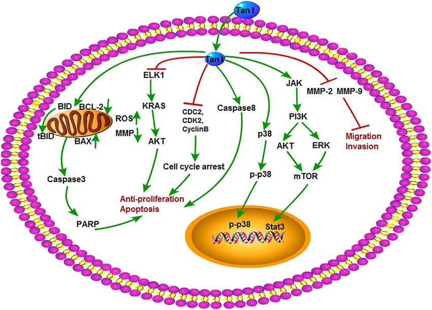
Fu et al.                                                                                                                      Antitumor Properties of Tanshinones

  FIGURE 1 | The chemical structure of four tanshinone monomers.

TABLE 1 | Basic physicochemical properties of tanshinone compounds.

Tanshinones            Molecular formula          Molecular weight      CAS           Detection method      Detection wavelength (nm)            Retention time (min)

Tanshinone IIA         C19 H18 O3                      294.33         568-72-9        HPLC                              220                               21.44
Dihydrodanshinone      C18 H12 O3                      278.3         87205-99-0       HPLC                              280                                7.79
Tanshinone I           C18 H12 O3                      276.29         568-73-0        HPLC                              220                               12.66
Cryptotanshinone       C19 H20 O3                      296.35        35825-57-1       HPLC                              220                               12.12

Chromatographic conditions, waters reversed-phase C18 symmetry column; mobile phase, acetonitrile: H2 O (65:35 vol/vol); temperature,   30◦ C;   flow velocity, 1 mL/min;
detection wavelength, 220 nm.

   Tumor is a group of cells/tissues that has lost its control                       effective antitumor activities. Their efficacy and the mechanisms
on the normal growth at the gene level due to the activation                         have been excavated gradually. This article summarizes the latest
of various oncogenes or inactivation of tumor suppressors                            researches on the antitumor effects and the mechanisms of the
(Noda et al., 1999; Zhang et al., 2013). According to the size                       four tanshinones (Tan IIA, DT, Tan I, and CT). Current study
and growth characteristics, tumor tissue can be divided into                         may provide reference for the research and development of
malignant (cancerous) and benign (non-cancerous) (Avgerinou                          tanshinone compounds.
et al., 2017). Malignant tumor grows rapidly and often infiltrate
to the surrounding tissues without envelops on the surface. The
pathological examination indicated that these cells often exhibit                    PHARMACOLOGICAL ACTIVITIES OF
atypical mitosis (Machado et al., 2016). Patients with advanced                      DIFFERENT TANSHINONE MONOMERS
cancer often exhibited severe systemic symptoms and high
recurrence rate after surgical excision, causing a big challenge                     Various studies showed that tanshinone compounds have a wide
for cancer therapy (Bax et al., 2016; Bendella et al., 2016;                         range of pharmacological effects, such as antibacterial, anti-
Frohman et al., 2016). In view of the enormous harm caused                           inflammation, antioxidation, and antithrombosis (Zhou et al.,
by cancer, the development of new antitumor treatments has                           2017; Luo et al., 2019). With the excavating study, people also
become a research hotspot. Natural products are a good resource                      found the unique effect of tanshinones in the treatment of some
of antitumor compounds. In recent years, tanshinones have                            diseases, such as diabetic cardiomyopathy (Tao et al., 2019),
drawn scientists’ attention because of its broad-spectrum and                        nephropathy (Liang et al., 2018), and diverse cancers. Here we

Frontiers in Pharmacology | www.frontiersin.org                                  2                                            March 2020 | Volume 11 | Article 193
Fu et al.                                                                                                         Antitumor Properties of Tanshinones

mainly summarized the antitumor effects of four tanshinone                 leading to the mitochondria-mediated apoptosis of cancer cells
monomers; detailed description is as follows:                              (He and Gu, 2018).
                                                                              The normal cell cycle circulation is driven by cyclin-dependent
The Antitumor Function and Mechanism                                       serine/threonine kinases and their regulated cyclin subunits.
of Tan IIA                                                                 These proteins consist of cyclin-dependent kinases (CDKs), such
Tanshinone IIA appears as a kind of red crystalline substance              as CDK2, CDK4, and CDK6, and cyclins, such as cyclin B,
that isolated from S. miltiorrhiza. Modern researches showed               cyclin D, and cyclin E (Thangaraj et al., 2019). The mutation
that Tan IIA has various physiological functions with extremely            and dysregulations of CDKs/cyclins lead to the uncontrolled
high medicinal value such as repair on myocardial damage,                  cell proliferation (Marion et al., 2015). Tan IIA suppressed the
improvement of microcirculation, and extensive antitumor                   growth of breast cancer MCF-7 cell line through arresting the S
activity (Lin et al., 2006; Wang J. et al., 2013; Wang et al., 2014;       and G2 phase cell cycle by inhibiting the phosphatidylinositol-
Yang X. J. et al., 2018; Zhang Y. et al., 2018). Meanwhile, as             3-kinase (PI3K), protein kinase B (Akt), mammalian target
one of the most severely studied monomers in S. miltiorrhiza,              of rapamycin (mTOR), protein kinase C (PKC), rheumatoid
the antitumor activities and functions of Tan IIA have drawn               arthritis (Ras), and mitogen-activated protein kinase (MAPK)
more and more scientists’ attention. The various antitumor                 signaling pathway. Interestingly, Tan IIA did not function
mechanisms of Tan IIA were shown in Figure 2 and discussed                 as an Hsp90 inhibitor but could act synergistically with the
in details as follows.                                                     Hsp90 inhibitors 17-AAG and ganetespib. Tan IIA inhibited
                                                                           the enzymatic activity of PKC especially the PKCζ and PKCε
Tan IIA Inhibits Tumor Cell Growth and Proliferation                       isoforms. Furthermore, the expression of antiapoptosis protein
Inhibition of tumor cell growth and proliferation is usually               Bcl-2 was decreased, and the levels of cleaved caspase-3 and
considered as the main strategy of anticancer compounds                    poly ADP-ribose polymerase (PARP) protein were increased after
on diverse tumors. In general, endoplasmic reticulum (ER)                  treatment with a certain concentration of Tan IIA for 24 h (Lv
stress, apoptosis induction, and cell cycle arrest can inhibit cell        et al., 2018). Similarly, Tan IIA also plays a critical inhibitory
proliferation. Previous studies reported that Tan IIA may inhibit          role on diverse lung cancer cells. For instance, Tan IIA induced
the growth and proliferation of various cancer cells, such as              apoptosis and S phase cell cycle arrest in lung cancer PC9 cells
lung, breast, liver, leukemia, and colon cancer (Liu et al., 2006;         by regulating the PI3K-Akt signaling pathway (Liao et al., 2019).
Chen et al., 2012; Zhao et al., 2013; Lin C. Y. et al., 2017; Lu           Besides, combination of Tan IIA and adriamycin significantly
et al., 2018; Wang et al., 2018; Zhou et al., 2018). The ER stress         up-regulated the expression of cleaved caspase-3 and Bax and
response is usually defined as the unfolded protein response,              down-regulated the expression of vascular endothelial growth
which is an imbalance between the accumulations of unfolded                factor (VEGF), VEGFR2, p-PI3K, p-Akt, Bcl-2, and caspase-3;
or misfolded proteins in the ER lumen (Chen W. et al., 2019).              induced apoptosis; and arrested cell cycle at the S and G2 phases
Sustained ER stress activates protein interacting with C-kinase            in A549 cells (Xie et al., 2016).
(PICK), inositol-requiring enzyme 1 (IRE1), and activating                    The antitumor efficacy of Tan IIA was also found in other
transcription factor 6 (ATF6), leading to the activation of the            malignancies. Signal transducer and activator of transcription 3
proapoptotic factor C/EBP homologous protein (CHOP), which                 (STAT3) is a member of signal-responsive transcription factors
further promotes the activation of caspase-dependent apoptosis             and plays a pivotal role in tumorigenesis (Chen et al., 2017;
(Zhu et al., 2019). Previously, Tan IIA increased expression               Wang et al., 2017). Activation of the STAT3 signal directly
of protein kinase RNA-like ER kinase (PERK), ATF6, inositol-               stimulates the expression of forkhead box M1 (FOXM1), which is
requiring enzyme 1α (IRE1α), caspase-12, and downstream                    a regulator of cell cycle (André et al., 2012; Tan et al., 2014). This
eukaryotic initiation factor 2α (eIF2α), phosphorylated c-Jun              evidence revealed that STAT3 constitutively activated in gastric
N-terminal kinase (p-JNK), and CHOP to activate ER-mediated                cancer. Zhang and his colleagues showed that Tan IIA could
apoptosis in human pancreatic cancer BxPC-3 cells in vitro (Chiu           suppress gastric cancer cells growth by down-regulating STAT3
and Su, 2017). Tan IIA also induced ER stress and apoptosis                and FOXM1 expression (Zhang Y. et al., 2018). Furthermore,
in human breast cancer BT-20 cells by increasing caspase-12,               Tan IIA could inhibit osteosarcoma MG-63 cell proliferation and
DNA damage-inducible gene 153 (GADD153), caspase-3, p-JNK,                 achieve its best inhibitory effect with 8.8 mg/L of Tan IIA (Zhang
phospho-p38 mitogen-activated protein kinases (p-p38), and Bcl-            et al., 2012b; Ma et al., 2016a,b). Tan IIA treatment also induced
2-Associated X protein (Bax) levels with decreased expression of           cell apoptosis and arrested cell cycle in human oral cancer KB
B-cell lymphoma extra large (Bcl-xl) and p-ERK in a time- and              cells line by mitochondrial pathway via activation of caspase-
dose-dependent manner (Yan et al., 2012). In another research,             3, caspase-9, and PARP (Tseng et al., 2014), by MDM4-IAP3
Tan IIA increased p53, p21, and Bax; decreased B-cell lymphoma-            signaling pathway in lung cancer H1299 cells (Zu et al., 2018),
2 (Bcl-2), cell division cycle gene 2 (cdc2), and cdc25 expression;        and by inducing the formation of cleaved caspase-8 and cleavage
and induced ER-related apoptosis in hepatocellular carcinoma               of RIP1, RIP3, and MLKL in human hepatocellular carcinoma
15 cells through the regulation of calreticulin, caspase-12, and           HepG2 cells (Lin C. Y. et al., 2016).
GADD153 expression (Cheng and Su, 2010). Additionally, Tan
IIA inhibited the protective mitophagy through the inhibition              Tan IIA Inhibits Tumor Cell Invasion and Migration
of the adenosine monophosphate-activated kinase (AMPK),                    It is well known that the recrudescence after operation and
S-phase kinase–associated protein 2 (Skp2), Parkin pathway,                metastasis are the major causes of death in cancer patients. The

Frontiers in Pharmacology | www.frontiersin.org                        3                                         March 2020 | Volume 11 | Article 193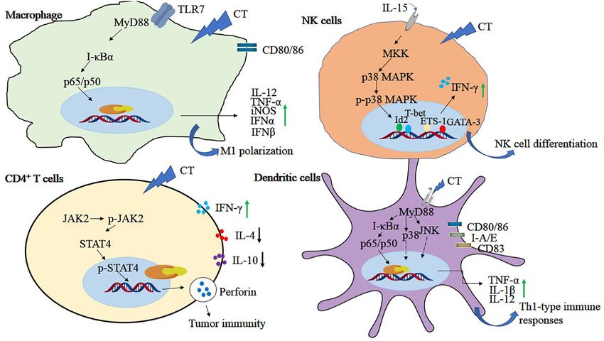
Fu et al.                                                                                                                        Antitumor Properties of Tanshinones

  FIGURE 2 | The antitumor mechanism of Tan IIA. Tanshinone IIA inhibits cancer cell proliferation, induces differentiation and apoptosis through MAPK,
  AMPK/Skp2/Parkin, ER stress and mitochondrial pathway, inhibits of invasion and migration through Notch1/NF-κB signaling, induces of autophagy in cancer cells
  through PI3K/Akt/mTOR signaling pathway, and inhibits angiogenesis by inhibiting the activity of VEGF. The arrow represents the promotion effect, and the T area
  represents the inhibition effect.

invasion and migration are two key factors that contributed                           Annese et al., 2019). Moreover, it also played a critical role in
to the recurrence and metastasis of cancer cells (Lin et al.,                         the progression of tumor formation, infiltration, invasion, and
2007; Nurnberg et al., 2011). Therefore, effective suppression                        metastasis (Carmeliet, 2006; Seano et al., 2014; Lii et al., 2016;
of tumor invasion and metastasis might be an important part                           Ao et al., 2017; Yang et al., 2017). Thus, inhibition of angiogenesis
for cancer therapy (Yang et al., 2015). Previous in vivo and                          has become an effective strategy for cancer therapy. VEGF is a
in vitro studies showed that Tan IIA could inhibit the invasion                       key factor in the production and release of angiogenesis in tumor
and migration of colon cancer cells (Su and Lin, 2008; Su et al.,                     tissues under hypoxia. There are eight members of the gene
2012). High dose of Tan IIA was shown to inhibit astrocytoma                          family: VEGF-A, VEGF-B, VEGF-C, VEGF-D, VEGF-E, VEGF-F,
migration through up-regulating transmembrane receptor notch                          and placenta growth factor-1 (PIGF-1) and PIGF-2. It has many
homolog 1 (Notch-1) pathway and down-regulating matrix                                functions, including stimulating angiogenesis, recruiting new
metalloproteinase-9 (MMP-9), cellular-myelocytomatosis viral                          blood vessels, inflammation, and vascular permeability through
oncogene (c-Myc), and Bcl-2 expression (Dong W. et al., 2018).                        angiogenic, which constitutes the most important signal pathway
FOXM1 is a member of the FOX family and associated with                               in tumor angiogenesis (Xie et al., 2017). In a recent study, Tan
cell fate decisions. Overexpression of FOXM1 promoted tumor                           IIA dramatically suppressed; VEGF promoted the migration and
progression and metastasis (Tan et al., 2014). Tan IIA could                          tube formation of human endothelia progenitor cells through
down-regulate FOXM1, MMP-2, and MMP-9 expression in                                   the phospholipase C (PLC), Akt, and JNK signaling pathways
gastric cancer SGC7901 cell line, resulting in the suppression                        without cytotoxic effect (Lee et al., 2017). Meanwhile, Tan
of proliferation and migration in a dose-dependent way                                IIA was found to effectively restrain β-catenin/VEGF-mediated
(Yu et al., 2017).                                                                    angiogenesis by targeting transforming growth factor-β (TGF-
                                                                                      β1) in normoxic and hypoxia-inducible factor 1α (HIF-1α) in
Tan IIA Inhibits Tumor Angiogenesis                                                   hypoxic microenvironments in human colorectal cancer (Sui
Angiogenesis is a vital step in the physiological process of tissue                   et al., 2017). Tan IIA exhibited antiangiogenic effects in vivo and
repair and regeneration, bone remodeling and reproduction, and                        in vitro by modulating the secretion of MMP-2 and TIMP-2 (Tsai
embryonic development (Folkman, 2006; Garona et al., 2018;                            et al., 2011). Other studies also suggested that Tan IIA could

Frontiers in Pharmacology | www.frontiersin.org                                   4                                              March 2020 | Volume 11 | Article 193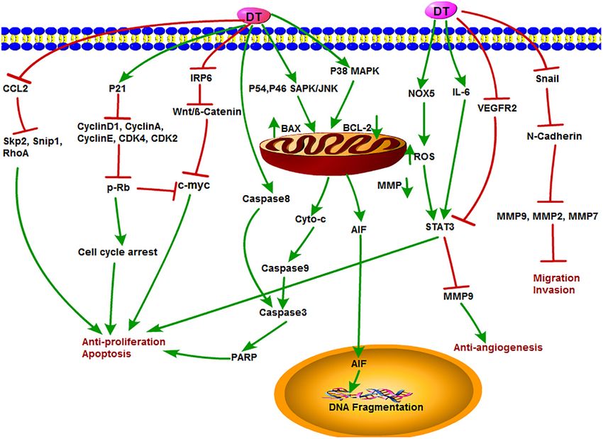
Fu et al.                                                                                                         Antitumor Properties of Tanshinones

inhibit angiogenesis in some cells, such as osteosarcoma cells,             The Antitumor Activities of Tan IIA in vivo
breast cancer cells, and vascular endothelial cells (Li et al., 2015;       To study the antitumor activity and function of a potential
Xing et al., 2015; Huang et al., 2017).                                     anticancer drug, in vivo experiments are often necessary and
                                                                            more convincing. In recent years, more and more in vivo studies
Tan IIA Induces Tumor Cell Autophagy                                        revealed the unique antitumor activity of Tan IIA. Here, the
Cell autophagy is an important physiological process in                     main antitumor studies of Tan IIA in vivo are summarized in
organism development. Basal autophagy is essential to the                   Table 2. Using Lewis lung cancer mice model, intraperitoneal
normal metabolism of cells, which process waste by removing                 injection of Tan IIA at 15 mg/kg significantly inhibited tumor
damaged organelles and protein aggregates (Qiu et al., 2017).               growth, neovascularization, and Bcl-2 expression and increased
The latest research focused on autophagy has made great                     the levels of CD4+ , CD4+ /CD8+ , and NK cells. Moreover,
progress in the understanding of the antitumor mechanisms                   combination of Tan IIA with cyclophosphamide (CTX) showed
of Tan IIA. Autophagy involves multiple signaling pathways,                 potent efficacy (Li et al., 2016). Endothelial progenitor cells
such as the AMPK and PI3K/Akt/mTOR signaling pathways                       (EPCs) usually derive from bone marrow that are generally
(Yang et al., 2019; Zhou et al., 2019). AMPK consists of a                  considered as the key regulator in tumor angiogenesis and
catalytic subunit (α 1, α2) and two regulatory subunits β (β1               metastasis (Asahara et al., 1999; Adams et al., 2012; Mund et al.,
and β2) and γ (γ1, γ2, and γ3). AMPK regulated variety of                   2012). Tan IIA was first proved to reduce EPC angiogenesis by
biochemical pathways, which control the signal of cellular energy           inhibiting PLC, Akt, and JNK signaling pathways in a chick
metabolism, whereas its dysfunction is associated with many                 embryo chorioallantoic membrane model and Matrigel plug
human diseases (Ge et al., 2019). mTOR is present in both                   assay in mice, indicating that Tan IIA might be the new potential
mTORC1 and mTORC2 complexes and has a negative regulatory                   treatment of angiogenesis-related cancers (Lee et al., 2017).
effect on autophagy. PI3K/Akt and AMPK pathways have                        In an acute promyelocytic leukemia (APL) NOD/SCID mice
positive and negative regulation of mTORC1 effect, respectively             model, Tan IIA was found to prolong the survival of APL-
(Yang et al., 2019).                                                        bearing mice, prevent APL-mediated body weight reduction,
    In another study, Tan IIA induced autophagic cell death                 and inhibit the proliferation of APL cells by inducing apoptosis
via activation of AMPK and ERK and inhibition of mTOR                       and differentiation (Zhang et al., 2016). Moreover, in NOD-
and rapamycin (mTOR), ribosomal protein S6 kinase (p70                      SCID mice xenografted with human osteosarcoma 143B cells, Tan
S6K) in KBM-5 leukemia cells (Yun et al., 2013; Han                         IIA inhibited the expression of CD31 and mitochondrial fusion
et al., 2018). Moreover, it was pointed that the survival of                proteins Mfn1/2 and Opa1, increased the expression of dynamic-
osteosarcoma cell could be inhibited by Tan IIA through the                 related protein 1 (Drp1), induced apoptosis and antiangiogenesis
PI3K/AKT signaling pathway, indicating that Tan IIA could                   (Huang et al., 2017). In gastric cancer AGS cell xenograft SCID
effectively induce autophagy in human osteosarcoma cells (Yen               mice model, results showed that treatment with Tan IIA for
et al., 2018). Analogously, Tan IIA induced autophagocytosis                8 weeks significantly reduced the protein expression levels of
of tumor cells by activating autophagic-related Beclin-1 and                epidermal growth factor receptor (EGFR), inverted gravity flame
light chain 3-II (LC3-II) expression in melanoma A375                       reactor (IGFR), PI3K, AKT, and mTOR and inhibited AGS cell
cells (Li et al., 2017). In addition, Tan IIA might induce the              proliferation by blocking the PI3K/AKT/mTOR pathway (Su
autophagy in oral squamous cell carcinoma by both activating                and Chiu, 2016). In BxPC-3-derived xenograft tumor model,
the Beclin-1/Atg7/Atg12-Atg5 pathway and inactivating the                   treatment with Tan IIA induced ER stress by up-regulating
PI3K/Akt/mTOR pathway (Ye et al., 2018). Another recent study               the levels of PERK, ATF6, caspase-12, IRE1α, eIF2α, p-JNK,
proved that Tan IIA suppressed colorectal cancer cell growth,               CHOP, and caspase-3 and inhibited the tumor growth in vivo
decreased mitochondrial membrane potential, and inhibited                   (Chiu and Su, 2017).
mitophagy through inactivation of AMPK/Skp1/Parkin pathway                     According to these studies, it can be concluded that Tan IIA
(He and Gu, 2018).                                                          is a promising natural product and deserves further study for
                                                                            cancer therapy. The further detailed research on the antitumor
Tan IIA Induces Tumor Immune Checkpoint Blockade                            mechanism of Tan IIA will have a clearer understanding about
Programmed cell death-ligand 1 (PD-L1) expressed on various                 the antitumor function, targets, and the whole regulation network
cancer cells played a protective role against the cytotoxicity              of Tan IIA. Based on this, it may provide a new and effective
of immune cells. It interacts with programmed cell death-1                  antitumor strategy for the treatment of cancer.
receptor (PD-1) to inhibit the cytotoxicity of T cells and block
the antitumor immune response. Thus, dismissing immune
suppression using immune checkpoint blockade agents is helpful              The Antitumor Function and Mechanism
for cancer therapy. Tan IIA was shown to inhibit breast                     of CT
cancer BT-20 cells by inhibiting the expression of PD-L1,                   CT is an important active component in S. miltiorrhiza and
cytotoxic T-lymphocyte-associated antigen 4 (CTLA-4), cluster               considered as one of the most effective antineoplastic constituents
of differentiation 80 [B7-1 (CD80)], and B7-2 (CD86) (Su, 2018).            in tanshinone compounds. Many studies showed that CT
In addition, Tan IIA enhanced interleukin 15 (IL-15)–mediated               significantly inhibited the growth of a variety of tumor cells in
natural killer (NK) cell differentiation via activation of p38MAPK          addition to its antibacterial and anti-inflammatory activities (Shi
pathway (Kim et al., 2012).                                                 et al., 2014; Yu et al., 2014; Hao et al., 2015; Zhou et al., 2016).

Frontiers in Pharmacology | www.frontiersin.org                         5                                        March 2020 | Volume 11 | Article 193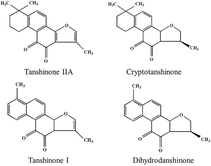
Fu et al. Antitumor Properties of Tanshinones
TABLE 2 | The antitumor experiment of tanshinone compounds in vivo.
Tanshinones Cancer Cell line Treatment Mechanism Signal pathway References
concentration
Tan IIA Breast cancer MCF-7 30 mg/kg Proliferation inhibition PKC↓, Ras/MAPK↓ Lv et al., 2018
Tan IIA Gastric cancer SNU-38, MKN1, 25–30 mg/kg Proliferation inhibition STAT3↓ Zhang Y. et al., 2018
AGS
Tan IIA Acute promyelocytic NB4 10, 100 mg/kg Proliferation inhibition – Zhang et al., 2016
leukemia
Tan IIA, CT Lung cancer Lewis tumor cells 15, 25 mg/kg Proliferation inhibition Bcl-2↓, Bax↑ Li et al., 2016
Tan IIA Osteosarcoma MG63 cell 2.5–20 mg/kg Apoptosis and Autophagy Beclin-1↑ Ma et al., 2016a
induction LC3II/LC3I↑
ATA Breast cancer MDA-MB-453 20, 60 mg/kg Proliferation inhibition NF-κBp65↓, Su et al., 2012
caspase-3↑
Tan IIA – Endothelial 0–10 µM Angiogenesis inhibition PLC/Akt↓ Yoder, 2012
progenitor cells
Tan IIA Osteosarcoma 143B cell 20 mg/kg Autophagy inhibition HGK-sestrin 2↑ Yen et al., 2018
SESN2↑
Tan IIA Melanoma A375 8, 25, 50 mg/kg Autophagy inhibition Beclin-1↑ LC3-II↑ Li et al., 2017
Tan IIA Oral squamous cell SCC-9 50 mg/kg Autophagy inhibition Beclin-1↑ Qiu et al., 2017
carcinoma
CT Liver cancer Bel-7404 100 mg/kg Apoptosis induction p-STAT3↓ Shen et al., 2017
CT Cholangiocarcinoma HCCC-9810 10, 25 mg/kg Cell circle arrest and JAK2/STAT3↓ Ke et al., 2017a
Apoptosis induction PI3K/Akt/NFκB↓
CT Glioma U87 30 mg/kg Proliferation inhibition STAT3↓ Cao et al., 2017
DT-I Hemangioma Endothelioma 10, 40 mg/kg Angiogenesis inhibition and PLC↓ Cai et al., 2018
(EOMA) Apoptosis induction
↓, down-regulated proteins; ↑, up-regulated proteins.
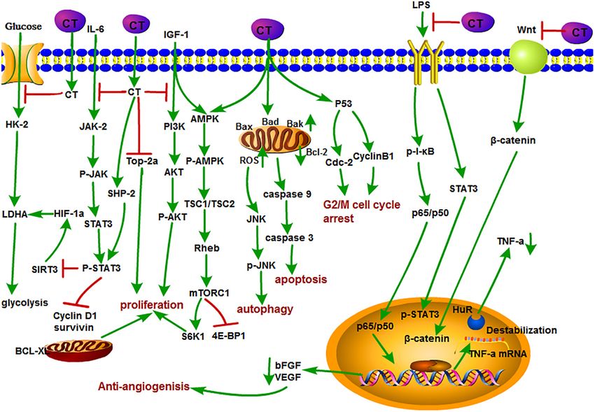
The antitumor mechanisms of CT are shown in Figure 3. STAT3 factor-κB (NF-κB) pathways. Their work may provide a possible
is one of the most frequently activated members of the STAT effective treatment for cholangiocarcinoma (Ke et al., 2017b).
family and plays an important role in the proliferation, survival, Aerobic glycolysis is a hallmark of cancer and also called Warburg
invasion, and angiogenesis signaling pathways of various tumors. effect. CT inhibited the expression of glycolysis-related proteins
Abnormal activation of JAK/STAT3 signaling is associated with including glucose transporter 1 (GLUT1), hexokinase 2 (HK2),
tumor progression, tumor microenvironment, and immune and lactate dehydrogenase A (LDHA) in ovarian cancer Hey
evasion (Chen et al., 2017; Ke et al., 2017a; Wang et al., 2017). cells and xenograft nude mice by repressing STAT3/SIRT3/HIF-
Interleukin 6 is a representative stimulant of STAT3 signaling 1α signaling pathway (Yang Y. et al., 2018). CT inhibited
pathway. CT was identified as a potent STAT3 inhibitor that p-STAT5 and p-STAT3, effectively blocked IL-6–mediated STAT3
inhibited the phosphorylation of STAT3 Tyr705 and the target activation, and reversed chronic myeloid leukemia (CML)
proteins such as survivin, Bcl-XL, and cyclin D1 through blocking fusion gene (BCR-ABL) kinase-independent drug resistance and
the dimerization in DU145 prostate cancer cells (Shin et al., inhibited key cell coproliferation and drug resistance pathway
2009). CT induced apoptosis of esophageal EC109 cancer cells of K562/ADR in CML (Dong B. et al., 2018). Another study
by inhibiting p-STAT3 (Tyr705) and p-JAK2 without effect on showed that CT induced cell cycle arrest and apoptosis of
the expression of the total STAT3 and JAK2 in vitro and in vivo multidrug-resistant leukemia cell line K562/ADM by inhibiting
(Ji et al., 2019). It is confirming that the antiesophageal cancer the expression of cyclin D1, Bcl-2, and eIF4E (Ge et al., 2012).
effect of CT was associated with the inhibition of IL-6–mediated Simultaneously, CT induced autophagic cell death in multidrug-
activation of JAK2/STAT3 signaling pathway (Ji et al., 2019). resistant colon cancer cell line SW620 Ad300 via ROS-p38
Similarly, CT was found to have strong inhibition effects on MAPK–NF-κB signaling pathway (Xu et al., 2017).
malignant gliomas (MGs) but also preliminarily explored its Inducing apoptosis of tumor cells is generally considered
potential mechanisms through a series of experiments in vivo to be a major mean for the efficacy of antitumor drugs, and
and in vitro. In this study, they elaborated that CT could inhibit it is no exception for CT. For example, CT inhibited the
the proliferation of MG by suppressing the phosphorylation proliferation of human cancer cell A549 and H1299. Detailed
of STAT3 Tyr705 through activating the tyrosine phosphate studies demonstrated that CT exerted its inhibition effect
activity of SHP-2 protein (Lu et al., 2017). Furthermore, CT by down-regulating the insulin-like growth factor 1 receptor
could significantly suppress the growth and colony-forming of (IGF-1R), PI3K/Akt signaling pathway. The insulin-like growth
HCCC-9810 and RBE cells by inducing apoptosis in a dose- factor 1(bIGF-1) induced IGF-1R and AKT phosphorylation,
dependent manner. The underlying mechanism contributed suggesting that it may become a potential clinical therapeutic
to the inhibition of the JAK2/STAT3 and PI3K/Akt/nuclear agent for the treatment of human lung cancer (Zhang J.
Frontiers in Pharmacology | www.frontiersin.org 6 March 2020 | Volume 11 | Article 193Fu et al. Antitumor Properties of Tanshinones FIGURE 3 | The antitumor mechanism of CT. Cryptotanshinone inhibits the proliferation of cancer cells and induces autophagy and apoptosis by inhibiting IGF-1, IL-6, and glycolysis and mediating the cell cycle and mitochondrial pathway and inhibits angiogenesis by inhibiting the LPS/Wnt signaling pathway. The arrow represents the promotion effect, and the T area represents the inhibition effect. et al., 2018). CT induced S-phase cell cycle arrest, apoptosis, metastasis. Therefore, it has been considered as a potential and mitochondrial fragmentation in osteosarcoma cells with target for cancer therapy (Yu et al., 2002; Guo et al., 2012; increased Bax, Bad, and Bak; decreased Bcl-2; and the activation Boreddy and Srivastava, 2013). Studies showed that CT of caspase-3, caspase-8, and caspase-9 expressions. Further also plays a unique role in this respect. CT inhibited tumor Data confirmed that CT directly promoted the interaction of angiogenesis in lipopolysaccharide (LPS)–induced neovascular Drp1with Bax, directly, which promoted the translocation of sprouts in zebrafish embryos and vascularization in mouse Bax from cytoplasm to mitochondria, resulting in the apoptotic Matrigel plug model. Moreover, CT could suppress VEGF- fragmentation of mitochondria (Yen et al., 2019). In a recent induced tube formation and sprout of human umbilical vein study, CT induced apoptosis in melanoma cell lines and endothelial cells (HUVECs) in vitro. Tumor necrosis factor increased the sensitivity of A375 cell line to tumor necrosis α is generally considered as a key angiogenic factor that is factor (TNF)–related apoptosis-inducing ligand (TRAIL), which related to both NF-κB and STAT3 pathways. Their further further led to the enhancement of cell death in melanoma cells study pointed out that CT could cause the reduction of (Radhika et al., 2018). DNA topoisomerase 2 is an important RNA-binding factor HuR stability and inhibited angiogenesis nuclear enzyme, which regulates cell proliferation by modulating through posttranscriptional mechanism of TNF-α mediated DNA topology and chromatid separation. Treatment with CT by NF-κB and STAT3 pathways (Zhu et al., 2016). Basic dramatically decreased the stabilization of topoisomerase 2a fibroblast growth factor (bFGF) is a proangiogenic factor at mRNA level and showed anticancer effect against human that stimulates the migration and spreading of endothelial prostate cancer in vitro and in vivo (Kim et al., 2017). cell invasion. CT suppressed bFGF-stimulated angiogenesis In addition, CT also inhibited mTORC1 expression through of bovine aortic endothelial cells in vitro. However, Tan activating AMPK-TSC2 axis in Rh30 cells (Chen W. et al., 2019). IIA showed no effect at the same concentration. CT and CT induced autophagy by activating JNK signaling through Tan IIA have similar structures except C-15 position of increasing intracellular reactive oxygen species (ROS) formation dihydrofuran ring. This might infer that the double bond (Hao et al., 2016b). at C-15 position of dihydrofuran ring contributed to the As we mentioned earlier, angiogenesis plays a key role antiangiogenesis efficacy (Hur et al., 2005). In another research, in providing oxygen and nutrition for tumor growth and CT inhibited tubular-like structure formation and decreased Frontiers in Pharmacology | www.frontiersin.org 7 March 2020 | Volume 11 | Article 193
Fu et al. Antitumor Properties of Tanshinones
VEGF expression and LiCl-induced β-catenin augmentation in infiltration of macrophages, CD45+ leukocyte, and CD8+ T
HUVECs. Thus, CT-mediated antiangiogenesis was associated cells into the tumor tissue. Importantly, combined treatment
with the inhibition of Wnt/β-catenin signaling pathway of CT with anti–PD-L1 successfully eradicated the tumor and
(Chen et al., 2014). showed a synergistic effect on the induction of Hepal-specific
Cryptotanshinone may also exert its anticancer function immunity responses and developed a long-term antitumor
by enhancing the activity of other antitumor drugs. As we immunity memory. Hepal-bearing mice cured by CT treatment
know, arsenic trioxide (ATO) is often used in the treatment of exhibited resistance to Hepal but not EG7 (another C57BL/6
advanced liver cancer. Combined treatment of ATO with CT lymphoma cell line) tumor (Han et al., 2019). In addition,
showed potent inhibition effect on the growth of Bel-7404 cells CT could promote the maturation of dendritic cells (DCs)
than ATO or CT alone. Meanwhile, the combination caused and stimulate DC to secrete proinflammatory cytokines TNF-
the obvious changes in the expression level of antiapoptotic α, IL-1β, and IL-12p70 through the activation of NF-κB, p38,
proteins (depressing XIAP, Bcl-2, and survivin) and apoptotic and JNK expression. CT -induced DC maturation is dependent
proteins (promoting Bak), which lead to the suppression of on MyD88 expression because the effects were compromised
tumor growth (Shen et al., 2017). In addition, CT could in MyD88−/− DC. Encouragingly, mice bearing established
enhance the sensitivity of ovarian cancer A2780 cells and showed Lewis lung tumors were cured by CT alone or more effective
favorable effect on various solid tumors and could sensitize when combined with anti–PD-L1. Data further demonstrated
A2780 cells to cisplatin treatment in a dose-dependent manner that CT plus low doses of anti–PD-L1 generated LLC-specific
(Jiang et al., 2017). antitumor immune response and immunological memory as
Immunosuppressive tumor microenvironment can lead to resultant tumor-free mice were resistant to rechallenge with LLC,
tumor-evading immunotherapy, so immunosuppression is a but not B16 melanoma (Liu et al., 2019). Natural killer cells are a
major problem in antitumor therapy. Tumor tissues are subset of lymphocytes crucial for innate and adaptive immune
infiltrated by immunosuppressive cells such as regulatory T cells responses that are triggered in the presence of IL-15. CT was
and myeloid-derived suppressor cells, and M2-polarized TAMs, shown to increase IL-15–induced NK cell differentiation through
which produce inhibitors such as PD-1/PD-L1, lymphocyte enhancing the phosphorylation of p38 MPAK and the expression
activation gene-3, IL-10, and TGF-β, inhibit the proliferation of transcription factors, such as T-box transcription factor TBX21
of CD4, CD8 cells, and their immune response (Han et al., (T-bet), GATA-binding protein 3 (GATA-3), inhibitor of DNA
2019; Liu et al., 2019; Yu et al., 2019). CD4+ regulatory biding 2 (Id2), and ETS proto-oncogene 1(ETS-1) (Kim et al.,
T cells possess immune functions that are associated with 2012). The antitumor immune mechanisms of CT in different
tumor cell immunosuppressive process. CT could increase the cells are shown in Figure 4.
cytotoxicity of CD4+ T cells without affecting the activity To sum up, as another lipid-soluble active component of
of CD8+ T cells. Further investigation indicated that CT S. miltiorrhiza, CT plays an important role in inducing tumor
activated the JAK 2/STAT 4 pathway in the CD4+ T cells, cell apoptosis; inhibiting tumor cell proliferation, invasion, and
thereby inhibiting the growth of small cell lung cancer (Yong angiogenesis; and enhancing the activity of other antitumor
et al., 2016). Perforin is one of the direct target genes of drugs. However, the study on the antitumor function and
STAT 4 and is activated by IL-12 (Yamamoto et al., 2002). mechanism of CT is still in its initial stage, and there are
CT might function like IL-12 to activate CD4+ T cells to some deficiencies in the analysis of the whole antitumor
secrete perforin through STAT 4 gene. CT inhibited breast regulatory network. It is still necessary to perform further studies
tumor MCF-7 growth by triggering proliferation and increasing on CT in terms of molecular biology, cell biology, receptor
perforin secretion of CD4+ T cells which is associated with pharmacology, and so on.
enhancement of the p-JAK 2 and p-STAT 4 expressions. However,
the efficacy was abrogated when treated with perforin inhibitor
concanamycin, suggesting that CT exhibited the anti–breast The Antitumor Function and Mechanism
tumor activity mainly by activating CD4+ T cells to secrete of Tan I
perforin (Zhou et al., 2017). Macrophages are heterogeneous and Tanshinone I is a kind of red crystalline powder and accounted for
exert contrast functions. M1 phenotype possess tumor inhibitory approximately 1.79% of the alcohol extract of S. miltiorrhiza roots
properties, whereas tumor-associated macrophages displaying (Lee et al., 2008). Modern researches showed that Tan I is mainly
the M2 phenotype promote tumor growth and metastasis (Sica used in the treatment of cardiovascular and cerebrovascular
and Bronte, 2007). Moreover, CT was revealed to activate diseases and also possesses broad-spectrum antitumor activity.
bone marrow–derived macrophages toward an M1 phenotype Recent studies showed that Tan I significantly inhibited the
with elevated expression of CD80, CD86, TNF-α, and IL- growth of osteosarcoma cell lines U2OS and MOS-J with IC50
12p40 through the TLR7/MyD88/NF-κB and the JAK2/STAT3 (half maximal inhibitory concentration) values around 1 to
signaling pathway in mouse hepatoma Hepa1-6 cells in vitro. 1.5 mol/L. It was further shown that Tan I induced apoptosis via
Further in vivo studies in a mouse Hepa1-6 model revealed up-regulation of Bax and down-regulation of Bcl-2 expression.
that CT treatment increased the levels of inducible nitric oxide Tan I also inhibited both the mRNA and protein expression of
synthase, TNF-α, interferon α (IFNα), IFNβ, and IL-12p40, MMP-2 and MMP-9, which are crucial for tumor metastasis.
but not IL-10 or TGFβ1. Flow cytometry revealed that CT The underlying mechanism can be concluded as the down-
enhanced antitumor T-cell responses with markedly increased regulation of the JAK/STAT3 signaling pathway (Wang et al.,
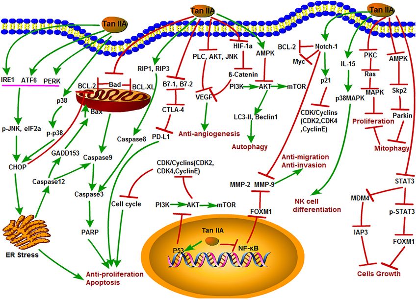
Frontiers in Pharmacology | www.frontiersin.org 8 March 2020 | Volume 11 | Article 193Fu et al. Antitumor Properties of Tanshinones FIGURE 4 | The antitumor immune mechanisms of CT in different cells. Cryptotanshinone plays a role in tumor immunity by regulating NF-κB, MAPK, JAK, and STAT4 signals in immune-related cells, including macrophage, NK cells, CD4+ T cells, and dendritic cells. 2019). Similarly, Tan I attenuated proliferation, colony formation, et al., 2008). The antitumor mechanisms of Tan I are shown and cisplatin resistance of cervical cancer. The expression of in Figure 5. p-AKT and kainate receptors (KARS) was markedly suppressed by Tan I treatment. In contrast, overexpression of KRAS or ETS-like 1 transcription factor (ELK1) markedly impaired the The Antitumor Function and Mechanism suppression of Tan I on HeLa cells (Dun and Gao, 2018). of DT In the research of Li et al., Tan I showed the most potent Dihydrotanshinone is also an important component of effect on the proliferation of lung cancer cells compared with tanshinone compounds. However, there is still less research CT and Tan IIA. Tan I also induced apoptosis and G2/M on its antitumor functions and mechanisms compared to the cell cycle arrest in vitro; inhibited the expression of Aurora other three monomers. DT exhibited special biological inhibitory A, survivin, cyclin B, cdc2, and CDK2; and increased the activities on diverse tumors (Chen X. et al., 2019). The antitumor ratio of Bax/Bcl-2. Aurora A–specific siRNA confirmed that mechanisms of DT are shown in Figure 6. DT inhibited Aurora A is a potential target for Tan I. In addition, H1299 the growth of liver cancer cells with an EC50 (50% effective xenograft mice oral gavaged with Tan I at a dose of 200 mg/kg concentration) value of 2.52 µM. In this study, DT induced exhibited the marked reduction of tumor weight, angiogenesis, caspase-3, -8, and -9 cleavages in a concentration-dependent and Aurora A expression in vivo (Li et al., 2013). Tan I was manner. Moreover, DT significantly induced phosphorylation explored that could cause the death of tumor multidrug resistance of P54 and P46 SAPK/JNK (Thr183/TYR185) and p38MAPK cells by inducing PARP, caspase-3, caspase-8, and caspase- (th180/bar182), increased Bax and decreased cytochrome c 9 cleavage and decreasing mitochondria membrane potential in mitochondria after p38 MAPK activation, but significantly without influence on drug transporter proteins P-glycoprotein inhibited PARP cleavage after p38 MPAK inhibition. This (P-gp) and multidrug resistance protein 1 (MRP1). It enhanced indirectly demonstrates that ROS-mediated phosphorylation the depression of p-705-STAT3 and the secondary activation of p38MAPK is involved in a certain extent in the apoptosis of p38-, AKT-, and ERK-involved signaling networks (Xu of human hepatoma HepG 2 cells induced by DT (Lee et al., et al., 2013). Moreover, Tan I was also proven to induce the 2009). Cancer stem cells (CSCs) play a key role in tumor apoptosis of prostate cancer cells and enhance its sensibility metastasis and recurrence. DT activated NOX5 to generate to TRAIL (Shin et al., 2018). In addition, Tan I could induce ROS and then phosphorylate STAT3 expression, reduce IL-6 apoptosis in both estrogen receptor positive MCF-7 cells and secretion, and induce CSC death (Kim et al., 2019). Similarly, negative MDA-MB-231 cells via the activation of caspase-3 and DT exhibited strong cytotoxicity in HCT116 p53(-/-) and Bax and the inhibition of Bcl-2 expression (Nizamutdinova HCT116 p53(+/+) colon cancer cells and induced PARP Frontiers in Pharmacology | www.frontiersin.org 9 March 2020 | Volume 11 | Article 193
Fu et al.                                                                                                                           Antitumor Properties of Tanshinones

  FIGURE 5 | The antitumor mechanism of Tan I. Tanshinone I inhibits proliferation and induces apoptosis of cancer cells by regulating cell cycle and mitochondrial
  pathway and inhibits cancer cell invasion and migration by inhibiting MMP-2 and MMP-9 signals. The arrow represents the promotion effect, and the T area
  represents the inhibition effect.

cleavage in a time-dependent manner. They further observed                              suggested that DT-I inhibited the proliferation, migration, and
that DT decreased mitochondria membrane potential and                                   invasion and induced apoptosis of osteosarcoma cells in vivo
stimulated mitochondria to produce ROS, causing a decrease                              and in vitro by inhibiting Wnt/β-catenin signaling pathway (Tan
in mitochondrial metabolites and ROS leakage. However, DT-                              et al., 2019). DT activated the activity of caspase-3, caspase-9,
induced apoptosis was inhibited by the ROS scavenger NAC or                             PARP, and cytochrome c release; inhibited proliferation of
catalase–PEG alone, confirming that DT impairs mitochondrial                            glioma cell; and induced apoptosis in SHG-44 cells (Cao et al.,
function. These results also suggested that DT induced apoptosis                        2017). Moreover, DT had an antiproliferative effect on human
of colon cancer cells through a p53-independent but ROS-                                hepatocellular carcinoma cells. DT could induce cell cycle arrest
dependent pathway (Wang L. et al., 2013). In multidrug-resistant                        of SK-HEP-1 cells at G0/G1 phase, which led to the inhibition of
colon cancer cell line SW620 Ad300, DT, and CT were shown                               tumor cell growth by down-regulating the expression of cyclin
to induce accumulation of LC3B-II and increase autophagy                                D1, cyclin A, cyclin E, CDK4, CDK2, c-Myc, and retinoblastoma
flux. In addition, the cytotoxic effects of the two tanshinones                         protein (p-Rb) expression with increased expression of the CDK
were independent of p53, suggesting that both DT and CT                                 inhibitor p21, suggesting that the antiproliferative activity of DT
inhibited the growth of multidrug-resistant colon cancer cells                          is related to the regulation of AMPK/AKT/mTOR and MAPK
by inducing autophagic cell death in a p53-independent manner                           signaling pathways (Hong et al., 2018). In addition, DT could
(Hu et al., 2015). Recently, DT-I decreased the expression of                           obviously inhibit the angiogenesis of infantile hemangioma by
MMP-9, MMP-2, MMP-7, Snail, and N-cadherin, thus inhibiting                             up-regulating several apoptosis-related proteins, such as caspase-
the migration and invasion of osteosarcoma cells. Besides, DT-I                         3, caspase-8, caspase-9, PARP, AIF, Bax, cytochrome c, and so
increased the expression of PARP and caspase-3, decreased the                           on (Cai et al., 2018). Lin et al. accessed data from the Taiwan
expression of Bcl-2, and induced apoptosis of osteosarcoma                              Computerized Insurance Reimbursement Claims Database and
cells through mitochondrial pathway. DT I down-regulated                                used National Health Insurance Research Database (NHIRD)
the expression of β-catenin, IRP6 (upstream of β-catenin),                              analysis to find out that S. miltiorrhiza has a protective effect on
c-Myc (downstream of β-catenin), and cyclin D1 protein and                              colon cancer patients in clinical practice. DT inhibited protein
suppressed Wnt/β-catenin signaling. In an in vivo mouse model,                          expression of Skp2, Smad nuclear interacting protein 1 (Snip1),
DT-I showed inhibited formation of osteosarcoma. These results                          and Ras homolog gene family member A (RhoA) and induced

Frontiers in Pharmacology | www.frontiersin.org                                    10                                              March 2020 | Volume 11 | Article 193
Fu et al.                                                                                                                       Antitumor Properties of Tanshinones

  FIGURE 6 | The antitumor mechanism of DT. Dihydrotanshinone inhibits cancer cell proliferation and induces apoptosis by mediating signals such as Wnt/β-Catenin,
  AMPK/Akt/mTOR, MAPK, CCL2, and p21 and inhibits the invasion and migration of cancer cells and angiogenesis by inhibiting MMP-2, MMP-9, and VEGFR2
  signals. The arrow represents the promotion effect, and the T area represents the inhibition effect.

apoptosis of HCT116 cells and HT-29 cells by reducing the                            cell cycle arrest at G1 phase; and decreased mitochondrial
secretion of CCL2 in macrophages and blocking the recruitment                        membrane permeability in MCF-7 cells. Phosphorylation of JNK,
of colon cancer cells. DT treatment also reduced tumor burden                        ERK, and p38 MAPK was induced by time and concentration
in xenograft nude mice (Lin Y. Y. et al., 2017).                                     dependence, and the MAPK signal was activated to inhibit the
                                                                                     proliferation of MCF-7 cells (Zhang et al., 2015). S222 and S439
                                                                                     are derivatives of Tan I that exhibited anti–multidrug resistance
The Antitumor Activities of Other                                                    and antiangiogenic properties. In a panel of 15 cancer cell lines,
Tanshinone Compounds                                                                 S222 and S439 inhibited STAT3 phosphorylation, induced DNA
There are also many other liposoluble compounds in                                   double-strand breaks, blocked cell cycle at G2/M phase, and
S. miltiorrhiza except those elaborated above, including Tan                         induced apoptosis (Tian et al., 2018). Acetyl tanshinone IIA
IIB, isocryptotanshinone, hydroxytanshinone, and so on                               (ATA) is a compound obtained by chemical modification of
(Li et al., 2018). These compounds also exhibit antitumor                            Tan IIA. Acetyl tanshinone IIA induced G1/S phase arrest and
activities on various cancers. For example, isocryptotanshinone                      apoptosis in HER2-positive MDA-MB-453, SK-BR-3, and BT-47
was proven as a STAT3 inhibitor that could induce the                                breast cancer cells by inhibiting receptor tyrosine kinases (RTKs)
apoptosis and autophagy of A549 lung cancer cells (Guo et al.,                       EGFR/HER2 and downstream survival signaling pathways.
2016). Isocryptotanshinone treatment down-regulated the                              Moreover, ATA significantly inhibited tumor growth in athymic
expression of cell cycle and apoptosis-related proteins cyclin D1,                   MDA-MB-453 xenograft mouse (Guerram et al., 2015). Acetyl
phosphorylated Rb, E2F transcription factor 1 (E2F1), myeloid                        tanshinone IIA inhibited the growth of breast cancer cells and
cell leukemia 1 (Mcl-1), Bcl-2, and survivin expression; inhibited                   the growth of xenografted mouse by inducing the production
the phosphorylation of STAT3; and induced cell cycle arrest                          of ROS and up-regulating the expression of Bax, cytochrome c,
at G1/G0 phase, thereby inhibiting the proliferation of gastric                      and caspase-3 in breast cancer cells (Tian et al., 2010). PTS33, a
cancer cells. In addition, isocryptotanshinone also inhibited                        sodium derivative of CT, could selectively inhibit prostate cancer
the gastric tumor growth of BALB/c nude mice in vivo (Chen                           cells growth by inhibiting the expression of androgen receptor
et al., 2018). Moreover, isocryptotanshinone down-regulated                          (AR) protein and blocking the expression of AR-regulated genes
the expression of Bcl-2 and Bcl-xl proteins; up-regulated the                        (Xu et al., 2012). Neo-tanshinlactone, a natural product isolated
expression of Bax, Bak, PARP, caspase-3, and caspase-9; induced                      from S. miltiorrhiza, could selectively inhibit the proliferation of

Frontiers in Pharmacology | www.frontiersin.org                                 11                                             March 2020 | Volume 11 | Article 193
Fu et al. Antitumor Properties of Tanshinones
estrogen receptor–positive breast cancer cells by inhibiting the The investigations on the signaling pathways help us to
synthesis of ESR1 mRNA and down-regulating the transcription have a better understanding on the antitumor mechanisms
of estrogen receptor α (Lin W. et al., 2016). DYZ-2-90 is a novel of tanshinones. However, process and pathways are usually
ring-opening compound modified by neo-tanshinlactone that extremely large and complicated; because of this, our
induced ERK-mediated cell division arrest and apoptosis in understanding is quite limited. Besides, studies have proven
human colorectal cancer cells by activating the stress-related CT having a potent antitumor immune function, whereas there
JNK pathway (Wang et al., 2012), although there are still few are few reports on other tanshinone compounds. Because of
studies on the anticancer effect of these liposoluble monomers. their similar chemical structures, it can be speculated that
We believe that with the deepening of research, their unique they may also possess the immunosuppressive effect. However,
antitumor functional activities will be gradually excavated. further investigation is needed. Meanwhile, the in vivo antitumor
experiments of the four tanshinones are quite few especially for
Tan I and DT. Besides, there is still no strong evidence whether it
THE CLINICAL CHALLENGES OF has cytotoxicity on normal cells. This might be a limit for further
TANSHINONES clinical application. Therefore, more preclinical or clinical trials
are also a direction of future research.
In recent years, there have been more and more studies on the
anticancer effects of tanshinones. However, because tanshinones
have a high hydrophobicity, it is difficult to prepare an injection,
and the absorption is poor at the time of oral administration
AUTHOR CONTRIBUTIONS
or injection. Poor bioavailability has been a major challenge LF consulted literatures and drafted the manuscript. BH, YZ,
for pharmaceutical development. Studies have shown that the JR, WC, and GP participated in manuscript sorting. JZ and GK
structural modification of tanshinone compounds can solve the revised the manuscript.
solubility problem to some extent and improve the limitation of
tanshinones in clinical application (Zhang et al., 2012a).
FUNDING
CONCLUSION AND PERSPECTIVES
This work was supported by the National Natural Science Fund
Traditional Chinese herbal medicine are important sources of China (81522049, 81670151, 31571735, and 31270007), the
of antitumor drugs. From their initial use (in the treatment “Dawn” Program of Shanghai Education Commission (16SG38),
of cardiovascular and cerebrovascular diseases) to the unique Open project of Shanghai Key Laboratory Atmospheric
antitumor activities, researches on tanshinone compounds have Particle Pollution Prevention (C-6105-17-029), Shanghai
made a great progress. In the last decade, numerous studies have Science and Technology Committee Project (17JC1404300
demonstrated the antitumor properties of tanshinones on various and 15430502700), Zhejiang Provincial Ten Thousands
tumors both in vitro and in vivo. Tanshinones exerted a broad Program for Leading Talents of Science and Technology
range of antitumor functions such as the induction of apoptosis Innovation, Zhejiang Provincial Program for the Cultivation
and autophagy; regulation of cell cycle; inhibition of proliferation, of High-level Innovative Health talents, Opening project of
invasion, metastasis, and angiogenesis; and enhancement of Zhejiang provincial preponderant and characteristic subject
immunology. It suggests that tanshinones especially Tan IIA and of Key University (Traditional Chinese Pharmacology), and
CT may become a potential antitumor agent and provide a new Zhejiang Chinese Medical University (ZYAOXYB2019005
therapeutic strategy for human cancers. and ZYAOX2018019).
REFERENCES Avgerinou, C., Giannezi, I., Theodoropoulou, S., Lazaris, V., Kolliopoulou,
G., Zikos, P., et al. (2017). Occupational, dietary, and other risk factors
Adams, E. L., Yoder, E. M., and Kasdan, M. L. (2012). Giant cell tumor of the for myelodysplastic syndromes in western greece. Hematology 22, 419–429.
tendon sheath: experience with 65 cases. Eplasty 12:e50. doi: 10.1080/10245332.2016.1277006
André, L. M., Renata, B., Gerson, M. F., Barbara, D. R., and Eliana, A. (2012). Bax, M. J., Johnson, T. M., Harms, P. W., Schwartz, J. L., Zhao, L., Fullen, D. R., et al.
Forkhead Box M1 (FoxM1) gene is a new STAT3 transcriptional factor target (2016). Detection of occult invasion in Melanoma in situ. JAMA Dermatol. 152,
and is essential for proliferation, survival and DNA repair of K562 cell line. PLoS 1201–1208. doi: 10.1001/jamadermatol.2016.2668
One 7:e48160. doi: 10.1371/journal.pone.0048160 Bendella, H., Brackmann, D. E., Goldbrunner, R., and Angelov, D. N. (2016).
Annese, T., Tamma, R., Ruggieri, S., and Ribatti, D. (2019). Erythropoietin in tumor Nerve crush but not displacement-induced stretch of the intra-arachnoidal
angiogenesis. Exp. Cell Res. 374, 266–273. doi: 10.1016/j.yexcr.2018.12.013 facial nerve promotes facial palsy after cerebellopontine angle surgery. Exp.
Ao, Z., Yu, S., Qian, P., Gao, W., Guo, R., Dong, X., et al. (2017). Tumor Brain Res. 234, 2905–2913. doi: 10.1007/s00221-016-4692-7
angiogenesis of SCLC inhibited by decreased expression of FMOD via Boreddy, S. R., and Srivastava, S. K. (2013). Deguelin suppresses pancreatic tumor
downregulating angiogenic factors of endothelial cells. Biomed. Pharmacother. growth and metastasis by inhibiting epithelial-to-mesenchymal transition
87, 539–547. doi: 10.1016/j.biopha.2016.12.110 in an orthotopic model. Oncogene 32, 3980–3991. doi: 10.1038/onc.201
Asahara, T., Yano, M., Fukuda, S., Fukuda, T., Nakahara, H., Katayama, K., 2.413
et al. (1999). Brain metastasis from hepatocellular carcinoma after radical Cai, Y., Lv, F., Kaldybayeva, N., Zhamilya, A., Wu, Z., and Wu, Y. (2018). 15, 16-
hepatectomy. Hiroshima J. Med. Sci. 48, 91–94. Dihydrotanshinone I inhibits hemangiomas through inducing pro-apoptotic
Frontiers in Pharmacology | www.frontiersin.org 12 March 2020 | Volume 11 | Article 193Fu et al. Antitumor Properties of Tanshinones and Anti-angiogenic mechanisms in vitro and in vivo. Front. Pharmacol. 9:25. Garona, J., Sobol, N. T., Pifano, M., Segatori, V. I, Gomez, D. E., Ripoll, G. V., et al. doi: 10.3389/fphar.2018.00025 (2018). Preclinical Efficacy of [V4Q5]dDAVP, a second generation vasopressin Cao, W., Wang, Y., Shi, M., Hao, X., Zhao, W., Wang, Y., et al. (2018). analog, on metastatic spread and tumor-associated angiogenesis in colorectal Transcription factor SmWRKY1 positively promotes the biosynthesis of cancer. Cancer Res. Treat. 51, 438–450. doi: 10.4143/crt.2018.040 tanshinones in salvia miltiorrhiza. Front. Plant Sci. 9:554. doi: 10.3389/fpls.2018. Ge, Y., Cheng, R., Zhou, Y., Shen, J., Peng, L., Xu, X., et al. (2012). 00554 Cryptotanshinone induces cell cycle arrest and apoptosis of multidrug resistant Cao, Y., Huang, B., and Gao, C. (2017). Salvia miltiorrhiza extract human chronic myeloid leukemia cells by inhibiting the activity of eukaryotic dihydrotanshinone induces apoptosis and inhibits proliferation of glioma initiation factor 4E. Mol. Cell. Biochem. 368, 17–25. doi: 10.1007/s11010-012- cells. Bosn J. Basic Med. Sci. 17, 235–240. doi: 10.17305/bjbms.2017.1800 1338-3 Carmeliet, E. (2006). Action potential duration, rate of stimulation, and Ge, Y., Sheng, Z., Yixuan, L., Zixu, W., Shuai, C., Tianwei, X., et al. (2019). Estrogen intracellular sodium. J. Cardiovasc. Electrophysiol. 17(Suppl. 1), S2–S7. doi: prevents articular cartilage destruction in a mouse model of AMPK deficiency 10.1111/j.1540-8167.2006.00378 via ERK-mTOR pathway. Ann. Transl. Med. 7:336. doi: 10.21037/atm.2019. Chen, J., Bi, Y., Chen, L., Zhang, Q., and Xu, L. (2018). Tanshinone IIA 06.77 exerts neuroprotective effects on hippocampus-dependent cognitive Guerram, M., Jiang, Z. Z., Yousef, B. A., Hamdi, A. M., Hassan, H. M., Yuan, Z. Q., impairments in diabetic rats by attenuating ER stress-induced apoptosis. et al. (2015). The potential utility of acetyltanshinone IIA in the treatment of Biomed. Pharmacother. 104, 530–536. doi: 10.1016/j.biopha.2018. HER2-overexpressed breast cancer: Induction of cancer cell death by targeting 05.040 apoptotic and metabolic signaling pathways. Oncotarget 6, 21865–21877. doi: Chen, J., Shi, D. Y., Liu, S. L., and Zhong, L. (2012). Tanshinone IIA induces growth 10.18632/oncotarget.4156. inhibition and apoptosis in gastric cancer in vitro and in vivo. Oncol. Rep. 27, Guo, G., Xu, J. H., Han, J. H., Liang, F., Zhang, Y., Zhang, Q., et al. (2012). Chinese 523–528. doi: 10.3892/or.2011.1524 herbal decoction Shiquan Dabu Tang inhibits tumor growth and angiogenesis Chen, Q., Zhuang, Q., Mao, W., Xu, X. M., Wang, L. H., and Wang, H. B. of metastasis after primary tumor surgical removal in mice. Zhong Xi Yi Jie He (2014). Inhibitory effect of cryptotanshinone on angiogenesis and Wnt/β- Xue Bao 10, 436–447. doi: 10.3736/jcim20120413 catenin signaling pathway in human umbilical vein endothelial cells. Chin J. Guo, J., Zhou, Y. J., Hillwig, M. L., Shen, Y., Yang, L., Wang, Y., et al. (2013). Integr. Med. 20, 743–750. doi: 10.1007/s11655-014-1810-x CYP76AH1 catalyzes turnover of miltiradiene in tanshinones biosynthesis and Chen, W., Pan, Y., Wang, S., Liu, Y., Chen, G., Zhou, L., et al. (2019). Correction to: enables heterologous production of ferruginol in yeasts. Proc. Natl. Acad. Sci. Cryptotanshinone activates AMPK-TSC2 axis leading to inhibition of mTORC1 U.S.A. 110, 12108–12113. doi: 10.1073/pnas.1218061110 signaling in cancer cells. BMC Cancer 19:257. doi: 10.1186/s12885-019- Guo, S., Luo, W., Liu, L., Pang, X., Zhu, H., Liu, A., et al. (2016). 5458-y Isocryptotanshinone, a STAT3 inhibitor, induces apoptosis and pro-death Chen, X., Yu, J., Zhong, B., Lu, J., Lu, J. J., Li, S., et al. (2019). autophagy in A549 lung cancer cells. J. Drug Target. 24, 934–942. doi: 10.3109/ Pharmacological activities of dihydrotanshinone I, a natural product from 1061186X.2016.1157882 salvia miltiorrhiza bunge. Pharmacol. Res. 145:104254. doi: 10.1016/j.phrs.2019. Han, D., Wu, X., Liu, L., Shu, W., and Huang, Z. (2018). Sodium tanshinone 104254 IIA sulfonate protects ARPE-19 cells against oxidative stress by inhibiting Chen, Z., Zhu, R., Zheng, J., Chen, C., Huang, C., Ma, J., et al. (2017). autophagy and apoptosis. Sci. Rep. 8:15137. doi: 10.1038/s41598-018-33552-2 Cryptotanshinone inhibits proliferation yet induces apoptosis by suppressing Han, Z., Shuo, L., Hongsheng, L., Anna, L. T., Sean, H., De, Y., et al. STAT3 signals in renal cell carcinoma. Oncotarget 8, 50023–50033. doi: 10. (2019). Inhibition of murine hepatoma tumor growth by cryptotanshinone 18632/oncotarget.18483 involves TLR7-dependent activation of macrophages and induction of adaptive Cheng, C. Y., and Su, C. C. (2010). Tanshinone IIA inhibits Hep-J5 cells by antitumor immune defenses. Cancer Immunol. Immunother. 68, 1073–1085. increasing calreticulin, caspase 12 and GADD153 protein expression. Int. J. doi: 10.1007/s00262-019-02338-4 Mol. Med. 26, 379–385. Hao, W., Chen, L., Wu, L. F., Yang, F., Niu, J. X., Kaye, A. D., et al. (2016a). Chiu, T. L., and Su, C. C. (2017). Tanshinone IIA increases protein expression Tanshinone IIA exerts an antinociceptive effect in rats with cancer-induced levels of PERK, ATF6, IRE1α, CHOP, caspase-3 and caspase-12 in pancreatic bone pain. Pain Phys. 19, 465–476. cancer BxPC-3 cell-derived xenograft tumors. Mol. Med. Rep. 15, 3259–3263. Hao, W., Zhang, X., Zhao, W., Zhu, H., Liu, Z. Y., Lu, J., et al. (2016b). doi: 10.3892/mmr.2017.6359 Cryptotanshinone induces pro-death autophagy through JNK signaling Dong, B., Liang, Z., Chen, Z., Li, B., Zheng, L., Yang, J., et al. (2018). mediated by reactive oxygen species generation in lung cancer cells. Anticancer Cryptotanshinone suppresses key onco-proliferative and drug-resistant Agents Med. Chem. 16, 593–600. doi: 10.2174/1871520615666150907093036 pathways of chronic myeloid leukemia by targeting STAT5 and STAT3 Hao, X., Shi, M., Cui, L., Xu, C., Zhang, Y., and Kai, G. (2015). Effects of phosphorylation. Sci. China Life Sci. 61, 999–1009. doi: 10.1007/s11427-018- methyl jasmonate and salicylic acid on tanshinone production and biosynthetic 9324-y gene expression in transgenic Salvia miltiorrhiza hairy roots. Biotechnol. Appl. Dong, W., Zhang, Y., Chen, X., and Jia, Y. (2018). High-Dose tanshinone Biochem. 62, 24–31. doi: 10.1002/bab.1236 IIA suppresses migration and proliferation while promoting apoptosis of He, L., and Gu, K. (2018). Tanshinone IIA regulates colorectal cancer apoptosis astrocytoma cells via notch-1 pathway. Neurochem. Res. 43, 1855–1861. doi: via attenuation of Parkinmediated mitophagy by suppressing AMPK/Skp2 10.1007/s11064-018-2601-0 pathways. Mol. Med. Rep. 18, 1692–1703. doi: 10.3892/mmr.2018.9087 Dun, S., and Gao, L. (2018). Tanshinone I attenuates proliferation and Hong, J. Y., Park, S. H., Park, H. J., and Lee, S. K. (2018). Anti-proliferative Effect chemoresistance of cervical cancer in a KRAS-dependent manner. J. Biochem. of 15, 16-Dihydrotanshinone I through cell cycle arrest and the regulation Mol. Toxicol. 33:e22267. doi: 10.1002/jbt.22267 of AMP-activated Protein Kinase/Akt/mTOR and mitogen-activated protein Folkman, J. (2006). Antiangiogenesis in cancer therapy–endostatin and its kinase signaling pathway in human hepatocellular carcinoma cells. J. Cancer mechanisms of action. Exp. Cell Res. 312, 594–607. doi: 10.1016/j.yexcr.2005. Prev. 23, 63–69. doi: 10.15430/JCP.2018.23.2.63 11.015 Hu, T., Wang, L., Zhang, L., Lu, L., Shen, J., Chan, R. L., et al. (2015). Sensitivity of Frohman, L., Wong, A. B., Matheos, K., Leon-Alvarado, L. G., and Danesh-Meyer, apoptosis-resistant colon cancer cells to tanshinones is mediated by autophagic H. V. (2016). New developments in giant cell arteritis. Surv. Ophthalmol. 61, cell death and p53-independent cytotoxicity. Phytomedicine. 22, 536–544. 400–421. doi: 10.1016/j.survophthal.2016.01.001 doi: 10.1016/j.phymed Gao, H., Liu, X., Sun, W., Kang, N., Liu, Y., Yang, S., et al. (2017). Total tanshinones Huang, S. T., Huang, C. C., Huang, W. L., Lin, T. K., Liao, P. L., Wang, P. W., exhibits anti-inflammatory effects through blocking TLR4 dimerization via the et al. (2017). Tanshinone IIA induces intrinsic apoptosis in osteosarcoma cells MyD88 pathway. Cell Death Dis. 8:e3004. doi: 10.1038/cddis.2017.389 both in vivo and in vitro associated with mitochondrial dysfunction. Sci. Rep. Gao, W., Hillwig, M. L., Huang, L., Cui, G., Wang, X., Kong, J., et al. (2009). 7:srep40382. doi: 10.1038/srep40382 A functional genomics approach to tanshinone biosynthesis provides Hur, J. M., Shim, J. S., Jung, H. J., and Kwon, H. J. (2005). Cryptotanshinone but stereochemical insights. Org. Lett. 11, 5170–5173. doi: 10.1021/ol90 not tanshinone IIA inhibits angiogenesis in vitro. Exp. Mol. Med. 37, 133–137. 2051 doi: 10.1038/emm.2005.18 Frontiers in Pharmacology | www.frontiersin.org 13 March 2020 | Volume 11 | Article 193
You can also read