Protective role of Chlorella vulgaris with Thiamine against Paracetamol induced toxic effects on haematological, biochemical, oxidative stress ...
←
→
Page content transcription
If your browser does not render page correctly, please read the page content below
www.nature.com/scientificreports

                OPEN             Protective role of Chlorella vulgaris
                                 with Thiamine against Paracetamol
                                 induced toxic effects
                                 on haematological, biochemical,
                                 oxidative stress parameters
                                 and histopathological changes
                                 in Wistar rats
                                 Amera Abd El Latif1, Doaa H. Assar2, Ebtihal M. Elkaw3, Hanafy A. Hamza3,
                                 Dalal Hussien M. Alkhalifah4, Wael N. Hozzein5,6 & Ragaa A. Hamouda3,7*

                                 Paracetamol is extensively consumed as an analgesic and antipyretic drug, but at a high dose
                                 level, it leads to deleterious side effects, such as hepatic and nephrotoxicity. This research aimed
                                 to estimate the prophylactic efficacy of Chlorella vulgaris and/or thiamine against paracetamol (P)
                                 induced hepatorenal and cardiac toxicity. Forty-eight female Wistar rats were randomly divided
                                 into eight equal groups (n = 6 rats). Group 1, normal control group. Group 2, Paracetamol group.
                                 Groups 3, 4 and 5 were treated with Silymarin drug, Chlorella vulgaris alga, Chlorella vulgaris alga
                                 supplemented with thiamine, respectively daily for 7 successive days, then all were administered
                                 Paracetamol (2gm/kg. bwt.). While, Groups 6, 7 and 8 were treated by Silymarin, Chlorella vulgaris
                                 alga, Chlorella vulgaris supplemented with thiamine, respectively daily for 7 successive days without
                                 paracetamol administration. Our results clarified that Paracetamol toxicity caused significant adverse
                                 effects on hematological, serum biochemical parameters, and oxidant -antioxidant status as well
                                 as histopathological picture of heart, liver, and kidney. However, in the Paracetamol intoxicated
                                 groups pretreatment either with Chlorella vulgaris alone or plus thiamine successfully improved the
                                 undesirable deleterious effects of paracetamol, and restored almost all variables to near their control
                                 levels. This study has finished to that oxidative stress participates in the pathogenesis of paracetamol-
                                 induced toxicity in rats and using Chlorella vulgaris alga either alone or plus thiamine alongside their
                                 health benefits can protect against oxidative harmful effects induced by paracetamol through their
                                 free radical scavenging and powerful antioxidant effects, and they can be used as propylactic agents
                                 against paracetamol-induced toxicity.

                                 Acetaminophen (paracetamol, N-acetyl p-aminophenol; APAP) is a non-toxic and active analgesic/antipyretic at
                                 therapeutic levels. Moreover, paracetamol is metabolized at therapeutic doses, by phase II conjugating enzymes,
                                 mostly UDP-glucuronosyl transferase (UGT) and sulfotransferase (SULT), changing it to safe compounds which

                                 1
                                  Department of Pharmacology, Faculty of Veterinary Medicine, Kafrelsheikh University, Kafrelsheikh,
                                 Egypt. 2Department of Clinical Pathology, Faculty of Veterinary Medicine, Kafrelsheikh University, Kafrelsheikh,
                                 Egypt. 3Department of Microbial Biotechnology, Genetic Engineering and Research Institute, University of Sadat
                                 City, Sadat City, Egypt. 4Biology Department, College of Science, Princess Nourah Bint Abdulrahman University,
                                 Riyadh, Saudi Arabia. 5Bioproducts Research Chair, Zoology Department, College of Science, King Saud University,
                                 Riyadh, Saudi Arabia. 6Botany and Microbiology Department, Faculty of Science, Beni-Suef University, Beni‑Suef,
                                 Egypt. 7Department of Biology, College of Sciences and Arts‑Khulais, University of Jeddah, Jeddah, Saudi
                                 Arabia. *email: ragaahom@yahoo.com

Scientific Reports |   (2021) 11:3911               | https://doi.org/10.1038/s41598-021-83316-8                                               1

                                                                                                                                          Vol.:(0123456789)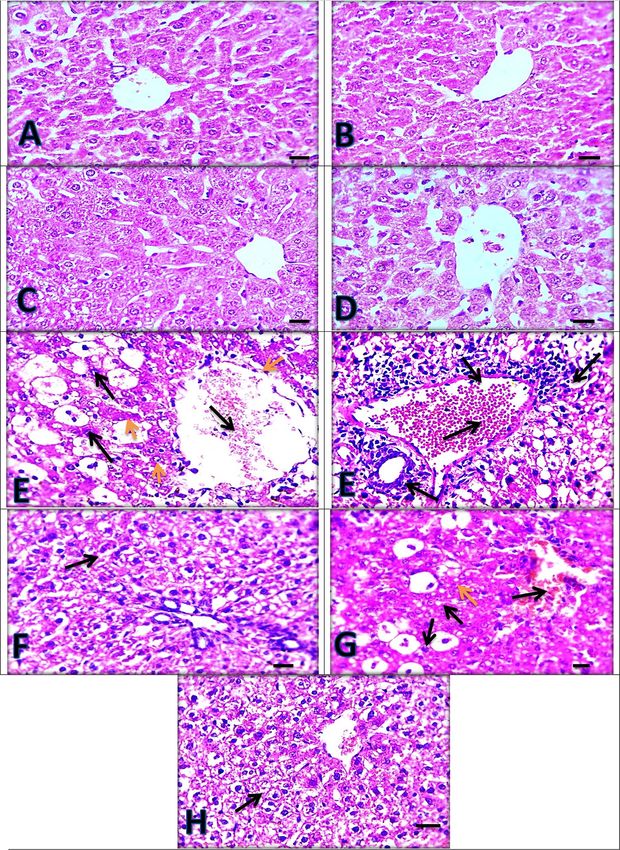
www.nature.com/scientificreports/
Parameter group Initial body wt Final body wt Body weight gain
G1 134.20 ± 2.22a 142.80 ± 2.63c 8.60 ± 0.07c
G2 133.80 ± 2.70a 143.40 ± 2.65c 9.60 ± 0.07c
G3 133.60 ± 2.24a 145.00 ± 2.23c 11.40 ± 0.07c
G4 133.40 ± 2.58a 147.20 ± 1.65b 13.80 ± 0.07b
G5 134.40 ± 2.33a 152.20 ± 3.13a 17.80 ± 0.07a
G6 134.40 ± 2.29a 145.20 ± 3.61c 10.80 ± 0.07c
G7 135.50 ± 2.54a 148.80 ± 1.68b 13.80 ± 0.07b
G8 134.80 ± 3.12a 153.20 ± 2.87a 18.40 ± 0.07a
Table 1. The changes in the body weight and body weight gain in the normal control and different
treated rat groups. G1 = control group, G2 = Paracetamol, G3 = Silymarin + Paracetamol, G4 = Chlorella
vulgaris + Paracetamol, G5 = Chlorella vulgaris + Thiamine + Paracetamol, G6 = Silymarin, G7 = Chlorella
vulgaris and G8 = Chlorella vulgaris + thiamine. Data are presented as means ± SEM (n = 6). Values having
different superscripts within same column are significantly different (p < 0.05).
are secreted via the kidney. Just a very little portion is expelled in the urine. The residual paracetamol about five
to nine percentage is biotransformed by the cytochrome P450 enzymes (CYPs), mostly CYP 2E1 into the highly
reactive intermediate metabolite N-acetyl-p-benzoquinone imine (NAPQI)1. When the toxic dose of paracetamol
is ingested, excessive NAPQ1 is produced and consequently causes serious GSH reduction as well as overproduc-
tion of reactive metabolites leading to covalent attachment of sulfhydryl groups in cellular proteins. This produces
disrupts homeostasis and starts apoptosis or programmed cell death, leading to tissue necrosis and eventually to
organ dysfunction which leads to liver oxidative stress1,2. Acute renal failure appears in nearly 1–2% of patients
with acetaminophen overdose, in addition to hepatic necrosis3,4. Recently, the usage of natural substances for
the prevention and treatment of liver disorders has increased5. Much attention has been pointed towards the
application of natural antioxidants originated from plants for alleviating the oxidative damages produced by free
radicals. Currently, numerous medicinal plants have shown such e ffectiveness6. Seeds of milk thistle (Silybum
marianum L. Gaertn) have been used the extraction of a mixture of flavonolignans (Silymarin). Silymarin is
a medicine used for the treatment of chronic and acute liver d iseases7. The main actions of Silymarin are the
scavenging of radical forms of oxygen and the stoppage of peroxynitrite creation8. Silymarin has been used as
a protective drug against paracetamol-induced hepatotoxicity and nephrotoxicity due to its anti-inflammatory
and antioxidant activities9–11.
Chlorella vulgaris is a single-cell green alga characterized by easy cultivation with high productivity and
composed of superior contents of chlorophyll, lutein, protein, and many other necessary micro-nutrients12,13,
C. vulgaris is documented as safe alga by the FDA14. It is considered as superfood including, 60% protein,
20 vitamins, 18 amino acids, and elements such as iron, potassium, calcium, phosphorous and m agnesium15.
Furthermore, there are many valuable antioxidants in microalgae, e.g., chlorophyll, carotenoids, astaxanthin,
lutein, and phycobiliproteins16,17. Chlorella sp. supplementation revealed beneficial physiological effects such as
antihypertensive18, antoxidative19, hypocholesterolemic20, and antitumor a ctivities21, hypoglycemic and hypolipi-
demic effects22,23 in animal, and human studies. Chorella had hepatoprotective effect against carbon tetrachloride-
induced liver damage in rats and m ice24,25. Another alga as Spirulina showed potential a hepatoprotective and
antioxidant activity against paracetamol-induced hepatic injury in r ats26.
Thiamine is the active form of vitamin B1 that assists as a coenzyme in a number of the main metabolic
pathways27. Zhou et al28 reported that, thiamine can reduce oxidative stress. Also, Asensi Fabado and Munne-
Bosch29, stated that, the antioxidant action of thiamine can be indirect, by offering NADH and NADPH to the
antioxidant network, or direct, by acting as an antioxidant. Thiamine Pyrophosphate proved to be as efficacious
as standard therapy and may be beneficial in APAP-induced h epatotoxicity30.
However, the hepatorenal protective activity of Chlorella vulgaris is not extensively s tudied31. Therefore,
the object of this study was to assess the protective effect or role of Chlorella vulgaris and/or thiamine against
Paracetamol induced toxicity in rats. For this purpose, hematological, serum biochemical, tissues’ lipid peroxida-
tion, and antioxidant biomarkers and histopathological examinations were estimated in Paracetamol intoxicated
Wistar rats pretreated either by C. vulgaris alga and /or thiamine.
Results and discussion
Body weight and weight gain changes. There was a significant (p ≤ 0.05) elevation in the final body
weight and body weight gain in G5 and G8 followed by G4, and G7 compared to the normal control group (G1).
While non-significant variations in the final body weight and body weight gain were seen in G2, G3 and G6
compared to the normal control group (G1) (Table 1).
Absolute and relative organ weights. As demonstrated in (Table 2), there was a significant (p ≤ 0.05)
increase in the absolute and relative weights of liver, kidney, and heart in paracetamol intoxicated group (G2) in
comparison with control normal group (G1). Meanwhile, a significant (p ≤ 0.05) decrease in these organ weights
was detected in G3, G4, and G5 compared with paracetamol intoxicated group (G2), the best reduction in these
Scientific Reports | (2021) 11:3911 | https://doi.org/10.1038/s41598-021-83316-8 2
Vol:.(1234567890)
www.nature.com/scientificreports/

                                                                      Absolute wt                                       Relative wt
                                   Parameter group   Final body wt    Liver          Kidney            Heart            Liver            Kidney             Heart
                                   G1                142.80 ± 2.63    5.26 ± 0.28b   1.52 ± 0.03d      1.15 ± 0.05d     3.68 ± 0.14b     1.07 ± 0.01d       0.80 ± 0.02d
                                   G2                143.40 ± 2.66    7.28 ± 0.28a   2.19 ± 0.07a      1.96 ± 0.05a     5.09 ± 0.23a     1.53 ± 0.03a       1.36 ± 0.03a
                                   G3                145.00 ± 2.24    5.66 ± 0.25b   1.85 ± 0.07bc     1.45 ± 0.09 cd   3.89 ± 0.15b     1.27 ± 0.03b       1.00 ± 0.06 cd
                                   G4                147.20 ± 1.66    6.05 ± 0.05b   1.94 ± 0.02ab     1.69 ± 0.10abc   4.11 ± 0.04b     1.32 ± 0.02b       1.14 ± 0.06abc
                                   G5                152.20 ± 3.14    5.91 ± 0.02b   1.84 ± 0.05bc     1.47 ± 0.07 cd   3.89 ± 0.09b     1.21 ± 0.02bc      0.97 ± 0.04 cd
                                   G6                145.20 ± 3.61    5.28 ± 0.32b   1.61 ± 0.07 cd    1.23 ± 0.09d     3.63 ± 0.13b     1.11 ± 0.02 cd     0.85 ± 0.05d
                                   G7                148.80 ± 1.69    5.65 ± 0.11b   1.58 ± 0.04 cd    1.26 ± 0.05d     3.79 ± 0.09b     1.06 ± 0.02d       0.85 ± 0.03d
                                   G8                153.20 ± 2.87    5.83 ± 0.05b   1.66 ± 0.09 cd    1.23 ± 0.04d     3.81 ± 0.04b     1.08 ± 0.04d       0.80 ± 0.03d

                                  Table 2.  The changes in the absolute and relative weight of different organs of normal control and different
                                  treated rat groups. G1 = control group, G2 = Paracetamol, G3 = Silymarin + Paracetamol, G4 = Chlorella
                                  vulgaris + Paracetamol, G5 = Chlorella vulgaris + Thiamine + Paracetamol, G6 = Silymarin, G7 = Chlorella
                                  vulgaris and G8 = Chlorella vulgaris + thiamine. Data are presented as means ± SEM (n = 6). Values having
                                  different superscripts within same column are significantly different (p < 0.05).

                                   Parameter group             RBCs (106/ul)               HB (g/dl)                    PCV (%)                          Plateletes (103/ul)
                                   G1                          7.86 ± 0.49a                13.76 ± 0.49ab               41.66 ± 1.69ab                   827.00 ± 35.75a
                                   G2                          5.01 ± 0.73c                10.86 ± 0.46c                32.94 ± 1.37c                    421.60 ± 28.68c
                                   G3                          7.39 ± 0.43a                12.92 ± 0.28b                39.48 ± 0.79b                    776.80 ± 56.17ab
                                   G4                          6.71 ± 0.21abc              12.12 ± 0.07bc               38.12 ± 0.18bc                   773.40 ± 43.45ab
                                   G5                          6.92 ± 0.54ab               13.00 ± 0.25b                40.32 ± 1.01b                    793.60 ± 49.64ab
                                   G6                          7.92 ± 0.54a                13.58 ± 0.52b                42.08 ± 1.42b                    803.80 ± 29.06ab
                                   G7                          8.08 ± 0.12a                14.04 ± 0.19a                41.22 ± 1.03b                    802.20 ± 32.92ab
                                   G8                          8.95 ± 0.30a                15.28 ± 0.37a                45.08 ± 0.82a                    847.40 ± 35.25a

                                  Table 3.  Erythrogram changes in the blood of normal control and different treated rat groups.
                                  G1 = control group, G2 = Paracetamol, G3 = Silymarin + Paracetamol, G4 = Chlorella vulgaris + Paracetamol,
                                  G5 = Chlorella vulgaris + Thiamine + Paracetamol, G6 = Silymarin, G7 = Chlorella vulgaris and G8 = Chlorella
                                  vulgaris + thiamine. RBCs = Red blood cells, HB = Hemoglobin, PCV = Packed cell volume. Data are presented
                                  as means ± SEM (n = 6). Values having different superscripts within same column are significantly different
                                  (p < 0.05).

                                   Parameter group             TLC (103/ul)               Neutrophils (%)                Lymphocytes (%)                      Monocytes (%)
                                   G1                          8.00 ± 0.56ab              22.20 ± 1.82b                  65.40 ± 1.74c                        5.800 ± 0.58a
                                   G2                          5.12 ± 1.02c               19.40 ± 0.92c                  73.20 ± 1.11a                        5.800 ± 0.58a
                                   G3                          8.32 ± 0.23ab              23.20 ± 1.82b                  68.80 ± 1.71bc                       5.000 ± 0.70a
                                   G4                          8.44 ± 0.70ab              23.20 ± 1.71b                  69.40 ± 0.24bc                       5.600 ± 0.92a
                                   G5                          8.62 ± 0.49ab              24.00 ± 1.34b                  66.20 ± 1.46c                        6.000 ± 0.70a
                                   G6                          8.68 ± 0.78ab              23.40 ± 1.88b                  66.40 ± 1.56c                        6.200 ± 0.86a
                                   G7                          8.20 ± 0.48ab              23.20 ± 1.59b                  66.60 ± 1.60c                        6.000 ± 0.70a
                                   G8                          8.98 ± 0.31a               26.40 ± 0.81a                  66.40 ± 1.53c                        5.800 ± 0.58a

                                  Table 4.  Leukogram changes in the blood of normal control and different treated rat groups. G1 = control
                                  group, G2 = Paracetamol, G3 = Silymarin + Paracetamol, G4 = Chlorella vulgaris + Paracetamol,
                                  G5 = Chlorella vulgaris + Thiamine + Paracetamol, G6 = Silymarin, G7 = Chlorella vulgaris and G8 = Chlorella
                                  vulgaris + thiamine. TLCs = Total leukocyte counts. Data are presented as means ± SEM (n = 6). Values having
                                  different superscripts within same column are significantly different (p < 0.05).

                                  organ weights was seen in G3 and G5. On the other hand, groups G6, G7, G8 showed non-significant changes in
                                  kidney, liver, and heart weights in comparison with control normal group (G1).

                                  Hematological parameters. The influences of paracetamol intoxication as well as the preventive effects
                                  of C. vulgaris and /or thiamine on hematological parameters of rats are shown in (Tables 3, 4). Paracetamol
                                  intoxication significantly (p ≤ 0.05) reduced RBCs count, Hb concentration, PCV%, platelets count, TLC, and

Scientific Reports |   (2021) 11:3911 |                 https://doi.org/10.1038/s41598-021-83316-8                                                                               3

                                                                                                                                                                            Vol.:(0123456789)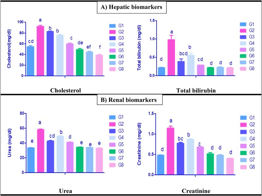
www.nature.com/scientificreports/

                                            Figure 1.  Serum biochemical parameters of liver enzymes and proteinogram of control and different
                                            treated rat groups. G1 = Control group, G2 = Paracetamol, G3 = Silymarin + Paracetamol, G4 = Chlorella
                                            vulgaris + Paracetamol, G5 = Chlorella vulgaris + Thiamine + Paracetamol, G6 = Silymarin, G7 = Chlorella vulgaris,
                                            G8 = Chlorella vulgaris + Thiamine. ALT = Alanine amino transferase, AST = Aspartate amino transferase. Data
                                            are presented as means ± SEM (n = 6). Different letter means significant difference effects in the same time
                                            period.

                                            neutrophils % with significant (p ≤ 0.05) rise in lymphocytes % in comparison with control normal group (G1).
                                            This picture was significantly (p ≤ 0.05) improved in the other treated groups compared with the paracetamol
                                            group (G2). The best improvement was detected in G3 and G5. Moreover, a significant increase in neutrophils %
                                            was observed in G8 compared with control (G1) and other treated groups.

                                            Serum biochemical parameters. The influences of paracetamol induced toxicity and the protective
                                            effects of C. vulgaris and /or thiamine on serum biochemical parameters are shown in (Figs. 1A,B, 2A,B). Par-
                                            acetamol exposed rats group (G2) revealed significantly increased serum transaminases activities (Fig. 1A), cho-
                                            lesterol, bilirubin levels (Fig. 2A) as well as elevated urea, and creatinine levels (Fig. 2B) with significant decline
                                            in serum total protein and albumin concentrations (Fig. 1B) in comparison with normal control rats group (G1).
                                            Moreover, a significant alleviation in the same parameters was seen in G3, G4 and G5 compared with paraceta-
                                            mol exposed group (G2), the best improvement was observed in G3 and G5. However, a significant decline in
                                            ALT activity was shown in G6, G7 and G8 compared with normal control rat (G1). Meanwhile, a significant
                                            reduction in cholesterol was seen in G7 and G8 in comparison to normal control rat group (G1).

                                            Hepatic renal and cardiac antioxidant status and lipid peroxidation. The influences of paraceta-
                                            mol induced toxicity and administration of C. vulgaris and /or thiamine on the lipid perioxidation and antioxi-
                                            dant enzymes of hepatic, renal, and cardiac tissues are shown in (Fig. 3A,B). MDA concentrations in hepatic,
                                            renal, and cardiac tissues were significantly elevated in paracetamol intoxicated group (G2) in comparison with
                                            the normal control group (G1) (Fig. 3A). Moreover, a significant decrease in hepatic, renal and cardiac MDA
                                            was observed in G3, G4, and G5 compared with paracetamol intoxicated group (G2). The superior reduction
                                            was observed in G3 and G5. On the other hand, paracetamol intoxication induced oxidative stress in liver, kid-
                                            ney and heart which resulted in the depletion of hepatic, renal and cardiac CAT activity (Fig. 3B). Furthermore,
                                            a significant elevation in catalase enzyme activity was detected in G3, G4 and G5 compared with paracetamol
                                            intoxicated group (G2), and the best was G5. Meanwhile, a significant increase in catalase activity was noticed
                                            in hepatic, renal, and cardiac tissues of rat groups administered C. vulgaris plus thiamine (G8) group compared
                                            to the normal control rat group (G1).

          Scientific Reports |   (2021) 11:3911 |                https://doi.org/10.1038/s41598-021-83316-8                                                      4

Vol:.(1234567890)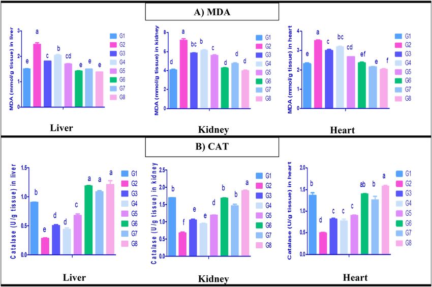
www.nature.com/scientificreports/
Figure 2. Serum biochemical parameters of hepatic and renal biomarkers of control and different
treated rat groups. G1 = Control group, G2 = Paracetamol, G3 = Silymarin + Paracetamol, G4 = Chlorella
vulgaris + Paracetamol, G5 = Chlorella vulgaris + Thiamine + Paracetamol, G6 = Silymarin, G7 = Chlorella vulgaris,
G8 = Chlorella vulgaris + Thiamine. Data are presented as means ± SEM (n = 6). Different letter means significant
difference effects in the same time period.
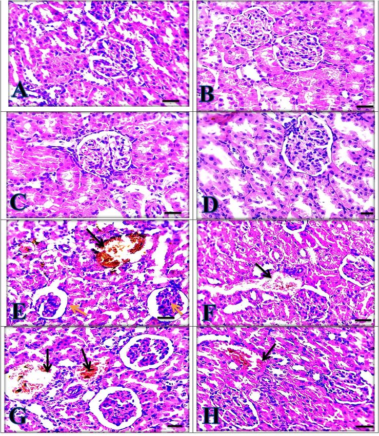
Histopathological findings. Normal control rat group liver sections (Fig. 4A) showed normal hepatic
architecture with no pathological changes. The same picture was seen in silymarin, C. vulgaris and C. vul-
garis + thiamine treated groups, respectively (Fig. 4B–D) confirming the hepatoprotective effects of silyma-
rin, C. vulgaris and thiamine when they were administered separately. Moreover, the paracetamol intoxicated
group (Fig. 4E) revealed severe congestion, and most of the centrilobular hepatocytes showed marked vacuolar
and ballooning degeneration, besides aggregation of lymphocytes in the portal area. The hepatic structure was
improved and looked close normal with mild hydropic degeneration in hepatocytes in silymarin + paracetamol
group and C. vulgaris + thiamine + paracetamol group (Fig. 4F,H). Moreover, paracetamol intoxicated rats given
C. vulgaris showed moderate congestion, vacuolar and ballooning degeneration in hepatocytes (Fig. 4G).
Kidney sections showed normal appearance of the glomerulus and tubules of control group (Fig. 5A). The
same picture was seen in silymarin, C. vulgaris and C. vulgaris + thiamine treated groups, respectively (Fig. 5B–D),
confirming the nephroprotective effects of silymarin, C. vulgaris, and thiamine when given separately. Paraceta-
mol intoxicated group (Fig. 5E) showed severe congestion, marked tubular dilation with loss of cellular boundary
and epithelial degeneration , glomerular shrinkage, bleeding and partial endothelial rupture in capsule. Sily-
marin and C. vulgaris + Thiamine administrations to paracetamol intoxicated groups revealed mild congestion
(Fig. 5F,H). While, C. vulgaris + paracetamol group was showed the moderate congestion beside the moderate
tubular dilation as in (Fig. 5G).
Heart sections showed the normal appearance of cardiomyocytes of control rat group (Fig. 6A). The same find-
ings were detected in silymarin, C. vulgaris and C. vulgaris + thiamine treated groups, respectively (Fig. 6B–D).
On the other hand, the paracetamol intoxicated group showed degeneration and vacuolation in cardiomyocytes
with severely congested cardiac blood vessels (Fig. 6E). This lesion was much improved by the administration of
either Silymarin or C. vulgaris plus Thiamine to the paracetamol intoxicated groups which showed mild conges-
tion, respectively (Fig. 6F,H). While moderate congestion was seen by C. vulgaris administration to paracetamol
intoxicated group (Fig. 6G).
Scientific Reports | (2021) 11:3911 | https://doi.org/10.1038/s41598-021-83316-8 5
Vol.:(0123456789)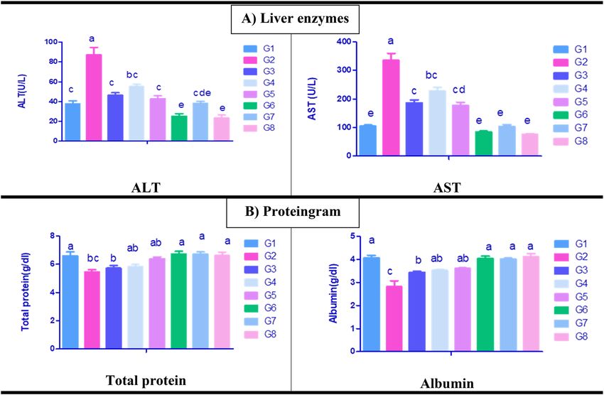
www.nature.com/scientificreports/
Figure 3. Oxidative stress and antioxidant status. (A) Malondialdehyde (MDA) (nmol/gm tissue), (B)
Catalase activity (CAT) (μmol/mg) of liver, kidney and heart tissues of control and different treated rat groups.
G1 = Control group, G2 = Paracetamol, G3 = Silymarin + Paracetamol, G4 = Chlorella vulgaris + Paracetamol,
G5 = Chlorella vulgaris + Thiamine + Paracetamol, G6 = Silymarin, G7 = Chlorella vulgaris, G8 = Chlorella
vulgaris + Thiamine. Data are presented as means ± SEM (n = 6). Different letter means significant difference
effects in the same time period.
FT‑IR. FT-IR technique was used for evaluation the type of organic and inorganic complexes in Chlorella
vulgaris and C. vulgaris supplemented with Thiamine. The FT-IR analyzes of C. vulgaris biomass without any
addition (control) and C. vulgaris supplemented with thiamin represented different absorption peeks. The peeks
with C. vulgaris control were 3404, 2970, 2925, 2856, 1655, 1549, 1408, 1384, 1054, 711 and 568 cm−1 which has
shifted to 3449, 2959,2954, 2853, 2768, 1646, 1384, 1076, 875, 831, 600 and 564 cm−1. The infra-red spectrum
displays a frequency ranges from 3500 to 3200 cm−1 indicating the O–H stretching vibration, existence of alco-
hols, phenols. The frequency ranges from, 3000–2850 cm−1peaks are representing in the C-H stretching vibra-
tion existence of alkenes. The wavenumber of some peaks of C. vulgaris biomass were decreased or increased
after supplemented with thiamine such as wavenumber peak at 3404 increased to 3449, the peak 2970 decreased
to 2959, peaks at 2925 increased to 2954, peak 2856 decreased to 2853, peak 1655 decreased to 1646, peak 1054
increased to 1076 respectively. There are some new peaks and also some peaks are disappeared as shown in
Table 5, these results showed the difference in the alga compositions when supplemented with thiamine and
hence its effect on oxidative stress induced by paracetamol.
Oxidative stress is a phenomenon caused by an imbalance between production and accumulation of oxygen
reactive species (ROS) in cells and tissues and the ability of a biological system to detoxify these reactive prod-
ucts. ROS can play, and in fact they do it, several physiological roles (i.e., cell signaling), and they are normally
generated as by-products of oxygen metabolism; despite this, environmental stressors (i.e., UV, ionizing radia-
tions, pollutants, and heavy metals) and xenobiotics (i.e., antiblastic drugs) contribute to greatly increase ROS
production, therefore causing the imbalance that leads to cell and tissue damage (oxidative stress)32.
Oxidative stress plays a vital role in the pathogenesis of paracetamol induced liver d amage33. This study dem-
onstrated that paracetamol intoxication caused deleterious impacts on hemopoietic organs, which represented by
lowered hematological parameters including, RBCs counts, Hb concentration, PCV%, TLC, Platelets count and
neutrophil%. These findings are consistent with that of Desnoyers34;Taylor & Dhupa35 who demonstrated that
the changes in the analyzed blood parameters might be due to the oxidative stress induced by paracetamol which
has a damaging effect on immune and hemopoietic organs and erythrocytes. Paracetamol inhibits hemopoesis
together with hematotoxicity, primarily methemoglobinemia and hemolytic anemia. This may be attributed to the
destruction of RBCs by increased lipid peroxidation in cell m embranes36. Moreover, uremia has a bad effect on
Scientific Reports | (2021) 11:3911 | https://doi.org/10.1038/s41598-021-83316-8 6
Vol:.(1234567890)www.nature.com/scientificreports/
Figure 4. Liver sections showing normal appearance in (A) Control group, (B) Silymarin group, (C)
Chlorella vulgaris group and (D) Chlorella vulgaris + thiamine group. (E) Paracetamol group showing severe
congestion (black thin arrow) with marked vacuolar (yellow arrowheads) and ballooning degeneration
in hepatocytes (black arrowheads) besides aggregation of lymphocytes in portal area (thick arrows). (F)
Silymarin + Paracetamol group and (H) Chlorella vulgaris + Thiamine + Paracetamol group showing mild
hydropic degeneration in hepatocytes (arrows). (G) Chlorella vulgaris + Paracetamol group showing moderate
congestion (black thin arrow) vacuolar (yellow arrowheads) and ballooning degeneration (black arrowheads) in
hepatocytes. (H) and (E) X: 400 bar 50.
Scientific Reports | (2021) 11:3911 | https://doi.org/10.1038/s41598-021-83316-8 7
Vol.:(0123456789)www.nature.com/scientificreports/
Figure 5. Kidney sections showing normal appearance in (A) control group, (B) Silymarin group, (C)
Chlorella vulgaris group and (D) Chlorella vulgaris + Thiamine group. (E) Paracetamol group showing severe
congestion (black arrow) and glomerular shrinkage (yellow arrows). (F) Silymarin + Paracetamol group and
(H) Chlorella vulgaris + Thiamine + Paracetamol group showing mild congestion (black arrow). (G) Chlorella
vulgaris + Paracetamol group showing moderate congestion (black arrow). (H) and (E) X: 400 bar 50.
blood platelets37. On the same line, Adedapo et al38, Daniel and C lement39, Biu et al40 reported that, xenobiotics
intoxication exhibited potential inhibition of erythropoietin release from damaged kidneys and susceptibility of
this highly proliferative tissue for toxicity. The current research declared that paracetamol stimulated hepatic renal
Scientific Reports | (2021) 11:3911 | https://doi.org/10.1038/s41598-021-83316-8 8
Vol:.(1234567890)www.nature.com/scientificreports/
Figure 6. Heart sections showing normal appearance in (A) Control group, (B) Silymarin group, (C) Chlorella
vulgaris group (D) Chlorella vulgaris + Thiamine group. (E) Paracetamol group showing severely congested
cardiac blood vessels (arrows) besides degeneration and vacuolation in cardiomyocytes (arrowheads). (F)
Silymarin + Paracetamol group and (H) Chlorella vulgaris + Thiamine + Paracetamol showing mildly congested
cardiac blood vessels (arrows). (G) Chlorella vulgaris + Paracetamol group showing moderately congested
cardiac blood vessels. (H) and (E) X: 400 bar 50.
Scientific Reports | (2021) 11:3911 | https://doi.org/10.1038/s41598-021-83316-8 9
Vol.:(0123456789)www.nature.com/scientificreports/
C. vulgaris C. vulgaris with thiamine Difference Frequency ranges(cm−1) Functional groups
O–H stretching vibration occurrence of alcohols,
3404 3449 54 3500–3200
phenols
2970 2959 − 11 3000–2850 C–H stretching vibration occurrence of alkenes
2925 2954 29 3000–2850 C–H stretching vibration occurrence of alkenes
2856 2853 −3 3000–2850 C–H stretching vibration occurrence of alkenes
– 2768 – 2925–2875 Aliphatic C–H Stretching vibration
1655 1646 −9 1680–1640 –C=C– stretching vibration
N–O asymmetric stretching vibration presence of
1549 – – 1550–1475
nitro compounds
1408 – – 1500–1400 C–C stretching vibration presence of aromatics
1384 1384 – 1390–1365 C–C stretching vibration presence of aromatics
1054 1076 22 1250–1020 C–N stretch stretching vibration
711 875–831 – 910–665 N–H wag stretching vibration
568 600–564 32–4 690–515 C–Br stretching vibration presence of alkyl halides
Table 5. The FT-IR frequency range and the following functional groups are present in the C. vulgaris and
with thiamine.
and cardiac damage which was represented by alterations in the serum biochemical parameters. These alterations
are implicated in a series of events leading to paracetamol mediated hepatic, renal and cardiac toxicities. Such
toxicities are the consequences of the oxidative injuries induced by excessive generation of ROS and the impair-
ment of the antioxidant enzyme activities. These results are in line with the previuos researches carried out by
Nikravesh et al41, Zhao et al42 who reported that, lipid peroxidation and oxidative stress are the early events related
to radicals generation during the hepatic metabolism of acetaminophen. Moreover, Du et al2 documented that the
intracellular mechanisms of paracetamol-induced hepatocytic injury is by mitochondrial dysfunction and exces-
sive ROS production causing severe oxidative stress. Paracetamol can stimulate liver injury by oxidative stress
and inflammation42,43. Gini and Muraleedhara44; Kanchana and Sadiq45 concluded that overdose of paracetamol
induces toxicity to the hepatocytes. Our results are also in harmony with Sabiu et al46 who indicated that cellular
leakage and loss of functional integrity of the liver cell membrane duo to paracetamol intoxication revealed a
significant increase in the serum enzyme activities of ALTand AST with elevation of bilirubin and cholesterol
levels. Moreover, the significant elevation in cholesterol level recorded after paracetamol administration may be
due to the imbalance between the normal rates of lipid synthesis, utilization and s ecretion47,48 or may be duo to
inhibition of bile acid synthesis as recorded by previous s tudies49–51.
The reduced serum total protein and albumin concentrations following paracetamol overdose exposure in
this study resulting from disturbance of protein synthesis as a consequence of altered hepatic function as a result
of inflammation52 or due to nephrotoxicity which leads to leakage of albumin in urine with decreasing of serum
albumin and total protein c oncentrations53.
Our study clearly demonstrates that acute acetaminophen toxicity enhanced renal MDA level, depleted the
renal CAT antioxidant activity leading to elevated serum urea and creatinine levels, reduced total protein and
deteriorated the renal architecture as confirmed by our histopathological observations. The end product of lipid
peroxidation is MDA, which is recognized as the second messenger of free radicals. The high concentration of
MDA in renal tissue denotes to renal toxicity54. Inconsistent with our results, Srinivasan et al33 who reported that,
increased ROS level and decreased enzymatic antioxidants considered as a mechanism by which several chemicals
can induce nephrotoxicity leading to disturbance of cell membrane integrity. Paracetamol nephrotoxicity occurs
as a result of its highly reactive metabolite- NAPQI- which acrylates proteins in the proximal tubule, initiating
renal tubular cells death55. In accordance with our results, Mandal et al54, Das et al56 who concluded that, aceta-
minophen overdose is often associated with elevation of urea and creatinine concentrations which are indicators
of drug-induced nephrotoxicity in animals. In line with our observation Cohen et al57 who demonstrated that
acetaminophen overdose decreased antioxidant enzymes in kidney tissues and enhanced lipid peroxidation.
Similary, Jones and Vale58 reported that paracetamol overdose induced hepatic and renal deleterious necrosis
in humans and experimental animals.
Several herbal and plant extracts derived compounds served as alternative therapeutic agents to counteract
the side effects of various d rugs59,60.
In the current study silymarin succeeded to overcome the deleterious impacts of paracetamol intoxication
on rat hematological, biochemical parameters and histopathological changes, reduced hepatic, renal and cardiac
oxidative damage and enhanced hepatic, renal and cardiac antioxidants. In consistent with our results, Papackova
et al8 who pointed that the main actions of silymarin are scavenging of radical forms of oxygen and inhibition
of peroxynitrite formation. Furthermore, Freitag et al10 stated that, the prophylactic activity of silymarin against
paracetamol-induced hepatotoxicity is generally attributed to its antioxidant and anti-inflammatory properties.
Several studies about the standard drug silymarin found that silymarin offered protection against chemical
hepatotoxins such as CCl4, ethanol, and paracetamol61. Moreover, Cacciapuoti et al62 mentioned that silymarin
is an effective remedy for decreasing hepatic steatosis in patients with non-alcoholic fatty liver disease. Silyma-
rin was approved for the treatment of the hepatotoxic doses of paracetamol. Therefore, in this research we used
silymarin as a standard control drug.
Scientific Reports | (2021) 11:3911 | https://doi.org/10.1038/s41598-021-83316-8 10
Vol:.(1234567890)www.nature.com/scientificreports/
Regarding to the effect of C. vulgaris algae on body weight, our results showed a significant increase in final
body weight and body weight gains in response to C. vulgaris algae administration in comparison to the control
and other treated groups. C. vulgaris is a rich source for chlorophyll pigment and vital amino acids; in addition
to considerable quantities of calcium, phosphorus, iodine, manganese, iron and vitamins such as A, B1, B2, B3,
B6, B12, C 67 and E63. In agreement with our results, Xu et al64 who stated that C. vulgaris can be a useful choice
as an additive for fish diets, they claimed that C. vulgaris could improve digestive the enzymes and enhance
growth performance and immunity due to its high concentrations of the crude protein, polysaccharides, lipid,
minerals and other bioactive components involved in many physiological activities. On the same line, Kang et al65
concluded that Chlorella additions to the diets of broiler chicks improved body weight.
Concerning to the effects of C. vulgaris against paracetamol intoxication, the current results demonstrated that
rats administered C. vulgaris at the chosen dose either alone or with thiamine succeeded to minimize the deleteri-
ous effects of paracetamol on rats’ hematological, biochemical, antioxidant status and histopathological findings,
suggested that C. vulgaris exhibits excellent hepato protective properties and has some role in maintaining the
structural integrity of the hepatocellular membrane, thus preventing the enzymes leakage into the blood circula-
tion, together with repairing of the hepatic tissue damage induced by paracetamol. This impact is in consistent
with Ahmed and Khater66, Pawlikowska-Pawlega et al67 who stated that serum levels of transaminases returned
to normal with the healing of hepatic parenchyma and the regeneration of hepatocytes. Moreover, Rodriguez-
Garcia and Guil-Guerrero68 reported that Chlorella vulgaris exhibited antioxidative and hepatoprotective effects.
Furthermore, Cheng et al69 recorded that the possible mechanism for C. vulgaris protection may be attributed
to its immunomodulatory potential, that may stimulate the lymphocytes propagation and phagocytic activities
of macrophages, promote the expressions of cytokines, improve the NK cells cytotoxicity, and ameliorate the
histological changes of the spleen.
Furthermore, C. vulgaris succeeded to restore the levels of urea, and creatinine close to normal values thus
preventing the kidney from damage. These restorative effects of C. vulgaris over the serum clinical chemistry
correlate with previous studies used C. vulgaris for treating oxidative stress31. On the same line several studies
declared that Chlorella vulgaris administration provided protection against membrane fragility with anti-inflam-
matory, antihypertensive, and antioxidative a ctivities18,19. As C. vulgaris microalgae, contains many valuable
antioxidants as chlorophyll, carotenoids, astaxanthin, lutein and phycobili-proteins17, with the highest amount
of chlorophyll than any known plant. Moreover, C. vulgaris prevented the lipid peroxidation in hepatic, renal
and cardiac tissues. In addition to, its ability to abolish the toxic effect of paracetamol on the examined tissues
through increasing the activities of antioxidant enzymes. The protective effects of C. vulgaris and its antioxidant
activity are attributed to their content of phenolic c ompounds70 as there is a close positive relationship or cor-
relation between the quantity of these compounds in C. vulgaris extract and their antioxidant activities due to
their redox properties that play a vital role in capturing and scavenging free radicals, oxygen suppression and
peroxide decomposition71–73. Furthermore, C. vulgaris extract significantly decreased the degree of lipid per-
oxidation and TBARS level in leukocytes in comparison to Ganoderma lucidum extract in vitro l ocation74. The
same results were detected when C. vulgaris is supplemented alone or with thiamine. In agreement with our
observation Zhou et al28 who reported that thiamine can reduce oxidative stress. Furthermore, Asensi Fabado
and Munne-Bosch29 stated that, the antioxidant activities of thiamine can be indirect, by providing NADH and
NADPH to the antioxidant network, or direct, by acting as an antioxidant.
The prophylactic effects of C. vulgaris against oxidative stress induced by paracetamol intoxication in our
study could be due to inhibition of lipid peroxidation and scavenging of free radicals as its administration was
responsible for the increased resistance against oxidative stress induced by paracetamol which consequently
plays a fundamental role in the pathogenesis of paracetamol induced liver d amage33,52. The elevated levels of
MDA demonstrated in the present study are in accordance with those of other investigators who reported the
association between paracetamol toxicity and MDA elevation75. Moreover, C. vulgaris and or thiamine prevented
the lipid peroxidation in hepatic, renal and cardiac tissues and improved the activities of antioxidant enzymes
in rats tissues, such effects could be the mechanisms of their hepatorenal protection. This is in agreement with
the report of Sabiu et al76 who stated that acetaminophen mediated hepatic oxidative insults in rats had induced
significant decrease in the activities of antioxidant enzymes.
Compared with the standard drug silymarin, no significant differences were detected in the protection
induced by silymarin treatment and C. vulgaris and /or thiamine treatment, suggesting that C. vulgaris either
alone or with thiamine succeeded to prevent disruption of organs function by protecting the lipids from peroxi-
dation by ROS under paracetamol toxicity and enhancing antioxidant enzymes activity.
Material and methods
Chemicals. Paracetamol tablets (each tablet contains 500 mg) was obtained from El-Nasr Pharmaceutical
Chemicals Co., Egypt. Paracetamol was suspended in pathogen-free normal distilled water prior usage. Silyma-
rin capsules (Legalon 140) each capsule contains 140 mg was purchased from Ced Pharmaceutical Co, Giza,
Egypt.
The diagnostic kits used for assaying hepatic and kidney performance tests, the levels of lipid peroxidation
and antioxidants were obtained from Bio-Diagnostic Co., Giza, Egypt. All other chemicals used throughout
the experiments were of high analytical grade. Thiamine powder was obtained from El-Nasr Pharmaceutical
Chemicals Co, Egypt.
Chlorella vulgaris alga (CV). Chlorella vulgaris alga was obtained from «Microbial Biotechnology Lap,
Genetic Engineering and Biotechnology Research Institute (GEBRI), University of Sadat City, Sadat City, Egypt».
BG11 nutritive culture was used as a medium for enrichment and growth of the tested alga.
Scientific Reports | (2021) 11:3911 | https://doi.org/10.1038/s41598-021-83316-8 11
Vol.:(0123456789)www.nature.com/scientificreports/
Chlorella vulgaris alga supplemented with Thiamine. Two hundred ml of the BG11 nutritive culture
medium were prepared and supplemented with 0.08 mg/L vitamin B1 (thiamine), after sterilization C. vulgaris
was inoculated. The culture was incubated at natural day light at temperature 30 ± 2 °C and shaken gently twice
a day to avoid clumping and enhance the growth. After 15 days of incubations, the culture was centrifuged,
washed by distilled water and dried through the hot air oven at 60 °C until the constant weight was obtained.
The required daily dose from Chlorella powder and Chlorella supplemented with thiamine administered to
the animals in this study were dissolved in sterilized distilled water to be in suspension format using Ultrasonic
homogenizer sonication (Biologics Inc. USA manufacturer and leading innovator)77.
FT‑IR analysis. FTIR spectroscopy is a technique required to define cell contents of microalgae78. FTIR
spectra illustrates the macromolecular composition of the algal biomass depending on the infrared absorp-
tion of functional groups. So, FTIR spectroscopy permits the revealing of changes in the relative abundance of
organic compounds such as carbohydrate, lipid and protein. This technique was used in many studies to define
the changes in the macromolecular composition of microalgae caused by nutrient s tress79–82. The change of func-
tional groups present in dry algal biomass control and that supplemented with Thiamine has been described by
Fourier transform infrared (FTIR) spectroscopy according to methods of J ebsen83.
Animals and experimental design. This study was approved by the Research Ethical Committee of the
Genetic Engineering and Research Institute, Sadat City University, Egypt. Forty eight female albino rats of Wistar
strain (130–150 g) were obtained from the Animal House of the Genetic Engineering and Research Institute,
University of Sadat City, Egypt and housed in well- ventilated plastic cages. The diet and water were provided
ad-libitum. All rats were housed under standard husbandry conditions (25 ± 2 °C temp, 60 ± 5% relative humid-
ity and 12 h photoperiod). Rats were kept untreated for two weeks for acclimatization prior treatment and were
weighed at the starting of research (initial weight). All animal handling procedures, sample collection and dis-
posal were according to the regulation of Institutional Animal Care and Use Committee (IACUC), Genetic Engi-
neering and Research Institute University of Sadat City, Egypt, under approval number (gebriUSC-009-1-19).
Forty eight female albino rats of Wistar strain were randomly divided into eight equal groups (n = 6 rats each)
as the following:
Group 1, Normal control group, it was administered distilled water only per os (0.5 ml/rat) daily for 7 suc-
cessive days.
Group 2, Paracetamol group, it was treated as normal control group for 7 successive days, then given Par-
acetamol per os once (2gm/kg.bwt.). according to S haroud26.
Group 3, Silymarin + Paracetamol group, it was treated with Silymarin drug (100 mg/kg. b.wt.) according
to Bektur et al9 per os daily for 7 successive days, then administered Paracetamol per os once (2gm/kg.bwt.).
Group 4, Chlorella vulgaris alga + Paracetamol group, it was treated with Chlorella vulgaris alga (500 mg/kg.
b.wt) according to Hsin-yi et al24; Sharoud et al26 per os daily for 7 successive days, then administered Paraceta-
mol per os once (2gm/kg.bwt.).
Group 5, Chlorella vulgaris alga + thiamine + Paracetamol group, it was treated with Chlorella vulgaris alga
plus thiamine (500 mg/kg. b.wt), respectively per os daily for 7 successive days, then administered Paracetamol
per os once (2gm/kg.bwt.).
Groups 6, Silymarin group, it was treated with Silymarin (100 mg/kg. b.wt) per os daily for 7 successive days
without paracetamol administration.
Group 7, Chlorella vulgaris alga group, it was treated with Chlorella vulgaris alga (500 mg/kg. b.wt.) per os
daily for 7 successive days without paracetamol administration.
Group 8, Chlorella vulgaris alga + thiamine group, it was treated with Chlorella vulgaris alga plus thiamine
(500 mg/kg.b.wt.) per os daily for 7 successive days without paracetamol administration.
Rats of all the experimental groups were anaesthetized and euthanized after 24 h of the last treatment for
samples collection.
Sampling. At the end of the experiment (24 h after paracetmol administration), rats in all groups were fasted
overnight and weighed to calculate the final body weights and weight gain. Then, blood samples were obtained
from each rat via retro orbital bleeding under light ether anaesthesia (Sigma Chem. Co., St Louis, Mo. U.S.A).
Two blood samples were taken from each rat. One sample was put into a tube containing heparin as anticoagu-
lant for hematological assessment. The other sample was put in a tube without heparin and allowed to coagulate,
then centrifuged at 3000 for 15 min. The clear sera were collected and kept at − 20 °C for subsequent biochemical
analysis. After blood samples collection, rats were euthanized by cervical dislocation for tissue samples col-
lection. Liver, kidney and heart from each rat were carefully excised, weighed and immediately cleaned with
normal saline solution (0.9% NaCl). Each tissue sample was divided into two parts. A part was kept at − 80 °C for
Malondialdehyde (MDA) and catalase (CAT) activity estimations. The other part was fixed in 10% neutral buffer
formalin solution for further histopathological examinations.
Absolute and relative body and organ weights. Just before killings the rats at 8th day of experiment,
final body weight of all rats in all experimental groups was calculated. The body gain was calculated from the
difference between the body weight at the beginning and at the end of experiment. Upon being sacrificed or
killed, the liver, kidney and heart were aseptically removed, weighed and their relative organ weights (ROW)
were determined according to the equation of Aniagu et al84.
Scientific Reports | (2021) 11:3911 | https://doi.org/10.1038/s41598-021-83316-8 12
Vol:.(1234567890)www.nature.com/scientificreports/
ROW = Absolute organ weight g /body weight of rat on sacrifice day g × 100.
Hematological analysis. The whole blood samples were utilized directly after collection for estimation of
hematological parameters including the red blood cells (RBCs), hemoglobin (Hb) concentration and hematocrit
value (PCV%), total leucocytes count (TLC), differential leukocyte counts and platelets (Plt) counts, by using
automated blood cells counter with an Auto Hematology Analyzer (Sysmex F-800, Japan)85.
Biochemical assays. The biochemical parameters of liver and renal injury biomarkers were estimated
in the collected serum samples according to the manufacturer protocol. Serum enzymatic activities of aspar-
tate amino transferase (AST) and alanine amino transferase (ALT) were assessed according to Reitman and
Frankel86. Albumin (Alb) and total proteins (TP) according to Henry et al87. Serum cholesterol was measured
according to Richmond88. Total bilirubin was analyzed according to Tietz89. Renal products; creatinine was esti-
mated according to Larsen90, and urea according to Coulombe and Favreau91.
Evaluation of oxidative stress and antioxidant biomarkers. Immediately after blood collection, the
animals were euthanzied by cervical dislocation, then the liver, kidney and heart from each rat were immedi-
ately dissected out and weighed. A part from each organ was homogenized using glass homogenizer with ice
cooled saline to prepare 25% W/V homogenate. This homogenate was centrifuged at 1700 rpm for 10 min; the
supernatant was stored at – 80 °C until analysis. This supernatant was used for the colorimetrical estimations of
hepatic, renal and cardiac malondialdehyde (MDA), the main end product of lipid peroxidation, according to
the protocol of Esterbauer et al92, and catalase activity according to the method of Sinha93.
Histopathological results. The other parts of liver, kidney and heart of the scarified rats were fixed in 10%
buffered formalin. Then, dehydration, clearance and processing in paraffin were carried out. Tissue sectioning
and staining with H&E were performed according to Bancroft et al94.
Statistical analysis. All data were expressed as means ± S.E. and statistically analyzed by one-way ANOVA
and Tukey’s post-hoc test multiple comparisons using Graphpad prism Version 5 software (Graph Pad Software
Inc., USA).. Statistical significance was acceptable to a level of p ≤ 0.05.
Conclusions
Oxidative stress plays essential role in paracetamol induced hepatorenal and cardiac toxicity. C. vulgaris is a
potent antioxidant agent that was indicated to protect intoxicated rats against oxidative stress induced by paracet-
amol. This study, revealed that paracetamol exposure resulted in varying degrees of lipid peroxidation, depletion
of the antioxidant enzymes activity and changes of hematological, biochemical parameters and histopathological
architectures of the examined tissues. C. vulgaris and /or thiamine pre-exposure offered near complete protection
in terms of blood and tissues changes, antioxidant enzymes activity and oxidative stress. Therefore, this study
suggested that C. vulgaris is a promizing protective agent against paracetamol induced toxicity as ROS scavenger
and a potential source of natural antioxidants.
Data availability
The research data used to support the findings of this study are included within the article (tables, figures).
Received: 8 June 2020; Accepted: 15 December 2020
References
1. Lancaster, E. M., Hiatt, J. R. & Zarrinpar, A. Acetaminophen hepatotoxicity: An updated review. Arch. Toxicol. 89, 193–1992. https
://doi.org/10.1007/s00204-014-1432-2 (2015).
2. Du, K., Ramachandran, A. & Jaeschke, H. Oxidative stress during acetaminophen hepatotoxicity: sources, pathophysiological role
and therapeutic potential. Redox Biol. 10, 148–156. https://doi.org/10.1016/j.redox.2016.10.001 (2016).
3. Abraham, P. Oxidative stress in paracetamol-induced pathogenesis: (I). Renal damage. Indian J. Biochem. Biophys. 42, 59–62 (2005).
4. Mazer, M. & Perrone, J. Acetaminophen-induced nephrotoxicity: Pathophysiology, clinical manifestations and management. J.
Med. Toxicol. 4, 2–6 (2008).
5. Jaeschke, H., McGill, M. R., Williams, C. D. & Ramachandran, A. Current issues with acetaminophen hepatotoxicity: A clinically
relevant model to test the efficacy of natural products. Life Sci. 88, 737–745. https://doi.org/10.1016/j.lfs.2011.01.025PMID:21296
090 (2011).
6. Khalafalla, M. M. et al. Active principle from Moringa oleifera Lam leaves effective against two leukemias and a hepatocarcinoma.
Afr. J. Biotech. 9, 8467–8471 (2010).
7. Abenavoli, L., Capasso, R., Milic, N. & Capasso, F. Milk thistle in liver diseases: Past present future. Phytother. Res. 24, 1423–1432
(2010).
8. Papackova, Z. et al. Silymarin prevents acetaminophen-induced hepatotoxicity in mice. PLoS ONE 13, e0191353. https://doi.
org/10.1371/journal.pone.0191353 (2018).
9. Bektur, N. E., Sahin, E., Baycu, C. & Unver, G. Protective effects of silymarin against acetaminophen-induced hepatotoxicity and
nephrotoxicity in mice. Toxicol. Ind. Health https://doi.org/10.1177/0748233713502841 (2013).
10. Freitag, A.F., Cardia, G.F., da Rocha, B.A., Aguiar, R.P., Silva-Comar, F.M., Spironello, R.A., Cuman, R.K. Hepatoprotective effect
of silymarin (silybum marianum) on hepatotoxicity induced by acetaminophen in spontaneously hypertensive rats. Evid. Based
Complem. Altern. Med. 538317 (2015).
Scientific Reports | (2021) 11:3911 | https://doi.org/10.1038/s41598-021-83316-8 13
Vol.:(0123456789)www.nature.com/scientificreports/
11. Ahmada, M. M., Rezkb, N. A., Fawzy, A. & Sabry, M. Protective effects of curcumin and silymarin against paracetamol induced-
hepatotoxicity in adult male albino rats. Gene 712, 143966 (2019).
12. Jeon, J. Y. et al. The production of Luteinenriched eggs with dietary Chlorella. Korean J. Food Sci. Anim. Resour. 32, 13–17 (2012).
13. Buono, S., Langellotti, A. L., Martello, A., Rinna, F. & Fogliano, V. Functional ingredients from microalgae. Food Funct. 5, 1669–1685
(2014).
14. Bauer, L. M., Vieira Costa, J. A., Conteno da Rosa, A. P. & Santos, L. O. Growth stimulation and synthesis of lipids, pigments and
antioxidants with magnetic fields in Chlorella kessleri cultivations. Bioresour. Technol. 244, 1425–1432 (2017).
15. Bengwayan, P. T. et al. A comparative study on the antioxidant property of Chlorella (Chlorella sp) tablet and glutathione tablet.
E. Int. Sci. Res. J. 2, 12–25 (2010).
16. Plaza, M., Herrero, M., Cifuentes, A. & Ibanez, E. Innovative natural functional ingredients from microalgae. J. Agric. Food Chem.
57, 7159–7170 (2009).
17. Ahmed, F. et al. Profiling of carotenoids and antioxidant capacity of microalgae from subtropical coastal and brackish waters. Food
Chem. 165, 300–306 (2014).
18. Sheih, I. C., Fang, T. J. & Wu, T. K. Isolation and characterisation of a novel angiotensin I-converting enzyme (ACE) inhibitory
peptide from the algaeprotein waste. Food Chem. 115, 279–284 (2009).
19. Ko, S. C., Kim, D. & Jeon, Y. J. Protective effect of a novel antioxidative peptide purified from a marine Chlorella ellipsoidea protein
against free radical-induced oxidative stress. Food Chem. Toxicol. 50, 2294–2302 (2012).
20. Cherng, J. Y. & Shih, M. F. Preventing dyslipidemia by Chlorella pyrenoidosa in rats and hamsters after chronic high fat diet treat-
ment. Life Sci. 76(2005), 3001–3013 (2005).
21. Wang, X. & Zhang, X. Separation, antitumor activities, and encapsulation of polypeptide from Chlorella pyrenoidosa. Biotechnol.
Prog. 29, 681–687 (2013).
22. Chovančíková, M. & Šimek, V. Effects of hight–fat and Chlorella vulgaris feeding on changes in lipid metabolism in mice. Biol.
Bratisl. 56, 661–666 (2001).
23. Cherng, J. Y. & Shih, M. F. Improving glycogenesis in Streptozocin (STZ) diabetic mice after administration of green algae Chlorella.
Life Sci. 78, 1181–1186 (2006).
24. Hsin-yi, P., Yu-chan, C., Shu-ju, C. & Su-tze, C. Hepatoprotection of chlorella against carbon tetrachloride-induced oxidative
damage in rats. In Vivo 23, 747–754 (2009).
25. Hyun-Kyung, K. et al. Protective effects of chlorella vulgaris extract on carbon tetrachloride-induced acute liver injury in mice.
Food Sci. Biotechnol. 18(5), 1186–1192 (2009).
26. Sharoud, M. N. M. Protective effect of Spirulinaagainst paracetamol-induced hepatic injury in rats. J. Exp. Biol. Agric. Sci. 3(1),
44–53 (2015).
27. Rapala-Kozik, M., Wolak, N., Kujda, M. & Banas, A. K. The upregulation of thiamine (vitamin B 1) biosynthesis in Arabidopsis
thaliana seedlings under salt and osmotic stress conditions is mediated by abscisic acid at the early stages of this stress response.
BMC Plant Biol. 12, 2–12 (2012).
28. Zhou, J., Sun, A. & Xing, D. Modulation of cellular redox status by thiamine-activated NADPH oxidase confers Arabidopsis resist-
ance to Sclerotinia sclerotiorum. J. Exp. Bot. 64, 3261–3272 (2013).
29. Asensi-Fabado, M. A. & Munne-Bosch, S. Vitamins in plants: occurrence, biosynthesis and antioxidant function. Trends Plant Sci.
15, 582–592 (2010).
30. Uysal, H. B. et al. Biochemical and histological effects of thiamine pyrophosphate against acetaminophen-induced hepatotoxicity.
Basic Clin. Pharmacol. Toxicol. 118, 70–76. https://doi.org/10.1111/bcpt.12496 (2016).
31. Li, W., Kim, Y. H. & Lee, Y. W. Chlorella vulgaris extract ameliorates carbon tetrachloride-induced acute hepatic injury in mice.
Exp. Toxicol. Pathol. 65, 73–80 (2013).
32. Pizzino, G. et al. Oxidative stress: Harms and benefits for human health. Oxidat. Med. Cell. Longev https : //doi.
org/10.1155/2017/8416763 (2017).
33. Srinivasan, C., Williams, W. M., Ray, M. B. & Chen, T. S. Prevention of acetaminophen-induced liver toxicity by 2(R, S)-n-
propylthiazolidine-4(R)- carboxylic acid in mice. Biochem. Pharmacol. 61, 245–252 (2001).
34. Desnoyers, M. Anemias associated with heinz bodies. In Schalm’s Veterinary Hematology (eds Feldman, B. F. et al.) 178–184 (Lip-
pincott WIlliams & Wilkins, Philadelphia, 2000).
35. Taylor, N. S. & Dhupa, N. Acetaminophen toxicity in dogs and cats. Compend. Contin. Educ. Gen. Dent. 12, 160–169 (2003).
36. Oyedeji, K. O., Bolarinwa, A. F. & Ojeniran, S. S. Effect of paracetamol (Acetaminophen) on hematological and reproductive
parameters in male albino Rat. IOSR J. Pharm. Biol. Sci. 4, 65–70 (2013).
37. Schoorl, M., Nube, M. J. & Bartels, P. C. Coagulation activation, depletion of platelet granules and endothelial integrity in case of
uraemia and haemodialysis treatment. BMC Nephrol. 14, 72 (2013).
38. Adedapo, A. A., Abatan, M. O. & Olorunsogo, O. O. Effects of some plants of the spurge family on haematological and biochemical
parameters in rats. Veterinarski Arhiv 77, 29–38 (2007).
39. Daniel, E. I. & Clement, O. N. Effect of ethanolic extract of Dennettia tripetala fruit on haematological parameters in albino Wista
rats. Niger. J. Physiol. Sci. 23, 13–17 (2008).
40. Biu, A. A., Yusufu, S. D. & Rabo, J. S. Studies on the effects of aqueous leaf extracts of Neem Azadirachta indica on haematological
parameters in chicken. Afr. Sci. 10, 189–192 (2009).
41. Nikravesh, H., Khodayar, M. J., Mahdavinia, M., Mansouri, E., Zeidooni, L., Dehbashi, F. Protective effect of gemfibrozil on hepa-
totoxicity induced by acetaminophen in mice: The importance of oxidative stress suppression. Adv. Pharm. Bull. 8, 331–339. https
://doi.org/10.15171/apb.2018.038 (2018).
42. Zhao, W., Zeng, C., Jiam, Q. & Yang, X. Effects of the Kunlun snow chrysanthemum polysaccharides on acetaminophen-induced
oxidative stress, inflammation and apoptosis using animal model. Pak. J. Pharm. Sci. 31, 985–990 (2018).
43. Yan, M., Huo, Y., Yin, S. & Hu, H. Mechanisms of acetaminophen-induced liver injury and its implications for therapeutic inter-
ventions. Redox. Biol. 17, 274–283. https://doi.org/10.1016/j.redox.2018.04.019 (2018).
44. Gini, C. K. & Muraleedhara, G. K. Hepatoprotective effect of Spirulina lonar on paracetamol induced liver damage in rats. Asian
J. Exp. Biol. Sci. 1, 614–623 (2010).
45. Kanchana, N. & Sadiq, A. M. Hepatoprotective effect of Plumbago zeylanica on paracetamol induced liver toxicity in rats. Int. J.
Pharm. Pharm. Sci. 3, 151–154 (2011).
46. Sabiu, S., Wudil, A. M. & Sunmonu, T. O. Combined administration of Telfaira occidentalis and Vernonia amygdalina leaf powders
ameliorates garlic-induced hepatotoxicity in Wistar rats. Pharmacologia 5, 191–198 (2014).
47. Glaser, G. & Mager, J. Biochemical studies on the mechanism of liver poisons. II. Induction of fatty liver. Biochem. Biophys. Acta.
261, 500 (1972).
48. Verma, P. K. et al. Hepatoprotective effect of Aheratum conyzoides L. on biochemical indices induced acetaminophen toxicity in
wistar rats. J. Appl. Pharm. Sci. 3, S23 (2013).
49. Oboh, G. Coagulants modulate the hypocholesterolemic effect of tofu (coagulated sonymilk). Afr. J. Biotech. 5, 290–294 (2006).
50. El-habib, E. M., Homeida, M. M. A. & Adam, S. E. I. Effect of combined paracetamol and Cuminum cyminum or Nigella Sativa
use in waster rats. J. Pharmacolo. Toxicol. 2, 653–659 (2007).
51. Yakubu, N., Oboh, G. & Olalekan, A.A. Antioxidant and hepatoprotective properties of Tofu (Curdle Soymilk) against acetami-
nophen-induced liver damage in rats. Biotechnol. Res. Int. ID 230142, 7 (2013).
Scientific Reports | (2021) 11:3911 | https://doi.org/10.1038/s41598-021-83316-8 14
Vol:.(1234567890)You can also read