Discovery of a redox-thiol switch regulating cellular energy metabolism - bioRxiv
←
→
Page content transcription
If your browser does not render page correctly, please read the page content below
bioRxiv preprint first posted online Jan. 14, 2019; doi: http://dx.doi.org/10.1101/520411. The copyright holder for this preprint
 (which was not peer-reviewed) is the author/funder, who has granted bioRxiv a license to display the preprint in perpetuity.
                                  It is made available under a CC-BY 4.0 International license.

            Discovery of a redox-thiol switch regulating cellular energy metabolism

   Xing-Huang Gao1, Ling Li2, Marc Parisien3, Matt Mcleod4, Jing Wu1, Ilya Bederman5,
   Zhaofeng Gao1, Dawid Krokowski1, Steven M Chirieleison6, Luda Diatchenko3, Derek
   Abbott6, Vivien Yee7, Charles L. Hoppel8, Richard G Kibbey9, Todd Holyoak4, Belinda
   Willard2, Peter Arvan10 and Maria Hatzoglou1*

   Author Affiliations
   1 Department of Genetics, Case Western Reserve University, Cleveland, OH, USA
   2 Mass Spectrometry Laboratory for Protein Sequencing, The Lerner Research Institute,
   Cleveland, OH, USA
   3 Alan Edwards Centre for Research on Pain, McGill University, Montreal, Canada
   4 Department of Biology, University of Waterloo, Waterloo, Canada
   5 Department of Pediatrics, Case Western Reserve University, Cleveland, OH, USA
   6 Department of Pathology, Case Western Reserve University, OH, USA
   7 Department of Biochemistry, Case Western Reserve University, OH, USA
   8 Department of Pharmacology, Case Western Reserve University, OH, USA
   9 Section of Endocrinology, Department of Internal Medicine, Yale School of Medicine,
   New Haven, CT, USA
   10 Department of Internal Medicine, University of Michigan, Ann Arbor, MI, USA

   * To whom correspondence should be addressed. Email: mxh8@case.edu

                                                                                                                                    1
bioRxiv preprint first posted online Jan. 14, 2019; doi: http://dx.doi.org/10.1101/520411. The copyright holder for this preprint
 (which was not peer-reviewed) is the author/funder, who has granted bioRxiv a license to display the preprint in perpetuity.
                                  It is made available under a CC-BY 4.0 International license.

        Abstract

        Previously, we reported that increased synthesis of the gas hydrogen sulfide
        (H2S) during the Integrated Stress Response (ISR) induced proteome-wide
        cysteine-sulfhydration with the predominant modified pathway being enzymes
        of cellular energy metabolism (Gao, et al. 2015). Using pancreatic beta cells and
        quantitative proteomics in this study, we identified a Redox Thiol Switch from S-
        glutathionylation to S-sulfhydration and we named it, RTSGS. About half of the
        identified proteins are involved in energy metabolism, and one novel target was
        the mitochondrial phosphoenolpyruvate carboxykinase 2 (PCK2) whose catalytic
        Cys306was targeted by both modifications. The enzymatic activity of PCK2 was
        inhibited by S-glutathionylation, and this inhibition was largely reversed by S-
        sulfhydration. S-sulfhydration also reversed the S-glutathionylation-mediated
        inhibition of glucose flux, indicating a broad metabolic significance. We propose
        that a Redox Thiol Switch from S-glutathionylation to S-sulfhydration is a key
        mechanism to fine tune cellular energy metabolism in response to different
        levels of oxidative stress.

                                                                                                                                    2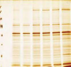
bioRxiv preprint first posted online Jan. 14, 2019; doi: http://dx.doi.org/10.1101/520411. The copyright holder for this preprint
 (which was not peer-reviewed) is the author/funder, who has granted bioRxiv a license to display the preprint in perpetuity.
                                  It is made available under a CC-BY 4.0 International license.

   Introduction

   Hydrogen sulfide (H2S) is a gas molecule that can be produced endogenously in many
   organisms from bacteria to mammals. H2S production at physiological concentrations
   is cytoprotective, as it reduces blood pressure (Yang et al., 2005), prevents
   neurodegeneration and extends lifespan (Hine et al., 2015; Kabil et al., 2011; Miller and
   Roth, 2007; Paul et al., 2014). H2S reacts with and modifies protein cysteine thiol
   groups to form persulfide bonds, known as protein S-sulfhydration. Because H2S is
   unstable, and its synthesis is regulated (Sbodio et al., 2018), protein S-sulfhydration
   serves as a mechanism to regulate protein functions with an impact in cellular
   metabolism (Mustafa et al., 2009, Gao et al., 2015).

   During oxidative stress Reactive Oxygen Species (ROS) often catalyze the non-
   enzymatic protein S-sulfhydraton (Mishanina et al., 2015; Paul and Snyder, 2015). In
   normal cells, in the absence of oxidative stress, protein S-sulfhydration can be the
   result of either local subcellular ROS fluctuations, or the enzymatic local H2S synthesis
   (Paul and Snyder, 2015). The latter likely involves the shunting of the SH group of the
   H2S to protein thiol groups, by the H2S-producing enzymes, as previously suggested for
   protein S-nitrosylation (Paul and Snyder, 2015). Among all amino acid residues in
   proteins, cysteine is the most susceptible to oxidative modifications in which a sulfur
   group is covalently bound to a ROS-derived electrophilic molecule (Reddie and Carroll,
   2008). Depending on the types and levels of ROS, a cysteine residue in a protein can
   be subjected to multiple types (Paul and Snyder, 2015) of redox-based
   posttranslational modifications (redox PTM). In addition to the already mentioned S-
   sulfhydration, other regulatory modifications are S-nitrosylation and S-glutathionylation
   (Gao et al., 2015, (Martinez-Ruiz and Lamas, 2007). S-nitrosylation is the covalent
   attachment of a nitroso group (-NO) to cysteine thiol of a protein to form an S-
   nitrosothiol (SNO). S-glutathionylation occurs when a protein cysteine residue forms an
   intermolecular disulfide bond with a glutathione to form protein-SSG.

   There is a growing body of evidence that the switch from one redox PTM to another in
   proteins, plays an important role in the regulation of various biological functions (Paul
   and Snyder, 2015). More importantly, various types of redox PTMs at the same
   cysteine residue in a protein yield diverse effects on the functions of the given protein.
   For instance, S-glutathionylation of a single redox sensitive cysteine in GAPDH inhibits
   its enzymatic activity, while S-sulfhydration can reverse this inhibitory effect and
   restore its activity (Gao et al., 2015). Similarly, S-nitrosylation of the p65 subunit of NF-
   kB inhibits its DNA binding activity, but S-sulfhydration increases the association of
   NF- kB with promoters of cytoprotective target genes in response to inflammation (Sen
   et al., 2012). Nonetheless, the ratio of different modifications on the same protein in a
   given cellular context determines the biological outcome. We focused on S-
   glutathionylation which is a pathophysiological redox protein regulation mechanism
   through the local subcellular fluctuations of GSH and ROS levels (Mieyal et al., 2008).
   The presence and extent of the conversion of S-glutathionylation to other oxidative
   PTMs in the proteome, is largely unknown. Furthermore, in the case of the conversion

                                                                                                                                    3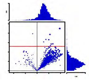
bioRxiv preprint first posted online Jan. 14, 2019; doi: http://dx.doi.org/10.1101/520411. The copyright holder for this preprint
 (which was not peer-reviewed) is the author/funder, who has granted bioRxiv a license to display the preprint in perpetuity.
                                  It is made available under a CC-BY 4.0 International license.

   of S-glutathionylation to S-sulfhydration, it is not clear if there is a switch of one
   modification to another on the same cysteine residue. The physiological consequences
   for such a protein Redox Thiol Switch (RTS) are also not well understood.

   It has been recognized as a great challenge to detect and quantify protein S-
   sulfhydration in vivo, due to the high reactivity and instability of the cysteine persulfide
   bond in sulfhydrated proteins (Mishanina et al., 2015; Paul and Snyder, 2015). Earlier,
   we developed a proteomics approach (Gao et al., 2015), the Biotin Thiol Assay (BTA) to
   identify sylfhydrated proteins. We used this approach to show that the increase of H2S
   synthesis during endoplasmic reticulum (ER) stress in pancreatic beta cells promotes
   selective S-sulfhydration of redox sensitive cysteine residues in proteins of specific
   metabolic pathways (Gao et al., 2015). The previous studies implicated H2S as a
   molecular switch in the regulation of energy metabolism.

   In this current study, we show the existence of an RTS, which involves H2S-mediated
   reversal of S-glutathionylation to S-sulfhydration (RTSGS) of specific cysteine residues
   in a subset of glutathionylated proteins. The iodoacetyl isobaric tandem mass tag
   system (iodoTMT) was combined with the previously reported (Gao et al., 2015) BTA
   assay (TMT-BTA), thus enabling the quantitative identification of sulfhydrated or RTSGS
   target proteins. We identified distinct patterns of the natural protein sulfhydromes in
   human pancreatic beta cells and hepatocytes, with hepatocytes exhibiting higher levels
   of sulfhydrated proteins compared to pancreatic beta cells. However, human
   pancreatic beta cells exhibited a unique landscape in sulfhydrated proteins, with the
   most enriched pathways being in intermediary metabolism. Furthermore, we identified
   insulin, the beta cell specific protein and master regulator of cellular metabolism, as a
   S-sulfhydration target. A novel RTSGS target in beta cells was the metabolic
   mitochondrial enzyme phosphoenolpyruvate carboxykinase 2 (PCK2) and known
   regulator (Stark et al., 2009) of glucose-stimulated insulin secretion (GSIS). The
   catalytic residue Cys306 was either sulfhydrated or glutathionylated. In vitro biochemical
   characterization showed that S-glutathionylation of the human recombinant PCK2
   protein inhibited its enzymatic activity, whereas S-sulfhydration reversed the inhibitory
   effect of S-glutathionylation on PCK2 activity. Finally, in order to understand the
   physiological significance of this RTSGS in cellular energy metabolism, we induced
   protein S-glutathionylation in pancreatic beta cells treated with diamide and measured
   metabolic flux of glucose. It is well known that protein S-glutathionylation can inhibit
   the activities of metabolic enzymes (Mieyal et al., 2008), and cause a decrease in the
   metabolic flux of glucose (Hiranruengchok and Harris, 1995). In agreement with the
   RTSGS mechanism contributing to regulation of energy metabolism, H2S, rescued the
   inhibited glucose flux in cells treated with diamide. We conclude that RTSGS, the redox
   thiol switch from S-glutathionylation to S-sulfhydration is a mechanism to fine tune
   cellular energy metabolism in response to oxidative stress.

                                                                                                                                    4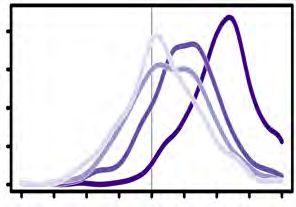
bioRxiv preprint first posted online Jan. 14, 2019; doi: http://dx.doi.org/10.1101/520411. The copyright holder for this preprint
 (which was not peer-reviewed) is the author/funder, who has granted bioRxiv a license to display the preprint in perpetuity.
                                  It is made available under a CC-BY 4.0 International license.

   Results

   We have used the previously reported by us, BTA assay (Gao et al., 2015), to develop a
   quantitative proteomics approach for profiling of the changes in the protein
   sulfhydrome in different cellular contexts. The utilization of tandem mass tags (TMT) is
   a well-recognized proteomics approach for the relative quantification of proteins and/or
   peptides (Thompson et al., 2003). We therefore have included the process of iodoTMT
   multiplex labeling of the peptides in the final step of the BTA assay (Fig 1A). The TMT-
   labeled sulfhydrated peptides can be combined for LC-MS identification and
   quantification.

   We first optimized the buffer system and the concentration of reducing agents (DTT or
   TCEP) in the elution buffer of the BTA assay, in order to maximize TMT-labeled
   peptides in the eluate. We treated mouse pancreatic beta cells (MIN6) with a
   commonly used H2S donor (NaHS, sodium hydrosulfide), which promotes protein S-
   sulfhydration both in vitro and in vivo (Paul and Snyder, 2015). The NaHS-treated MIN6
   cells were lysed and subjected to the BTA assay. The sulfhydrated peptides were
   eluted in different buffer systems (A-D, Supplemental Fig 1) followed by the iodoTMT
   labeling and LC-MS analysis (Supplemental Fig 1). The elution buffer containing 50 mM
   TEAB (triethylammonium bicarbonate) and 10 mM TCEP (tris-carboxyethyl phosphine
   hydrochloride) provided the highest labeling efficiency of sulfhydrated peptides
   (Supplemental Fig 1). As a first step, in order to identify the range of protein targets in
   the entire cellular proteome that are potentially subjected to RTSGS in living cells, we
   treated MIN6 cell lysates with NaHS and then applied the optimized TMT-BTA assay
   (Fig 1B, left panel). Elution was performed either with a buffer containing TCEP, or
   NaCl (-TCEP) as a negative control. Eluates were labeled with iodoTMT reagents,
   followed by LC-MS analysis (Fig 1B, left panel). About 200 sulfhydrated peptides were
   identified and quantified (Fig 1B, right panel). Compared to the NaCl control, TCEP-
   eluted samples exhibited significantly higher ratios of sulfhydrated peptides (Fig 1C).
   These data verify that the TMT-BTA assay is a highly selective method to identify the
   protein sulfhydromes in vitro.

   We next determined the changes in the cellular sulfhydrome in vivo under exposure to
   increasing H2S levels. The intracellular levels of H2S are on the order of nanomolar to
   micromolar, depending on cell types and cellular contexts (Filipovic et al., 2018). Under
   the highly reducing intracellular environments, protein S-sulfhydration is unlikely the
   predominant modification over the vast majority of protein free thiol groups in the
   proteome. Therefore, to determine if the TMT-BTA assay can quantify stoichiometric
   changes in the protein sulfhydrome in cells, we incubated MIN6 cells with increasing
   concentrations of the H2S donor (NaHS, 1-200 μM) followed by the TMT-BTA of MIN6
   cell extracts (Fig 1D). The vast majority (95%) of the identified peptides by the TMT-
   BTA assay contained cysteine residues, confirming that this method is highly selective
   in the identification of cysteine persulfides (Fig 1E). As expected, the increase in the
   levels of protein S-sulfhydration was dependent on the NaHS concentration (Fig 1F-H).
   Moreover, we found that all MS-identified sulfhydrated peptides were labeled by the
                                                                                                                                    5
bioRxiv preprint first posted online Jan. 14, 2019; doi: http://dx.doi.org/10.1101/520411. The copyright holder for this preprint
 (which was not peer-reviewed) is the author/funder, who has granted bioRxiv a license to display the preprint in perpetuity.
                                  It is made available under a CC-BY 4.0 International license.

   TMT tags (data not shown). Taken together, these data show that the TMT-BTA assay
   is a tool to quantitatively study changes of the S-sulfhydrated proteome in live cells
   and cell extracts under different redox manipulations.

   Next we determined the ability of the TMT-BTA assay to distinguish the levels of
   sulfhydromes between different cell types. It has been reported that protein S-
   sulfhydration is a prevalent redox PTM in certain tissues; for example, about 10–25%
   of mouse hepatic proteins are estimated to be sulfhydrated (Mustafa et al., 2009). The
   high abundance of protein S-sulfhydration in liver cells made this cell type ideal for our
   purpose of comparing sulfhydromes between cell types. To this aim, we used the TMT-
   BTA assay to analyze the protein sulfhydrome in immortalized human hepatocytes, IHH,
   (Ulatowski et al., 2012) and in human beta cells, EndoC-BH3 (Fig 2A). 447 sulfhydrated
   peptides from 307 proteins were identified and quantified (Fig 2B). Among all identified
   proteins, 33 of them were sulfhydrated specifically in EndoC-BH3 cells with ratios
   greater than 1.5-fold (Fig 2C-D). As expected, human hepatocytes exhibited
   significantly higher levels of sulfhydrated peptides as compared to EndoC-BH3 cells
   (Fig 2B-D). Bioinformatics clustering with a pathway annotation program (DAVID) of the
   identified shared sulfhydrome between hepatocytes and EndoC-BH3 cells, revealed an
   enrichment of those proteins involved in ribosome and metabolic pathways including
   glycolysis/gluconeogenesis (Fig 2E). Furthermore, proteins involved in mRNA
   translation and metabolic pathways are highly susceptible to S-sulfhydration, in
   agreement with our previous studies showing that the stress-induced inhibition of
   glycolytic flux was reversed by increasing H2S biosynthesis in the presence of stress
   (Gao et al., 2015). The higher levels of protein S-sulfhydration in hepatocytes as
   compared to the EndoC-BH3 cells, was further supported by the higher expression
   levels of the H2S-generating enzymes in these cells (Fig 2F). De novo synthesis of H2S
   is mediated by three enzymes: CTH (cystathionine γ lyase), CBS (cystathionine β
   synthase) and 3-MST (3-mercaptopyruvate sulfurtransferase) (Sbodio et al., 2018).
   Therefore, the levels of sulfhydrated proteins are in good correlation with the levels of
   the H2S-generating enzymes in these cells (Fig 2).

   We hypothesized that the shared hepatic and beta cell sulfhydrome overlaid on their
   proteomes should show differential pathway enrichments. We first performed a label-
   free MS quantification experiment to determine the absolute protein abundance in the
   cellular proteomes (Fig 3A). With this analysis, almost 4500 proteins were identified and
   their abundance was quantified and compared between the two cell lines (Fig 3B).
   About 2-3% of these proteins were solely identified either in the EndoC-BH3 cells, or in
   the hepatocytes; 10-13% of the proteins exhibited 2-fold differences between the two
   cell types, including CBS and CTH, which were in agreement with the Western blot
   analyses shown in Fig 2F. To further validate the label-free MS data, we used Western
   blot assays to test the expression of two proteins, MANF (mesencephalic astrocyte-
   derived neurotrophic factor) and PCK2 (mitochondrial phosphoenolpyruvate
   carboxykinase), which exhibited differential protein abundance in the two cell lines. In
   agreement with the MS data, EndoC-BH3 cells had higher expression of the MANF
   protein (Supplemental Fig 3). In contrast, no significant change was found in the

                                                                                                                                    6
bioRxiv preprint first posted online Jan. 14, 2019; doi: http://dx.doi.org/10.1101/520411. The copyright holder for this preprint
 (which was not peer-reviewed) is the author/funder, who has granted bioRxiv a license to display the preprint in perpetuity.
                                  It is made available under a CC-BY 4.0 International license.

   metabolic enzyme PCK2 levels between hepatocytes and EndoC-BH3 cells, in
   agreement with the MS data (Fig 3B). Pathway analysis of the proteins with relative
   abundance greater than 2-fold between the two cell types (Fig 3B), revealed that the
   branched-chain amino acid degradation and ribosome pathways were enriched in
   EndoC-BH3 cells and hepatocytes, respectively (Fig 3D). Next, we compared the
   relative changes in sulfhydromes between the two cell types, over their proteome
   abundance. To this end, we overlaid the protein abundance map with the natural
   sulfhydrome datasets (Fig 3E). This analysis showed that 63 sulfydrated proteins had a
   relative high abundance (log2 ratios greater than 1) in hepatocytes and 32 sulfhydrated
   proteins had relative high abundance (log2 ratios less than -1) in EndoC-BH3 cells.
   Among the 62 proteins, the enriched pathways were the ribosome and
   gluconeogenesis/glycolysis, whereas no pathway enrichment was noted in the 32
   proteins from the EnoC-BH3 cells. Furthermore, the remaining sulfhydrated proteins
   identified in both cell types showed the proteasome as an enriched pathway. Together,
   these data suggest that cellular sulfhydromes exhibit differential pathway enrichments
   as compared to their proteomes. A few metabolic proteins, such as lactase
   dehydrogenase (LDHA and LDHB), fructose-bisphosphate aldolase (ALDOA) and
   enoase 1 (ENO1) (Fig 3F), were in relatively lower abundance, but were more
   sulfhydrated in the EndoC-BH3 cells than the hepatocytes. Overall, the evaluation of
   the relative abundance of sulfhydromes described here, can also be a very useful tool
   for identifying changes in the redox-mediated cellular responses in a single cell type
   under different intracellular and extracellular cues.

   Next, we used the TMT-BTA assay to determine the regulation of the sulfhydrome in a
   single cell line under genetic manipulation of de novo H2S synthesis. We chose INS1
   cells because the levels of the H2S-producing enzyme CTH, was undetectable in the
   absence of ER stress (Fig 4E) and thus made this cell line a good candidate to
   determine the effects of CTH expression on the sulfhydrome. We have previously
   shown that during chronic ER stress in MIN6 cells, increased protein S-sulfhydration
   was in good correlation with increased expression of the transcription factor ATF4 and
   its transcription target, the CTH gene (Sbodio et al., 2018). In order to get more insights
   on the regulation of the sulfhydromes by the ATF4/CTH axis-mediated de novo H2S
   synthesis (Fig 4A), we overexpressed the transcription factor ATF4 or GFP (green
   fluorescent protein) as a negative control in INS1 cells and applied the TMT-BTA assay
   to cell lysates. The TMT-BTA assay identified and quantified over 2000 sulfhydrated
   peptides (Fig 4B). Cysteine S-sulfhydration levels were significantly increased by the
   forced ATF4 expression but not by GFP expression (Fig 4B-C), in agreement to our
   previous study (Gao et al., 2015). However, ATF4 can induce expression of many
   genes that can potentially increase the sulfhydrome (Gao et al., 2015; Mieyal et al.,
   2008). To identify the subgroup of CTH-mediated sulfhydrated proteins (Fig 4A), we
   looked for proteins with significant decreases in S-sulfhydration in response to
   depletion of CTH in ATF4-expressing INS1 cells. To this end, we employed the
   CRISPR-Cas9 technique to knock out the CTH-encoding gene in INS1 cells. We
   designed two single guide RNA sequences to target the translation start codon of the
   rat CTH gene (Fig 4D, Supplemental Fig 4-1A), and then screened for individual INS1

                                                                                                                                    7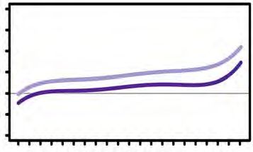
bioRxiv preprint first posted online Jan. 14, 2019; doi: http://dx.doi.org/10.1101/520411. The copyright holder for this preprint
 (which was not peer-reviewed) is the author/funder, who has granted bioRxiv a license to display the preprint in perpetuity.
                                  It is made available under a CC-BY 4.0 International license.

   clones to identify the deficiency of CTH expression induced by ATF4 (Fig 4E,
   Supplemental Fig 4-1(B, C, D) and Supplemental Fig 4-2). INS1 cells deficient for CTH,
   were infected with adenovirus that expressed ATF4, followed by the TMT-BTA assay.
   As expected, CTH depletion resulted in significant decreases of the protein
   sulfhydrome in INS1 cells expressing ATF4 (Fig 4F-I), confirming that regulation of
   protein S-sulfhydration by ATF4, is mediated in part via the induction of CTH. Among
   about 2000 sulfhydrated peptides, 157 peptides (corresponding to 130 proteins)
   exhibited more than 1.5-fold decrease in S-sulfhydration with the loss of CTH
   induction. Notably, insulin exhibited a significant CTH-dependent decrease in S-
   sulfhydration, suggesting that insulin may be a specific substrate of CTH in INS1 cells
   expressing ATF4. Western blot analysis using antibodies for proinsulin revealed that
   the protein expression levels were not affected by CRISPR-mediated CTH knockout in
   INS1 cells expressing ATF4 (Fig 4J), further supporting that the decreased insulin S-
   sulfhydration is not due to changes in intracellular insulin content.

   There are three disulfide bonds that are essential for the mature insulin to maintain the
   correct structure (Fig 4K). To verify whether the enzymatic activity of CTH can regulate
   insulin S-sulfhydration in vitro, we incubated recombinant human insulin with either
   active, or heat-inactivated recombinant CTH protein, followed by LC-MS analysis.
   Consistent with the in vivo data from INS1 cells, human insulin was sulfhydrated by
   H2S generated from the human recombinant CTH, but this modification was abolished
   by the inactivation of the CTH enzyme (Fig 4L). Treatment of recombinant insulin with
   NaHS gave similar results (Fig 4L). The combined data suggest that insulin S-
   sulfhydration is the result of an enzymatic reaction that requires the presence of active
   CTH to catalyze protein cysteine persulfidation.

   We next tested the hypothesis that S-sulfhydration may be used as a regulatory
   mechanism of the enzymatic activities of metabolic proteins under oxidative stress
   conditions. It is known that redox sensitive cysteine glutathionylation inhibits the
   activities of metabolic enzymes (Mieyal et al., 2008). We have also previously shown
   that S-glutathionylation of GAPDH at the catalytic cysteine residue inhibits its
   enzymatic activity, and it can be restored by H2S treatment (Gao et al., 2015),
   suggesting that the S-glutathionylation to S-sulfhydration RTS switch (Fig 5A) could be
   a regulatory mechanism of energy metabolism.

   In order to test our hypotheses, we first evaluated whether the PTM switch from S-
   glutathionylation to S-sulfhydration could occur on the scale of the cellular proteome. It
   has been reported that protein S-glutathionylation can be promoted by treatment with
   oxidized glutathione (GSSG) in vitro (Mieyal et al., 2008; Zaffagnini et al., 2012). We
   therefore incubated protein lysates isolated from mouse livers with TCEP in order to
   remove reversible cysteine modifications in the proteome. Subsequently, we treated
   lysates with GSSG to introduce S-glutathionylation. Of note, GSSG-treatment may also
   result in disulfide bond formation via the initial S-glutathionylation (Mieyal et al., 2008).
   The GSSG-treated lysates were subsequently subjected to an assay (Anastasiou et al.,
   2011) referred as the Biotin Switch Technique (BST) to identify the proteins targeted by

                                                                                                                                    8
bioRxiv preprint first posted online Jan. 14, 2019; doi: http://dx.doi.org/10.1101/520411. The copyright holder for this preprint
 (which was not peer-reviewed) is the author/funder, who has granted bioRxiv a license to display the preprint in perpetuity.
                                  It is made available under a CC-BY 4.0 International license.

   S-glutathionylation (Fig 5B). We found that the levels of glutathionylated proteins
   increased by the GSSG treatment (Fig 5C, Supplemental Fig 5), consistent with
   previous reports (Zaffagnini et al., 2012). MS analysis identified more than 2300 protein
   targets for S-glutathionylation, including GAPDH (Fig 5C), which supports our previous
   observation of S-glutathionylation of GAPDH by GSSG treatment (Gao et al., 2015).
   Western blot analysis confirmed GAPDH and as an additional control, MAT1A
   (methionine adenosyltransferase 1A) as protein targets being modified by GSSG in vitro
   (Fig 5D).

   Does exposure of cell extracts to H2S, can convert the proteome-wide S-
   glutathionylation to S-sulfhydration and what proteins respond to this PTM switch? To
   address this question, lysates isolated from mouse livers were exposed to TCEP
   followed by GSSG treatment. After desalting to remove excess GSSG, protein free
   cysteine thiol groups in the lysates were alkylated with NEM (N-ethylmaleimide) to
   block remaining free thiol groups. The lysates were incubated with NaHS, then
   subjected to the BTA assay which was further analyzed by SDS-PAGE (Fig 6A). As
   shown in Fig 6B, NaHS treatment increased the levels of S-sulfhydrated proteins from
   the GSSG-treated proteome compared to the untreated control; a dose-dependent
   increase in the levels of sulfhydrated proteins by H2S was also observed (Fig 6C).
   These data provide strong support for the notion that the PTM switch from S-
   glutathionylation to S-sulfhydration occurs on a proteome-wide scale. We named this
   protein cysteine PTM inter-conversion as Redox Thiol Switch S-Glutathionylation to S-
   Sulfhydration (RTSGS).

   Pancreatic beta cells are highly sensitive to oxidative stress and the redox status of
   intracellular glutathione (GSH/GSSG) is important for their metabolism and glucose-
   stimulated insulin secretion (GSIS) (Gerber and Rutter, 2017). We have previously
   shown that H2S-treatment of MIN6 cells pre-exposed to oxidative stress can reverse
   the stress-induced inhibitory effects on glucose metabolism (Gao et al., 2015).
   Therefore, MIN6 cells appeared a good experimental system to test the existence of
   the RTSGS in living cells and its significance on the cellular metabolism. We
   hypothesized that small changes in intracellular H2S levels can be sensed by metabolic
   enzymes via the RTSGS mechanism and thus adjust their glucose flux under oxidative
   stress (Fig 7A, top panel). We therefore tested this concept, by exposing INS1 cells to
   a protein S-glutathionylation-promoting oxidant for a short time and measuring the
   effects of H2S on the oxidant-induced changes of glucose flux. Diamide is a commonly
   used oxidant, whose addition to culture medium for short time induces oxidation of
   glutathione and increases protein S-glutathionylation (Fratelli et al., 2002). Treatment of
   INS1 cells with diamide for 1 hour, increased the GSSG levels without any global
   changes in protein synthesis rates (Supplemental Fig 7-1A,B). Diamide-treated INS1
   cells were washed to remove diamde, then exposed to NaHS (10 µM) in the presence
   of glucose [U-13C6] (Fig 7A, bottom panel). As we previously reported treatment of S-
   glutathionylated GAPDH with NaHS at concentrations 10-50 μM reversed the inhibition
   of glutathionylation on the GAPDH activity (Gao et al., 2015). Therefore we considered
   the concentration of 10 μM of NaHS within the physiological range for the regulation of

                                                                                                                                    9
bioRxiv preprint first posted online Jan. 14, 2019; doi: http://dx.doi.org/10.1101/520411. The copyright holder for this preprint
 (which was not peer-reviewed) is the author/funder, who has granted bioRxiv a license to display the preprint in perpetuity.
                                  It is made available under a CC-BY 4.0 International license.

   metabolic activities of enzymes, in vivo. As expected, diamide-treatment alone
   significantly reduced glycolytic and mitochondrial TCA flux as shown by the low 13C
   label incorporation in glyceraldehyde 3-phosphate (G3P), alanine, lactate, citrate,
   glutamate, succinate, fumarate, malate, oxaloacetic acid (OAA) and aspartic acid (Fig
   7A, bottom panel). This inhibition of the metabolic flux was correlated with increased
   levels of oxidized metabolic enzymes including GAPDH and PCK2 determined by the
   BST assay (Fig 7B). When the diamide-treated cells were exposed to NaHS, the
   glycolytic and mitochondrial TCA flux were restored, as shown by the increase in 13C-
   labeling of G3P, alanine, lactate, citrate, glutamate, succinate, malate, OAA. These
   data suggest that the H2S-mediated RTSGS mechanism sustains glycolytic and
   mitochondrial metabolism under oxidative stress.

   Based on data in Fig 7A, the concentration of 10 μM NaHS was able to reverse the
   inhibitory effects of diamide in glucose metabolism. We therefore considered the
   sulfhydromes formed at 10 μM NaHS treatment as the physiological cellular response
   to the RTSGS mechanism of regulation. We also hypothesized that the increasing
   concentrations of NaHS will affect the physiological RTSGS protein sulfhydrome in a
   manner dependent on the sensitivity of the redox cysteine residues to the RTSGS PTM.
   We tested this hypothesis in lysates isolated from MIN6 cells. As previously mentioned,
   the identification of the RTSGS target proteins depends on the sequential conversion of
   the proteome from S-glutathionylation to S-sulfhydration. Lysates from MIN6 cells
   were exposed to TCEP and GSSG, followed by the incubation with NaHS at different
   concentrations 10, 50 and 100 µM, then subjected to the TMT-BTA assay for LC-MS
   analysis (Fig 7C,D). Quantification of the sulfhydromes between the 10 and 100 μM
   NaHS treatment, revealed 990 RTSGS protein targets corresponding to 1535 cysteine-
   containing peptides. About 18% of these peptides exhibited substantial increases (1.5-
   fold, log2 ratio) in S-sulfhydration (Fig 7E), validating the results observed in the SDS
   acrylamide gel (Fig 6C). Interestingly, we observed that 8% (124 peptides) of the total
   identified RTSGS cysteine peptides exhibited substantially decreased (1.5-fold, log2
   ratio) S-sulfhydration, and at least 21 peptides (~17%) contained two cysteine residues
   in their amino acid sequences. By contrast, only 5 dithiol peptides (~1.8%) were found
   in the group of RTSGS targets (277 peptides) with increased S-sulfhydration. These data
   suggest that the group of peptides with decreased S-sulfhydration contain a higher
   percentage of two cysteine-containing peptides, as compared to the increased S-
   sulfhydration group. Why did we observe this enrichment in the decreased S-
   sulfhydration group? Our explanation is that two oxidized cysteine residues in a single
   peptide may have different sensitivity and reactivity to H2S. It is therefore possible that
   at higher concentrations of H2S, one cysteine in a glutathionylated protein is switched
   to a persulfide by H2S, but the second cysteine residue may be reduced to a free thiol,
   resulting in the retention of the peptide on the avidin column and decrease of the
   peptide in the eluate (Supplemental Fig 7-2). This can explain the enrichment of dithiol
   peptides in the decreased S-sulfhydration group. Taken together, we conclude that the
   concentration of H2S in cells under oxidative stress, can be a determinant for the
   function of the RTSGS mechanism in the regulation of energy metabolism.

                                                                                                                                    10
bioRxiv preprint first posted online Jan. 14, 2019; doi: http://dx.doi.org/10.1101/520411. The copyright holder for this preprint
(which was not peer-reviewed) is the author/funder, who has granted bioRxiv a license to display the preprint in perpetuity.
It is made available under a CC-BY 4.0 International license.
Among the 1535 RTSGS peptides, 425 peptides did not change in S-sulfhydration levels
with increasing concentration of NaHS (Fig 7D, within the red dotted line frame). This
finding suggests that a large number of the RTSGS target proteins have redox cysteine
residues that exhibit greater sensitivity to H2S. Low levels of H2S lead to the complete
conversion from S-glutathionylation to S-sulfhydration (Supplemental Fig 7-2). We
propose that highly sensitive RTSGS proteins may serve as H2S sensors that can quickly
switch their oxidized cysteine residues to persulfides under a small magnitude changes
of intracellular H2S levels in response to oxidative stress. Bioinformatics analysis
showed that these protein targets contain NAD(P)H binding motif and they are
prevalent in the glycolytic and mitochondrial TCA (tricarboxylic acid) cycle pathways
(Fig 7F). The data further suggest that the RTSGS may regulate energy metabolism
under oxidative stress conditions. One of those targets is the mitochondrial PEPCK
(phosphoenolpyruvate carboxykinase)- (PCK2). Most cells express a cytosolic form of
PEPCK (PCK1), an enzyme involved in gluconeogenesis (Yang et al., 2009). The
function of PCK2 is not well understood (Yang et al., 2009). PCK2 is the only isoform of
PEPCK expressed both in human and mouse pancreatic beta cells (Stark et al., 2009).
It has been proposed that the function of PCK2 in beta cells is to regulate GSIS via the
mitochondrial GTP-mediated PEP synthesis (Stark et al., 2009). The identification of
PCK2 as an RTSGS target strongly suggests its involvement in the redox-mediated
regulation of energy metabolism and GSIS.
PEPCK proteins display conserved amino acid sequences across species; they contain
a catalytic conserved cysteine residue at the active site (Supplemental Fig 8-1A),
suggesting that the activity of PEPCK may be regulated by redox modifications. PCK2
was found as an RTSGS target with high sensitivity to H2S (Fig 7D). We therefore tested
if PCK2 can be glutathionylated by GSSG, and whether H2S can convert S-
glutathionylation to S-sulfhydration in the PCK2 protein. We expressed and purified
human recombinant PCK2 from E. coli, and determined if the enzymatic activity of
recombinant PCK2 can be altered by GSSG-mediated S-glutathionylation, or H2O2-
mediated cysteine oxidation sulfenic acid (-SOH), sulfinic acid (-SO2H) or sulfonic acid
(-SO3H). As shown in Figure 8A, both treatments reduced PCK2 enzymatic activity in
vitro; LC-MS analysis confirmed that the catalytic cysteine 306 in human recombinant
PCK2 was predominantly modified by glutathione in response to the GSSG treatment
(Supplemental Fig 8-1B-D). Treatment of the glutathionylated PCK2 protein with NaHS,
restored its activity (Fig 8B). Furthermore, NaHS-treatment reversed PCK2 S-
glutathionylation to S-sulfhydration (Fig 8C). It is shown that the levels of
glutathionylated PCK2 were decreased, while the levels of S-sulfhydrated PCK2 were
increased by NaHS treatment, confirming the proteomics data (Fig 8C and
Supplemental Fig 8-1). These data suggest that S-glutathionylation at the conserved
(Supplemental Fig 8-1D) catalytic cysteine 306 of PCK2, has an inhibitory effect on its
enzymatic activity.
We finally tested the sulfhydration status of PCK2 during chronic ER stress, a condition
that associates with increased ROS levels, and increased H2S synthesis (Gao et al.,
2015). We previously showed that chronic ER stress in MIN6 cells promotes protein S-
11bioRxiv preprint first posted online Jan. 14, 2019; doi: http://dx.doi.org/10.1101/520411. The copyright holder for this preprint
(which was not peer-reviewed) is the author/funder, who has granted bioRxiv a license to display the preprint in perpetuity.
It is made available under a CC-BY 4.0 International license.
sulfhydration which restores stress-induced inhibition of glycolysis (Gao et al., 2015).
MIN6 cells were treated with the chemical stressor thapsigargin (Tg) to induce chronic
ER stress (Guan et al., 2017). Treatment with Tg induces the ATF4/CTH axis of protein
S-sulfhydration (Gao et al., 2015). After incubation for 6, 12 and 18h, the cells were
lysed and subjected to the BTA assay. As expected, the levels of sulfhydrated PCK2
were increased in response to ER stress (Fig 8D). LC-MS analysis confirmed that the
catalytic cysteine 306 was sulfhydrated from the immunepurified FLAG-PCK2 protein
expressed in Tg-treated MIN6 cells for 18h (Supplemental Fig 8-2A-C). This prompted
the question as to whether PCK2-mediated flux of PEP (phosphoenolpyruvate) is
regulated by the H2S-dependent modification of this enzyme. PCK2 converts OAA to
PEP using GTP as a cofactor (Fig 8E). To measure the changes in PCK2-mediated PEP
flux, MIN6 cells were treated with Tg for 18h; the growth media was changed to
contain the stable isotope [3-13C] pyruvate during the last 3.5 h of treatment. Tg-
treatment augmented total and M+1 PEP in MIN6 cells (Fig 8F). These data were
consistent with our previous observation that H2S synthesis during chronic ER stress
restores stress-induced inhibition of glycolytic flux (Gao et al., 2015). We conclude that
enzymes in energy metabolism sense increased H2S levels under conditions of chronic
oxidative stress and via the RTSGS mechanism rescue stress-induced deficits in energy
metabolism.
Discussion
The gas hydrogen sulfide (H2S), induces S-sulfhydration of redox sensitive cysteine
residues in the proteome and thus regulates protein functions. The development of
quantitative tools that can profile proteome-wide S-sulfhydration is crucial to identify
the major protein targets and determine their relationship with the biological effects of
this gas. Here, we introduced the TMT-BTA assay, which allowed the quantitative
assessment of changes in protein-cysteine sulfhydration in vivo and in vitro under
various cellular contexts. Using multiple validations, our data confirm the specificity
and suitability of the TMT-BTA assay for the analysis of protein S-sulfhydration. With
this new quantitative proteomics approach, we have demonstrated a good correlation
of the S-sulfhydration status of intracellular proteins with the expression of H2S-
generating enzymes. This PTM occurs only on redox sensitive cysteine residues of
specific proteins. Our findings suggest the protein S-sulfhydration mechanism as a dial
switch in the regulation of cellular metabolism in pathophysiological states. Glycolytic
proteins are the most highly sulfhydrated proteins in EndoC-BH3 cells, including
(ALDOA), an enzyme that converts fructose 1,6 bisphosphate (FBP) into
dihydroxyacetone phosphate (DHAP) and glyceraldehyde 3 phosphate (G3P). The
regulation of FBP levels via synthesis and degradation is critical to control GSIS
(Kebede et al., 2008), the most important function of beta cells. ALDOA exhibits
conserved amino acid residues across species, including the two cysteine residues
(Cys73 and Cys178) which were extensively sulfhydrated in human pancreatic beta cells
(Fig 3F). A previous study showed that plant cytosolic ALDOA is subject to both S-
glutathionylation and S-nitrosylation at the same cysteine residues, both resulting in
12bioRxiv preprint first posted online Jan. 14, 2019; doi: http://dx.doi.org/10.1101/520411. The copyright holder for this preprint
(which was not peer-reviewed) is the author/funder, who has granted bioRxiv a license to display the preprint in perpetuity.
It is made available under a CC-BY 4.0 International license.
inhibition of its enzymatic activity (van der Linde et al., 2011). Given this correlation, we
speculate that S-sulfhydration of Cys73 and or Cys178 might regulate ALDOA enzymatic
activity, change glycolytic flux, and affect insulin secretion in pancreatic beta cells.
There are three H2S-generating enzymes, CTH, CBS and MST, in mammalian cells
(Sbodio et al., 2018). Unlike the other two, CTH protein expression is transcriptionally
induced by the stress-induced master transcription factor-ATF4, thus linking H2S
synthesis to diverse physiological and pathological stress conditions (Paul and Snyder,
2015; Sbodio et al., 2018). The ATF4/CTH-mediated changes in the sulfhydrome are
shown here for the first time. Pancreatic beta cells require the presence of active stress
response mechanisms for their function, due to the easily disturbed proteostasis in the
ER, during fluctuations of proinsulin synthesis (Liu et al., 2010). Biochemical studies
with recombinant insulin showed that the insulin B chain was a target for the
ATF4/CTH-mediated S-sulfhydration, and thus prompted the question on the
physiological significance of this regulation. Pancreatic beta cells synthesize proinsulin
on ER-associated ribosomes. Proinsulin is then folded and processed to produce
insulin which is stored in secretory granules and is secreted in response to nutrients,
such as glucose (Liu et al., 2010). Increased H2S levels repressed GSIS in INS1 cells
treated with H2S donors (Supplemental Fig 9) (Yang et al., 2007). The mechanism via
which H2S decreases GSIS is not well understood. It has been shown that S-
sulfhydration of the KATP channel increased the activity of the ion channels, resulting in
an inhibition of GSIS in pancreatic beta cells (Yang et al., 2005). We have shown here
that in addition to this mechanism, S-sulfhydration of insulin (specifically the B chain)
may also contribute to inhibition of GSIS. Insulin B chain cysteine sulfhydration is
expected to break the disulfide bonds and unfold its structure (Fig 4K), reducing the
folded insulin in the secretory granules. This may consist a novel physiological
mechanism for the fine-tuning of GSIS in pancreatic beta cells. Impairment of GSIS in
pancreatic beta cells is associated with the development of diabetes in humans. A
recent study showed that the insulin B chain fragment triggered an activation of
immune T cells by enhancing the pathogenic B chain peptide loading to antigen-
presenting cells, which contributed to the development of type 1 diabetes (Wan et al.,
2018). Animal and cell culture models have showed that elevated H2S production is
associated with the development of diabetes (Szabo, 2012). Indeed, whether S-
sulfhydration of the insulin B chain in islets is an onset to trigger T cell activation
leading to beta cell destruction remains to be further investigated.
We discovered that a large number of protein targets (~990 targets) were subjected to
the RTSGS PTM switch from S-glutathionylation to S-sulfhydration mediated by
hydrogen sulfide. A key feature of this PTM switch is that both modifications occur on
the same cysteine residue. The RedoxDB database, indicated about 9% of the
identified RTSGS peptides are known to be modified by cysteine-based PTMs including
disulfides, S-nitrosylation, S-glutathionylation and sulfenic acid. This is in agreement to
our functional annotation analysis that RTSGS proteins contain redox sensitive cysteine
residues which are highly susceptible to oxidative stress associated PTMs. Pathway
analyses also showed that RTSGS targets with the highest sensitivity to H2S were
13bioRxiv preprint first posted online Jan. 14, 2019; doi: http://dx.doi.org/10.1101/520411. The copyright holder for this preprint
(which was not peer-reviewed) is the author/funder, who has granted bioRxiv a license to display the preprint in perpetuity.
It is made available under a CC-BY 4.0 International license.
involved in energy metabolism. We can therefore propose that the RTSGS mechanism
may regulate energy metabolism under chronic oxidative stress. Five RTSGS targets
involved in glycolysis and the mitochondrial TCA cycle were found: Glyceraldehyde 3-
phosphate dehydrogenase (GAPDH), pyruvate kinase 2 (PKM2), isocitrate
dehydrogenase (IDH), oxoglutarate dehydrogenase (OGDH) and pyruvate
dehydrogenase (PDH). The enzymatic activities of those enzymes are inhibited by
redox modifications under increased ROS levels (Mailloux et al., 2014). Thus, H2S-
mediated RTSGS of these metabolic enzymes is likely part of the mechanism to reverse
the ROS-induced inhibition of glycolysis and the TCA cycle and restore metabolic flux
under oxidative stress conditions.
One novel RTSGS target was the mitochondrial enzyme PCK2. This enzyme converts
OAA to PEP and promotes insulin secretion in pancreatic beta cells (Stark et al., 2009).
Both isoforms of PEPCK (PCK1 and PCK2) displayed a conserved catalytic cysteine at
their active site (Supplemental Fig 7-1D). In human and rodent pancreatic beta cells,
only the mitochondrial PCK2 enzyme is expressed, whereas other tissues express both
PCK2 and PCK1. It is known that oxidative stress is a hallmark of ER stress (Cao and
Kaufman, 2014). PCK2 is a redox sensitive enzyme regulated by the RTSGS described
in the current study. This regulation mediated by H2S might indicate a protective
mechanism to sustain mitochondrial function and restore insulin secretion in pancreatic
beta cells under chronic ER stress. With higher ROS production in response to ER
stress, mitochondrial oxidative phosphorylation and TCA metabolic rates are
decreased to preserve mitochondria (Gao et al., 2015). The oxidation of the PCK2
enzyme results in an accumulation of OAA within the mitochondria due to the reduction
of PEP cycling (Gao et al., 2015). The restoration of PCK2 activity triggered by
increased H2S under chronic ER stress could give beta cells the capability to increase
ATP turnover and normalize the redox state. This in turns helps to reduce oxidative
stress imposed on the cells. We therefore view that the H2S-mediated RTSGS switch in
mitochondrial PCK2 is an adaptive mechanism for promoting beta cell survival, along
with the improved function of pancreatic beta cells.
The H2S-mediated protective effect against oxidative stress-mediated-deregulations of
biological activities, is an evolutionary conserved process (Filipovic et al., 2018). For
example, endogenous H2S production in multiple bacterial species offers resistance to
oxidative stress and various classes of antibiotics (Mironov et al., 2017). H2S reverses
the age-associated impairment of the formation of new blood vessels in muscle (Das et
al., 2018), and the increase in H2S production in endothelial cells induced by sulfur
amino acid restriction promotes glucose uptake and ATP production via glycolysis
(Longchamp et al., 2018). However, the protective effects of H2S on cellular metabolism,
may also enhance tumor growth, which is not a desired outcome for cancer therapeutics.
Our studies can transform our current thinking of cancer cell metabolism. Tumors
exhibit increased ATP production through glycolysis (Vander Heiden et al., 2009) and
high H2S levels in tumors are associated with tumor growth (Szabo et al., 2013). In
addition, unique splicing isoforms of key metabolic enzymes contribute to increased
glycolytic flux in tumors (Luo and Semenza, 2012). For example, tumors express an
14bioRxiv preprint first posted online Jan. 14, 2019; doi: http://dx.doi.org/10.1101/520411. The copyright holder for this preprint
(which was not peer-reviewed) is the author/funder, who has granted bioRxiv a license to display the preprint in perpetuity.
It is made available under a CC-BY 4.0 International license.
alternatively spliced form of pyruvate kinase, PKM2, which enhances aerobic glycolysis
and provides tumor growth advantage (Anastasiou et al., 2011). PKM2 is a redox
sensitive protein with cysteine oxidation being inhibitory of its activity. Given that we
have identified PKM2 as an RTSGS target, we propose that the H2S-mediated PTM
switch on metabolic enzymes such as PKM2, is a potential mechanism for tumor
growth.
In the current study, we have developed an experimental tool, named the TMT-BTA
assay, for selectively trapping and tagging cysteine persulfides in the proteome of cells.
Combined with click chemistry and quantitative MS proteomics, the TMT-BTA assay
revealed the natural protein sulfhydrome of two human cell lines, and identified insulin
as a novel sulfhydration substrate in pancreatic beta cells. With this approach, we have
identified biological pathways being targets for RTSGS, a specific reversible thiol
modification in cysteine residues under oxidative stress conditions. The findings
suggest that the levels of H2S-producing enzymes control the sulfhydromes of cells,
thus modulating energy metabolism. The identified pool of protein targets subjected to
the RTSGS can serve as a valuable resource to study the effects of H2S on redox
homeostasis and metabolism in physiological and pathological states. Although in
physiological states, H2S may protect normal cells from ROS-induced metabolic
deregulation, in cancer cells, it may be an unwanted passenger. Cancer cells are
known to reroute signaling pathways to gain a metabolic advantage to prevent
oxidative damage under stress conditions, which is a signature phenotype associated
with most tumors (Vander Heiden et al., 2009). We therefore view that the H2S-
mediated RTSGS may be a major contributing mechanism to reprogram cancer cell
metabolism for their proliferation and survival. In this case, it would provide a rationale
for cancer therapeutics to modulate the activity of the H2S-producing enzymes in
tumor cells.
15bioRxiv preprint first posted online Jan. 14, 2019; doi: http://dx.doi.org/10.1101/520411. The copyright holder for this preprint
(which was not peer-reviewed) is the author/funder, who has granted bioRxiv a license to display the preprint in perpetuity.
It is made available under a CC-BY 4.0 International license.
Acknowledgements
We thank Dr. Ruma Banerjee for providing recombinant CTH used in this study. We
thank Dr. Danny Manor for providing IHH cells used in this study. We also thank Dr.
Renliang Zhang for assisting with advice on the PEP flux analysis. We thank Ken
Farabough for provided editorial assistance. We acknowledge the Univercell
Biosolutions (Paris, France) providing the EndoC-BH3 cells used in this study. This
work was supported by NIH grants R37-DK060596, R01-DK053307 (to MH), American
Diabetes Association Postdoctoral Fellowship 1-16-PDF-018 (to X.-H. G). The Orbitrap
Elite and Fusion Lumos LC-MS instruments were purchased via an NIH shared
instrument grants 1S10RR031537-01, 1S10OD023436-01 (to BW).
Author Contributions
Xing-Huang Gao and Maria Hatzoglou conceived the study; designed and performed
the experiments; wrote the manuscript and analysed and interpreted the data. Ling Li
and Belinda Willard, performed the proteomics analysis with LC-MS. Marc Parisien and
Luda Diatchenko, performed bioinformatics tasks and assisted in data
interpretation.Matt Mcleod, expressed and purify human recombinant PCK2
protein.Jing Wu, performed Western blot analysis.Ilya Bederman, performed and
analysed the metabolic flux studies. Zhaofeng Gao, performed bioinformatics tasks
and assisted in data interpretation and manuscript editing. Dawid Krokowski, assisted
in data interpretation and manuscript editing. Steven M Chirieleison and Derek Abbott,
contributed the Crispr-Cas9 experiments. Vivien Yee, adviced on structural modeling
of PCK2. Charles L. Hoppel, provided expertise on cellular metabolism, mitochondrial
function and metabolic data analysis. Richard G Kibbey, provided expertise on the PEP
flux study.Todd Holyoak, advised on biochemical characterization of the human
recombinant PCK2. Peter Arvan, provided expertise on beta cell biology and glucose
stimulated insulin secretion assays.
16bioRxiv preprint first posted online Jan. 14, 2019; doi: http://dx.doi.org/10.1101/520411. The copyright holder for this preprint
(which was not peer-reviewed) is the author/funder, who has granted bioRxiv a license to display the preprint in perpetuity.
It is made available under a CC-BY 4.0 International license.
Figure 1. Quantitative proteomics approach to profile the protein sulfhydrome in
vivo. (A) Graphic representation of the Biotin Thiol Assay (BTA) (Gao et al., 2015)
combined with alkylation of free SH groups with the tandem mass tags sixplex system
(iodoTMT). Sulfhydrated cysteine residues in proteins from 6 samples were biotinylated
with maleimide-PEG2-biotin (NM-Biotin). Subsequently, samples were processed with
trypsin, passed through an avidin column and eluted with DTT or TCEP, which reduced
the persulfide bonds and produced eluates containing the peptides with free SH
groups. After desalting, free SH groups were alkylated with iodoTMT tags and the
TMT-labeled samples were combined for LC-MS analysis. (B) Schematic of workflow
to determine the specificity of the TMT-BTA assay (left panel). Protein extracts from
mouse pancreatic beta cells (MIN6) pretreated with an H2S donor (NaHS, 0.2 mM) were
lysed, then subjected to the TMT-BTA assay. After trypsin digestion of the biotinylated
proteome, the peptide sample was divided into six equal fractions. Each fraction was
incubated with the same amount of avidin beads, then eluted without (-) or with (+)
TCEP followed by the TMT labeling. The TMT-labeled samples were combined and
subjected to LC-MS. Curve plot showing the distribution of relative changes of ratios
(+/-TCEP) of the sulfhydrated peptides against the number (#) of identified peptides
(right panel). Triplicates of each condition were used (C) Volcano plot showing the P
value distribution against the relative changes of ratios (+/-TCEP) of the sulfhydrated
peptides identified from the experimental data in (B). (D) Schematic workflow to
evaluate the TMT-BTA assay for the quantification of sulfhydrated proteins in vivo.
MIN6 cells were treated with the H2S donor, NaHS, at the indicated concentrations for
1h, then subjected to the TMT-BTA assay. LC-MS analysis was used to identify and
quantify sulfhydrated peptides. (E) Pie chart illustrating that 95% of the identified
peptides in (D), were containing cysteine residues. (F) Bar plot illustrating the dose-
dependent increases of the average values of relative ratios (log2) of sulfhydrated
peptides identified in (D). (G) Heat map showing the relative sulfhydrated peptide
enrichment ratios in MIN6 cells treated with the H2S donor at the indicated
concentrations, as shown in (D). (H) (Left panel) Curve plot showing the distribution of
relative sulfhydrated peptide enrichment ratios against the number of identified
peptides from the experimental data in (D), (Right panel) a histogram chart for the left
panel.
Figure 2. Quantitative profiling of the natural protein sulfhydrome in human
hepatocytes and pancreatic beta cells by the TMT-BTA assay. (A) Schematic of
workflow for profiling the sulfhydrome in the two cell lines. Protein extracts from human
hepatocytes (IHH) and pancreatic beta cells (EndoC-BH3) were subjected to the TMT-
BTA assay, and the TMT-labeled peptides were mixed and analyzed by LC-MS.
Triplicates of each cell line were used. (B) (Left panel) Curve plot showing the
distribution of relative changes (log2 IHH/EndoC-BH3 ratios) of the sulfhydrated
peptides in IHH (light purple) and EndoC-BH3 cells (deep purple). (Right panel) Boxplot
showing comparisons of the average levels of relative ratios (log2) of sulfhydrated
17bioRxiv preprint first posted online Jan. 14, 2019; doi: http://dx.doi.org/10.1101/520411. The copyright holder for this preprint
(which was not peer-reviewed) is the author/funder, who has granted bioRxiv a license to display the preprint in perpetuity.
It is made available under a CC-BY 4.0 International license.
peptides between the two cell lines. Triplicates of each cell line were used. (C) Heat
map of the relative ratios of the sulfhydrated peptides identified in experimental data in
(B), illustrating the differences in the patterns of the natural protein sulfhydrome
between IHH and EndoC-BH3 cells. (D) Volcano plot showing the P value distribution
against the fold change of ratios (log2 IHH/EndoC-BH3 ratios) of the sulfhydrated
peptides identified from the experimental data in (B). (E) Gene ontology biological
pathways for all sulfhydrated proteins. PPP: pentose phosphate pathway. (F)
Evaluation of levels of H2S-generating proteins (CTH, CBS and 3-MST) in IHH and
EndoC-BH3 by Western blot analysis.
Figure 3. Protein S-sulfhydration does not correlate with relative protein
abundance between IHH and EndoC-BH3 cells. (A) Identification and quantification
of the full proteome from human hepatocytes (IHH) and EndoC-BH3 cells by LC-MS
analysis. Cell extracts from both cell lines were resolved by reducing SDS–PAGE and
stained with Coomassie blue. Gel lane was cut into fractions for in-gel digestions with
trypsin. The peptides from each fraction were submitted for LC-MS identification and
quantification. A total of 4664 proteins from hepatocytes and 4050 proteins from
EndoC-BH3 cells were identified and quantified by LC-MS. (B) Comparison of protein
abundance between human hepatocytes and EndoC-BH3 cells in a curve plot showing
the cell-type specific differences. Red dotted lines marks the sulfhydration ratio
(IHH/EndoC-BH3, log2) greater or less than 1. (C) Pie charts of the data described in A
and B. (D) Gene ontology biological pathways for the relative most abundant proteins (>
1 in Fig 3B) identified from IHH (blue bars) and EndoC-BH3 cells (orange bars). (E)
Comparison of the natural protein sulfhydrome in Fig 2B with the full proteome reveals
that a large fraction of sulfhydrated proteins have a moderate abundance in the cells.
(F) The TMT-BTA profiling of proteins containing cysteine residues with different
reactivity to H2S from IHH and EndoC-BH3 cells. LC-MS-MS(2) profiles for cysteine-
containing peptides from LDHB, ALDOA, LDHA and ENO1 proteins are shown.
Figure 4. The TMT-BTA assay reveals insulin as a CTH-mediated sulfhydrated
protein, in INS1, rat pancreatic beta cells. (A) Schematic representation of the ATF4-
induced CTH gene expression leading to increased intracellular H2S synthesis and S-
sulfhydration of proteins. (B) Relative distribution of peptides containing sulfhydrated
cysteine residues as determined by the TMT-BTA assay of cell extracts isolated from
INS1 cells overexpressing ATF4 or GFP for 36 h. The relative changes (log2 value) of
TMT ratios are plotted against the number of identified peptides. (C) Volcano plot
showing the relative change of the sulfhydration ratios (ATF4/GFP, log2) of modified
peptides plotted against the –log10 P value (n=3). Red line indicates the P value ≤ 0.05.
(D) Schematic representation of CRISPR-cas9 mediated CTH knockout (KO) in INS1
cells. Two sgRNA sequences were designed for targeting exon 1 of the rat CTH gene.
sgRNA targeting sequences are in bold. The translation initiation codon is shown in red.
(E) Western blot analysis of the effect of ATF4-overexpression in individual CRISPR-
mediated CTH knockout INS1 cells. Clones 1, 2 and 7 did not express CTH protein. (F)
Heat map showing the relative enrichment values (log2 fold change) of sulfhydrated
18You can also read