Intrauterine growth restriction decreases NF- B signaling in fetal pulmonary artery endothelial cells of fetal sheep
←
→
Page content transcription
If your browser does not render page correctly, please read the page content below
Am J Physiol Lung Cell Mol Physiol 315: L348–L359, 2018.
First published May 3, 2018; doi:10.1152/ajplung.00052.2018.
RESEARCH ARTICLE
Intrauterine growth restriction decreases NF-B signaling in fetal pulmonary
artery endothelial cells of fetal sheep
R. Blair Dodson,1,2,3,4,6 Kyle N. Powers,1,2,3 Jason Gien,2,4 Paul J. Rozance,4 Gregory Seedorf,2,5
David Astling,6 Kenneth Jones,6 Timothy M. Crombleholme,1,3 Steven H. Abman,2,4 and
X Cristina M. Alvira7
1
Laboratory for Fetal and Regenerative Biology, University of Colorado Denver Anschutz Medical Campus, Aurora,
Colorado; 2Pediatric Heart Lung Center, University of Colorado Denver Anschutz Medical Campus, Aurora, Colorado;
3
Department of Surgery, University of Colorado Denver Anschutz Medical Campus, Aurora, Colorado; 4Department of
Pediatrics, University of Colorado Denver Anschutz Medical Campus, Aurora, Colorado; 5Department of Biochemistry and
Molecular Genetics, University of Colorado Denver Anschutz Medical Campus, Aurora, Colorado; 6United Therapeutics,
Regenerative Medicine Laboratory, Research Triangle Park, Durham, North Carolina; and 7Department of Pediatrics,
Stanford University School of Medicine, Palo Alto, California
Submitted 31 January 2018; accepted in final form 2 May 2018
Dodson RB, Powers KN, Gien J, Rozance PJ, Seedorf G, INTRODUCTION
Astling D, Jones K, Crombleholme TM, Abman SH, Alvira CM.
Intrauterine growth restriction decreases NF-B signaling in fetal Intrauterine growth restriction (IUGR) is a multifactorial
pulmonary artery endothelial cells of fetal sheep. Am J Physiol Lung disease resulting from maternal, placental, or fetal factors,
Cell Mol Physiol 315: L348 –L359, 2018. First published May 3, which culminate in decreased nutrient and oxygen supply and
2018; doi:10.1152/ajplung.00052.2018.—Intrauterine growth restric- hemodynamic stress to the developing fetus (20). IUGR is the
tion (IUGR) in premature newborns increases the risk for broncho- second leading cause of prematurity and increases the risk of
pulmonary dysplasia, a chronic lung disease characterized by dis- additional adverse perinatal and neonatal outcomes, including
rupted pulmonary angiogenesis and alveolarization. We previously
stillbirth, necrotizing enterocolitis (48, 65), retinopathy of
showed that experimental IUGR impairs angiogenesis; however,
mechanisms that impair pulmonary artery endothelial cell (PAEC) prematurity (18, 37, 42, 48), and bronchopulmonary dysplasia
function are uncertain. The NF-B pathway promotes vascular growth (BPD) (11, 62, 67). The growth-restricted fetus is exposed to
in the developing mouse lung, and we hypothesized that IUGR significant metabolic, hormonal, and hemodynamic alterations
disrupts NF-B-regulated proangiogenic targets in fetal PAEC. of the intrauterine environment that have detrimental effects on
PAECs were isolated from the lungs of control fetal sheep and sheep fetal organ development and increase the risk for adult disor-
with experimental IUGR from an established model of chronic ders, including diabetes mellitus, hypertension, and coronary
placental insufficiency. Microarray analysis identified suppression artery disease (20, 66).
of NF-B signaling and significant alterations in extracellular Previous studies have shown that physiological changes in
matrix (ECM) pathways in IUGR PAEC, including decreases in
collagen 4␣1 and laminin ␣4, components of the basement mem-
the fetal lung that are associated with IUGR are likely related
brane and putative NF-B targets. In comparison with controls, to disruption of several developmental pathways, including
immunostaining of active NF-B complexes, NF-B-DNA bind- surfactant synthesis (15, 36, 44) and decreased epithelial cell
ing, baseline expression of NF-B subunits p65 and p50, and differentiation (17). In addition, rats with experimental IUGR
LPS-mediated inducible activation of NF-B signaling were de- have decreased alveolar and vascular growth, which may be
creased in IUGR PAEC. Although pharmacological NF-B inhi- due to downregulation of vascular endothelial growth factor
bition did not affect angiogenic function in IUGR PAEC, angio- (VEGF), a potent proangiogenic factor (47) and elastin depo-
genic function of control PAEC was reduced to a similar degree as sition (33). In a fetal sheep model of IUGR induced by
that observed in IUGR PAEC. These data identify reductions in
endothelial NF-B signaling as central to the disrupted angiogen-
utero-placental embolization, lung diffusion capacity and com-
esis observed in IUGR, likely by impairing both intrinsic PAEC pliance are significantly decreased in 8-wk-old sheep (34), and
angiogenic function and NF-B-mediated regulation of ECM com- fewer and larger alveoli are observed in IUGR sheep at 2 yr of
ponents necessary for vascular development. These data further age (51). Similarly, clinical studies have shown that IUGR
suggest that strategies that preserve endothelial NF-B activation adversely affects both short- and long-term respiratory out-
may be useful in lung diseases marked by disrupted angiogenesis comes. In premature infants, growth restriction is an indepen-
such as IUGR. dent risk factor for the development of BPD (11, 60, 62) and
alveolarization; angiogenesis; bronchopulmonary dysplasia; extracel- may represent an even stronger risk factor than extreme pre-
lular matrix maturity for BPD and respiratory disease during early child-
hood (67). Lower birth weight has also been associated with an
increased risk of asthma in children and chronic obstructive
pulmonary disease in adults, suggesting an incomplete capacity
Address for reprint requests and other correspondence: R. B. Dodson, 2 of lung growth to compensate for antenatal disruptions in lung
Maughan Dr., Durham, NC 27709 (e-mail: bdodson@unither.com). development because of IUGR (5, 21, 49, 53).
L348 1040-0605/18 Copyright © 2018 the American Physiological Society http://www.ajplung.org
Downloaded from journals.physiology.org/journal/ajplung at Stanford Univ Med Ctr (171.066.010.174) on January 1, 2021.DECREASED NF-B IN IUGR PAEC L349
Growth of the pulmonary vasculature is essential for distal lung sedated by ketamine (17.5 mg/kg iv) and diazepam (0.2 mg/kg iv) and
growth during late development. Inhibiting angiogenesis in new- euthanized by an injection of Fatal-Plus (Vortec Pharmaceuticals,
born rats decreases pulmonary artery density and impairs alveo- Dearborn, MI) (85 mg/kg iv). In addition, the fetus was euthanized by
larization (31). Previous work by our group has shown that an intra-cardiac injection of Fatal-Plus (250 mg/kg). Lungs were
harvested for either histology or the isolation of PAECs, as previously
alveolarization and pulmonary vascular density are both de-
published (61). The number of individual animals and technical
creased in a sheep model of IUGR (61). Although angiogenesis is replicates used for each experiment are detailed in the figure legends.
a complex process coordinated by several growth and transcrip- Lung fixation and immunofluorescent staining. Lung tissue was
tional factors, hemodynamic stimuli, and signals from the extra- inflation fixed with 4% paraformaldehyde (wt/vol) in phosphate-
cellular matrix (ECM), we found that experimental IUGR has buffered saline at 30 cmH2O pressure. Lungs were ligated while under
discrete, direct effects on pulmonary artery endothelial cell pressure and stored in 70% ethanol. Fixed lung tissue was paraffin
(PAEC) function. PAECs derived from IUGR fetal sheep dem- embedded and sectioned. Tissue sections were deparafinized and
onstrate impaired proliferation, migration, and tube formation in rehydrated. Lung tissue immunofluorescent staining was performed as
association with downregulation of VEGF and nitric oxide sig- previously described using primary antibodies to detect NF-B p65
naling pathways (61). However, molecular mechanisms that alter (1:200; Santa Cruz Biotechnology, Santa Cruz) and the endothelial
specific marker, CD31 (1:200 Dianova), and cell nuclei were stained
PAEC function and disrupt pulmonary angiogenesis in IUGR
with DAPI (30). Fluorescent images were captured using a Leica
remain incompletely understood. DM5500 upright microscope and a CoolSNAP fluorescent camera
Accumulating evidence suggests that the NF-B signaling using HC Plan Apo 25-mm objective at ⫻10 magnification, and
pathway may be a key driver of angiogenesis in the developing images were processed using Metamorph image analysis software.
lung (30). The NF-B family of transcription factors is ubiq- Sections obtained from three to four animals per experimental group
uitously expressed but remains sequestered in the cytoplasm in were immunostained, and 5 fields of view/animal were imaged at ⫻20
an inactive state in association with inhibitory IB proteins (1). and ⫻40 magnification.
Activation of NF-B by a complex of kinases, including IB PAEC isolation and culture. Primary PAECs were isolated from
kinase ␣ and  (IKK␣ and IKK), causes degradation of the the left and right pulmonary proximal arteries as previously described
IB proteins and rapid nuclear translocation of active NF-B (61). Endothelial cell phenotype was confirmed by positive immuno-
complexes. Although best recognized for its role in regulating staining for the endothelial specific markers von Willebrand factor,
vascular-endothelial cadherin, and endothelial nitric oxide synthase
innate immunity and inflammation, the NF-B pathway also and negative staining for the smooth muscle cell markers, ␣-smooth
has important roles during development (7, 28, 56, 74). Our muscle cell actin and desmin. PAECs from passages 3–6 were used
prior studies showed that endogenous activation of the NF-B for all experiments. PAECs obtained from individual animals were
pathway in the developing mouse lung promotes pulmonary kept separate throughout all passages and for all experiments de-
angiogenesis and alveolarization and regulates VEGF receptor scribed. Cells were cultured in DMEM with 10% fetal bovine serum
2 (VEGFR2) expression in the pulmonary endothelium (30). (FBS) and 1% antibiotic, antimicrobial.
However, whether disrupted NF-B signaling in pulmonary Gene array analysis and RNA validation. Total RNA was isolated
endothelium contributes to decreased angiogenesis and alveo- from subconfluent PAECs, control (n ⫽ 3, 2 male, 1 female) and
larization associated with IUGR is not known. IUGR (n ⫽ 4, 2 male, 2 female), using TRIzol phenol extraction.
To identify signaling pathways that are dysregulated and may RNA concentration and purity was determined by NanoDrop analysis
(Thermo Fisher Scientific, Waltham MA) and 250 ng of RNA was
contribute to abnormal endothelial function in the IUGR lung, we used for sheep-specific gene microarrays (Affymetrix, Santa Clara,
performed unbiased, transcriptomic profiling of fetal PAECs de- CA). Data were analyzed using R statistical software with the Bio-
rived from an experimental model IUGR induced by chronic conductor Gene Set Enrichment Analysis (GSEA) (normalized en-
placental insufficiency (59, 72). Using this approach, we identified richment score to compare differences in gene sets and a false
marked decreases in NF-B signaling in fetal IUGR PAECs and discovery rate ⬍25%) plugin and ingenuity pathway analysis (ana-
show that this reduction in NF-B signaling is due to decreased lyzed upstream regulator analysis, mechanistic networks, causal net-
gene expression of key NF-B subunits and suppression of work analysis, and downstream effects analysis, P ⬍ 0.001).
NF-B inducible activation. We further found altered expression Based on GSEA analysis, specific genes were confirmed by quan-
of ECM proteins that are regulated by NF-B, suggesting that titative PCR, using the primers listed in Table 1. cDNA was amplified
disruption of NF-B-mediated ECM remodeling may contribute using Transcriptor First Strand cDNA Synthesis Kit (Roche, Mann-
heim, Germany) in a standard thermocycler. cDNA samples were then
to abnormal lung and vascular development in IUGR. assayed in triplicate using FastStart Essential DNA Green Master Kit
METHODS (Roche) in a Lightcycler 96 (Roche) and the respective 2(-⌬⌬CT)
values were calculated using S15 as a housekeeping gene.
Experimental sheep model of IUGR. Pregnant Columbia-Ram- Electrophoretic mobility shift assays. Nuclear extracts were ob-
bouillet mixed-breed ewes (singleton pregnancies) were purchased tained from cultured PAECs (n ⫽ 4 for control PAECs, 1 male and 3
from Nebeker Ranch (Lancaster, CA), housed, cared for, managed in females; n ⫽ 5 for IUGR PAECs, 1 male and 4 females) using the
compliance with the United States Department of Agriculture, the NE-PER Kit (Pierce, Rockford, IL). The protein concentration was
American Association for Accreditation for Laboratory Animal Care determined by the Bradford method. Ten micrograms of nuclear
at the Perinatal Research Center at the University of Colorado (Au- protein were used for binding reactions using ␥-32P-labeled oligonu-
rora, CO), and approved by University of Colorado Institutional cleotides containing the B consensus sequence (Promega, Madison,
Animal Care and Use Committee (101924). The previously described WI) in a binding buffer containing 500 ng of salmon sperm DNA,
sheep model of IUGR was created by exposing pregnant ewes to 0.01 U of poly(dI-dC), and 0.5 mM DTT (EMD Chemicals) as
ambient hyperthermia (40°C for 12 h and 35°C for 12 h) from day 35 previously described (30). Supershift experiments were conducted
to day 110 during the 148-day gestation. Control animals were with the addition of 2 l of anti-p65, p50, p52, cRel, and RelB
maintained in normothermic conditions at 25°C ambient temperature antibodies (Santa Cruz Biotechnology) to the nuclear extracts 30 min
in otherwise identical living conditions. Food intake was matched to before the addition of the radiolabeled probe. Nuclear extracts were
the IUGR group. On day 135 (⫾2 days) of gestation, the ewe was incubated with radiolabeled B oligonucleotides at room temperature
AJP-Lung Cell Mol Physiol • doi:10.1152/ajplung.00052.2018 • www.ajplung.org
Downloaded from journals.physiology.org/journal/ajplung at Stanford Univ Med Ctr (171.066.010.174) on January 1, 2021.L350 DECREASED NF-B IN IUGR PAEC
Table 1. Primer sequences used for qPCR validation of genes identified in the microarray analysis
Gene RefSeq Amplicon Length, base pairs Sequence 5=¡3=
S15 XM_015096022.1 1–146 146 FWD ATC ATT CTG CCC GAG ATG GTG
REV CGG GCC GGC CAT GTC TTA CG
BIRC2 (cIAP1) XM_004015974.2 2314–2393 99 FWD GGT GGC TTA AGG TGT TGG GA
REV CTC CTG CCC TTT CAT GCG TA
BIRC3 (cIAP2) XM_012095321.1 96–250 176 FWD CTC CTT AGG CCG GTC TCC T
REV TCC TCT CTT TTG TAA AAC GGC A
TNFAIP3 (A20) XM_012131627.1 2455–2714 260 FWD GGA CTG AAG AGC AGC TGA GGT
REV GCC ATT GCA CTT GGT GTT GC
CLFAR (cFLIP) XM_004004820.2 2588–2807 239 FWD GCT CCT GGA ACT CCA CAC TG
REV TCA AAG TCC CTC TGC TCC AC
NFKB1 (p50) XM_004009667.2 3442–3551 109 FWD CTT CCA TCC TGG AAC CAC TAA A
REV CAC CTC TCT GTC ATC ACT CTT G
RelA XM_012102355.1 1107–1198 91 FWD GTT CAG GGC TTC CTT CCT TAT T
REV GTA AAT CGG AAC TCT GGG AGC
MALT1 XM_012120865.1 1124–1347 243 FWD2 AGT GGA ATG CAC CGA AGA TGA
REV2 ACG GCG TTA CGC ATC TCA TA
BTK XM_012146742.1 1122–1347 245 FWD GCA GTG GAA TGC ACC GAA GA
REV ACG GCG TTA CGC ATC TCA TA
LAMA4 XM_012182567 968–1104 136 FWD TGG CAC AGA AGA TGC TTG AGG AGA
REV AGT GTC ATT GTA CTG CCT CTG CCA
COL4A1 XM_015098012.1 430–617 187 FWD TGG CAT CAA AGG AGA AGC AGG TCT
REV CCA CAT CGC CCT TTG AAC CTT TCT
COL1A1 XM_015098715.1 889–981 92 FWD AAT GGA GCT CCT GGT CAG AT
REV ATC ATT TCC TCG AGC ACC AG
BIRC3, baculoviral IAP repeat containing protein 3; CFLAR, CASP8 and FADD-like apoptosis regulator; COL1A1, collagen 1␣1; Col4␣1, collagen 4␣1;
Lama4, laminin ␣4; MALT1, mucosa associated lymphoid tissue lymphoma translocation gene 1; NFKB1, NF-B1; TNFAIP3, TNF-␣-induced protein 3; BTK,
Bruton’s tyrosine kinase.
for 30 min and electrophoresed on 6% polyacrylamide gels. To hibitors (PhosSTOP, Roche). Whole cell protein, cytoplasmic protein,
distinguish nonspecific binding of the nuclear proteins, competition and nuclear extract samples were quantified by Bradford DC protein
reactions were performed by adding a 100-fold excess nonradiola- assay (Bio-Rad, Hercules, CA). For analysis, a 30-g sample was
beled B probe as done previously (2). loaded into a Novex 4 –12% gradient Bis-Tris gel (Thermo Fisher
PAEC growth assay. Fetal PAECs were plated at 1 ⫻ 105 cells/well Scientific) and separated by electrophoresis. Proteins were transferred
into six-well plates and allowed to adhere overnight in DMEM with from the gel to PVDF membranes (Thermo Fisher Scientific). Blots
10% FBS. The following day (day 0), the cells were washed three were blocked in Li-Cor Odyssey blocking buffer (Li-Cor Biosciences,
times with PBS. DMEM with 2.5% FBS was added, and cells were Lincoln, NE) for 1 h at room temperature and washed in Tris-buffered
incubated in room air (21% oxygen). Media was changed daily, and saline-Tween 20. Immunoblot detection of IKK␣ (1:1,000; Cell Sig-
cell counts were performed on day 0 and day 5 after removing cells naling, Danvers, MA; no. 2682), IKK (1:1,000; Cell Signaling; no.
with trypsin digestion. All conditions were run in triplicate for each 2678), p65 (1:1,000; Cell Signaling; no. 8242), p50 (1:1,000; Abcam,
animal cell line (control: n ⫽ 3, 1 male and 2 females; IUGR: n ⫽ 3, Cambridge, MA; no. ab7971), and -actin (1:10,000; MP Biomedi-
1 male and 2 females). cals, Santa Ana, CA; no. 8691001) were detected using Li-Cor
In vitro angiogenesis assay. The ability of fetal PAECs (control: anti-mouse (no. 925-32212) and anti-rabbit (no. 925-68071) antibod-
n ⫽ 3, 1 male and 2 females; IUGR: n ⫽ 3, 1 male and 2 females) to ies at concentrations of 1:7,500 in Odyssey buffer. The blots were
form vascular structures in vitro was assayed by plating PAECs on a imaged using the Odyssey CLx (Li-Cor Biosciences) and analyzed by
cross-linked collagen gel. PAECs were seeded at a density of 5 ⫻ 104 Image Studio for Odyssey for CLx (Li-Cor Biosciences). Raw values
cells/well in 0.5% FBS DMEM, and each condition was tested in were normalized to their respective -actin loading control values and
quadruplicate for each animal. PAECs are incubated for 24 h in room then reported as fold-change over control.
air (21% oxygen). Branch-point counting was performed in blinded IKK activation assay. Identification of phosphorylated (active)
fashion under ⫻10 magnification from each of four wells, with 3 to 4 levels of IKK protein were determined by ELISA assays to detect
fields of view obtained per well. phosphorylation at Ser177 and Ser181 using the PathScan Phospho-
Modulation of NF-B activity. NF-B activity was modulated IKK (Ser177/Ser181) Sandwich ELISA Kit no. 7080 (Cell Signaling
using two approaches. First, NF-B activity was pharmacologically Technology) per manufacturer’s protocols. Spectrophotometric values
inhibited using BAY 11-7082 (BAY), a targeted IKK␣ and IKK (absorbance at 450 nm) were determined in control and IUGR PAECs
inhibitor, as previously reported (30). Second, NF-B activity was at baseline and 1 and 2 h after treatment with LPS (10 ng/ml).
induced in fetal control and IUGR sheep PAECs (control: n ⫽ 3, 1 Statistical analysis. The number of enriched gene sets that were
male and 2 females; IUGR: n ⫽ 3, 1 male and 2 females) by significant were indicated by a false discovery rate of ⬍25%, follow-
incubating the cells with LPS (10 ng/ml; Escherichia coli 055:B55, ing the recommendation by GSEA computation tool. Those gene sets
no. L2880 diluted into culture media; Sigma Chemical, St. Louis, with a nominal P value of ⬍1% received further investigation (68).
MO) for 1 or 2 h (70). NF-B and IKK activation were then The ingenuity pathway analysis was completed using a suite of
assessed by Western blot analysis to detect nuclear p50 and p65 or algorithms and tools for inferring and scoring regulator networks as
determination of IKK activation by ELISA, as described in detail previously described (38). For all other studies, results are expressed
below. as means ⫾ SE. Differences between two groups were determined
Western immunoblot analysis. PAECs were lysed in ice-cold lysis using a t-test for normally distributed samples and Mann-Whitney for
buffer (50 mM Tris, pH 7.4, 1 mM EDTA, and 1 mM EGTA) with samples without normal distribution. For analysis with two indepen-
dissolved protease (Roche, Basel, Switzerland) and phosphatase in- dent variables, statistical significance was determined using two-way
AJP-Lung Cell Mol Physiol • doi:10.1152/ajplung.00052.2018 • www.ajplung.org
Downloaded from journals.physiology.org/journal/ajplung at Stanford Univ Med Ctr (171.066.010.174) on January 1, 2021.DECREASED NF-B IN IUGR PAEC L351
Table 2. Altered pathways of interest identified by microarray analysis
Enrichment/Z-Score P Value Method
Collagen fibril organization 2.35 ⬍0.01 Gene ontology analysis
Extracellular matrix 2.04 ⬍0.01 Gene ontology analysis
IGF-1 signaling 0.82 ⬍0.01 Ingenuity pathway analysis
VEGF signaling ⫺0.45 ⫽0.02 Ingenuity pathway analysis
NF-B ⫺1.63 ⬍0.01 Gene set enrichment analysis
IKK2 signaling ⫺1.87 ⬍0.01 Gene ontology analysis
Glucose metabolism disorder ⫺3.05 ⬍0.01 Ingenuity pathway analysis
analysis of variance, followed by Sidak’s multiple comparisons anal- ing collagen 4␣1 (Col4␣1) and laminin ␣4 (Lama4). In addi-
ysis. Statistical significance was defined as a P value of ⬍0.05. All tion, analyses of the microarray data identified significant
nonbioinformatics statistical analysis was done using GraphPad Prism suppression of NF-B signaling, including decreased expres-
version 6 (GraphPad Software, LaJolla, CA). sion of individual NF-B subunits (e.g., p50 and p65) and
RESULTS decreased gene expression of prosurvival NF-B downstream
targets (e.g., baculoviral IAP repeat containing protein 3 and
Microarray analysis identifies abnormalities in NF-B sig- CASP8 and FADD-like apoptosis regulator). Expression of the
naling and ECM remodeling in IUGR PAECs. We previously genes in the GSEA pathway showed NF-B genes to be
found that the angiogenic function of PAECs obtained from minimally expressed as shown in heat map form (Fig. 1A, red
IUGR fetal sheep was significantly decreased compared with is the normalized high gene expression, and blue is the nor-
PAECs from control sheep (61). However, the molecular malized low gene expression).
mechanisms accounting for this angiogenic defect are not Using quantitative PCR, we confirmed specific dysregula-
known. To address this question, we performed unbiased gene tion of the genes identified by the microarrays in these path-
expression profiling in control and IUGR PAECs using sheep ways of interest and found strong agreement between the two
specific microarrays. Gene ontogeny and pathway analyses of the assays, thereby confirming significant downregulation of the
differentially expressed genes identified the enrichment of genes NF-B subunits p50 and p65 and the ECM components
in association with specific metabolic, angiogenic, and ECM Col4␣1 and Lama4 (Fig. 1B).
pathways (Table 2). Differences in gene expression included NF-B expression and activity are decreased in IUGR fetal
decreases in glucose metabolism and VEGF signaling, including sheep and PAECs. We previously showed that endogenous
Vegfa and Vegfr2, consistent with abnormalities in these same NF-B activity is high in the pulmonary endothelium of mice
pathways we described previously (61). We also identified in- at the onset of alveolarization, and inhibiting NF-B compro-
creases in ECM and collagen fibril organization pathways in the mises angiogenic function of primary pulmonary endothelial
IUGR PAECs that are similar to changes that we observed in the cells obtained from the early alveolar murine lung (30). There-
systemic vasculature in our fetal sheep IUGR model (23). These fore, we hypothesized that suppression of NF-B signaling in
include an increased expression of fibrosis-related genes, such as IUGR fetal sheep may contribute to impaired angiogenesis
collagen1␣1, and decreased basement membrane genes, includ- in this model. Thus, we assessed the degree of active NF-B in
Fig. 1. Microarray analysis identifies abnormalities
in NF-B signaling and extracellular matrix path-
ways in intrauterine growth restriction (IUGR) pul-
monary artery endothelial cells (PAECs). A: heat
map of hierarchical cluster analysis showing dys-
regulated genes in the NF-B pathway in control
and IUGR PAECs. Light shading indicates in-
creased expression, whereas dark shading indicates
decreased expression. B: validation of dysregulated
genes identified by microarray (dark bars) using
quantitative PCR (qPCR) (light bars). Data pre-
sented are means ⫾ SE, with control n ⫽ 3 and
IUGR n ⫽ 4 for the microarray and n ⫽ 4 animals
with 3 replicates of each for the qPCR. *P ⬍ 0.05,
**P ⬍ 0.01, and ***P ⬍ 0.001. BIRC3, baculoviral
IAP repeat containing protein 3; CFLAR, CASP8
and FADD-like apoptosis regulator; COL1A1, col-
lagen 1␣1; COL4⟨1, collagen 4␣1; CTL, control;
LAMA4, laminin ␣4; MALT1, mucosa associated
lymphoid tissue lymphoma translocation gene 1;
TNFAIP3, TNF-␣-induced protein 3; RELA, v-rel
avian reticuloendotheliosis viral oncogene homolog
A; BTK, Bruton’s tyrosine kinase.
AJP-Lung Cell Mol Physiol • doi:10.1152/ajplung.00052.2018 • www.ajplung.org
Downloaded from journals.physiology.org/journal/ajplung at Stanford Univ Med Ctr (171.066.010.174) on January 1, 2021.L352 DECREASED NF-B IN IUGR PAEC
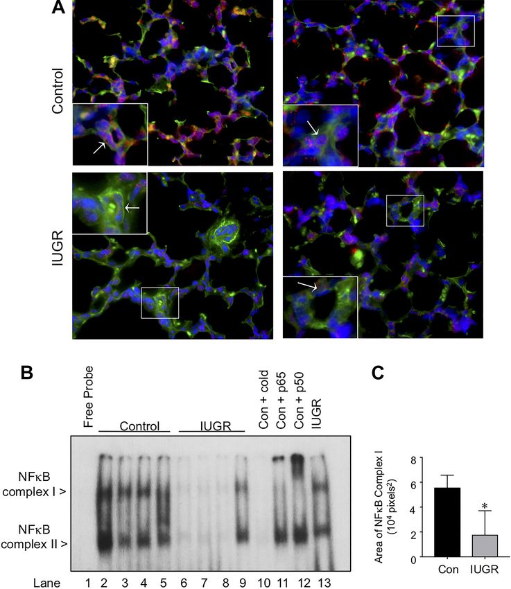
control and IUGR fetal sheep lungs in situ by immunostaining members p50 and p65 before incubation with the radiolabeled
to detect active nuclear NF-B complexes. Similar to past NF-B oligonucleotide. Using this method, we found that the
findings in the developing murine lung, there was striking upper complex was shifted upward with the addition of anti-
evidence of active nuclear NF-B complexes throughout the bodies against either the p65 or the p50 subunit, identifying
lungs of control fetal sheep (Fig. 2), including strong expres- this complex as the heterodimer of p65p50.
sion within CD31 positive endothelial cells in the distal pul- Inducible activation of IKKs is impaired in IUGR compared
monary vasculature (Fig. 2, high magnification inset). In con- with control PAECs despite similar levels of IKK␣ and IKK.
trast, staining for NF-B complexes was greatly diminished in To explore mechanisms underlying the difference in NF-B
the lungs of IUGR fetal sheep, including a paucity of nuclear activation between control and IUGR PAECs, we first com-
staining within the endothelial cells of the distal vasculature pared the expression of the two main NF-B activating ki-
(Fig. 2). nases, IKK␣ and IKK. Using Western blot analysis, we found
We confirmed this difference in endothelial NF-B activa- that the baseline levels of IKK␣ and IKK were no different in
tion by performing electrophoretic mobility shift assays on IUGR compared with control PAECs (Fig. 3A and B). We then
nuclear extracts obtained from PAECs isolated from control assessed IKK activation with an ELISA that detects phos-
and IUGR fetal sheep to detect NF-B-DNA binding. NF-B- phorylation of IKK at serine residues Ser177 and Ser181,
DNA binding was present in all the control PAECs, consisting which are key steps in IKK activation (14). IKK activity
of both an upper and lower complex (Fig. 2B). In contrast, was similar in unstimulated control and IUGR PAECs at
NF-B-DNA binding was completely absent in three primary baseline (Fig. 3C). In response to LPS stimulation, both control
cell lines of IUGR PAECs and diminished in the remaining two and IUGR PAECs activated IKK at 1 h. Although IKK
cell lines. Quantification of these differences by densitometry activity remained elevated in control PAECs at 2h, it was
identified a 68% reduction in NF-B-DNA binding in the significantly lower in IUGR PAECs at this time point.
IUGR PAECs as compared with control PAECs (P ⬍ 0.05) Baseline expression and LPS-mediated nuclear transloca-
(Fig. 2C). We also performed supershift analysis to identify the tion of NF-B subunits p65 and p50 are decreased in IUGR
composition of the two complexes present by preincubating the PAECs. We next determined nuclear and cytoplasmic levels of
nuclear extracts with antibodies specific for the NF-B family p65 and p50 in control and PAECs at baseline and in response
Fig. 2. Active NF-B is higher in the lungs and pulmo-
nary artery endothelial cells (PAECs) of control fetal
sheep as compared with intrauterine growth restriction
(IUGR) fetal sheep. A: representative immunofluores-
cent images from control and IUGR fetal lung from two
separate animals per group, stained to detect the NF-B
subunit, p65 (red), the endothelial specific marker,
CD31 (green), and chromatin (blue). The insets show
higher magnification images of distal vessels in the four
animals. B: EMSA was performed using nuclear ex-
tracts obtained from control (lanes 2–5) and IUGR
PAECs (lanes 6 –9), incubated with radiolabeled B
oligonucleotides. Specificity of the bands in control
PAECs (lane 2 sample) was confirmed by the disap-
pearance of the bands with the addition of 100-fold
excess of cold oligonucleotide (lane 10). Supershift
analysis of the NF-B complexes present in control
PAECs (lane 2 sample) was then performed by prein-
cubating the nuclear extracts with antibodies against
p65 (lane 11) and p50 (lane 12). For Complex I, a shift
upward was produced by the addition of p65 antibodies
and p50 antibodies. C: quantification of EMSA by
Image J to measure the total area the Complex I band in
each sample. Data presented are means ⫾ SE, with n ⫽
4 for control PAECs and n ⫽ 5 for IUGR PAECs *P ⫽
0.02 vs. control. Con, control.
AJP-Lung Cell Mol Physiol • doi:10.1152/ajplung.00052.2018 • www.ajplung.org
Downloaded from journals.physiology.org/journal/ajplung at Stanford Univ Med Ctr (171.066.010.174) on January 1, 2021.DECREASED NF-B IN IUGR PAEC L353
Fig. 3. Inducible activation of IKK is impaired in intrauterine growth restriction (IUGR) compared with control pulmonary artery endothelial cells (PAECs)
despite similar levels of IKK␣ and IKK. IKK␣ (A) and IKK (B) protein levels were detected by western immunoblot, with n ⫽ 3 animals with 3 technical
replicates per group. Relative expression of IKK␣ and IKK were normalized to -actin and expressed as fold-change over control. Data presented are
means ⫾ SE, with n ⫽ 3 animals for each group. Phosphorylation of IKK (C) was determined by ELISA in control and IUGR PAECs at baseline and at 1 or
2 h after LPS (10 ng/ml) stimulation. Data presented are means ⫾ SE, with n ⫽ 3 animals with 2 technical replicates for each group. ###P ⬍ 0.001 vs. 0 h, and
**P ⫽ 0.002 vs. control. Con, control.
to stimulation with LPS, an activator of NF-B signaling. In unbiased gene expression profiling to compare differences in
control PAECs, cytoplasmic levels of p65 (Fig. 4A) and p50 PAECs from normal fetal sheep and an established model of
(Fig. 4B) are decreased 1 h after stimulation with LPS, and IUGR. Using this strategy, we found that IUGR significantly
nuclear levels of both subunits are increased at 1 and 2 h (Fig. suppresses endogenous NF-B activation in fetal pulmonary
4C and D), which is consistent with translocation of active endothelium, and we attributed this decrease in pathway acti-
p65p50 heterodimers into the nucleus. In contrast, IUGR vation to both decreases in the expression of key NF-B
PAECs had significantly lower amounts of cytoplasmic p65 subunits (p65 and p50) and to defects in IKK-mediated,
and p50 than control PAECs at baseline and marked reductions inducible NF-B activation. IUGR also decreased the expres-
in nuclear levels of p65 and p50 1 and 2 h after LPS stimula- sion of Col4␣1 and Lama4, putative downstream targets of
tion, consistent with reduced inducible activation of NF-B in NF-B that are essential basement membrane components. The
IUGR PAECs. importance of endogenous NF-B activation in the fetal sheep
Angiogenic functions are suppressed in IUGR PAECs to PAECs was further highlighted by demonstrating that inhibit-
levels measured in control PAECs after treatment with BAY ing IKK in control PAECs disrupts angiogenic function to a
11-7082, an inhibitor of NF-B signaling. We next compared degree similar to the impaired angiogenesis observed in the
the capacity of the control and IUGR PAECs to proliferate and IUGR PAECs in baseline conditions. In concert with our prior
form tube structures in vitro at baseline and after treatment studies, this report provides additional evidence in a robust
with a selective and pharmacological inhibitor of the IKK animal model of disrupted alveolarization and angiogenesis
activating kinases, BAY 11-7082 (2, 29, 30). At baseline, that endogenous NF-B activity in pulmonary endothelial cells
proliferation of IUGR PAECs in response to DMEM was is essential for late lung development.
significantly lower than that observed in the control PAECs Pulmonary angiogenesis is essential for the growth and
(Fig. 5A). Treatment of the control PAECs with BAY resulted development of the distal lung. In premature infants dying
in dose-dependent decreases in PAEC proliferation. In con- from BPD, angiogenic factors are decreased, and the pulmo-
trast, BAY treatment had no effect on the proliferation of the nary vasculature is dysmorphic (8, 19, 26). Inhibition of
IUGR PAECs. We also performed in vitro tube formation angiogenesis impairs lung growth, decreases pulmonary artery
assays and found that tube formation was impaired in vehicle- density, and causes pulmonary hypertension (31), whereas
treated IUGR PAECs as compared with control PAECs (Fig. augmenting lung angiogenesis with inhaled nitric oxide (45) or
5B). BAY treatment also markedly decreased tube formation in exogenous VEGF (39, 40, 71) preserves alveolarization in
control PAECs, suppressing branch points to values obtained experimental models of BPD. Mechanisms through which
with IUGR PAECs at baseline. In contrast, BAY treatment had VEGF enhances alveolar growth include the release of angio-
no further reduction on tube formation in the IUGR PAECs. crine factors that act on the developing alveolar epithelium,
DISCUSSION
such as retinoic acid (76) and hepatocyte growth factor (64).
In addition, our prior studies identified high levels of endog-
To determine potential molecular mechanisms associated enous activation of NF-B in the pulmonary endothelium of
with abnormal lung endothelial function in IUGR, we used the early alveolar mouse lung in vivo and showed that inhibi-
AJP-Lung Cell Mol Physiol • doi:10.1152/ajplung.00052.2018 • www.ajplung.org
Downloaded from journals.physiology.org/journal/ajplung at Stanford Univ Med Ctr (171.066.010.174) on January 1, 2021.L354 DECREASED NF-B IN IUGR PAEC
Fig. 4. Baseline cytoplasmic expression and LPS-mediated nuclear translocation of NF-B subunits p65 and p50 are decreased in intrauterine growth restriction
(IUGR) pulmonary artery endothelial cells (PAECs). Representative images and densitometric quantification of western immunoblot analysis was used to detect
the levels of cytoplasmic p65 (A), cytoplasmic p50 (B), nuclear p65 (C), and nuclear p50 (D). Data presented are means ⫾ SE, with n ⫽ 3– 4 per group with
values for each target protein normalized to -actin at time 0 (t0). **P ⫽ 0.002 and ##P ⫽ 0.006 vs. control PAECs at 0 h (A); ***P ⬍ 0.001, #P ⫽ 0.02, and
##P ⫽ 0.002 vs. control PAECs at 0 h (B); **P ⫽ 0.007 vs. control PAECs at 1 h, ###P ⬍ 0.001 and ##P ⫽ 0.004 vs. control PAECs at 0 h, and ⫹P ⫽ 0.04
vs. IUGR PAECs at 0 h (C); and *P ⫽ 0.01 vs. control PAECs at 1 h, **P ⫽ 0.001 vs. control PAECs at 2 h, ##P ⫽ 0.001 and ###P ⬍ 0.001 vs. control PAECs
at 0 h (D).
AJP-Lung Cell Mol Physiol • doi:10.1152/ajplung.00052.2018 • www.ajplung.org
Downloaded from journals.physiology.org/journal/ajplung at Stanford Univ Med Ctr (171.066.010.174) on January 1, 2021.DECREASED NF-B IN IUGR PAEC L355
Fig. 5. Inhibition of NF-B reduces control of pulmonary artery endothelial cell (PAEC) angiogenic function to levels similar to the impaired angiogenesis
observed in intrauterine growth restriction (IUGR) PAECs. A: proliferation assays were performed on control (dark bars) and IUGR (light bars) PAECs in
response to DMEM ⫹ vehicle. In similar studies, proliferation was measured in control and IUGR PAECs treated with increasing doses of the IKK inhibitor,
BAY 11-7082 (BAY). Data shown are means ⫾ SE, with n ⫽ 3 cell lines from different animals with 3– 4 replicates performed per cell isolation. *P ⬍ 0.05
and **P ⬍ 0.01 vs. control PAECs treated with the same stimulus, and #P ⬍ 0.05 and ###P ⬍ 0.001 vs. control DMEM-treated. B: in vitro tube formation assays
were performed by plating control and IUGR PAECs on cross-linked collagen. At 18 h, images were obtained, and the number of branch points per ⫻10 field
magnification were determined in a blinded fashion. Data presented are means ⫾ SE, with n ⫽ 18 –30 fields counted from PAECs isolated from 3 separate
animals per experimental group. ***P ⬍ 0.001 and ###P ⬍ 0.001 vs. vehicle treated control PAECs.
tion of NF-B in primary murine pulmonary endothelial cells decreased. In addition, the expression of p65 and p50, compo-
decreases VEGFR2 expression and reduces PAEC function in nents of the transcriptionally active p65p50 heterodimer, was
vitro (30). These original findings are supported by observa- significantly decreased in IUGR PAECs. Consistent with these
tions that lung growth in transgenic mice with sustained NF-B results, prosurvival (e.g., baculoviral IAP repeat containing
signaling is preserved in response to hyperoxia-induced injury protein 3 and CASP8 and FADD-like apoptosis regulator) and
(52). In addition, female newborn mice exposed to hyperoxia proinflammatory [e.g., TNF-␣-induced protein 3 (TNFAIP3)]
have better lung structure when compared with male mice, genes, which are downstream of NF-B, are decreased in
which is associated with enhanced NF-B signaling (46). This IUGR PAECs.
latter observation may partly explain clinical observations of In addition, immunofluorescent staining of nuclear (active)
sex-related differences for BPD risk and severity in premature p65 in situ was decreased in IUGR lungs in comparison with
infants (9, 16). Moreover, a recent study that performed pro- controls. Assessment of NF-B-DNA binding by EMSA fur-
teomic analysis of murine lungs from embryonic to adult stages ther confirmed a significant reduction in the binding of p65p50
of development identified NF-B-regulated networks as a key heterodimers, which are NF-B complexes that we previously
signaling pathway increased during the transition from the identified as playing a key role to upregulate VEGFR2 expres-
prenatal to the postnatal period (55). In the present report, we sion in the developing mouse endothelium (30). Overall, these
further report suppression of the IKK/NF-B signaling path- data are consistent with our hypothesis that reduced activation
way in a fetal sheep model of IUGR that is characterized by of the NF-B pathway is an important mechanism underlying
decreased fetal lung vascular and alveolar growth, which is the angiogenic defect induced by IUGR.
independent of hyperoxia exposure. These findings provide In addition to alterations in the NF-B signaling pathway,
additional support for the hypothesis that early disruption of microarray analysis also identified alterations in genes in-
NF-B signaling impairs pulmonary angiogenesis and alveo- volved in the ECM and collagen fibril organization pathways,
larization across multiple species and in response to both including decreased Col4␣1 and Lama4 expression, which are
prenatal and postnatal injury and for the paradigm that NF-B both key components of the basement membrane (BM) and
is a central regulator of pulmonary vascular development. regulators of angiogenesis. The ECM is continuously remod-
We found that combined gene set enrichment, gene ontol- eled during lung development (69), serving as a scaffold that
ogy, and ingenuity pathway analyses implicated NF-B as a influences numerous cellular processes, and dysregulated ECM
top decreased pathway in IUGR PAECs when compared with remodeling has been demonstrated in experimental models of
controls. Although IKK␣ and IKK gene expression was not aberrant lung development (54). BM components can provide
different in control versus IUGR PAECs, genes both upstream either pro- or antiangiogenic cues. Although mature networks
and downstream of the IKKs were significantly decreased in of BM maintain blood vessel quiescence by inhibiting endo-
IUGR cells. These data suggest that while IKK gene expres- thelial proliferation and migration, during remodeling, the
sion is preserved in IUGR PAECs, IKK activity may be exposure of different domains modulates endothelial cell phe-
AJP-Lung Cell Mol Physiol • doi:10.1152/ajplung.00052.2018 • www.ajplung.org
Downloaded from journals.physiology.org/journal/ajplung at Stanford Univ Med Ctr (171.066.010.174) on January 1, 2021.L356 DECREASED NF-B IN IUGR PAEC
notype to promote angiogenesis (35). In human microvascular PAECs: 1 male and 2 females). Future studies designed spe-
endothelial cells, type IV collagen secretion increases during cifically to determine the influence of sex on suppressed
angiogenesis (4). In addition, type IV collagen promotes NF-B signaling in IUGR would be necessary to know
neovessel length and stability in a dose-dependent fashion in whether there are similar sex-specific differences in the lungs
aortic explants (10). Simultaneous disruption of Lama4 and of fetal sheep. NF-B is a complex signaling pathway consid-
Lama1 in zebrafish impairs development of the intersegmental ered a “master regulator” because of its ability to regulate
vessels (57), and Lama4 deletion in mice impairs corneal diverse cellular responses by promoting or repressing the
angiogenesis and microvascular maturation (73). Moreover, transcription of hundreds of target genes in a cell- and stimu-
disruption of these key BM components may directly affect lus-specific fashion. Furthermore, the NF-B activating ki-
lung development. Although mice with homozygous deletions nases, IKK␣ and IKK, have been shown to exert NF-B
of both Col4a1 and Col4a2 die during midembryogenesis independent effects, altering the activation states of other
because of widespread impairments of basement membrane transcription factors and influencing transcription by altering
stability (58), mice with heterozygous mutations in Col4a1 chromatin remodeling (1). In this study, it is not possible to
frequently die in the perinatal period, exhibiting respiratory differentiate between NF-B-mediated effects resulting from
distress and cyanosis and compact lungs that contain few the decreased expression of p65 and p50 and IKK-mediated,
terminal airspaces (27). Col4a1 mutant mice that survive the NF-B-independent effects resulting from the decreased acti-
perinatal period have lungs with simplified alveoli at P6, and vation of IKK. Future studies employing ChIP-Seq strategies
emphysematous changes at P30, in association with alterations to identify specific p50 and p65 regulated genes in the fetal
in myofibroblast proliferation and differentiation (50). Taken sheep PAECs may be helpful in further delineating the specific
together, these data suggest that the decreases of Col4␣1 and effects of the two limbs of this pathway. Although we show
Lama4 expression in our model could be directly contributing that inhibition of NF-B signaling in control PAECs inhibits
to the impaired angiogenesis and alveolarization observed in angiogenesis, definitive experiments to rescue the impaired
this experimental model of IUGR. NF-B signaling in the IUGR PAECs were not performed.
Moreover, the alterations in NF-B and ECM signaling Given that these cells exhibit both a defect in IKK activation
observed in IUGR may be interrelated. For example, the ECM and repressed expression of the p65 and p50 subunits, an
can influence NF-B activation by changing the clustering and effective rescue strategy would necessarily entail the overex-
engagement of specific cell surface integrins. Specifically, the pression of a constitutively active IKK construct and p65 and
␣v3 integrin appears to play an important role in mediating p50 constructs. The downstream effects of NF-B activation
endothelial angiogenic functions, in part through NF-B-me- are cell- and stimulus-specific and are influenced by timing of
diated effects. Integrin ␣v3 is highly expressed by angiogenic activation and development. Thus, a more granular understand-
but not quiescent blood vessels (12), and blocking ␣v3 ing of the mechanisms allowing for physiological effects for
integrin signaling with antibodies disrupts both embryonic and NF-B in the developing pulmonary endothelium lung is
tumor angiogenesis, in part by inducing endothelial cell apo- essential, given that pathological activation of NF-B can have
ptosis (13). In rat aortic endothelial cells, ␣v3 promotes detrimental effects (1, 70). Although, many therapeutic strate-
endothelial cell survival by activating NF-B (63). Although gies aimed at limiting pathological NF-B activation have been
intact type IV collagen binds ␣11 integrins, proteolytic cleav- developed (3, 6), the development of strategies to enhance
age during ECM remodeling reveals a cryptic site that allows NF-B has not received similar focus. Potential strategies that
binding of ␣v3 integrins (75). Similarly, Lama4 mediates could be effective in enhancing NF-B-mediated angiogenesis
endothelial cell adhesion through the ␣v3 integrin, and this in the pulmonary vasculature include the modulation of micro
interaction promotes angiogenesis in chick chorioallantoic RNAs that modulate NF-B subunit expression (22, 77); in-
membrane assays (43) and in Matrigel plug assays performed in creasing the activity of factors which alter the acetylation of
nude mice (25). Both Col4␣1 and Lama4 contain B binding p65, allowing for nuclear retention of NF-B complexes (41);
sequences in their promoters, indicating that they are putative and high throughput screens of the Food and Drug Adminis-
NF-B transcriptional targets. Taken together, these data raise the tration approved drug, compound, and/or siRNA libraries to
possibility that in this IUGR model, decreased expression of identify novel activators of NF-B in reporter cell lines.
Col4␣1 and Lama4 could contribute to the suppression of NF-B In summary, the present study identifies suppression of
activation, or, alternatively, that the downregulation of NF-B IKK/NF-B signaling as a key mechanism contributing to the
transcriptional activity decreases the expression of these specific impaired pulmonary endothelial angiogenic function observed
ECM components. in IUGR. In addition to decreasing the expression of key
The limitations of this study must be acknowledged. Cryo- NF-B subunits, IUGR also impaired inducible activation of
preserved cell lines from previous studies were used for the the NF-B pathway and decreased the expression of numerous
array analysis, without matching sexes in the samples. Re- putative NF-B downstream targets, including BM compo-
search has shown sex-related differences in NF-B signaling in nents important angiogenesis. These data add to our prior work
the developing mouse lung (46). Although in this study the identifying endogenous activation of NF-B as an important
samples were not specifically matched based on sex, similar regulator of pulmonary angiogenesis during late lung develop-
ratios of male and female animals were distributed between the ment and are the first, to our knowledge, to identify endothe-
two experimental groups (gene array analysis, control PAECs: lial-specific suppression of NF-B signaling in an established,
2 males and 1 female and IUGR PAECs: 2 males and 2 experimental model of impaired angiogenesis and alveolariza-
females; EMSA, control PAECs: 1 male and 3 females and tion induced by IUGR. Despite advances in the treatment of
IUGR PAECs: 1 male and 4 females; and in vitro and western infants with IUGR, pulmonary outcomes remain a major clin-
analyses, control PAECs: 1 male and 2 females and IUGR ical problem with little change in incidence in recent decades
AJP-Lung Cell Mol Physiol • doi:10.1152/ajplung.00052.2018 • www.ajplung.org
Downloaded from journals.physiology.org/journal/ajplung at Stanford Univ Med Ctr (171.066.010.174) on January 1, 2021.DECREASED NF-B IN IUGR PAEC L357
(24, 32). Given that alveolarization continues for years after bronchopulmonary dysplasia. Am J Respir Crit Care Med 164: 1971–
birth, we speculate that the suppression of NF-B activity in 1980, 2001. doi:10.1164/ajrccm.164.10.2101140.
9. Binet ME, Bujold E, Lefebvre F, Tremblay Y, Piedboeuf B; Canadian
response to IUGR could contribute to the higher incidence of Neonatal Network™. Role of gender in morbidity and mortality of
BPD and long-term deficits in pulmonary function observed in extremely premature neonates. Am J Perinatol 29: 159 –166, 2012. doi:
premature patients with IUGR. Thus, the development of 10.1055/s-0031-1284225.
targeted strategies to selectively restore NF-B activity in the 10. Bonanno E, Iurlaro M, Madri JA, Nicosia RF. Type IV collagen
developing pulmonary endothelium may represent a potential modulates angiogenesis and neovessel survival in the rat aorta model. In
Vitro Cell Dev Biol Anim 36: 336 –340, 2000. doi:10.1290/1071-
strategy to enhance pulmonary angiogenesis and alveolariza- 2690(2000)036⬍0336:TICMAA⬎2.0.CO;2.
tion in patients with IUGR. 11. Bose C, Van Marter LJ, Laughon M, O’Shea TM, Allred EN, Karna
P, Ehrenkranz RA, Boggess K, Leviton A; Extremely Low Gestational
ACKNOWLEDGMENTS Age Newborn Study Investigators. Fetal growth restriction and chronic
We thank the Genomics and Microarray Core at the University of Colorado lung disease among infants born before the 28th week of gestation.
Anschutz Medical Campus for their assistance in preparing and running Pediatrics 124: e450 –e458, 2009. doi:10.1542/peds.2008-3249.
mRNA arrays. Dr. Dodson is currently at United Therapeutics. The work 12. Brooks PC, Clark RA, Cheresh DA. Requirement of vascular integrin
contained was completed at the University of Colorado Denver, Anschutz alpha v beta 3 for angiogenesis. Science 264: 569 –571, 1994. doi:10.1126/
Medical Campus and Stanford University. science.7512751.
13. Brooks PC, Montgomery AM, Rosenfeld M, Reisfeld RA, Hu T, Klier
GRANTS G, Cheresh DA. Integrin alpha v beta 3 antagonists promote tumor
regression by inducing apoptosis of angiogenic blood vessels. Cell 79:
This work was supported by grants from the Children’s Hospital Colorado 1157–1164, 1994. doi:10.1016/0092-8674(94)90007-8.
Research Scholar Award (to R. B. Dodson); the Entelligence Young Investi- 14. Carter RS, Geyer BC, Xie M, Acevedo-Suárez CA, Ballard DW.
gator Award (to R. B. Dodson); Stanford Child Health Research Institute Persistent activation of NF-kappa B by the tax transforming protein
Tashia and John Morgridge Faculty Scholar Award (to C. M. Alvira); NIH involves chronic phosphorylation of IkappaB kinase subunits IKKbeta and
National Heart, Lung, and Blood Institute (NHLBI) Grant R01-HL-122918 (to IKKgamma. J Biol Chem 276: 24445–24448, 2001. doi:10.1074/jbc.
C. M. Alvira); NIH National Institute of Diabetes and Digestive and Kidney C000777200.
Diseases Grant R01-DK-088139 (to P. J. Rozance); and NIH NHLBI Grant 15. Chen CM, Wang LF, Su B. Effects of maternal undernutrition during late
R01-HL-68702 (to S. H. Abman). gestation on the lung surfactant system and morphometry in rats. Pediatr
Res 56: 329 –335, 2004. doi:10.1203/01.PDR.0000134254.83113.8E.
DISCLOSURES 16. Costeloe K, Hennessy E, Gibson AT, Marlow N, Wilkinson AR; for
No conflicts of interest, financial or otherwise, are declared by the authors. the EPICure Study Group. The EPICure study: outcomes to discharge
from hospital for infants born at the threshold of viability. Pediatrics 106:
AUTHOR CONTRIBUTIONS 659 –671, 2000. doi:10.1542/peds.106.4.659.
17. Curle DC, Adamson IY. Retarded development of noenatal rat lung by
R.B.D., J.G., T.M.C., S.H.A., and C.M.A. conceived and designed research; maternal malnutrition. J Histochem Cytochem 26: 401–408, 1978. doi:10.
R.B.D., K.N.P., G.S., and C.M.A. performed experiments; R.B.D., K.N.P., 1177/26.5.659840.
J.G., P.J.R., G.S., D.A., K.J., T.M.C., S.H.A., and C.M.A. analyzed data; 18. Dailey WA, Gryc W, Garg PG, Drenser KA. Frizzled-4 variations
R.B.D., K.N.P., P.J.R., T.M.C., S.H.A., and C.M.A. interpreted results of associated with retinopathy and intrauterine growth retardation: a potential
experiments; R.B.D., S.H.A., and C.M.A. prepared figures; R.B.D. and C.M.A. marker for prematurity and retinopathy. Ophthalmology 122: 1917–1923,
drafted manuscript; R.B.D., K.N.P., J.G., P.J.R., D.A., T.M.C., S.H.A., and 2015. doi:10.1016/j.ophtha.2015.05.036.
C.M.A. edited and revised manuscript; R.B.D., K.N.P., J.G., P.J.R., G.S., D.A., 19. De Paepe ME, Mao Q, Powell J, Rubin SE, DeKoninck P, Appel N,
K.J., T.M.C., S.H.A., and C.M.A. approved final version of manuscript. Dixon M, Gundogan F. Growth of pulmonary microvasculature in
ventilated preterm infants. Am J Respir Crit Care Med 173: 204 –211,
REFERENCES 2006. doi:10.1164/rccm.200506-927OC.
20. Devaskar SU, Chu A. Intrauterine growth restriction: hungry for an
1. Alvira CM. Nuclear factor-kappa-B signaling in lung development and
answer. Physiology (Bethesda) 31: 131–146, 2016. doi:10.1152/physiol.
disease: one pathway, numerous functions. Birth Defects Res A Clin Mol
00033.2015.
Teratol 100: 202–216, 2014. doi:10.1002/bdra.23233.
21. Dezateux C, Lum S, Hoo AF, Hawdon J, Costeloe K, Stocks J. Low
2. Alvira CM, Abate A, Yang G, Dennery PA, Rabinovitch M. Nuclear
birth weight for gestation and airway function in infancy: exploring the
factor-kappaB activation in neonatal mouse lung protects against lipopo-
fetal origins hypothesis. Thorax 59: 60 –66, 2004.
lysaccharide-induced inflammation. Am J Respir Crit Care Med 175:
805–815, 2007. doi:10.1164/rccm.200608-1162OC. 22. Dharap A, Pokrzywa C, Murali S, Pandi G, Vemuganti R. MicroRNA
3. Awasthee N, Rai V, Chava S, Nallasamy P, Kunnumakkara AB, miR-324-3p induces promoter-mediated expression of RelA gene. PLoS
Bishayee A, Chauhan SC, Challagundla KB, Gupta SC. Targeting One 8: e79467, 2013. doi:10.1371/journal.pone.0079467.
IappaB kinases for cancer therapy. Semin Cancer Biol S1044- 23. Dodson RB, Rozance PJ, Fleenor BS, Petrash CC, Shoemaker LG,
579X(17)30046-9, 2018. doi:10.1016/j.semcancer.2018.02.007. Hunter KS, Ferguson VL. Increased arterial stiffness and extracellular
4. Bahramsoltani M, Slosarek I, De Spiegelaere W, Plendl J. Angiogen- matrix reorganization in intrauterine growth-restricted fetal sheep. Pediatr
esis and collagen type IV expression in different endothelial cell culture Res 73: 147–154, 2013. doi:10.1038/pr.2012.156.
systems. Anat Histol Embryol 43: 103–115, 2014. doi:10.1111/ahe.12052. 24. Fanaroff AA, Stoll BJ, Wright LL, Carlo WA, Ehrenkranz RA, Stark
5. Barker DJ, Godfrey KM, Fall C, Osmond C, Winter PD, Shaheen SO. AR, Bauer CR, Donovan EF, Korones SB, Laptook AR, Lemons JA,
Relation of birth weight and childhood respiratory infection to adult lung Oh W, Papile LA, Shankaran S, Stevenson DK, Tyson JE, Poole WK;
function and death from chronic obstructive airways disease. BMJ 303: NICHD Neonatal Research Network. Trends in neonatal morbidity and
671–675, 1991. doi:10.1136/bmj.303.6804.671. mortality for very low birthweight infants. Am J Obstet Gynecol 196:
6. Begalli F, Bennett J, Capece D, Verzella D, D’Andrea D, Tornatore 147.e1–147.e8, 2007. doi:10.1016/j.ajog.2006.09.014.
L, Franzoso G. Unlocking the NF-B conundrum: embracing com- 25. Gonzalez AM, Gonzales M, Herron GS, Nagavarapu U, Hopkinson
plexity to achieve specificity. Biomedicines 5: 50, 2017. doi:10.3390/ SB, Tsuruta D, Jones JC. Complex interactions between the laminin
biomedicines5030050. alpha 4 subunit and integrins regulate endothelial cell behavior in vitro and
7. Belvin MP, Anderson KV. A conserved signaling pathway: the Drosoph- angiogenesis in vivo. Proc Natl Acad Sci USA 99: 16075–16080, 2002.
ila toll-dorsal pathway. Annu Rev Cell Dev Biol 12: 393–416, 1996. doi:10.1073/pnas.252649399.
doi:10.1146/annurev.cellbio.12.1.393. 26. Gorenflo M, Vogel M, Obladen M. Pulmonary vascular changes in
8. Bhatt AJ, Pryhuber GS, Huyck H, Watkins RH, Metlay LA, Manis- bronchopulmonary dysplasia: a clinicopathologic correlation in short- and
calco WM. Disrupted pulmonary vasculature and decreased vascular long-term survivors. Pediatr Pathol 11: 851–866, 1991. doi:10.3109/
endothelial growth factor, Flt-1, and TIE-2 in human infants dying with 15513819109065482.
AJP-Lung Cell Mol Physiol • doi:10.1152/ajplung.00052.2018 • www.ajplung.org
Downloaded from journals.physiology.org/journal/ajplung at Stanford Univ Med Ctr (171.066.010.174) on January 1, 2021.You can also read