INPP4B promotes PI3K α-dependent late endosome formation and Wnt/ β-catenin signaling in breast cancer - Nature
←
→
Page content transcription
If your browser does not render page correctly, please read the page content below
ARTICLE
                  https://doi.org/10.1038/s41467-021-23241-6                 OPEN

                  INPP4B promotes PI3Kα-dependent late endosome
                  formation and Wnt/β-catenin signaling in breast
                  cancer
                  Samuel J. Rodgers1,2, Lisa M. Ooms 1,2, Viola M. J. Oorschot3,10, Ralf B. Schittenhelm 4,
                  Elizabeth V. Nguyen1,2, Sabryn A. Hamila1,2, Natalie Rynkiewicz2,11, Rajendra Gurung1,2, Matthew J. Eramo1,2,
                  Absorn Sriratana1,2, Clare G. Fedele2,12, Franco Caramia 5, Sherene Loi 5, Genevieve Kerr6,7,
1234567890():,;

                  Helen E. Abud 6,7, Georg Ramm 1,2,3, Antonella Papa 1,2, Andrew M. Ellisdon1,2,8, Roger J. Daly 1,2,
                  Catriona A. McLean 9 & Christina A. Mitchell 1,2 ✉

                  INPP4B suppresses PI3K/AKT signaling by converting PI(3,4)P2 to PI(3)P and INPP4B
                  inactivation is common in triple-negative breast cancer. Paradoxically, INPP4B is also a
                  reported oncogene in other cancers. How these opposing INPP4B roles relate to PI3K reg-
                  ulation is unclear. We report PIK3CA-mutant ER+ breast cancers exhibit increased INPP4B
                  mRNA and protein expression and INPP4B increased the proliferation and tumor growth of
                  PIK3CA-mutant ER+ breast cancer cells, despite suppression of AKT signaling. We used
                  integrated proteomics, transcriptomics and imaging to demonstrate INPP4B localized to late
                  endosomes via interaction with Rab7, which increased endosomal PI3Kα-dependent PI(3,4)
                  P2 to PI(3)P conversion, late endosome/lysosome number and cargo trafficking, resulting in
                  enhanced GSK3β lysosomal degradation and activation of Wnt/β-catenin signaling.
                  Mechanistically, Wnt inhibition or depletion of the PI(3)P-effector, Hrs, reduced INPP4B-
                  mediated cell proliferation and tumor growth. Therefore, INPP4B facilitates PI3Kα crosstalk
                  with Wnt signaling in ER+ breast cancer via PI(3,4)P2 to PI(3)P conversion on late endo-
                  somes, suggesting these tumors may be targeted with combined PI3K and Wnt/β-catenin
                  therapies.

                  1 Cancer Program, Biomedicine Discovery Institute, Monash University, Clayton, VIC, Australia. 2 Department of Biochemistry and Molecular Biology, Monash

                  University, Clayton, VIC, Australia. 3 Monash Ramaciotti Centre for Cryo Electron Microscopy, a Node of Microscopy Australia, Monash University,
                  Victoria, Australia. 4 Monash Proteomics and Metabolomics Facility, Monash Biomedicine Discovery Institute and Department of Biochemistry and
                  Molecular Biology, Monash University, Clayton, VIC, Australia. 5 Peter MacCallum Cancer Centre, University of Melbourne, Melbourne, VIC, Australia.
                  6 Development and Stem Cells Program, Biomedicine Discovery Institute, Monash University, Clayton, VIC, Australia. 7 Department of Anatomy and

                  Developmental Biology, Monash University, Clayton, VIC, Australia. 8 Australian Research Council Centre of Excellence in Advanced Molecular Imaging,
                  Monash University, Clayton, VIC, Australia. 9 Department of Anatomical Pathology, Alfred Hospital, Prahran, VIC, Australia. 10Present address: Electron
                  Microscopy Core Facility, European Molecular Biology Laboratory, Heidelberg, Germany. 11Present address: Babraham Institute, Cambridge, UK. 12Present
                  address: Peter MacCallum Cancer Centre, University of Melbourne, Melbourne, VIC, Australia. ✉email: christina.mitchell@monash.edu

                  NATURE COMMUNICATIONS | (2021)12:3140 | https://doi.org/10.1038/s41467-021-23241-6 | www.nature.com/naturecommunications                               1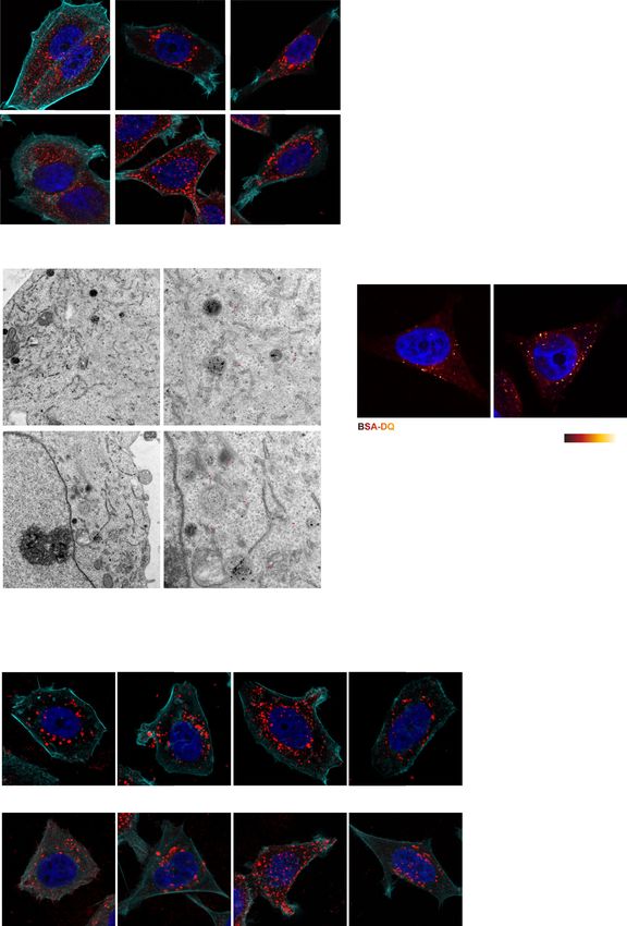
ARTICLE                                                                 NATURE COMMUNICATIONS | https://doi.org/10.1038/s41467-021-23241-6

T
       he phosphoinositide 3-kinase (PI3K) signaling pathway is              Results
       frequently hyperactivated in cancer. Class I PI3K tran-               INPP4B expression is increased in PIK3CA-mutant ER+ breast
       siently generates phosphatidylinositol 3,4,5-trisphosphate            cancer. Recently INPP4B was identified as a bona fide marker for
(PI(3,4,5)P3) on the inner leaflet of the plasma membrane in                  ER-positivity in human breast cancer24. INPP4B expression is lost
response to extracellular stimulation, which is rapidly converted            in triple-negative breast cancers, however, its emergence as a
to phosphatidylinositol 3,4-bisphosphate (PI(3,4)P2) by inositol             potential oncogene in other cancers led us to examine its relative
polyphosphate 5-phosphatases1. PI(3,4,5)P3 and PI(3,4)P2 toge-               expression and function in ER+ breast cancers. Here, INPP4B
ther increase the recruitment and activation of the protein kinase           protein expression was examined by immunohistochemistry
AKT2,3, the central effector of oncogenic PI3K signaling. PI(3,4,5)          using a validated monoclonal INPP4B (3D5) antibody6 on two
P3/PI(3,4)P2 signaling activates numerous other effectors at the             independent tissue cohorts; one tissue microarray containing 224
plasma membrane, including the protein kinase PDK1, which in                 primary human breast cancers with 32 tumor-adjacent normal
turn phosphorylates AKT and other AGC kinases such as SGK34.                 breast tissues (US Biomax) (Supplementary Fig. 1a), and a second
The tumor suppressor, phosphatase and tensin homolog (PTEN),                 cohort of 107 primary breast cancers collected as part of the
converts PI(3,4,5)P3 to PI(4,5)P2 thereby inhibiting PI3K/AKT                Melbourne Collaborative Cohort Study (MCCS)6. Loss of
signaling. Inositol polyphosphate 4-phosphatase type II (INPP4B)             INPP4B protein expression was associated with triple-negative
dephosphorylates PI(3,4)P2 downstream of class I PI3K to form                breast cancers (Fig. 1a, b), as we previously reported6. However,
phosphatidylinositol 3-phosphate (PI(3)P), also suppresses AKT               an increase in INPP4B protein expression was observed in
signaling, and is proposed to function as a tumor suppressor in              14–40% of breast cancers relative to normal tissue associated with
some cancers5,6.                                                             ER/PR-positivity and the luminal (ER+ and/or PR+) breast
   PIK3CA, which encodes the p110α subunit of PI3Kα, is                      cancer subtype (Fig. 1a, b and Supplementary Fig. 1b, c). As
mutated in up to 40% of breast cancers, most frequently in                   mRNA expression data were not available for these cohorts,
estrogen receptor-positive (ER+) tumors7,8. Mutant PIK3CA is                 INPP4B mRNA expression was examined using Tissue Scan
not an independent marker of prognosis when corrected for                    Breast Cancer cDNA arrays I–IV (OriGene) of 130 primary
favorable-risk variables such as ER-positivity9,10, but is a pre-            human breast cancers and 16 normal breast tissues (Fig. 1c).
dictive biomarker for improved response to combined endocrine                Decreased INPP4B expression was associated with triple-negative
and PI3K therapy (alpelisib-fulvestrant) in advanced ER+ breast              breast cancers, and notably increased INPP4B expression was
cancers11. Interestingly, PIK3CA-mutant ER+ breast cancers                   demonstrated in 25% of breast cancers associated with ER/PR-
display minimal AKT activation and little dependence on AKT                  positivity and the luminal subtype (Fig. 1c, d and Supplementary
signaling compared to tumors with other PI3K pathway altera-                 Fig. 1d, e).
tions, such as PTEN loss4,12.                                                   Mutations in PI3K pathway genes PIK3CA, PTEN, and AKT1
   We and others have identified that INPP4B inhibits AKT                     are commonly associated with ER+ breast cancers12. Using the
signaling and exhibits tumor suppressor activity in triple-negative          METABRIC and TCGA datasets8,25, we found that only 1% of
(ER−/PR−/HER2−) and basal-like breast cancers5,6,13. In addi-                breast cancers exhibited INPP4B genetic alterations such as
tion, many subsequent reports have shown that INPP4B protein                 mutations, truncations, amplifications, or deletions. INPP4B
expression is reduced in melanoma, ovarian, and prostate                     mRNA expression did not associate with PTEN or AKT1
cancers5,14,15. In murine models, Inpp4b ablation increases                  mutations, but positively correlated with PIK3CA-mutation status
mammary tumor penetrance in cooperation with Tp53/Brca1                      (Fig. 1e and Supplementary Fig. 1f). INPP4B expression was also
deletion13, and promotes thyroid tumorigenesis and metastasis                significantly higher in breast cancers with multiple PIK3CA
in vivo in cooperation with Pten heterozygous loss16,17. Notably,            mutations (Supplementary Fig. 1g). Further stratification revealed
INPP4B suppresses localized AKT2 signaling on early endosomes                high INPP4B expression was specifically associated with the
and EGFR degradation by degrading PI(3,4)P213,16,18.                         PIK3CA-mutant ER+ breast cancer subset (Fig. 1f).
   Paradoxically, more recent studies have indicated a possible
oncogenic role for INPP4B in acute myeloid leukemia (AML)
associated with chemoresistance, as well as in melanoma and                  INPP4B enhances PIK3CA-mutant ER+ breast cancer and
colon cancer19–22. Several molecular mechanisms for INPP4B                   mammary epithelial cell proliferation. To determine the func-
oncogenic function have been proposed via regulation of PTEN                 tional consequences of increased INPP4B expression in PIK3CA-
protein stability21, or SGK3 activation22,23, and may be context-            mutant ER+ breast cancer, GFP-INPP4B was expressed in MCF-7
dependent. For example, in breast cancer SGK3 is amplified and                and T47D ER+ breast cancer cells (Supplementary Fig. 2a), which
its kinase activity is dependent on oncogenic PI3K and INPP4B23.             harbor hyperactivating PIK3CAE545K and PIK3CAH1047R muta-
Recently INPP4B was identified as the top gene associated with                tions, respectively. The ability of cancer cells to undergo
ER+ breast cancers and tumor grade24.                                        anchorage-independent cell growth is a hallmark of cellular
   Here, we further explored the role INPP4B plays in ER+ breast             transformation. GFP-INPP4B significantly increased colony size
cancer revealing a cohort of PIK3CA-mutant ER+ breast cancers                of MCF-7 (2.1-fold) and T47D (1.8-fold) cells in soft agar but had
exhibit increased INPP4B expression. We demonstrate INPP4B                   no effect on colony number (Fig. 2a–c). Expression of Myc-
overexpression enhances the proliferation and tumor growth of                INPP4BWT, but not a PI(3,4)P2 4-phosphatase-dead Myc-
PIK3CA-mutant ER+ breast cancer cell lines, despite concurrent               INPP4BC842A mutant19 (Supplementary Fig. 2b), increased
AKT signaling suppression. To investigate this apparent paradox,             MCF-7 cell colony size (2.1-fold) in soft agar but did not affect
we utilized an integrative approach featuring proteomics, tran-              colony number (Supplementary Fig. 2c–e), consistent with
scriptomics, and advanced cellular imaging to elucidate the                  increased cell proliferation. GFP-INPP4B enhanced MCF-7 cell
molecular mechanisms by which INPP4B promotes tumorigen-                     proliferation (1.7-fold) following serum deprivation relative to
esis of PIK3CA-mutant ER+ breast cancers. Our findings show                   vector controls (Fig. 2d, e), but did not affect the proliferation of
INPP4B stimulates a PI3Kα-dependent signaling hub on late                    MCF-7 cells grown in serum-containing media (Supplementary
endosomes that directs Wnt/β-catenin activation. These studies               Fig. 2f). EGF-stimulated AKTS473 and AKTT308 phosphorylation
reveal a mechanism for crosstalk between two oncogenic signal-               levels were modestly reduced in MCF-7 cells overexpressing GFP-
ing pathways that promotes cell proliferation and tumor growth.              INPP4B (Fig. 2f–h), revealing that INPP4B promotes cell

2                NATURE COMMUNICATIONS | (2021)12:3140 | https://doi.org/10.1038/s41467-021-23241-6 | www.nature.com/naturecommunications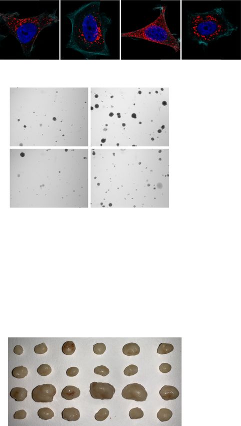
NATURE COMMUNICATIONS | https://doi.org/10.1038/s41467-021-23241-6                                                                                                                                                                                                                 ARTICLE

        a                                                                    Tissue microarrays (n=224 cases)                                                      b                 MCCS cohort (n=107 cases)
                                                                                              Luminal                                                                                                                           Luminal
                                                                                                                                       INPP4B staining                                                                                                                  INPP4B staining
                                                                                                                                            0 (37%)                                                                                                                             0 (21%)
                                                                                                                                            1 (28%)                                                                                                                             1 (14%)
                                                                                                                                            2 (21%)                                                                                                                             2 (25%)
                                                                                                                                            3 (14%)                                                                                                                             3 (40%)

                                                                                                            p=0.0038                                                                                                                         p=0.035

                                                                                                                           p lif 2 -
                                                                                                                                ie d
                                                                                                                         am ER
                                                                                               p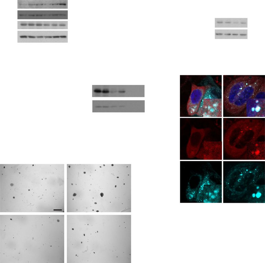
ARTICLE                                                                                                 NATURE COMMUNICATIONS | https://doi.org/10.1038/s41467-021-23241-6

             a                        GFP-vector                     GFP-INPP4B                                   b                                                     GFP-vector                                                              c                                              GFP-vector
                                                                                                                                                                        GFP-INPP4B                                                                                                             GFP-INPP4B

                                                                                                                                                         1.2                                                                                                                    p=0.0004

                                                                                                                                Relative colony number
                                                                                                                                                                                                                                                                       2.5
                                                                                                                                                                                                                                                                                                                        p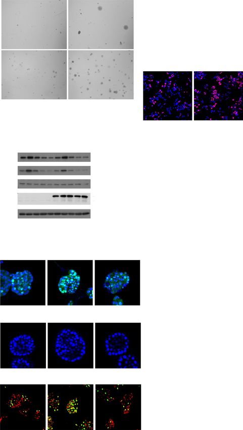
NATURE COMMUNICATIONS | https://doi.org/10.1038/s41467-021-23241-6 ARTICLE
Fig. 2 INPP4B promotes PIK3CA-mutant ER+ breast cancer and mammary epithelial cell proliferation. a–c MCF-7 or T47D cells expressing GFP-INPP4B
or GFP-vector were suspended in 0.3% soft agar and cultured for 4 weeks to allow anchorage-independent cell growth (a). Data represent relative number
of colonies (b), and relative colony size (c) (n > 50 colonies/experiment) ± SD (n = 3 experiments in triplicate). d, e MCF-7 cells expressing GFP-INPP4B or
GFP-vector were serum-starved for 24 h, incubated with BrdU overnight then fixed and immunostained with BrdU antibodies and DAPI (d). Data represent
mean percentage of BrdU-positive cells ± SD (n = 3 experiments, >300 cells/experiment) (e). f–h MCF-7 cells expressing GFP-INPP4B or GFP-vector were
serum-starved overnight, then stimulated with EGF (100 ng/mL) for the indicated times. Cells were lysed and immunoblotted with pAKTS473, pAKTT308,
or AKT(pan) antibodies or GAPDH antibodies as a loading control (f). Data represent the mean relative pAKTS473 (g) or pAKTT308 (h) levels relative to
AKT(pan) ± SD (n = 3 experiments). i–k MCF-10A cells expressing Myc-INPP4BWT, Myc-INPP4BC842A, or empty vector were cultured in Matrigel for
7 days, then fixed and immunostained with Ki67 antibodies and DAPI (i). Data represent mean percentage of Ki67-positive cells ± SD (n = 3 experiments,
>200 cells/experiment) (j), and relative acini area ± SD (n = 3 experiments, 15 largest acini scored/experiment) (k). l–n MCF-10A cells expressing
INPP4BWT, INPP4BC842A, or empty vector were cultured in Matrigel for 14 days, then fixed and immunostained with DAPI (l). Data represent relative acini
area (m) and number of cells per acinus (n) ± SD (n = 3 experiments, 15 largest acini scored/experiment). o, p MCF-10A cells expressing Myc-INPP4BWT,
Myc-INPP4BC842A, or empty vector were serum-starved for 48 h, then incubated with BrdU overnight. Cells were fixed and immunostained with BrdU
antibodies and propidium iodide (o). Data represent mean percentage of BrdU-positive cells ± SD (n = 3 experiments, >300 cells/experiment) (p). Scale
bars, 50 µm (i, l), 100 µm (d, o), 1 mm (a). p values determined by two-tailed unpaired t-test are indicated in c, e, g, h, or by one-way ANOVA with Tukey
post hoc test in j, m, n, p.
7 days in culture, Myc-INPP4BWT but not Myc-INPP4BC842A (PI endogenous Rab7 (Fig. 3c). Rab7 regulates late endosome for-
(3,4)P2 4-phosphatase-dead) expressing MCF-10A acini (Supple- mation, motility, and fusion with lysosomes, and its localization
mentary Fig. 2k) displayed a significant increase in the percentage and function is controlled by GTP/GDP loading, whereby it
of proliferating cells compared to vector control acini (1.5-fold) localizes to late endosomes in its active GTP-bound state and
(Fig. 2i, j). No significant changes to acini size were observed at then dissociates following hydrolysis of GTP to GDP27. Immu-
day 7 (Fig. 2k). By day 14, Myc-INPP4BWT but not Myc- noprecipitation of HA-Rab7WT, HA-Rab7Q67L (constitutively
INPP4BC842A expressing MCF-10A cells formed larger acini (1.8- GTP-bound) or HA-Rab7T22N (constitutively GDP-bound),
fold), with an increased number of cells per acinus (1.4-fold) showed that endogenous INPP4B was able to bind both GTP and
compared to vector controls (Fig. 2l–n). After 21 days, Myc- GDP-loaded Rab7 (Supplementary Fig. 3a). Notably, in a purified
INPP4BWT overexpressing MCF-10A cells formed mature acini in vitro component assay recombinant Rab7 significantly
with a single layer of epithelial cells surrounding a hollow lumen enhanced the PI(3,4)P2 4-phosphatase activity of recombinant
(Supplementary Fig. 2l). Myc-INPP4BWT expression did not INPP4B in a dose-dependent manner (Supplementary Fig. 3b, c).
affect the apical distribution of the Golgi marker GM130 Endogenous INPP4B was diffusely distributed in the cytosol of
(Supplementary Fig. 2m), or the percentage of cleaved caspase- MCF-7 cells (Supplementary Fig. 3d). However, removal of
3-positive acini (Supplementary Fig. 2n, o). This suggests that cytoplasmic proteins by saponin treatment revealed a pool of
INPP4B overexpression promotes MCF-10A cell proliferation but INPP4B that in part co-localized with CD63-positive late
does not affect apico-basal polarity nor apoptosis. Consistent with endosomes, the site of Rab7 intracellular localization, and this
this, Myc-INPP4BWT but not Myc-INPP4BC842A overexpression was most apparent following EGF stimulation (Supplementary
enhanced MCF-10A cell proliferation in monolayer cultures Fig. 3e). INPP4B localization to late endosomes was further
following serum deprivation (1.7-fold) (Fig. 2o, p). EGF- analyzed by immuno-electron microscopy, which revealed GFP-
stimulated AKTS473 phosphorylation was decreased in Myc- INPP4B localized to the limiting and internal membranes of late
INPP4BWT but not Myc-INPP4BC842A overexpressing MCF-10A endosomes (Fig. 3d), a site where previous studies have identified
cells (Supplementary Fig. 2p, q). This data is consistent with an both PI(3,4)P2 and PI(3)P28,29. In saponin-treated MCF-7 cells,
interpretation that INPP4B promotes the proliferation of endogenous INPP4B was observed to co-localize with HA-
PIK3CA-mutant ER+ breast cancer and PIK3CA-wild-type Rab7WT or HA-Rab7Q67L on CD63-positive late endosomes
mammary epithelial cells in a PI(3,4)P2 4-phosphatase- (Fig. 3e). Expression of HA-Rab7T22N, a dominant-negative
dependent manner. mutant that does not localize to late endosomes (Supplementary
Fig. 3f), also prevented the recruitment of endogenous INPP4B to
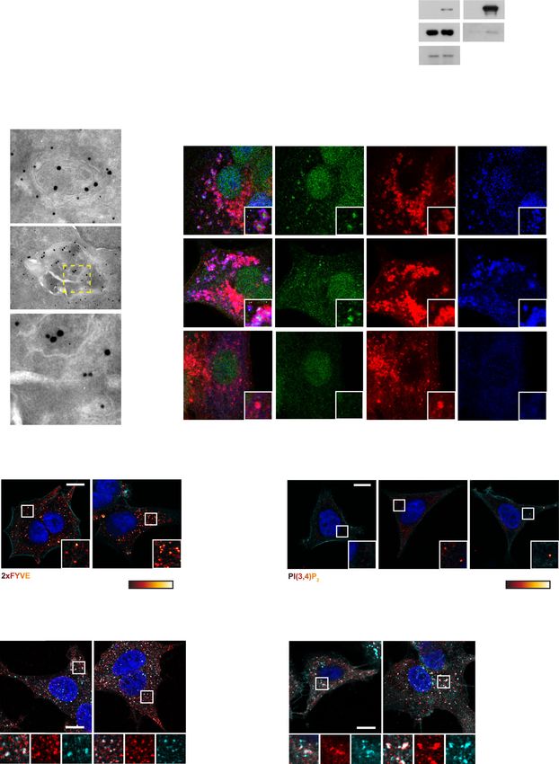
INPP4B promotes PI(3,4)P2 to PI(3)P conversion on late this compartment (Fig. 3e), suggesting that active GTP-bound
endosomes. We utilized several unbiased proteomics-based Rab7 is required for INPP4B subcellular localization to late
techniques to characterize the downstream signaling functions endosomes.
of INPP4B in PIK3CA-mutant ER+ breast cancer. First, whole As Rab7 enhances INPP4B 4-phosphatase activity towards PI
cell proteomics using liquid chromatography-tandem mass (3,4)P2, we predicted that INPP4B may promote PI(3,4)P2
spectrometry (LC-MS/MS) was undertaken to identify variations conversion to PI(3)P on late endosomes. PI(3)P resides on early
in protein levels (>2-fold) in GFP-INPP4B versus GFP-vector and late endosome membranes, and was detected here using a
expressing MCF-7 cells. Functional annotation analysis revealed 2xFYVE/Hrs recombinant biosensor28. GFP-INPP4B significantly
that the upregulated proteins in INPP4B-overexpressing cells increased intracellular PI(3)P levels (1.8-fold) (Fig. 3f). PI(3,4)P2,
were strongly associated with the endolysosomal system, the which also resides on early and late endosomes18,29, was
vesicular trafficking pathway that mediates the internalization examined by antibody staining in MCF-7 cells but specific
and degradation of extracellular receptors and cargo (Fig. 3a). staining was not apparent, we assume due to either low signal or
Furthermore, immunoprecipitation-mass spectrometry (IP-MS) antibody affinity. However, MCF-7 cells that expressed INPP4B
analysis of GFP-INPP4B protein complexes was performed on shRNAs (Supplementary Fig. 3g, h) showed more prominent
MCF-7 cells stimulated with EGF to activate class I PI3K sig- intracellular PI(3,4)P2 puncta suggesting that INPP4B depletion
naling. The most-enriched binding partner identified from this suppressed PI(3,4)P2 degradation on endosomes resulting in its
analysis was RAB7A (Rab7), a small GTPase that localizes to late accumulation (Fig. 3g). To examine the site of PI(3,4)P2
endosomes (Fig. 3b). Binding between INPP4B and Rab7 was conversion to PI(3)P by INPP4B, 2xFYVE/Hrs co-localization
confirmed by co-immunoprecipitation of GFP-INPP4B with with the early endosome marker EEA1 and the late endosome
NATURE COMMUNICATIONS | (2021)12:3140 | https://doi.org/10.1038/s41467-021-23241-6 | www.nature.com/naturecommunications 5ARTICLE NATURE COMMUNICATIONS | https://doi.org/10.1038/s41467-021-23241-6
a Enriched pathways (GFP-INPP4B/GFP-vector) b c
p=0.05
5% Input IP: GFP
Cytosol 2.5
INPP4B + − + − GFP-vector
Lysosome
2.0 HSPA1A − + − + GFP-INPP4B
Trans-Golgi network
P value (-log10)
HSPA8
Clathrin-coated vesicle RAB7A 130 kDa GFP-INPP4B
1.5 p=0.05
Intracellular protein transport
25 kDa Rab7
Cell junction assembly 1.0 (endogenous)
Golgi apparatus
0.5 GAPDH
Clathrin adaptor complex 35 kDa
Vesicle-mediated transport
0.0
0 1 2 3 4 -3 -2 -1 0 1 2 13 14
p-value (-log10) log2-fold change (GFP-INPP4B/GFP-vector)
d Late endosomes e + Saponin
INPP4B Merged INPP4B CD63 HA-Rab7
HA-Rab7WT
CD63
HA-Rab7Q67L
CD63
HA-Rab7T22N
INPP4B
GFP-INPP4B 10 nm INPP4B CD63 HA-Rab7
CD63 15 nm
GFP-vector
f GFP-INPP4B g
GFP-vector GFP-INPP4B p=0.0011 NT shRNA INPP4B shRNA #1 INPP4B shRNA #2
140
120
PI(3)P+ puncta/cell
100
80
60
40
20
F-actin DAPI Low High 0 F-actin DAPI Low High
h i
PI(3)P-positive early endosomes PI(3)P-positive late endosomes
GFP-vector GFP-INPP4B GFP-vector GFP-vector
GFP-vector GFP-INPP4B
GFP-INPP4B GFP-INPP4B
p=0.025
80 25
% PI(3)P+ early endosoomes
% PI(3)P+ late endosomes
60 20
15
40
10
20
5
0 0
2xFYVE EEA1 DAPI 2xFYVE CD63 DAPI
marker CD63 was investigated, revealing GFP-INPP4B did not INPP4B promotes PI3Kα-dependent late endosome formation.
alter the proportion of PI(3)P-positive early endosomes, but Endomembrane PI(3)P signaling is essential for binding and
significantly increased PI(3)P-positive late endosomes (1.7-fold) localizing FYVE or phox homology (PX) domain-containing
(Fig. 3h, i). In contrast, INPP4B shRNA depletion decreased PI(3) proteins to endosomal membranes to regulate numerous pro-
P-positive late endosomes (Supplementary Fig. 3i), supporting a cesses including early endosome fusion, early-to-late endosome
role for INPP4B in promoting the conversion of PI(3,4)P2 to PI maturation, and cargo sorting30. Examination of the endolyso-
(3)P on late endosomes. somal compartments of MCF-7 cells showed that GFP-INPP4B
6 NATURE COMMUNICATIONS | (2021)12:3140 | https://doi.org/10.1038/s41467-021-23241-6 | www.nature.com/naturecommunicationsNATURE COMMUNICATIONS | https://doi.org/10.1038/s41467-021-23241-6 ARTICLE Fig. 3 INPP4B promotes localized PI(3,4)P2 conversion to PI(3)P on late endosomes. a MCF-7 cells expressing GFP-INPP4B or GFP-vector were lysed, then subjected to whole cell proteomic analysis by LC-MS/MS. DAVID functional annotation bioinformatics microarray analysis was performed on upregulated proteins in GFP-INPP4B expressing cells versus GFP-vector cells (n = 3 experiments). b MCF-7 cells expressing GFP-INPP4B or GFP-vector were serum-starved overnight then stimulated with EGF (100 ng/mL) for 5 min. Cells were lysed and subjected to immunoprecipitation using GFP-Trap beads. Bound proteins were identified by mass spectrometry using a cut-off of >2-fold change and p < 0.05 (n = 2 experiments). c MCF-7 cells expressing GFP-INPP4B or GFP-vector were lysed and subjected to immunoprecipitation using GFP-Trap beads. Bound fractions and soluble lysates (5% of input) were subjected to immunoblotting to detect endogenous Rab7. d MCF-7 cells expressing GFP-INPP4B were serum starved overnight then stimulated with EGF (100 ng/mL) for 15 min. Cells were fixed and subjected to immuno-electron microscopy analysis using GFP (10 nm) and CD63 (15 nm) antibodies. Representative electron micrographs are shown. Arrows show GFP-INPP4B (white) and CD63 (black) localization. Yellow box indicates area where higher magnification micrograph was captured. e MCF-7 cells expressing HA-Rab7WT, HA-Rab7Q67L, or HA-Rab7T22N were serum-starved overnight then stimulated with EGF (100 ng/mL) for 15 min. Cells were treated with saponin (0.02% w/v) to remove cytoplasmic proteins, then fixed and immunostained using INPP4B, CD63, and HA antibodies. f MCF-7 cells expressing GFP-INPP4B or GFP-vector were fixed and immunostained using purified recombinant GST-2xFYVE, DAPI, and phalloidin. Data represent the number of 2xFYVE+ puncta per cell ± SD (n = 3 experiments, >50 cells per experiment). g MCF-7 cells expressing NT, INPP4B #1, or INPP4B #2 shRNA were fixed and immunostained with PI(3,4)P2 antibodies, DAPI, and phalloidin. h, i MCF-7 cells expressing GFP-INPP4B or GFP-vector were fixed and immunostained using recombinant GST-2xFYVE and EEA1 (h) or CD63 (i) antibodies, and co- stained with DAPI. Data represent the percentage of 2xFYVE+ early endosomes (h) or late endosomes (i) ±SD (n = 3 experiments, >30 cells per experiment). The inset panels at the lower right or under each image are higher power regions of the boxed areas. Scale bar is 200 nm (d), 10 µm (e–i). p values determined by a modified Fisher’s exact test are indicated in a, or by two-tailed unpaired t-test in b, f, i. did not affect EEA1-positive early endosomes, but significantly formation by generating PI(3,4)P2, which is then hydrolyzed by increased the number of CD63-positive late endosomes (2.5-fold) INPP4B to PI(3)P on late endosomes. and LAMP1-positive lysosomes (1.7-fold) (Fig. 4a, b), suggesting that INPP4B promotes the formation of late endosomes/lyso- INPP4B promotes late endosome-mediated cell proliferation somes. Ultrastructural analysis using electron microscopy was and tumor growth. PI(3)P signaling regulates late endosome undertaken using BSA-gold labeling of endolysosomal compart- formation by recruiting Hrs, a member of the endosomal sorting ments of MCF-7 cells. GFP-INPP4B increased the number of complex required for transport (ESCRT), that facilitates intra- BSA-positive vacuolar structures that resembled late endosomes luminal vesicle (ILV) formation35. Hrs shRNA depletion attenu- (Fig. 4c). In contrast, INPP4B shRNA depletion reduced late ates the proliferation and xenograft tumor growth of HeLa cells36. endosomes (Supplementary Fig. 4a–c), suggesting INPP4B reg- We hypothesized that by generating PI(3)P on late endosomes, ulates late endosome formation. Cargo trafficking to lysosomes INPP4B promotes late endosome formation via Hrs. Notably was examined using dye-quenched BSA (BSA-DQ) that is shRNA-mediated depletion of Hrs (Supplementary Fig. 6a, b), incorporated into endosomes by fluid-phase endocytosis, where resulted in fewer CD63-positive late endosomes in GFP-vector lysosomal hydrolases subsequently promote de-quenching and cells and reverted the increased late endosome formation in GFP- activation of the fluorescent signal. GFP-INPP4B significantly INPP4B cells to GFP-vector control levels (Fig. 5a, b). This was increased the number of BSA-DQ-positive structures (2-fold) confirmed at the ultrastructural level by transmission electron (Fig. 4d, e), revealing INPP4B enhances the trafficking of endo- microscopy (Supplementary Fig. 6c), revealing that INPP4B cytosed cargo via late endosomes to the lysosome. contributes to PI(3)P-driven Hrs-mediated late endosome PI(3)P-dependent late endosome formation is catalyzed by formation. Vps3431. This class III PI3K phosphorylates PI to PI(3)P. We also investigated whether INPP4B was dependent on Hrs- However, our findings suggest PI(3,4)P2 conversion to PI(3)P mediated late endosome formation for the observed increased by INPP4B also contributes to late endosome formation in breast proliferation of MCF-7 cells. In anchorage-independent cell cancer cells. In agreement with this, expression of Myc- growth assays, Hrs shRNA depletion had no effect on the size of INPP4BWT but not PI(3,4)P2 phosphatase-dead Myc- GFP-vector soft agar colonies, but reduced the increased colony INPP4BC842A increased the number of CD63-positive late size of GFP-INPP4B cells consistent with Hrs-dependent cell endosomes (1.5-fold) in MCF-10A cells (Supplementary Fig. 4d, proliferation (Fig. 5c, d). To test Hrs-dependence in vivo for e). Endosomal PI(3,4)P2 is generated by the sequential actions of xenograft tumor growth, MCF-7 cells were injected into the class I PI3K, which synthesizes PI(3,4,5)P3, and 5-phosphatases fourth inguinal mammary fat pad of female BALB/c nude mice. that hydrolyze PI(3,4,5)P3 to PI(3,4)P2, which is then trafficked to GFP-INPP4B expression significantly increased in vivo tumor early endosomes18,32. Endosomal PI(3,4)P2 is also generated from volume and ex vivo tumor weight (3-fold) compared to GFP- PI(4)P by the class II PI3K PI3KC2α, or the liver-specific vector (Fig. 5e–g). Although Hrs knockdown did not affect the PI3KC2γ33,34. To determine the upstream pathways that generate size or weight of GFP-vector tumors, the enhanced growth of the pool of PI(3,4)P2 degraded by INPP4B, we used shRNA or GFP-INPP4B tumors was suppressed by Hrs depletion (Fig. 5e– inhibitors of candidate PI3Ks. Depletion of PIK3C2A using two g), indicating that INPP4B promotes the proliferation and tumor distinct shRNAs (Supplementary Fig. 5a, b) did not affect late growth of PIK3CA-mutant ER+ breast cancer cells in an Hrs- endosome formation in GFP-vector or GFP-INPP4B MCF-7 cells dependent manner. (Supplementary Fig. 5c, d). In contrast, inhibition of class I PI3K signaling using the pan-class I PI3K inhibitor BKM120 (Supplementary Fig. 5e), the PI3Kα-specific inhibitor BYL719 INPP4B promotes PI3K-dependent Wnt/β-catenin signaling (Supplementary Fig. 5f) or PIK3CA depletion using two distinct by enhancing lysosomal degradation of GSK3β. Late endosome siRNAs (Supplementary Fig. 5g, h) suppressed the increased late trafficking has the potential to affect a number of different pro- endosome formation observed in GFP-INPP4B cells (Fig. 4f, g proliferative pathways by facilitating activation or degradation of and Supplementary Fig. 5i, j). These results suggest a model endocytosed signaling receptors and effector proteins. To address whereby class I PI3K signaling facilitates late endosome this, we examined known cancer pathways in GFP-INPP4B NATURE COMMUNICATIONS | (2021)12:3140 | https://doi.org/10.1038/s41467-021-23241-6 | www.nature.com/naturecommunications 7
ARTICLE NATURE COMMUNICATIONS | https://doi.org/10.1038/s41467-021-23241-6
a b
GFP-vector
p=0.0007 GFP-INPP4B
EEA1 CD63 LAMP1
120
GFP-vector
100 p=0.0099
80
Puncta/cell
60
40
20
GFP-INPP4B
0
EEA1+ CD63+ LAMP1+
early late lysosomes
endosomes endosomes
F-actin DAPI
c d
GFP-vector GFP-INPP4B
GFP-vector
DAPI Low High
e
50 GFP-vector
p=0.0043
GFP-INPP4B
GFP-INPP4B
BSA-DQ+ puncta/cell
40
30
20
10
0
BSA-Au 10 nm
f g GFP-vector
GFP-vector GFP-INPP4B GFP-INPP4B
p=0.0081 p=0.0019
DMSO BKM120 DMSO BKM120
100
CD63+ puncta/cell
75
50
25
0
BKM120 0 0.5 0 0.5
(μM)
DMSO BYL719 DMSO BYL719
100 p=0.0001 p50% of human breast cancers37,38. Increased GFP-INPP4B (Supplementary Fig. 7a), demonstrating that
mRNA expression of the Wnt/β-catenin target genes AXIN2 (1.8- INPP4B enhances Wnt/β-catenin signaling in PIK3CA-mutant
fold), LEF1 (5.6-fold), DKK1 (3.3-fold), and MYCN (2-fold) in ER+ breast cancer cells. AXIN2 mRNA levels were increased in
8 NATURE COMMUNICATIONS | (2021)12:3140 | https://doi.org/10.1038/s41467-021-23241-6 | www.nature.com/naturecommunicationsNATURE COMMUNICATIONS | https://doi.org/10.1038/s41467-021-23241-6 ARTICLE Fig. 4 INPP4B promotes PI3Kα-dependent late endosome formation. a, b MCF-7 cells expressing GFP-INPP4B or GFP-vector were fixed and immunostained using EEA1, CD63, or LAMP1 antibodies, and co-stained with DAPI and phalloidin (a). Data represent the number of EEA1 + , CD63 + , or LAMP1 + puncta per cell ± SD (n = 3 experiments, >50 cells per experiment) (b). c MCF-7 cells expressing GFP-INPP4B or GFP-vector were serum-starved for 1 h, then growth media with BSA-gold (10 nm) was added for 3 h. Cells were fixed and subjected to electron microscopy. Representative electron micrographs of lower and higher magnification are shown. Yellow boxes indicate area where higher magnification micrographs were captured. d, e MCF-7 cells expressing GFP-INPP4B or GFP-vector were incubated with BSA-DQ (40 µg/mL) for 5 h, then fixed and stained with DAPI (d). Data represent the number of BSA-DQ + puncta per cell ± SD (n = 3 experiments, >50 cells per experiment) (e). f, g MCF-7 cells expressing GFP-INPP4B or GFP-vector were treated with BKM120 (0.5 µM) or BYL179 (1 µM) for 24 h, or DMSO as a vehicle control, then fixed and stained with CD63 antibodies and co-stained with DAPI and phalloidin (f). Data represent the number of CD63 + puncta per cell ± SD (n = 3 experiments, >50 cells per experiment) (g). Scale bar is 10 µm (a, d, f), 2 µm (c). p values determined by two-tailed unpaired t-test are indicated in b, e, or by one-way ANOVA with Tukey post hoc test in g. Myc-INPP4BWT but not Myc-INPP4BC842A expressing MCF- Previously reported gene expression analysis of 249 ERα+ 10A cells (Supplementary Fig. 7b), suggesting that increased Wnt/ breast cancers revealed upregulation of several Wnt pathway β-catenin activation is dependent on INPP4B PI(3,4)P2 4- genes (TCF7L2, MSX2, WNT5A, and TNFRSF11B) in PIK3CA- phosphatase activity. mutant versus wild-type tumors44, suggesting there may be a Wnt/β-catenin pathway activation requires inhibition of functional link between PI3Kα and Wnt signaling in ER+ breast GSK3β, a member of the β-catenin destruction complex (AXIN, cancer. Using the METABRIC dataset, we assessed the expression APC, CK1, PP2A, and β-TrCP). In the absence of Wnt/β-catenin of a panel of 17 common Wnt/β-catenin pathway genes, activation, GSK3β phosphorylates β-cateninS33/S37/T41 leading to including several Wnt target genes, in 1459 ER+ breast cancers β-catenin proteasomal degradation which results in inhibition of (Fig. 7a). PIK3CA-mutant cancers displayed higher expression of Wnt/β-catenin signaling. When Wnt/β-catenin signaling is 12 of the 17 Wnt genes compared to PIK3CA wild-type tumors, activated, GSK3β is recruited to endosomes where it is including five Wnt target genes (LEF1, MYCN, FZD7, SFRP2, and sequestered within the lumen of late endosomes and degraded TNFRSF11B) and seven Wnt signaling components (TCF7L1, by lysosomes39,40. This allows β-catenin to accumulate in the TCF7L2, CTNNB1, FZD4, SFRP1, WNT5A, and MSX2). We also nucleus where it binds TCF/LEF transcription factors and examined the expression of these genes in a separate cohort of promotes Wnt target gene transcription. Wnt/β-catenin signaling 698 luminal (ER/PR+) breast cancers using the TCGA dataset was activated by Wnt3a and R-spondin treatment, and under (Supplementary Fig. 8), where PIK3CA-mutant cancers displayed these conditions GFP-INPP4B cells exhibited increased AXIN2 significantly higher expression of eight Wnt genes (LEF1, mRNA expression and non-phospho-β-cateninS33/S37/T41 (active- TCF7L1, TCF7L2, CTNNB1, LRP6, SFRP2, WNT5A, and β-catenin) levels compared to GFP-vector controls (Fig. 6c, d). In MSX2). Therefore PIK3CA-mutant ER+ breast cancers, which addition, GFP-INPP4B expression reduced GSK3β protein levels exhibit increased INPP4B expression, display a gene expression under basal and Wnt-stimulated conditions, consistent with profile consistent with increased Wnt/β-catenin signaling. To enhanced destruction of GSK3β (Fig. 6d). No changes in GSK3B determine whether INPP4B promotes crosstalk between PI3K mRNA levels were observed in GFP-INPP4B cells (Supplemen- and Wnt/β-catenin pathways, we treated GFP-vector and GFP- tary Fig. 7c). Decreased GSK3β protein levels were restored upon INPP4B MCF-7 cells with increasing doses of the PI3K inhibitor inhibition of late endosome formation by Hrs shRNA depletion BKM120 (0.5 and 1 µM) and measured the mRNA expression of (Fig. 6f, g). GSK3β sequestration within late endosomes was the Wnt target genes AXIN2, LEF1, and MYCN (Fig. 7b–d). In examined using a protease protection assay, where following GFP-vector cells, a low dose of BKM120 had minimal effect on digitonin permeabilization, cytoplasmic proteins are sensitive to Wnt target gene expression, whereas at a higher dose Wnt gene proteinase K degradation but proteins within organelles are expression was reduced. In GFP-INPP4B expressing cells, a low protected41. GSK3β is not readily detectable in late endosomes in BKM120 partially rescued the increased Wnt gene expression, the absence of Wnt stimulation40,42, however, GFP-INPP4B but and this was further reduced at a higher concentration. Together, not GFP-vector cells exhibited clear GSK3β protease protection these findings are consistent with an interpretation that INPP4B consistent with enhanced GSK3β trafficking to late endosomes promotes Wnt/β-catenin signaling and thereby the proliferation (Fig. 6h). This was confirmed by immunofluorescence where co- of PIK3CA-mutant ER+ breast cancer cells via enhanced late localization of RFP-GSK3 with LysoTracker, a dye that endosome formation (Fig. 7e). accumulates in late endosomes/lysosomes, was observed in GFP-INPP4B but not GFP-vector cells (Fig. 6i). Together, this data suggests that INPP4B promotes GSK3β trafficking and Discussion degradation via the late endosome-lysosome compartments. Here, we identify that INPP4B promotes late endosome/lysosome To assess whether INPP4B promotes the proliferation of formation and trafficking that activates Wnt/β-catenin signaling PIK3CA-mutant ER+ breast cancer cells via Wnt/β-catenin in PIK3CA-mutant ER+ breast cancer. Increased INPP4B mRNA signaling, assays were performed in the presence of the small and protein expression was detected in 14–40% of breast cancers molecule Porcupine (Porcn) inhibitors IWP-2 or LGK-974, which associated with ER-positivity and mutant PIK3CA expression. inhibit Wnt ligand secretion, or the Tankyrase (TNKS) inhibitor INPP4B increased the proliferation and tumor growth of IWR-1-endo, which promotes constitutive β-catenin destruction PIK3CA-mutant ER+ breast cancer cells, despite reduced AKT complex stabilization43. Inhibitors were titrated to determine the activation. INPP4B promoted PI(3,4)P2 conversion to PI(3)P on maximum concentration that had no effect on the proliferation of late endosomes, acting downstream of PI3Kα to increase late MCF-7 cells expressing GFP-vector. Treatment of GFP-INPP4B endosome formation and trafficking. In this context, INPP4B MCF-7 cells with IWP-2, LGK-974, or IWR-1 reduced cell increased the lysosomal degradation of GSK3β leading to proliferation to a level comparable to vehicle-treated GFP-vector enhanced Wnt/β-catenin signal activation. We therefore propose cells (Supplementary Fig. 7d, e). Furthermore, in anchorage- INPP4B facilitates crosstalk between PI3K and Wnt/β-catenin independent cell growth assays, IWP-2 treatment reduced the signaling pathways on a late endosome signaling hub in PIK3CA- colony size of GFP-INPP4B cells (Fig. 6j, k). mutant ER+ breast cancer. NATURE COMMUNICATIONS | (2021)12:3140 | https://doi.org/10.1038/s41467-021-23241-6 | www.nature.com/naturecommunications 9
ARTICLE                                                                                              NATURE COMMUNICATIONS | https://doi.org/10.1038/s41467-021-23241-6

      a                                                                                                                                                                                b                                              GFP-vector
                                                    GFP-vector                                          GFP-INPP4B                                                                                                                    GFP-INPP4B
                                    NT shRNA                     Hrs shRNA                NT shRNA                                           Hrs shRNA                                                                      p
NATURE COMMUNICATIONS | https://doi.org/10.1038/s41467-021-23241-6 ARTICLE Fig. 5 INPP4B promotes late endosome-dependent cell proliferation and tumor growth. a, b MCF-7 cells co-expressing GFP-INPP4B or GFP-vector, and NT or Hrs shRNA, were fixed and immunostained using CD63 antibodies, and co-stained with DAPI and phalloidin (a). Data represent the number of CD63 + puncta per cell ± SD (n = 3 experiments, >50 cells per experiment) (b). c, d MCF-7 cells co-expressing GFP-INPP4B or GFP-vector, and NT or Hrs shRNA, were suspended in 0.3% soft agar and cultured for 4 weeks to allow anchorage-independent colony growth (c). Data represent the relative colony size ± SD (n = 3 experiments in triplicate, n > 50 colonies/experiment) (d). e–g 1 × 10⁶ MCF-7 cells co-expressing GFP-vector;NT shRNA (n = 8 mice), GFP-vector:Hrs shRNA (n = 8 mice), GFP-INPP4B;NT shRNA (n = 7 mice), or GFP-INPP4B;Hrs shRNA (n = 7 mice) were mixed with Matrigel and injected into the fourth mammary fat pad of female BALB/c nude mice. From 2 weeks postinjection, tumor size was measured using callipers three times a week. Data represent tumor size measured from 2 weeks postinjection over the course of the experiment ±SD (e). Data represent extracted mammary tumor weight ex vivo at 42 days postinjection ±SD (f). Comparison of six representative extracted mammary tumors ex vivo at 42 days postinjection (g). Scale bar 10 µm (a), 1 mm (c), 1 cm (g). p values determined by one-way ANOVA with Tukey post hoc test are indicated in b, d, by two-way ANOVA with Tukey post hoc test in e, or by one-way ANOVA in f. is due to INPP4B-mediated degradation of one phosphoinositide PI3K signaling at the plasma membrane18,29,32. Here we identi- signal, PI(3,4)P2, which activates oncogenic Akt signaling, to fied INPP4B-mediated late endosome formation was PI3Kα- generate another signal, PI(3)P, which we show here activates dependent, but independent of PI3KC2α, suggesting PI3Kα is the Wnt signaling. Interestingly, INPP4B also enhanced Wnt/β- primary source of PI(3,4)P2 on late endosomes, that is in turn catenin activation and proliferation of MCF-10A cells, which are hydrolyzed by INPP4B at this site. As PI(3,4,5)P3 synthesis occurs ER− and express wild-type PI3Kα, suggesting that ER-positivity primarily at the plasma membrane, class I PI3K-dependent PI or mutant-PIK3CA are dispensable for INPP4B oncogenic sig- (3,4)P2 may be generated locally by 5-phosphatases and the lipid naling although these factors have the potential to enhance sig- is internalized via endocytosis, or generated by 5-phosphatases on naling. In ER+ breast cancer, reduced dependence on AKT endomembranes. Spatiotemporal analysis of class I PI3K- signaling by PIK3CA-mutant breast cancers has been associated generated PI(3,4)P2 signals is required to elucidate this mole- with increased SGK3 signaling4, which can also be activated by cular pathway in future studies. INPP4B23. INPP4B can also regulate AKT activation via PTEN Hyperactivation of Wnt/β-catenin signaling occurs in 50% of destabilization in colon cancer21,23. However, we found no evi- breast cancers37, but this rarely occurs via mutations in Wnt dence of enhanced SGK3 activation or PTEN degradation in pathway components, although this remains the dominant INPP4B-overexpressing MCF-7 cells under the conditions used in mechanism of activation in other cancers38. Late endosomes and our experiments, which may relate to the relative levels of lysosomes are critical components of Wnt/β-catenin signaling as INPP4B overexpression or specific agonist stimulus used to this endosomal compartment sequesters and degrades GSK3β, activate these signaling pathways. preventing its phosphorylation and inactivation of β-catenin40. Here we demonstrated endogenous and ectopically-expressed Endosome acidification, MITF-dependent lysosome biogenesis, INPP4B localized to late endosomes of ER+ breast cancer cells. and ESCRT proteins Hrs and Vps4 are all required for Wnt/β- INPP4B interacted with Rab7 which facilitated INPP4B recruit- catenin activation40,48–51, but the role of late endosome- ment to late endosomes and increased its PI(3,4)P2 4-phosphatase dependent Wnt activation in cancer pathogenesis has not been activity. These findings are reminiscent of the related 4-phos- well defined. Notably, depletion of Hrs, a known regulator of phatase, INPP4A, that binds the early endosome protein Rab5, Wnt/β-catenin signaling40, rescued INPP4B-driven in vivo tumor which activates INPP4A PI(3,4)P2 4-phosphatase activity to growth consistent with an interpretation that pathway activation regulate the early stages of endocytosis32,47. In ER+ breast cancer occurs at the level of late endosome formation. Although we cells, INPP4B functions at the later stages of endosomal traf- cannot exclude the possibility that INPP4B affects other ficking to regulate Hrs-dependent late endosome formation, endosome-dependent signaling pathways, such as EGFR recycling suggesting that distinct PI(3,4)P2 to PI(3)P conversion events or degradation, the pro-proliferative function of INPP4B was regulate different stages of endosomal trafficking. As INPP4A is rescued by small molecule Wnt inhibitors demonstrating late not expressed in breast cancer cells6, endosomal PI(3,4)P2 to PI endosome-mediated Wnt signaling as an oncogenic mechanism (3)P conversion in breast cancer is most likely dependent on of action of INPP4B. Recent studies have also found that Wnt INPP4B. A recent report found INPP4B inhibits receptor tyrosine signaling increases macropinocytosis and the number of endo- kinase (RTK) trafficking from early to late endosomes in MCF- lysosomes to promote protein uptake and degradation42,50. 10A mammary cells by unknown mechanisms13, and it is possible Macropinocytosis enhances the proliferation and drug resistance the role INPP4B plays in late endosome formation and trafficking of breast cancer cells52, thus INPP4B regulation of Wnt signaling contributes to RTK trafficking. Other reports have shown and lysosome activity has the potential to regulate cancer cell INPP4B is enriched on early endosomes of thyroid cancer cells, metabolism and should be investigated in future studies. ER+ where it suppressed localized AKT2 activation16,18. We cannot PIK3CA-mutant breast tumors display upregulation of several exclude the possibility that INPP4B localization to early versus Wnt pathway genes (TCF7L2, MSX2, WNT5A, and late endosomes may be cell type dependent or defined by yet to be TNFRSF11B)44,53. Our independent analysis of primary ER+ determined interactions. PIK3CA-mutant breast cancers, which exhibit high INPP4B Although endosomal PI(3)P synthesis is primarily attributed to expression, identified these and additional upregulated Wnt genes phosphorylation of PI by the Class III PI3K, Vps3431, our find- (LEF1, MYCN, FZD7, SFRP2, TCF7L1, CTNNB1, FZD4, and ings demonstrate that PI(3,4)P2 hydrolysis by INPP4B is an SFRP1). Crosstalk between class I PI3K and Wnt/β-catenin sig- alternate mechanism of PI(3)P synthesis on late endosomes. A naling has been previously reported54,55, although the mechan- number of other studies have shown PI(3,4)P2 and PI(3)P levels isms remain poorly understood. One proposed mechanism of on endosomes are partially dependent on class I and class II PI3K crosstalk is via GSK3β, which is phosphorylated on its N-terminal signaling18,29,32,33. PI(3,4)P2 is locally synthesized by PI3KC2α on tail region by AKT, however, GSK3β binding to AXIN in the β- endocytic pits during clathrin-mediated endocytosis33, but catenin destruction complex prevents AKT-dependent phos- endosomal PI(3,4)P2 synthesis also requires activation of class I phorylation at this site56. Our data supports a model whereby NATURE COMMUNICATIONS | (2021)12:3140 | https://doi.org/10.1038/s41467-021-23241-6 | www.nature.com/naturecommunications 11
ARTICLE NATURE COMMUNICATIONS | https://doi.org/10.1038/s41467-021-23241-6
a b 10 c GFP-vector p=0.0019
Wnt genes (GFP-INPP4B/GFP-vector) p=0.033 GFP-vector 20
GFP-INPP4B
Relative AXIN2 expression
GFP-INPP4B
Relative mRNA expression
Gene Ratio Target gene 8
16
AXIN2 2.82 Yes
6 12 p=0.044
CREBBP -2.22 No
MAPK10 2.53 p=0.0001
No
4 8
SMAD3 -2.02 No p=0.016 p=0.001
SMAD4 -2.02 No 4
2
WNT5A 2.06 No
0
WNT7B -2.07 No 0 L-cell Wnt3a Wnt3a +
AXIN2 DKK1 LEF1 MYCN MYC CCND1 control R-spondin
Wnt/β-catenin target genes
Wnt/β-catenin activation
d GFP-vector GFP-INPP4B
e p=0.0079
1.4 p=0.011
− + + − + +
Relative GSK3B/GAPDH
Wnt3a p=0.0075
1.2
R-spondin − − + − − + f
1.0 GFP- GFP-
100 kDa Active-β-catenin vector INPP4B
0.8
100 kDa β-catenin 0.6 shRNA NT Hrs NT Hrs
0.4 GSK3β
GSK3β 40 kDa
40 kDa 0.2
GAPDH 0.0 GAPDH
35 kDa
35 kDa L-cell Wnt3a Wnt3a +
Wnt/β-catenin control R-spondin
activation
Wnt/β-catenin activation
g h i GFP-vector GFP-INPP4B
GFP-vector
GFP-vector + − + − + −
GFP-INPP4B
GFP-INPP4B − + − + − +
1.2 p=0.0004 p=0.021
Relative GSK3B/GAPDH
GSK3β
Merged
1.0 40 kDa
0.8
GAPDH
0.6
35 kDa
0.4 − − + + + +
Digitonin
0.2 Proteinase K − − + + + +
0.0 − − − − + +
RFP-GSK3
Triton X-100
shRNA NT Hrs NT Hrs
j
GFP-vector GFP-INPP4B
Lysotracker
DMSO
RFP-GSK3 LysoTracker DAPI
k p=0.0007 p=0.0392
3
GFP-vector
Relative colony size
GFP-INPP4B
IWP-2
2
1
0
IWP-2 0 2 0 2
(μM)
INPP4B regulates crosstalk between PI3K and Wnt/β-catenin alteration in ER+ breast cancers7,8, and are a target for emerging
pathways by promoting the endosomal sequestration and lyso- PI3K inhibitor therapies. Recently, alpelisib (PI3Kα inhibitor) in
somal degradation of GSK3β (Fig. 7e). combination with fulvestrant (endocrine therapy) was approved
Endocrine therapy remains the standard first line treatment for for use in patients with PIK3CA-mutant, hormone receptor-
hormone receptor-positive breast cancers, although inherent and positive, HER2-negative advanced breast cancer11. Double
acquired resistance does occur in a subset of patients leading to PIK3CA mutations predict improved response to taselisib
advanced disease57. PIK3CA mutations are the most common (PI3Kα/γ/δ inhibitor) in combination with fulvestrant in ER+
12 NATURE COMMUNICATIONS | (2021)12:3140 | https://doi.org/10.1038/s41467-021-23241-6 | www.nature.com/naturecommunicationsNATURE COMMUNICATIONS | https://doi.org/10.1038/s41467-021-23241-6 ARTICLE
Fig. 6 INPP4B promotes Wnt/β-catenin activation. a Differentially expressed (>2-fold) Wnt pathway genes identified from nanoString RNA profile
analysis of GFP-INPP4B versus GFP-vector expressing MCF-7 cells using the nCounter PanCancer Pathways codeset. Wnt/β-catenin target genes are
indicated. b RNA was extracted from MCF-7 cells expressing GFP-INPP4B or GFP-vector, and two-step quantitative RT-PCR was performed using primers
for AXIN2, LEF1, DKK1, MYCN, MYC, or CCND1. Expression was normalized to GAPDH. Expression was determined using the ΔΔCt method and expressed
relative to GFP-vector control cells (±SD), which were assigned an arbitrary value of 1 (n = 3 experiments). c–e MCF-7 cells expressing GFP-INPP4B or
GFP-vector were stimulated with 50% Wnt3a-conditioned media ± 10% R-spondin-conditioned media or 50% L-cell control media. c RNA was extracted
and two-step quantitative RT-PCR was performed using primers for AXIN2 and expression was normalized to GAPDH. Expression was determined using the
ΔΔCt method and expressed relative to L-cell control media-treated GFP-vector control cells (±SD) which were assigned an arbitrary value of 1 (n = 3
experiments). d Cells were lysed and subjected to immunoblotting with non-phospho β-cateninS33/S37/T41 (active-β-catenin), β-catenin or GSK3β
antibodies, and GAPDH antibodies were used as loading control. e Data represent the mean GSK3β levels relative to GAPDH ± SD (n = 3 experiments). f, g
MCF-7 cells co-expressing GFP-INPP4B or GFP-vector, and NT or Hrs shRNA, were lysed and subjected to immunoblotting with GSK3β antibodies or
GAPDH antibodies as a loading control (f). Data represent the mean GSK3β levels relative to GAPDH ± SD (n = 4 experiments) (g). h MCF-7 cells
expressing GFP-INPP4B or GFP-vector were permeabilized with 6.5 μg/mL digitonin, then treated with 1 µg/mL proteinase K in the presence or absence of
0.1% Triton X-100. Cells were lysed and immunoblotted with GSK3β antibodies, and GAPDH antibodies were used as loading control. Yellow box indicates
GSK3β protein protected from proteinase K treatment. i MCF-7 cells co-expressing RFP-GSK3 and GFP-INPP4B or GFP-vector were incubated with 50 nM
LysoTracker Deep Red for 1 h, then fixed and stained with DAPI. The inset panels at the lower right of each image are higher power regions of the boxed
areas. j, k MCF-7 cells expressing GFP-INPP4B or GFP-vector were suspended in 0.3% soft agar in the presence of 2 µM IWP-2, or DMSO as a vehicle
control, and cultured for 4 weeks to allow anchorage-independent colony growth (j). Data represent the relative colony size ± SD (n = 3 experiments in
triplicate, 50 colonies/experiment) (k). Scale bar is 10 µm (i), 1 mm (j). p values determined by two-tailed unpaired t-test are indicated in b, by one-way
ANOVA in c, e, g, or by one-way ANOVA with Tukey post hoc test in k.
metastatic breast cancer58. However, the clinical outcomes using AJA3197). Slides were imaged by brightfield microscopy using an Aperio Scan-
PI3K inhibitors have been limited as tumors invariably progress, scope microscope (Leica Microsystems), and analyzed using Aperio Imagescope
version 12.4.0.5043 software (Leica Microsystems, https://www.leicabiosystems.
and develop resistance through a variety of mechanisms including com) (Monash Histology Platform, Monash University, Australia). INPP4B
acquired PTEN loss or insulin feedback59,60. Identification of staining intensity was scored blindly by Prof Catriona McLean. INPP4B staining
additional biomarkers and novel combination therapies are were scored as negative (0), low (1), medium (2), or high (3) using normal adjacent
therefore essential for improving patient outcomes and slowing breast tissue as a control for INPP4B levels. Fisher’s exact test was used to deter-
disease progression. Our findings that ER+ PIK3CA-mutant mine the significance of the contingency between two categorical groups.
tumors exhibit increased INPP4B expression leading to enhanced
Wnt/β-catenin activation suggest these tumors may benefit from Cell culture. MCF-7 (Cat # HTB-22), T47D (Cat # HTB-133), MCF-10A (Cat #
combined treatment with PI3K and Wnt/β-catenin inhibitors. CRL-10317), and HEK293T (Cat # CRL-3216) cells were purchased from ATCC.
MCF-10A cells were cultured in DMEM/F12 supplemented with 5% (v/v) horse
serum, 2 mM L-glutamine, 100 units/mL penicillin, 1% (v/v) streptomycin, 0.5 μg/
mL hydrocortisone, 100 ng/mL cholera toxin, 10 μg/mL insulin, and 5 ng/mL EGF.
Methods MCF-7 cells were cultured in DMEM supplemented with 10% (v/v) FCS, 2 mM L-
Tissue Scan Breast Cancer cDNA arrays (I–IV). Breast cancer cDNA arrays (176
glutamine, 100 units/mL penicillin, 1% (v/v) streptomycin, and 10 μg/mL insulin.
breast cancers and 16 tumor adjacent normal breast tissues) were purchased from
T47D cells were cultured in RPMI supplemented with 10% (v/v) FCS, 2 mM L-
OriGene (BCRT101, BCRT102, BCRT103, and BCRT104). qRT-PCR reactions
glutamine, 100 units/mL penicillin, 1% (v/v) streptomycin, and 10 μg/mL insulin.
were performed with INPP4B and β-ACTIN primers (Supplementary Table 1)
HEK293T were cultured in DMEM supplemented with 10% (v/v) FCS, 2 mM L-
according to the manufacturer’s instructions in an MX3000P qPCR system
glutamine, 100 units/mL penicillin and 1% (v/v) streptomycin. All cells were
(Stratagene). INPP4B mRNA expression was normalized to β-ACTIN using the
maintained in a 5% CO2 humidified 37 °C incubator. All aseptic culture techniques
ΔΔCt method61. INPP4B expression levels in the breast cancer samples were cal-
were performed in a class II biohazard hood. Cells were routinely tested to confirm
culated relative to the mean of the normal tissue samples. Statistical significance
the absence of mycoplasma contamination. For EGF stimulation, cells were washed
was determined by performing a Mann–Whitney test assuming non-Gaussian
once with PBS then incubated overnight with phenol red-free DMEM without FCS.
distribution for two groups or a Kruskal–Wallis test assuming non-Gaussian dis-
The following day, 100 ng/µL EGF (Gibco, Cat # 354052) diluted in phenol red-free
tribution for more than two groups. Fisher’s exact test was used to determine the
DMEM without FCS was added for the indicated time points.
significance of the contingency between two categorical groups.
Three dimensional MCF-10A acini cell culture. Three-dimensional culture of
Immunohistochemical analysis of INPP4B expression in primary human MCF-10A cells was carried out in chamber slides as previously described26. Eight-
breast tissue microarrays. Primary human breast cancer tissue microarrays (224 well chamber slides were coated with 45 μL of Matrigel (Corning, Cat # 356231) per
breast cancers and 32 tumor-adjacent normal breast tissues) were purchased from well which was allowed to set at 37 °C. Cells were detached with trypsin/EDTA
US Biomax (BR1921a and BR2082a). Immunohistochemistry was performed using solution and suspended in 5 mL of DMEM/F12 supplemented with 20% (v/v) horse
an INPP4B antibody generated by the host laboratory6. Paraffin embedded tissues serum, 2 mM L-glutamine, 100 units/mL penicillin, and 1% (v/v) streptomycin,
were dewaxed in three changes of xylene then rehydrated in three changes of then centrifuged at 230 × g for 3 min. After removal of the supernatant, cells were
ethanol. Heat induced antigen retrieval was performed in a pressure cooker for 3 resuspended in DMEM/F12 supplemented with 2% (v/v) horse serum, 2 mM L-
mins at 120 °C in Novocastra Epitope Retrieval solution pH 9 (Leica Microsystems, glutamine, 100 units/mL penicillin, 1% (v/v) streptomycin, 0.5 μg/mL hydro-
Cat # RE7119-CE). Sections were incubated in 1% (w/v) BSA, 50 mM Tris pH 8, cortisone, 100 ng/mL cholera toxin, 10 μg/mL insulin, 5 ng/mL EGF, and 2% (v/v)
150 mM NaCl for 1 h to block nonspecific antibody binding then incubated with Matrigel with doxycycline (Sigma, Cat # D9891, 1 μg/mL). Cells were seeded into
INPP4B primary antibodies (Supplementary Table 2) diluted in blocking buffer Matrigel coated chamber slides at 5000 cells per well in 400 μL DMEM/
overnight at 4 °C. Sections were washed three times with 50 mM Tris pH 8, 150 F12 supplemented with 2% (v/v) horse serum, 2 mM L-glutamine, 100 units/mL
mM NaCl then endogenous peroxidase activity was quenched with 0.3% (v/v) penicillin, 1% (v/v) streptomycin, 0.5 μg/mL hydrocortisone, 100 ng/mL cholera
hydrogen peroxide for 10 min. Sections were washed three times in 50 mM Tris pH toxin, 10 μg/mL insulin, 5 ng/mL EGF, 1 µg/mL doxycycline, and 2% (v/v)
8, 150 mM NaCl. Immunoreactivity was detected using the Envision+ HRP mouse Matrigel. Cells were incubated in a 5% CO2 humidified 37 °C incubator, with media
kit (Dako, Cat # K4001). Post-primary block was added to slides for 10 min at being replaced every 3–4 days.
room temperature. Sections were washed twice in 50 mM Tris pH 8, 150 mM NaCl
before HRP conjugated polymer was added for 15 min. Sections were washed twice
in 50 mM Tris pH 8, 150 mM NaCl. DAB chromogen was added to the sections for Generation of stable cell lines by viral transduction. To generate pBMN-HA-
10 min, and sections were washed in dH2O for 30 s. Slides were counterstained vector, pBMN-HA-Rab7WT, pBMN-HA-Rab7Q67L, and pBMN-HA-Rab7T22N
with haematoxylin for 30 s, then rinsed under running tap water for 30 s. Slides constructs, the pBMN-Z retroviral vector (Gary Nolan, Stanford University,
were washed with acid alcohol for 2 s, then rinsed under running tap water for 30 s. Addgene # 1734) was digested with Sal1/BamH1 before cloning using the HiFi
Slides were then washed 3 times in ethanol for 1 min each, then 3 times in xylene DNA Assembly 1232 Kit (New England Biolabs, Cat # E5520S) according to the
for 1 min each. Cover slips were mounted onto slides using DPX (Labchem, Cat # manufacturer’s instructions. pBMN-HA vector, pBMN-HA-Rab7WT, pBMN-HA-
NATURE COMMUNICATIONS | (2021)12:3140 | https://doi.org/10.1038/s41467-021-23241-6 | www.nature.com/naturecommunications 13You can also read