Molecular Evolution of the Yap/Yorkie Proto-Oncogene and Elucidation of Its Core Transcriptional Program
←
→
Page content transcription
If your browser does not render page correctly, please read the page content below
Molecular Evolution of the Yap/Yorkie Proto-Oncogene and
Elucidation of Its Core Transcriptional Program
Aissam Ikmi,1 Bjoern Gaertner,1 Christopher Seidel,1 Mansi Srivastava,2 Julia Zeitlinger,1,3 and
Matthew C. Gibson*,1,4
1
 Stowers Institute for Medical Research, Kansas City, MO
2
 Whitehead Institute for Biomedical Research, Cambridge, MA
3
 Department of Pathology and Laboratory Medicine, University of Kansas School of Medicine, Kansas City, KS
4
 Department of Anatomy and Cell Biology, University of Kansas School of Medicine, Kansas City, KS
*Corresponding author: E-mail: MG2@stowers.org.

                                                                                                                                                            Downloaded from http://mbe.oxfordjournals.org/ at Ernst Mayr Library of the Museum Comp Zoology, Harvard University on October 2, 2014
Associate editor: Stuart Newfeld

Abstract
Throughout Metazoa, developmental processes are controlled by a surprisingly limited number of conserved
signaling pathways. Precisely how these signaling cassettes were assembled in early animal evolution remains
poorly understood, as do the molecular transitions that potentiated the acquisition of their myriad developmental
functions. Here we analyze the molecular evolution of the proto-oncogene yes-associated protein (Yap)/Yorkie, a
key effector of the Hippo signaling pathway that controls organ size in both Drosophila and mammals. Based on
heterologous functional analysis of evolutionarily distant Yap/Yorkie orthologs, we demonstrate that a structurally
distinct interaction interface between Yap/Yorkie and its partner TEAD/Scalloped became fixed in the eumetazoan
common ancestor. We then combine transcriptional profiling of tissues expressing phylogenetically diverse forms of
Yap/Yorkie with ChIP-seq validation to identify a common downstream gene expression program underlying the
control of tissue growth in Drosophila. Intriguingly, a subset of the newly identified Yorkie target genes are also
induced by Yap in mammalian tissues, thus revealing a conserved Yap-dependent gene expression signature likely to
mediate organ size control throughout bilaterian animals. Combined, these experiments provide new mechanistic
insights while revealing the ancient evolutionary history of Hippo signaling.
Key words: Yap, growth control, target genes, Metazoa, evolution.

Introduction                                                                           At the molecular level, Yap contains multiple functional
                                                                                    domains, including TEAD-binding (TBD), WW, coiled-coil,
Recent advances in comparative genomics have potentiated
                                                                                    and PDZ-binding motifs (Wang et al. 2009). To promote
new insights into the evolution of animal multicellularity
                                                                                    growth, Yap interacts with Scalloped/TEAD and other
(Putnam et al. 2007; King et al. 2008; Srivastava et al. 2008,

                                                                                                                                                        Article
                                                                                    DNA-binding partners to drive the expression of cell cycle
2010), with a primary focus on molecules that maintain
stable cell–cell interactions and cell differentiation (King                        regulators and cell death inhibitors (Huang et al. 2005; Goulev
et al. 2003; Nichols et al. 2006; Abedin and King 2008;                             et al. 2008; Wu et al. 2008; Zhang, Ren, et al. 2008; Zhao et al.
Larroux et al. 2008; Sebe-Pedros et al. 2010). Although                             2008; Peng et al. 2009; Oh and Irvine 2011). These interactions
these fundamental processes are essential to form an epithe-                        require Yap’s TBD and WW motifs (Zhao et al. 2009; Zhang,
lium composed of different cell types, the evolution of animal                      Milton, et al. 2009). The growth-promoting activity of Yap is
complexity must have also required the incorporation of                             in turn constrained through phosphorylation by Warts/Lats
novel tissue growth-regulatory mechanisms. In both insects                          (Huang et al. 2005; Dong et al. 2007), a member of an ancient
and mammals, the Hippo tumor suppressor pathway serves                              eukaryotic kinase cassette including Hippo/Mst (Sebe-Pedros
such a function by antagonizing the growth-promoting ac-                            et al. 2012). Yap phosphorylation induces cytoplasmic reten-
tivity of a transcriptional coactivator known as Yorkie (Yki) in                    tion by recruiting 14-3-3 proteins (Camargo et al. 2007; Dong
Drosophila and yes-associated protein (Yap) in mammals                              et al. 2007; Zhao et al. 2007), which then limit the ability of
(Dong et al. 2007; Oh and Irvine 2010; Zhao et al. 2010).                           Yki/Yap to complex with its DNA-binding partners.
Importantly, consistent with its function in control of                                Recently, the identification and functional analysis of Yap
tissue growth, Yap is a candidate oncogene in human disease                         and TEAD from the amoeba Capsaspora owczarzaki suggests
(Overholtzer et al. 2006; Zender et al. 2006). In addition,                         that the capacity to control tissue growth may have emerged
several lines of evidence suggest that Yap also plays a critical                    through co-option of a preexisting Hippo-Yap regulatory ar-
role in other biological processes, including cell fate determi-                    chitecture. However, unlike Human Yap, the Capsaspora
nation, stem cell proliferation, and regeneration (Zhao et al.                      ortholog alone is not sufficient to induce tissue overgrowth
2011; Liu et al. 2012).                                                             in Drosophila (Dong et al. 2007; Sebe-Pedros et al. 2012).

ß The Author 2014. Published by Oxford University Press on behalf of the Society for Molecular Biology and Evolution.
This is an Open Access article distributed under the terms of the Creative Commons Attribution Non-Commercial License (http://
creativecommons.org/licenses/by-nc/3.0/), which permits non-commercial re-use, distribution, and reproduction in any medium,
provided the original work is properly cited. For commercial re-use, please contact journals.permissions@oup.com                 Open Access
Mol. Biol. Evol. 31(6):1375–1390 doi:10.1093/molbev/msu071 Advance Access publication February 8, 2014                                          1375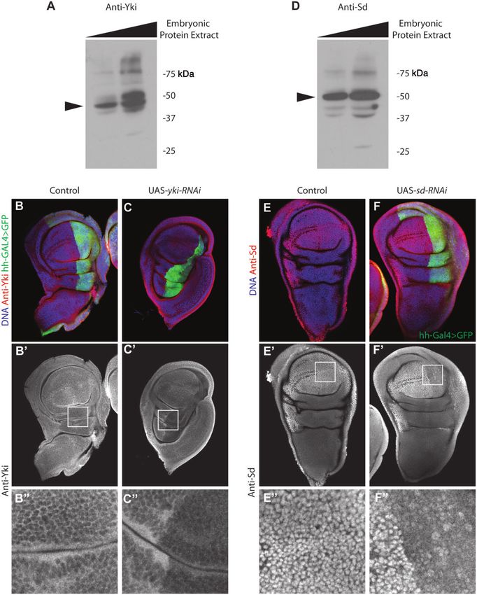
Ikmi et al. . doi:10.1093/molbev/msu071                                                                                   MBE
This raises the question of how and when the Yap-TBD                 et al. 2010) was conserved in Eumetazoa, this protein–protein
changed during evolution. Here, we compare the structure             interaction domain diverged in Amphimedon, exhibiting an
of the TBD from phylogenetically informative lineages includ-        additional a3 helix motif (fig. 1C and D). Beyond metazoans,
ing multiple early branching metazoan species, as well as the        the most conspicuous conserved motifs were the Yap WW
closest unicellular relatives of Metazoa. We then use a heter-       and coiled-coil domains (fig. 1A), indicating that this struc-
ologous expression assay to 1) directly compare the growth-          tural combination was found in the unicellular ancestors of
promoting activity of divergent Yap orthologs and 2) identify        animals. Intriguingly, we manually identified a1 and a2 helix
a downstream transcriptional profile induced by select vari-         motifs as a cryptic TBD in the N-terminus of Capsaspora Yap
ants in the Drosophila eye disc. Combined, these results dem-        (fig. 1C and D). This may represent a transitional state that
onstrate that the Yap-TEAD interaction interface became              existed before evolution of the complete a1-loop-a2 struc-

                                                                                                                                         Downloaded from http://mbe.oxfordjournals.org/ at Ernst Mayr Library of the Museum Comp Zoology, Harvard University on October 2, 2014
stabilized sometime after the divergence of sponges from             ture in Eumetazoa. In addition, a highly divergent Yap TBD
the eumetazoan common ancestor. In addition, coupled                 containing only the a1 helix motif was found in Monosiga (fig.
with Chip-seq validation of Yki/Scalloped binding sites, our         1D). Combined, these results suggest a remarkable structural
comparative analysis identifies multiple novel Yap/TEAD              plasticity of the TBD during early Yap evolution, followed by
targets in Drosophila while hinting at the existence of a con-       fixation of the a1-loop-a2 structure throughout Eumetazoa.
served bilaterian gene expression program downstream of
Yap/TEAD.                                                            Heterologous Functional Analysis of Yap/Yki
                                                                     Orthologs in Drosophila
Results
                                                                     We next set out to determine the functional implications of
Evolutionary Changes in Yap/Yki Protein Architecture                 the evolutionary changes in Yap protein architecture. Because
To determine the extent to which the structural features of          experimental tools are a limiting factor in most nonmodel
Yap are conserved between animals and their unicellular rel-         organisms, we took advantage of Drosophila genetics to
atives, we performed a detailed domain composition analysis          directly compare the activities of representative Yap ortho-
of Yap orthologs from species occupying key phylogenetic             logs in a uniform in vivo system. We first generated transgenic
positions (fig. 1A). In addition to Drosophila and Homo,             flies carrying inducible forms of yap from Monosiga,
these included the cnidarian Nematostella vectensis, the             Amphimedon, Trichoplax, and Nematostella, as well as induc-
placozoan Trichoplax adhaerens and the demosponge                    ible forms of Drosophila yki and Homo yap for experimental
Amphimedon queenslandica, which are modern representa-               controls. The phiC31-attP-attB site-specific integration tech-
tives of the earliest branching Metazoa (Putnam et al. 2007;         nique was employed to insert all transgenes into a specific
Srivastava et al. 2008, 2010). We also analyzed the domain           genomic site, ensuring identical transgene expression levels
structure of Yap orthologs from the genome of three                  (Groth et al. 2004; Bischof et al. 2007). Because Yap activity is
nonmetazoan species, the amoeba C. owczarzaki, and the               regulated by Lats/Warts phosphorylation, we also gener-
choanoflagellates Monosiga brevicollis and Salpingoeca rosetta       ated hypothetically nonphosphorylatable forms of
(King et al. 2008; Suga et al. 2013). Consistent with prior          each Yap ortholog by modifying the appropriate serine res-
findings (Sebe-Pedros et al. 2012), our phylogenetic analyses        idues to create constitutively active Yap variants (fig. 2A).
showed a well-supported monophyletic group that included             Although the mutated serine for each metazoan Yap
the known bilaterian Yap protein together with a single pu-          matched the position of the critical phosphorylation site of
tative Yap protein from each analyzed genome (supplemen-             Yki/Yap (Ser168/127), the only candidate residue within an
tary fig. S1, Supplementary Material online). This contrasts         optimal 14-3-3 binding motif for the Monosiga protein was
with vertebrate genomes, which contain a paralogous copy             Ser48.
of Yap (Taz), proposed to have originated from a gene dupli-             Yki/Yap misexpression is sufficient to induce tissue over-
cation event early in vertebrate evolution (Hilman and               growth in Drosophila (Huang et al. 2005; Dong et al. 2007). We
Gat 2011).                                                           therefore performed a heterologous overexpression assay to
   Interestingly, the most critical Lats/Warts phosphorylation       compare the activity of each Yap ortholog in the Drosophila
site in Yap (corresponding to Ser127 in Homo) was conserved          eye, using Glass Multiple Reporter-Gal4 (GMR-Gal4) to drive
in all metazoan species analyzed (fig. 1B). It was also present in   tissue-specific expression. Contrasting with the effects of
the Monosiga, Salpingoeca, and Capsaspora Yap proteins,              Monosiga Yap, the Amphimedon, Trichoplax, and
although the associated regulatory 14-3-3 binding motif was          Nematostella variants elicited distinct degrees of overgrowth
incomplete (fig. 1B). These observations are consistent with         (fig. 2A and B). Importantly, each of these orthologs exhibited
conservation of the Yap-Lats regulatory interaction through-         roughly equivalent protein stability in Drosophila S2 cells, in-
out animal evolution.                                                dicating that the observed phenotypic variability arose from
   As expected, the Yap orthologs we identified shared many          protein-intrinsic properties (fig. 2C). Trichoplax Yap produced
additional features. However, they also displayed some critical      an exceptionally severe overgrowth phenotype with enlarged,
structural variations. Most prominently, these included alter-       folded adult eyes. This effect was even further enhanced in
ations in the architecture of the TBD (fig. 1A and supplemen-        animals overexpressing Trichoplax YapS81A (fig. 2A and B). We
tary fig. S2, Supplementary Material online). Although the           also found evidence for phosphoregulation of Yap from
TBD a1-loop-a2 secondary structure (Chen et al. 2010; Li             Amphimedon and Nematostella; both showed similar

1376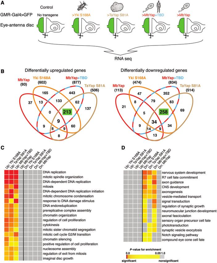
Molecular Evolution of the Yap/Yorkie Proto-Oncogene . doi:10.1093/molbev/msu071                                                         MBE

                                                                                                                                                         Downloaded from http://mbe.oxfordjournals.org/ at Ernst Mayr Library of the Museum Comp Zoology, Harvard University on October 2, 2014
FIG. 1. Domain architecture of evolutionary distant Yap orthologs. (A) A simplified metazoan phylogeny including the unicellular species Monosiga
brevicollis, Capsaspora owczarzaki, and Saccharomyces cerevisiae. Domain composition and protein size for each Yap ortholog are indicated. The
annotated domains include the TBD, which indicates a complete TEAD binding domain containing two short helices (a1 and a2) and a loop (L) in
between. This protein interaction domain is divergent in Amphimedon, Monosiga, and Capsaspora. Also indicated are the WW1 and WW2 domains
(WW), coiled-coil domains (C-C), and the PDZ binding motif. (B) The most critical phosphorylation site of Yap (red arrowhead) is conserved in all
indicated species. The HXRXXS motif associated with this site is incomplete in Monosiga and Capsaspora. (C) Alignments of the N-terminal regions of
Yap orthologs. Secondary structural elements are labeled as following: a1 helix (green), loop (red), and a2 helix (brown). The complete a1-loop-a2 TBD
is conserved from placozoans to humans. (D) Predicted 3D structures of five metazoan Yap TBDs as well as two nonmetazoan Yap TBDs. The Monosiga
TBD lacks both the loop and a2 helix, while the Capsaspora is missing only the loop. The Amphimedon TBD possesses an additional helix (a3) instead of
a loop. The loop-containing motif is shorter in Trichoplax (PXP) but longer in Nematostella (PXXXXP), when compared with that of Drosophila and
human (PXXP). X: any residue; : hydrophobic residue; P: proline.

increases in eye size that were enhanced by mutation of the                   phosphorylation of Drosophila Yki (supplementary fig. S3,
conserved phosphorylation sites (fig. 2A and B). Together,                    Supplementary Material online).
these results not only show that basal metazoan versions of                      Surprisingly, in contrast to the metazoan Yaps, overexpres-
Yap possess potent growth-promoting activity in Drosophila                    sion of Monosiga Yap or its phosphomutant form resulted in
but also that they are regulated by phosphorylation via similar               significantly reduced eye size (fig. 2A and B). Although the
mechanisms to those of Drosophila Yki and human Yap. This                     proximal cause of this reduced eye phenotype is still un-
implies that the Hippo/Wts cassette may function similar to                   known, cell clones overexpressing Monosiga Yap exhibited
phosphoregulate Yap in early branching metazoans.                             normal growth and morphology (data not shown). These
Consistent with this, we cloned a Warts/Lats ortholog from                    findings exclude the possibility that Monosiga Yap acts as a
Nematostella and found that it was sufficient to rescue the                   dominant negative to inhibit cell proliferation through effects
corresponding Drosophila mutant, presumably through                           on endogenous Yki activity.

                                                                                                                                                1377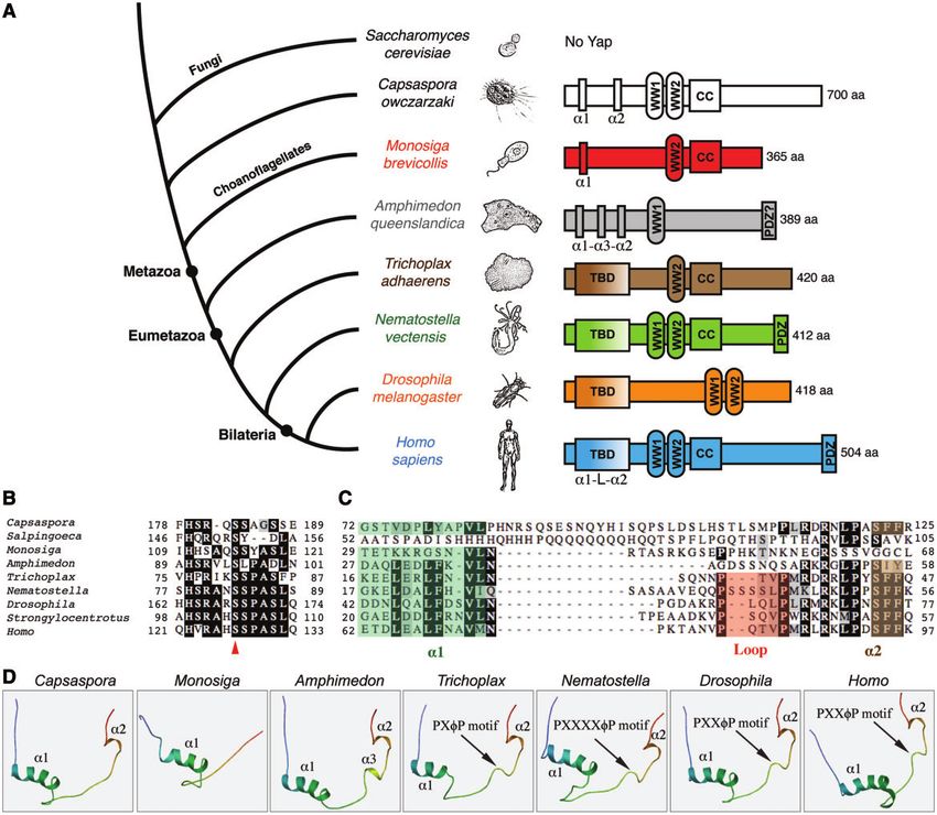
Ikmi et al. . doi:10.1093/molbev/msu071                                                                                                      MBE

                                                                                                                                                             Downloaded from http://mbe.oxfordjournals.org/ at Ernst Mayr Library of the Museum Comp Zoology, Harvard University on October 2, 2014
FIG. 2. In vivo functional analysis of Yap orthologs in Drosophila. (A) Representative adult female heads from flies overexpressing either wild-type (top
row) or phosphomutant (bottom row) forms of the indicated Yap orthologs under the control of GMR-Gal4. Control is GMR-Gal4/ + . Besides
differences in eye size, we also observed changes in eye pigmentation, although the flies carrying each UAS-transgene originally displayed a similar eye
color (inset boxes). (B) Quantification of adult eye widths for each overexpression condition. Statistical analysis was performed using a Student’s t-test
(n = number of analyzed adult eyes; *P < 0.05; **P < 0.01). (C) Constructs encoding C-terminal HA fusion proteins of each Yap ortholog were
transfected into Drosophila S2 cells and expressed under the control of a heat-shock promoter. After heat shock, cell lysates were collected at the
indicated times. Anti-HA western blots were used to show the protein levels of each Yap ortholog. Anti-a-tubulin was used as a loading control.

Cellular and Molecular Basis for Yap/Yki                                        human YapS127A (fig. 3A), resulting in extra bristles and inter-
Ortholog-Induced Overgrowth                                                     ommatidial cells (IOCs) in the adult eyes (fig. 3D and E).
To determine the cellular basis for eye size increases                          Consistent with a Yki-like transcriptional output of
induced by each Yap ortholog, we next performed EdU                             Trichoplax and Nematostella Yap, both induced expression of
incorporation assays to directly measure cell proliferation.                    Cyclin E and the cell death inhibitor diap1 (fig. 3B, C, and C0 ).
As expected from our analysis of adult eye size, Trichoplax                     These findings show that Nematostella and Trichoplax Yap are
YapS81A induced the most extensive cell proliferation in the                    able to induce cell proliferation and survival, behaving like their
GMR-Gal4 domain (fig. 3A). As a consequence of this dra-                        Drosophila and human counterparts.
matic overgrowth, the eye disc epithelium was extensively                          Contrasting with their eumetazoan counterparts,
folded and disorganized (fig. 3D and E). Nematostella                           Amphimedon YapS95A and Monosiga YapS48A did not
YapS83A induced significant but moderate ectopic cell pro-                      induce Cyclin E, diap1, or cell proliferation as indicated by
liferation when compared with Drosophila YkiS168A and                           EdU incorporation (fig. 3A, B, C, and C0 ). Further, overgrowth

1378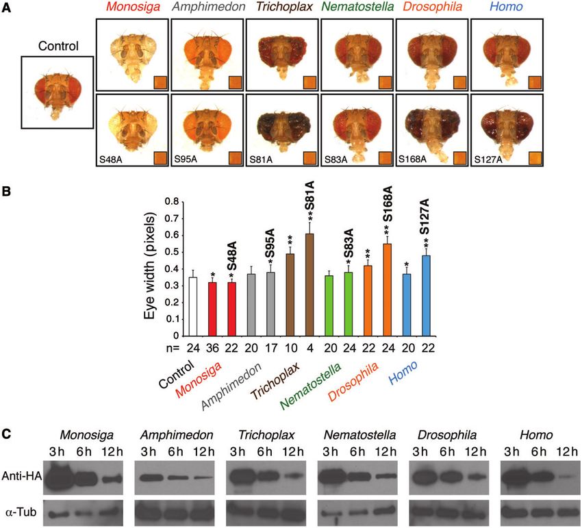
Molecular Evolution of the Yap/Yorkie Proto-Oncogene . doi:10.1093/molbev/msu071                                                   MBE

                                                                                                                                                   Downloaded from http://mbe.oxfordjournals.org/ at Ernst Mayr Library of the Museum Comp Zoology, Harvard University on October 2, 2014

FIG. 3. Cellular and molecular mechanisms underlying Yap ortholog-induced overgrowth in Drosophila. (A) Edu labeling in eye discs overexpressing
hypothetically nonphosphorylatable forms of Yap from the indicated species under the control of GMR-Gal4. Note the dramatic induction of
proliferation by Trichoplax yap and the absence of ectopic proliferation caused by the Monosiga and Amphimedon orthologs. (B)
Immunostaining of Cyclin E in eye discs of the same genotypes indicated above. Arrowheads indicate the position of the morphogenetic furrow.

                                                                                                                                    (continued)
                                                                                                                                           1379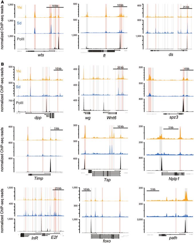
Ikmi et al. . doi:10.1093/molbev/msu071                                                                                                  MBE
was not induced following misexpression in other imaginal                     eumetazoan Yap proteins. Although the evolutionary advan-
discs (data not shown). Intriguingly, pupal retinae of GMR-                   tages of this modification remain unknown, we speculate that
Gal4>Amphimedon YapS95A animals exhibited an elevated                         it may have served as an important adaptation of Yap to
number of IOCs, which are normally eliminated by develop-                     critical new roles in the growth control in the multicellular
mentally programmed apoptosis (fig. 3D and F) (Wolff and                      context.
Ready 1991). Following expression of Amphimedon YapS95A,
these cells failed to undergo normal differentiation of the lens              The Transcriptional Output of Yap/Yki Orthologs in
cuticle and retained a pupal-like appearance in adult animals                 Drosophila
(fig. 3E). More pronounced defects in the differentiation and                 In vivo, Yap acts through modulation of target gene expres-

                                                                                                                                                         Downloaded from http://mbe.oxfordjournals.org/ at Ernst Mayr Library of the Museum Comp Zoology, Harvard University on October 2, 2014
patterning of ommatidial cells were observed in retinae over-                 sion (Overholtzer et al. 2006; Dong et al. 2007; Hao et al. 2008;
expressing Monosiga Yap, as the normal hexagonal topology                     Lu et al. 2010). To leverage the evolutionary diversity of se-
of ommatidial subunits was abolished (fig. 3D and E). These                   lected Yki/Yap orthologs and identify novel transcriptional
results suggest a defect in retinal differentiation and are con-              targets of the Hippo pathway in Drosophila, we performed
sistent with severe reductions of eye pigmentation observed                   RNA sequencing analysis (RNA-seq) on total RNA isolated
in flies overexpressing Monosiga and Amphimedon proteins                      from GMR-Gal4>Yap eye discs (fig. 4A and supplementary
(fig. 2A).                                                                    fig. S6A–E, Supplementary Material online). We first deter-
    The Amphimedon and Monosiga Yap proteins both pos-                        mined the endogenous targets of GMR-Gal4-driven
sessed an incomplete a1-loop-a2 TBD (fig. 1C), and thus their                 Drosophila Yki and then utilized the highly active
inability to drive proliferative growth was most likely due to a              Trichoplax Yap and Monosiga Yap containing the human
failure to recognize the endogenous Drosophila TEAD                           TBD (Yap+TBD) to define core target genes activated in
(Scalloped, Sd). To test this hypothesis, we first constructed                common. In addition, we used Monosiga Yap, which was
chimeric forms of Amphimedon and Monosiga Yap bearing                         not able to induce ectopic proliferation, as a negative control
the 50-amino acid human TBD. Strikingly, in both cases, this                  to remove the transcriptional background resulting from pro-
single change in protein architecture was sufficient to induce                tein overexpression. For each of these conditions, the tran-
extensive ectopic proliferation in the eye disc (fig. 3G and H).              scriptional profile was compared with that of a GMR-
Second, we found that Amphimedon Yap was able to form a                       Gal4>UAS-GFP control.
functional protein complex with Amphidedon TEAD in                                RNA-seq analysis identified a robust gene expression sig-
Drosophila S2 cells (supplementary fig. S4, Supplementary                     nature common to eumetazoan Yaps as well as the form of
Material online). In parallel, as expected from our in vivo                   Monosiga Yap+TBD (fig. 4B; supplementary table S1,
heterologous expression assay, we did not detect a physical                   Supplementary Material online). Validating our approach,
interaction between Amphimedon Yap and Drosophila Sd                          this signature comprised 213 commonly upregulated targets
(supplementary fig. S4, Supplementary Material online), de-                   that included almost all previously known Yki target genes
spite the fact that TEAD proteins exhibit few differences in                  (cyclin E [Tapon et al. 2002], expanded [Hamaratoglu et al.
their Yap-binding interfaces (supplementary fig. S5,                          2006], e2f1 [Goulev et al. 2008], wingless [Cho et al. 2006], dally
Supplementary Material online). These findings suggest a crit-                [Baena-Lopez et al. 2008], kibra [Genevet et al. 2010], and vein
ical functional divergence in Yap-TEAD pairwise interactions                  [Zhang, Ji, et al. 2009]), along with 258 commonly downreg-
during early animal evolution. Consistent with this scenario,                 ulated factors (fig. 4B). Interestingly, most of these genes were
Capsaspora Yap cannot induce tissue overgrowth in                             conserved in the Trichoplax and Monosiga genomes (supple-
Drosophila unless it is co-overexpressed with the endogenous                  mentary table S2, Supplementary Material online), perhaps
Capsaspora TEAD (Sebe-Pedros et al. 2012). In contrast to the                 representing an ancient Yap-dependent gene expression sig-
Amphimedon Yap, Nematostella Yap was able to physically                       nature. Among the 213 commonly upregulated targets, 16
interact with both Nematostella TEAD and Drosophila Sd                        genes were subsequently validated by real-time quantitative
(supplementary fig. S4A, Supplementary Material online).                      PCR analysis (supplementary fig. S7, Supplementary Material
Thus, these findings not only show the deep evolutionary                      online). Although we observed a highly significant overlap
origins of Yap-TEAD activity but also reveal that the interac-                between eumetazoan Yap target genes in Drosophila, only
tion interface between Yap and TEAD changed during early                      4% and 11% of these genes were correspondingly up- and
metazoan evolution and ultimately became fixed in                             downregulated by Monosiga Yap, respectively (fig. 4B). This

FIG. 3. Continued
Scale bar = 50 mm. (C-C0 ) diap1-lacZ expression (red) in eye discs overexpressing the corresponding Yap orthologs in clones (GFP+, green) and stained
with Hoechst (blue). Except for AqYapS95A and MbYapS48A, elevated diap1-lacZ expression was detected in all Yap ortholog-overexpressing clones
(yellow arrowheads). Scale bar = 50 mm. (D) Pupal retinae from the genotypes indicated above, stained with anti-Armadillo antibody to visualize cell
outlines at 42 h after puparium formation. Scale bar = 10 mm. (E) Corresponding scanning electron micrographs (SEM) of adult retinae.
(F) Quantification of IOCs per ommatidia in controls (w, GMR-Gal4/ + ; n = 20) and following expression of Amphimedon Yap
(GMR-Gal4>AqYapS95A; n = 20). Statistical analysis was performed using Student’s t-test (*P < 0.001). (G, H) Edu incorporation assay in eye discs
overexpressing chimeric constructs of Monosiga Yap (G) and Amphimedon Yap (H) with the Homo TBD. Addition of the human TBD to either variant
resulted in a strong capacity to induce proliferation.

1380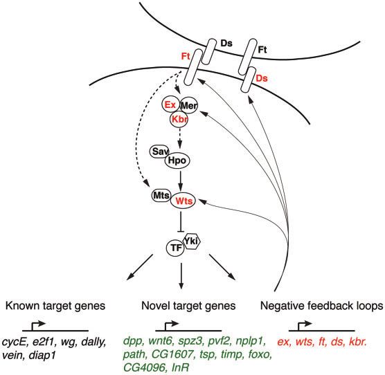
Molecular Evolution of the Yap/Yorkie Proto-Oncogene . doi:10.1093/molbev/msu071                                                       MBE

                                                                                                                                                       Downloaded from http://mbe.oxfordjournals.org/ at Ernst Mayr Library of the Museum Comp Zoology, Harvard University on October 2, 2014

FIG. 4. The transcriptional program downstream of Yap ortholog induction. (A) Experimental design for RNA-seq experiment. Drosophila Yki (YkiS168A),
Trichoplax Yap (TaYapS81A), chimeric Monosiga Yap with the human TBD (MbYap+TBD), and Monosiga Yap (MbYap) were co-overexpressed with
UAS-GFP using GMR-Gal4. Control is GMR-Gal4>UAS-GFP. For each experimental condition, total RNA was extracted from surgically dissected GMR
expression domains (GFP + ). (B) Four-way Venn diagrams of differentially upregulated (left) and downregulated (right) genes in each overexpression
condition. The number of genes up- and downregulated is indicated between brackets under each transgene name. The numbers of commonly up- and
downregulated genes in YkiS168A, TaYapS81A, and MbYap+TBD are indicated in green boxes. (C, D) Matrices of gene ontology of biological processes
analysis for genes upregulated (Up) and downregulated (Dn) in each overexpression condition.

                                                                                                                                              1381
Ikmi et al. . doi:10.1093/molbev/msu071 MBE
indicates that the transcriptional outputs of Yap with and for the Sd motif (supplementary fig. S10B, Supplementary
without the a1-loop-a2 TBD are fundamentally distinct. One Material online). Among the 1,238 genes bound by Sd, 97%
notable exception was that expanded (ex), a well-defined were also bound by Yki (supplementary fig. S10C and D,
target of Yki in Drosophila, was 1.4-fold upregulated following Supplementary Material online), and this high-confidence
Monosiga Yap expression compared with more than 3-fold subset included all previously known target genes (supple-
induction by metazoan Yap variants (supplementary table S1, mentary fig. S11, Supplementary Material online).
Supplementary Material online). To identify novel Yki target genes, we next focused on the
In agreement with the phenotypes induced by Yki/Yap overlap between our RNA-seq and ChIP-seq data sets (sup-
expression (figs. 2A and 3A), GO term analysis indicated plementary fig. S10C and D, Supplementary Material online).
that a significant number of commonly upregulated genes We found that Yki and Yki/Sd peaks were highly enriched
Downloaded from http://mbe.oxfordjournals.org/ at Ernst Mayr Library of the Museum Comp Zoology, Harvard University on October 2, 2014
were involved in DNA replication, cell cycle, and growth reg- near upregulated genes (the common set from figure 4B,
ulation processes (fig. 4C). Interestingly, we found that the P < 3 1016; Fisher test) but not near downregulated
average expression level of the top 100 commonly upregu- genes (P = 0.3 for Yki and P = 0.08 for Yki/Sd). Interestingly,
lated genes induced by Trichoplax Yap and Monosiga Yap+TBD the genes induced in common by different Yap orthologs
was higher than that induced by Drosophila Yki (supplemen- showed a higher enrichment of Yki and Yki/Sd peaks (78%
tary fig. S6F, Supplementary Material online). These quantita- and 32%, respectively) compared with the analysis of solely
tive differences in target gene expression level may account Yki-induced genes (64% for Yki peaks and 24% for Yki/Sd
for the more extensive cell proliferation induced by these Yap peaks). These results corroborate the predominant function
variants (fig. 3A). By contrast to the enrichment of upregu- of Yki/Yap as a transcriptional activator and led us to the
lated genes for cell proliferation processes, downregulated identification of several novel putative targets. Most promi-
genes were enriched for the process of nervous system devel- nently, the Hippo pathway components warts (wts), fat (ft),
opment (fig. 4D). This is consistent with the observed delays and dachsous (ds) were commonly upregulated targets and
in cell cycle exit and retinal determination/differentiation as a exhibited both Yki and Sd peaks, revealing the existence of
consequence of Yap expression in the eye (fig. 3D and E). novel negative feedback loops controlling Yki activity (fig. 6A).
Except in the case of Monosiga Yap+TBD, we did not identify Another class of putative direct target genes included the
significant Yap ortholog-specific enrichment for biological signaling ligands decapentaplegic (dpp), wnt6, spatzle 3
processes (supplementary fig. S8, Supplementary Material (spz3), vascular endothelial growth factor 2 (pvf2), thrombos-
online). pondin (tsp), tissue inhibitor of metalloprotease (Timp), and
neuropeptide-like precursor 1 (nplp1) (fig. 6B). Our analysis
Genome-Wide Distribution of Yki and Sd on also highlighted the amino acid transporter pathetic (path)
Chromatin and two insulin-signaling components as potential growth-
regulatory factors targeted by Yki/Sd activity (insulin receptor
In principle, the Yki/Yap dependent gene expression profiles (InR) and forkhead box protein O (foxo); fig. 6B). In addition to
described above could be the result of primary, secondary, or these Yki/Sd target genes, our ChIP-seq data clearly indicate
tertiary transcriptional events. To extend the results of our the existence of an Sd-independent downstream program
comparative studies and determine which induced genes that is most likely the result of Yki’s putative interactions
were most likely direct targets, we performed a ChIP-Seq ex- with other DNA-binding partners, such as Homothorax,
periment to identify the genome-wide occupancy of Teashirt, and Mad (Peng et al. 2009; Oh and Irvine 2011).
Drosophila Yki in proliferating cells. For chromatin immuno- Indeed, using a recent ChIP-Seq analysis of Homothorax in
precipitation, we generated a novel antibody that specifically eye discs (Slattery et al. 2013), we found that 40% of these Sd-
detects Yki protein (fig. 5A, B-B00 , and C-C00 ) and analyzed independent targets were cobound by Yki and Homothorax
dissected eye discs of the genotype GMR-Gal4>UAS- (supplementary table S3, Supplementary Material online).
YkiS168A. In parallel, we performed a ChIP-Seq experiment Together, these findings reveal a complex network of factors
using Polymerase II (Pol II) antibody as a control. Similar to downstream of Yap activation and directly link Hippo signal-
recent ChIP-Seq analyses of wild-type Yki in wing and eye ing to the regulatory architecture for a wide variety of pro-
discs (Oh et al. 2013; Slattery et al. 2013), Yki was enriched cesses required for tissue growth in vivo.
with high confidence at a large number of sites throughout
the genome (using Model-based Analysis for ChIP-Seq
[MACS], P < 0.001; supplementary fig. S9, Supplementary A Yap/Yki-Induced Transcriptional Program Shared by
Material online). Binding peaks were found in proximity to Drosophila and Vertebrates
5,732 genes (supplementary table S1, Supplementary Material In vertebrates, Yap-induced genes have been identified by
online) and a large number of them were localized within microarray profiling of hepatocytes, fibroblasts, and breast
promoter regions (supplementary fig. S10A, Supplementary epithelial cells (Overholtzer et al. 2006; Dong et al. 2007;
Material online). To focus our analysis on bona fide targets of Hao et al. 2008; Lu et al. 2010). To interpret our transcriptional
the Drosophila Yki/Sd complex, we also generated a novel profiling results at an evolutionary level and investigate the
antibody directed against Sd and performed ChIP-seq analysis extent to which Yap’s transcriptional output is conserved, we
under the same conditions (fig. 5D, E-E00 , and F-F00 ). The Sd took advantage of the OrthoDB catalog of eukaryotic ortho-
binding peaks were specific because they were highly enriched logs (Waterhouse et al. 2011) to test for orthologous
1382Molecular Evolution of the Yap/Yorkie Proto-Oncogene . doi:10.1093/molbev/msu071 MBE
Downloaded from http://mbe.oxfordjournals.org/ at Ernst Mayr Library of the Museum Comp Zoology, Harvard University on October 2, 2014
FIG. 5. Validation of anti-Yki and anti-Sd antibodies. (A) Western blot analysis shows that anti-Yki antibody detects a strong signal at the expected
molecular weight (arrowhead, 45 kDa). (B-B00 ) Control wing disc stained with Hoechst (blue) and anti-Yki (red). The posterior compartment is marked
by the expression of hh-Gal4>UAS-GFP (green). Anti-Yki detects ubiquitous expression of Yki. (C-C00 ) Wing disc overexpressing UAS-yki-RNAi under the
control of hh-Gal4. The clear reduction of anti-Yki staining in the posterior compartment (inset box) confirms that our antibody recognizes Drosophila
Yki. (D) Anti-Sd detects a specific band at the expected molecular weight (arrowhead, 50 kDa). (E-E00 ) Control wing disc stained with Hoechst (blue) and
anti-Sd (red). Endogenous sd expression is elevated in the wing pouch and margin, which is consistent with our Anti-Sd staining. (F-F00 ) A wing disc
expressing UAS-sd-RNAi under the control of hh-Gal4 shows a strong reduction of Sd staining in the posterior compartment (inset box), confirming the
specificity of Anti-Sd.
1383Ikmi et al. . doi:10.1093/molbev/msu071 MBE
Downloaded from http://mbe.oxfordjournals.org/ at Ernst Mayr Library of the Museum Comp Zoology, Harvard University on October 2, 2014
FIG. 6. Chromatin localization of Yki and Sd on novel target genes. Plots of Yki (orange), Sd (blue), and PolII (black) occupancy peaks in selected
commonly upregulated genes from the RNA-seq data. (A) Target genes belonging to novel negative feedback loops. (B) Novel target genes.
Transcriptional units of target genes are highlighted in black and their neighbor genes are in gray. Pink and gray bars indicate significant Yki/Sd
and Yki peaks, respectively.
relationships between genes induced by Yap in mouse liver Yap-induced genes from the mouse experiments were also
(Dong et al. 2007) and those in our Drosophila RNA-seq data. significantly upregulated in Drosophila (supplementary table
Strikingly, despite the cell type differences and the substantial S4, Supplementary Material online). At least 63 and 31 of
evolutionary distance between these two organisms, 74 these 74 genes show Yki/Yap binding in Drosophila and
1384Molecular Evolution of the Yap/Yorkie Proto-Oncogene . doi:10.1093/molbev/msu071 MBE
mouse, respectively (supplementary table S4, Supplementary Our results also define the Yap-TEAD interaction as a new
Material online; [Lian et al. 2010]). Although most of these model system for understanding the coevolution of protein
bilaterian Yap targets belonged to the core DNA replication complexes during the emergence of animal multicellularity.
and cell cycle machinery, we noted the following key con- Interestingly, a similar evolutionary scenario was recently de-
served targets: dpp/Bmp4, dally/Gpc3, wts/Lats2, fat/Fat4, scribed for another central growth regulator, the Myc-Max
foxo/Foxo3a, and Timp/Timp2. Consistent with this, Bmp4 complex. However, cross-species interactions between
was recently validated as a key transcriptional target of the Monosiga and human Myc and Max were not detected
Hippo pathway in mammary cells (Lai and Yang 2013). (Young et al. 2011). Thus, we propose that the structural
However, only 28 of these 74 common genes were upregu- changes in these protein complexes (Yap-TEAD and Myc-
lated following Warts loss of function in Drosophila, and only Max) may have served as critical adaptations for multicellular
Downloaded from http://mbe.oxfordjournals.org/ at Ernst Mayr Library of the Museum Comp Zoology, Harvard University on October 2, 2014
18 were induced following Mst1-2 loss of function in mouse growth control during their co-option into an ancient meta-
(supplementary table S4, Supplementary Material online; [Oh zoan gene regulatory architecture.
et al. 2013; Lu et al. 2010]). While allowing for substantial The presence of the most critical Warts/Lats phosphory-
context-specific transcriptional effects, our results show the lation site in all Yap orthologs (fig. 1B) suggests the conser-
potential utility of comparative methodologies and hint at vation of Lats-Yap phosphoregulation. Consistent with this,
the existence of an ancient growth-promoting gene expres- we observed an enhancement of Yap activity following the
sion profile downstream of Yap throughout Bilateria. modification of this critical site in the metazoan forms of Yap
(fig. 2A and B). In addition, it has been reported that the
Discussion activity of Capsaspora Yap is regulated through phosphory-
In a search for the origins of animal complexity, comparative lation, pushing back the origin of Lats-Yap phosphoregulation
genomic and evolutionary analyses have generated a wealth to the unicellular ancestors of animals (Sebe-Pedros et al.
of knowledge about the potential genetic toolkit of the meta- 2012). Because it is thought that the modern Hippo/Lats
zoan common ancestor (King 2004; Putnam et al. 2007; King pathway responds to extracellular cues to regulate tissue
et al. 2008; Srivastava et al. 2008, 2010). Generally, these stud- growth, it remains unclear what function this pathway may
ies emphasize the evolution of molecules that direct cell–cell have served in a hypothetical unicellular ancestor of animals.
adhesion, cell differentiation, and body axis formation One possibility is that the pathway regulated growth in multi-
(Nichols et al. 2006; Abedin and King 2008; Larroux et al. cellular aggregates or in response to local density of specific
2008; Richards et al. 2008; Sebe-Pedros et al. 2010; Holstein cell types. Analyses that examine the downstream transcrip-
et al. 2011). Here, we contribute a new perspective: that the tional output of Yap TEAD signaling in close metazoan rela-
development and diversification of complex animal body tives could shed light on this important issue.
plans must have also required the incorporation of new Beyond the evolutionary implications of these results, our
mechanisms to coordinately control the patterns of cell functional phylogenomic approach also provided mechanis-
growth, proliferation, and survival in a multicellular context. tic insights into the contemporary Hippo pathway itself.
We substantiate this view with a detailed functional analysis Although recent studies have analyzed the genome-wide oc-
of the evolution of a critical growth regulator, the Hippo cupancy of Yki during normal development (Oh et al. 2013;
pathway effector Yap, a transcriptional coactivator whose Slattery et al. 2013), here we used a combination of RNA-seq
activity is mediated by its evolutionary conserved DNA-bind- and ChIP-seq experiments to identify novel Yki and Sd target
ing partner TEAD. genes induced during cell proliferation. Indeed, we report the
Using Drosophila as a uniform in vivo experimental system, existence of a core gene expression signature underlying the
we compared the activity of representative Yap orthologs control of tissue growth in Drosophila (fig. 4B and supple-
from major animal lineages and their closest unicellular rela- mentary table S1, Supplementary Material online). This sig-
tives, thus providing insight into the evolutionary history of nature includes novel target genes that could mediate cross
Yap protein structure and function throughout 700 My of talk with key signaling pathways, as well as multiple feedback
evolution. Although directed studies will be required to test loops controlling Yap activity (fig. 7). Further experimental
the taxon-specific requirements for Yap in Metazoa and its analyses would be required to know when and where Yki
closest unicellular relatives, our results nevertheless provide regulates these novel targets during normal development.
clear experimental support for the ubiquity of the Yap-TEAD On a broader note, although it is widely accepted that the
complex as a key functional unit that possesses growth-pro- incredible diversity of animal development is directed by a
moting activity. Importantly, the variable effects of different limited number of conserved signaling pathways (Pires-
Yap orthologs in Drosophila could be attributed to numerous daSilva and Sommer 2003), it remains unclear whether
structural differences that could enhance or reduce their ac- these pathways act, at least partially, through conserved
tivity or regulation. downstream genetic programs. By comparing the transcrip-
As evidence for coevolution of the Yap-TEAD complex, we tional output of Yap in Drosophila and mouse, we identified a
report that the eumetazoan interaction interfaces are distinct conserved set of target genes commonly induced in these
from those in the premetazoan and sponge complexes evolutionarily distant species (supplementary table S4,
(fig. 1C). This suggests strong evolutionary constraints and Supplementary Material online). These targets may represent
highlights the importance of these transcriptional cofactors an ancestral gene-expression signature of Yap and constitute
as a building block for the evolution of animal growth control. candidate effectors of Yap-TEAD activity in other metazoans.
1385Ikmi et al. . doi:10.1093/molbev/msu071 MBE
the SWISS-MODEL automated comparative protein-model-
ing server (http://swissmodel.expasy.org, last accessed
February 23, 2014). Yap orthologs were aligned using
MUSCLE (Edgar 2004) with default settings. For phylogenetic
analyses, the alignment was manually curated to only retain
the two well-conserved WW domains. Neighbor-joining trees
were generated using Phylip with default settings and 10,000
bootstrap replicates. Maximum likelihood analyses were run
with 1,000 bootstrap replicates using PhyML with the Whelan
and Goldman (WAG) model of amino acid substitution, four
Downloaded from http://mbe.oxfordjournals.org/ at Ernst Mayr Library of the Museum Comp Zoology, Harvard University on October 2, 2014
substitution rate categories, and the proportion of invariable
sites estimated from the data set. Bayesian inference methods
were implemented using MrBayes v3.1.2 with a mixed amino
acid model prior and a variable rate prior.
Cloning
The full-length coding sequence of Nematostella Yap was
amplified from larval cDNA using the following primers:
Nematostella-YapF: 50 -TTC ACA ATG GAA AGG AAA AAC
FIG. 7. Novel downstream target genes of Yki in Drosophila. Schematic
representation of the Fat-Hippo pathway in Drosophila. In response to A-30 ; Nematostella-YapR: 50 -TCG GAC TAC AAC CAA GTT
dachsous (Ds) binding, fat (Ft) protocadherin activates the Hippo path- AAA AA-30 . The amplified cDNA was cloned into the
way, potentially through expanded and warts (dashed line). pCRTM4-TOPO vector (Invitrogen). The coding sequences
The Expanded-Merlin-Kibra complex (Ex-Mer-Kbr) also activates the of Yap from Monosiga, Amphimedon, Trichoplax,
kinase cascade leading from the Hippo/Warts (Hpo/Wts) kinases and Drosophila, and Human were synthesized by GenScript
their scaffolding proteins Salvador (Sav) and Mod as tumor suppressor (Piscataway, NJ). Amphimedon Yap was also cloned from
(Mats) to Yki and its transcriptional cofactors (TF). Listed below are larval cDNA into the pCRTM4-TOPO vector using the fol-
examples of putative target genes that met the dual criteria of Yki/Sd lowing primers: Amphimedon-YapF: 50 -ATG ACT GAT ATT
chromatin association and were upregulated in common following ATC AAT ACG AAT TCC CCT TCC-30 ; Amphimedon-YapR:
YkiS168A, TaYapS81A, and MbYap+TBD overexpression. Yap ortholog-
50 -CAC CCA AGT ATT ACT ACC AAA CAT TCC-30 . To gen-
induced genes were divided into three classes: known target genes,
novel target genes (green), and candidate negative feedback loop
erate the hypothetically nonphosphorylatable form of each
components (red). protein, serine to alanine mutations were introduced by
primer-mediated site-directed mutagenesis. Chimeric con-
structs of Monosiga Yap and Amphimedon Yap with the
Additional analyses would be required to characterize these
human TBD were synthesized by GenScript. All GenScript
potentially conserved targets in depth. Combined, these re-
constructs were cloned into the pUC57 plasmid EcoRV site.
sults illustrate the power of comparative studies to provide
For phiC31-mediated site-specific transformation, all con-
both evolutionary and mechanistic insight into fundamental
structs were cloned into the pUAST-attB vector using BglII-
biological processes.
NotI or NotI-XbaI sites. Nematostella warts (Nvwarts) was
amplified from larval cDNA and cloned into the pCRTM4-
Materials and Methods TOPO vector using the following primers: Nematostella wartF:
Bioinformatics 50 -TGG CCC TCA ACA TAC CAA GGA GTA AG-30 ;
We identified Yap genes using the basic alignment sequence Nematostella wartR: 50 -AAG AAT GCA TGT TCT GGA
tool (Blast: TBlastN, TBlastX, and BlastP) with human CGA TGG TT-30 . We then cloned Nvwarts into NotI-digested
Yap as a query. The genomes of N. vectensis, T. adhaerens, pCaSpeR-hs.
A. Queenslandica, and M. brevicollis are available in http://
genome.jgi-psf.org/Nemve1, http://genome.jgi-psf.org/Triad1 Protein Stability of Yap/Yki Orthologs
(last accessed February 23, 2014) (spongezome.metazo- To generate C-terminal human influenza hemagglutinin
me.net), and http://genome.jgi-psf.org/Monbr1 (last accessed (HA) fusion proteins of Yap orthologs, the coding sequences
February 23, 2014). Capsaspora owczarzaki and S. rosetta of each protein were cloned in frame with the HA coding
genome assemblies were examined on the Broad Institute sequences in pHWH (Drosophila Genomics Resource Center)
web site (http://www.broadinstitute.org/, last accessed using Gateway cloning (Invitrogen). Yap-HA constructs were
February 23, 2014). Reciprocal best Blast searches and protein transfected into Drosophila S2 cells using Effectene
domain structure analyses (Pfam: http://pfam.sanger.ac.uk/ Transfection Reagent Kit (Qiagen) following the manufac-
search [last accessed February 23, 2014] and SMART: http:// turer’s instructions. Transfected S2 cells were incubated for
smart.embl-heidelberg.de [last accessed February 23, 2014] 24 h and heat shocked for 1 h to induce Yap-HA expression.
were used to screen for positive hits. To identify the TBD, Cell lysates were collected 3, 6, and 12 h postheat shock and
we conducted a protein structure homology analysis using analyzed by Western blotting with Anti-HA (Sigma-Aldrich,
1386Molecular Evolution of the Yap/Yorkie Proto-Oncogene . doi:10.1093/molbev/msu071 MBE
1:4,000) and anti-a-tubulin (Sigma-Aldrich, 1:500) as a loading Generation of Yki and Sd antibodies
control. Custom-made polyclonal rabbit anti-Yki and anti-Sd antibo-
dies were generated and affinity purified by GenScript. They
Coimmunoprecipitation were raised against the N-terminal 243 amino acids of Yki and
the full-length protein of Sd. For Western blots, anti-Yki and
We generated N-terminal HA and C-terminal Flag fusion pro- Anti-Sd dilutions were 1:2,000 and 1:4,000, respectively.
teins of TEAD and Yap, respectively. The full coding se-
quences of TEAD/Sd from Amphimedon, Nematostella, and Immunocytochemistry
Drosophila were amplified from their corresponding adult
Imaginal discs and pupal retina were fixed and processed
cDNAs and cloned in frame with the HA coding sequences
Downloaded from http://mbe.oxfordjournals.org/ at Ernst Mayr Library of the Museum Comp Zoology, Harvard University on October 2, 2014
according to standard protocols. Primary antibodies used
in pAHW (Drosophila Genomics Resource Center) using the
were anti-Cyclin E (Santa Cruz Biotechnology, 1:500), anti-
Gibson assembly kit (NEB). Using the same approach, we also
b-galactosidase (Sigma-Aldrich, 1:1,000), anti-Armadillo
cloned the coding sequences of the phosphomutant forms of
(Developmental Studies Hybridoma Bank, 1:400), anti-Yki
Yap/Yki in frame with the coding sequence of Flag in pAWF
(1:1,000), and anti-Sd (1:1,000).
(Drosophila Genomics Resource Center). Drosophila S2 cells
were transiently transfected with these constructs as indi-
RNA Extraction, Library Preparation, Sequencing, and
cated above. After 3 days, cells were lysed (lysate buffer:
50 mM Tris pH7.4, 150 mM NaCl, 5 mM MgCl2, 5% glycerol,
RNA-Seq Analysis
0.5% Triton X-100, and 1 protease inhibitor cocktail Total RNA was recovered from surgically isolated GMR ex-
[Roche]) and centrifuged at 16,000 g for 10 min. pression domains (GFP+) from 25 to 30 third instar eye discs
Coimmunoprecipitations were performed using Dynabeads using a RNeasy kit (Qiagen). For each genotype, total RNA
Protein G immunoprecipitation kit (Life Technologies) fol- extraction was conducted in triplicate. Total RNA (400 ng)
lowing the manufacturer’s instructions. was enriched for poly(A) + RNA by oligo(dT) selection. The
Poly(A) + RNA was then fragmented, and first-strand cDNA
synthesis was performed using random hexamer priming in
Drosophila Stocks the Stowers Institute Molecular Biology Core facility, where all
All transgenic flies carrying UAS-attB transgenes were created subsequent steps were conducted. Following second-strand
by phiC31-mediated site-specific transformation using the synthesis, the ends were cleaned up, a nontemplated 30
attP2 site (Groth et al. 2004; Bischof et al. 2007). These trans- adenosine was added, and Illumina indexed adapters were
genes were overexpressed using either GMR-Gal4 or GMR- ligated to the ends. The libraries were enriched by 15
Gal4 with UAS-EGFP. The expression of diap1 was monitored rounds of polymerase chain reaction (PCR). The purified li-
using thi5c8 (diap1-lacZ) (Hay et al. 1995). To test the specifi- braries were quantified with the high-sensitivity DNA assay
city of anti-Yki and anti-Sd antibodies in vivo, UAS-yki-RNAi on an Agilent 2100 Bioanalyzer. Equal molarities of individual
(Bloomington, 34067) and UAS-sd-RNAi (Bloomington, libraries were pooled together (five libraries per pool) for
35481) were overexpressed using hh-Gal4. For the multiplex sequencing. Pooled libraries were sent to Tufts
Drosophila warts rescue experiment, we crossed yw, eyeless- (TUCF) for single read sequencing (50 nt) on a HiSeq 2000,
FLP; FRT82B/TM6 Tb (Bloomington, 5620) to w, hs-Nvwarts/ and fastq files were returned.
hs-Nvwarts; wtsX1 FRT82B/TM6 Sb Tb. Expression of hs- For analysis, sequence reads in fastq format were mapped
Nvwarts was induced by heat shock for 1 h every day from to the Drosophila genome using tophat-1.0.14 (Trapnell et al.
the second instar larval stage until eclosion. wtsX1 is described 2009) against Flybase 5.29 (dm3 compatible). Flybase tran-
in Xu et al. (1995). scripts (v5.29) were quantified and compared between con-
trol and each overexpression condition using cuffdiff-1.0.2
(Trapnell et al. 2010). Genes with adjusted P values of less
Eye Width Measurement than 0.05 were selected for functional annotation based on
For measuring adult eyes, flies were decapitated using a sharp Gene Ontology (GO Consortium 2006). Enrichment analysis
razor blade. The heads were imaged using a Leica MZ 16F was performed using Gitools (http://www.gitools.org, last
microscope. To determine eye width, two parallel lines were accessed February 23, 2014) to identify biological processes
drawn at the edges of each eye. The distance separating these that were enriched among up- or downregulated genes.
two lines was measured using ImageJ.
Quantitative Real-Time PCR
To independently validate the RNA-seq results, total RNA
Edu Incorporation was isolated as described above. Five independent RNA ex-
EdU incorporation was detected using the Click-iT Alexa tractions were performed for each genotype. Following cDNA
Fluor 488 imaging kit (Invitrogen). Third instar eye imaginal generation, each real-time quantitative PCR (qPCR) reaction
discs were incubated for 30 min with 300 mM EdU in Ringer’s contained 0.33 ng of cDNA and a PCR master mix including
solution. After fixation, samples were stored for at least 1 h in 0.5 mM of each primer and 1 PerfeCTa SYBR Green FastMix
100% methanol at 20 C. All subsequent steps were con- from Quanta Biosciences (cat. no. 95072-250) in 10 ml total
ducted according to the manufacturer’s protocol. reaction using a CAS-4200 qPCR loading robot from Corbett
1387Ikmi et al. . doi:10.1093/molbev/msu071 MBE
Life Science. qPCR reactions were performed in 384-well instructions. Concentration and size distribution of the librar-
format on a 7900HT Real-Time PCR Detection System from ies were assessed on an Agilent 2100 Bioanalyzer (high-sensi-
Applied Biosystems. Results were analyzed using qBase Plus tivity DNA assay chip). Sequencing was performed on an
software from Biogazelle. actin, GAPDH, and tbp genes were Illumina HiSeq2500 instrument, with 50 bp single reads in
used as endogenous controls and the calibrated normalized the high-output mode.
relative quantity (CNRQ) values were calculated for each All sequence reads were filtered to include only those
tested gene. Primers are available on request. passing the standard Illumina quality filter and then aligned
to the Drosophila melanogaster reference genome (UCSC
ChIP-Seq dm3 release) using Bowtie version 0.12.9, with the following
parameters: -v 2 -k 1 -m 3 –best –strata. Peaks were called
Downloaded from http://mbe.oxfordjournals.org/ at Ernst Mayr Library of the Museum Comp Zoology, Harvard University on October 2, 2014
Chromatin immunoprecipitation from eye discs was per-
with MACS v2.0.10.20120913 (Zhang, Liu, et al. 2008), using an
formed using a modified protocol from Gaertner et al.
adjusted P value of 0.001 and 0.01 for Yki and Sd, respectively.
(2012). First, third instar larvae were dissected in phosphate
To assign a peak to its nearest gene, the following criteria were
buffered saline (PBS) (pH 7.4) such that only eye discs and
used: If the peak overlapped a gene, it was assigned to that
brain remained attached to the mouth hooks. The dissected
gene regardless of where the overlap occurred; otherwise, it
material was subsequently fixed in 1 ml fixation buffer
was assigned to the gene if the peak summit occurred within
(50 mM 4-(2-hydroxyethyl)-1-piperazineethanesulfonic acid
1,500 bp upstream of the transcription start site.
[HEPES], pH 7.5; 1 mM ethylenediaminetetraacetic acid ChIP-seq and RNA-seq data are available under Gene
[EDTA]; 0.5 mM ethylene glycol tetraacetic acid [EGTA]; Expression Omnibus (GEO) accession number GSE54603.
100 mM NaCl; 2% formaldehyde) for 30 min at room tem-
perature. After four washes (PBS, pH 7.4; 0.1% Triton X-100;
0.1% Tween-20), eye discs were hand-dissected and combined Supplementary Material
into pools of 200 discs in 300 ml buffer A2 (15 mM HEPES, pH Supplementary figures S1–S11 and tables S1–S4 are available
7.5; 140 mM NaCl; 1 mM EDTA; 0.5 mM EGTA; 1% Triton at Molecular Biology and Evolution online (http://www.mbe.
X-100; 0.1% sodium deoxycholate; 0.1 % sodium sodecyl sul- oxfordjournals.org/).
fate [SDS]; 0.5 % N-lauroylsarcosine; 1 Roche complete
protease inhibitor cocktail, cat. no. 5056489001). Sonication
was performed in a Bioruptor sonicator for 30 min (30 s on/ Acknowledgments
off cycle at the “high” setting). Following centrifugation The authors thank B. Degnan, L. Grice, C. Conaco and K. Kosik
(16,000 g; 10 min at 4 C), the supernatant containing sol- for providing the Amphimedon cDNA; A. Peak and K.
uble chromatin was transferred to fresh tubes, and 50 ml was Zueckert-Gaudenz for RNA-seq library preparation and se-
set aside as whole cell extract (WCE; input). quencing; K. Walton and A. Perera for ChIP-seq library se-
Per ChIP, 10 mg antibodies were added to 450 ml chromatin quencing; B. Fleharty and W. McDowell for real-time
(corresponding to approximately 300 discs) and incubated quantitative PCR analysis; J. Johnston for assistance with bio-
overnight at 4 C with rotation. We used the following anti- informatic analysis; T. Akiyama for his suggestions on the co-
bodies: anti-Pol II (Covance 8WG16, cat. no. MMS-126R; immunoprecipitation experiments; and L. Gutchewsky for
mouse monoclonal antibody), anti-Sd, and anti-Yki. administrative support. Financial support was provided by
Immunocomplexes were purified by adding 50 ml prewashed the Stowers Institute for Medical Research and from a
Dynabeads coated with protein A/protein G (Life Burroughs Wellcome Fund Career Award in Biomedical
Technologies, cat. no. 10002D and 10004D) for 4 h, rotating Sciences to M.C.G.
at 4 C. The beads were washed three times in radioimmu-
noprecipitation assay buffer (RIPA) buffer (50 mM HEPES, pH
7.5; 1 mM EDTA; 0.7% sodium deoxycholate; 1% NP-40; References
500 mM LiCl) and once in TE. Immunoprecipitated DNA Abedin M, King N. 2008. The premetazoan ancestry of cadherins. Science
was eluted twice in 75 ml elution buffer (50 mM Tris, pH 319:946–948.
Baena-Lopez LA, Rodriguez I, Baonza A. 2008. The tumor suppressor
8.0; 10 mM EDTA; 1% SDS) at 65 C to maximize yields. genes dachsous and fat modulate different signalling pathways by
Crosslinks of ChIP and WCE DNA were reversed overnight regulating dally and dally-like. Pro Natl Acad Sci U S A. 105:
at 65 C. DNA was purified by RNAse A (Sigma, cat. no. 9645–9650.
R6513; [0.2 mg/ml]; 1 h at 37 C) and proteinase K (Life Bischof J, Maeda RK, Hediger M, Karch F, Basler K. 2007. An optimized
Technologies, cat. no. AM2546; [0.2 mg/ml]; 2 h at 55 C) treat- transgenesis system for Drosophila using germ-line-specific phiC31
integrases. Proc Natl Acad Sci U S A. 104:3312–3317.
ment followed by phenol/phenol–chloroform–isoamylalco- Camargo FD, Gokhale S, Johnnidis JB, Fu D, Bell GW, Jaenisch R,
hol extractions and ethanol precipitation. The precipitated Brummelkamp TR. 2007. YAP1 increases organ size and expands
DNA was resuspended in 30 ml 10 mM Tris buffer (pH 8). undifferentiated progenitor cells. Curr Biol. 17:2054–2060.
For ChIP-seq library preparation, 30 ml ChIP DNA and Chen L, Chan SW, Zhang X, Walsh M, Lim CJ, Hong W, Song H. 2010.
100 ng WCE DNA were used to construct ChIP-Seq libraries Structural basis of YAP recognition by TEAD4 in the Hippo pathway.
Genes Dev. 24:290–300.
with the NEBNext ChIP-seq Library Prep Master Mix Set (cat Cho E, Feng Y, Rauskolb C, Maitra S, Fehon R, Irvine KD. 2006.
no. E6200L) and the NEBNext Multiplex Oligos (cat. no. Delineation of a fat tumor suppressor pathway. Nat Genet. 38:
E7335S and E7500S) for Illumina, following the manufacturer’s 1142–1150.
1388You can also read