Cassiosomes are stinging-cell structures in the mucus of the upside-down jellyfish Cassiopea xamachana - Nature
←
→
Page content transcription
If your browser does not render page correctly, please read the page content below
ARTICLE
                  https://doi.org/10.1038/s42003-020-0777-8                OPEN

                  Cassiosomes are stinging-cell structures in the
                  mucus of the upside-down jellyfish Cassiopea
                  xamachana
1234567890():,;

                  Cheryl L. Ames1,2,3,12*, Anna M.L. Klompen 3,4,12, Krishna Badhiwala5, Kade Muffett3,6, Abigail J. Reft3,7,
                  Mehr Kumar3,8, Jennie D. Janssen 3,9, Janna N. Schultzhaus1, Lauren D. Field1, Megan E. Muroski1,
                  Nick Bezio3,10, Jacob T. Robinson5, Dagmar H. Leary11, Paulyn Cartwright4, Allen G. Collins 3,7 &
                  Gary J. Vora11*

                  Snorkelers in mangrove forest waters inhabited by the upside-down jellyfish Cassiopea
                  xamachana report discomfort due to a sensation known as stinging water, the cause of which
                  is unknown. Using a combination of histology, microscopy, microfluidics, videography,
                  molecular biology, and mass spectrometry-based proteomics, we describe C. xamachana
                  stinging-cell structures that we term cassiosomes. These structures are released within
                  C. xamachana mucus and are capable of killing prey. Cassiosomes consist of an outer epi-
                  thelial layer mainly composed of nematocytes surrounding a core filled by endosymbiotic
                  dinoflagellates hosted within amoebocytes and presumptive mesoglea. Furthermore, we
                  report cassiosome structures in four additional jellyfish species in the same taxonomic group
                  as C. xamachana (Class Scyphozoa; Order Rhizostomeae), categorized as either motile
                  (ciliated) or nonmotile types. This inaugural study provides a qualitative assessment of the
                  stinging contents of C. xamachana mucus and implicates mucus containing cassiosomes and
                  free intact nematocytes as the cause of stinging water.

                  1 National Academy of Sciences, National Research Council, Postdoctoral Research Associate, US Naval Research Laboratory, Washington, DC 20375, USA.
                  2 Graduate   School of Agricultural Science, Tohoku University, Sendai 980-8572, Japan. 3 Department of Invertebrate Zoology, National Museum of Natural
                  History, Smithsonian Institution, Washington, DC 20560, USA. 4 Department of Ecology and Evolutionary Biology, University of Kansas, Lawrence, KS
                  66049, USA. 5 Department of Bioengineering, Rice University, Houston, TX 77005, USA. 6 Texas A&M University at Galveston, Galveston, TX 77553, USA.
                  7 National Systematics Laboratory of the National Oceanic Atmospheric Administration Fisheries Service, National Museum of Natural History, Smithsonian

                  Institution, Washington, DC 20560, USA. 8 Stanford University, Stanford, CA 94305-2004, USA. 9 National Aquarium, Baltimore, MD 21202, USA.
                  10 California State University, Monterey Bay, CA 93955, USA. 11 Center for Bio/Molecular Science and Engineering, US Naval Research Laboratory,

                  Washington, DC 20375, USA. 12These authors contributed equally: Cheryl L. Ames, Anna M. L. Klompen. *email: ames.cheryl.lynn.a1@tohoku.ac.jp;
                  gary.vora@nrl.navy.mil

                  COMMUNICATIONS BIOLOGY | (2020)3:67 | https://doi.org/10.1038/s42003-020-0777-8 | www.nature.com/commsbio                                             1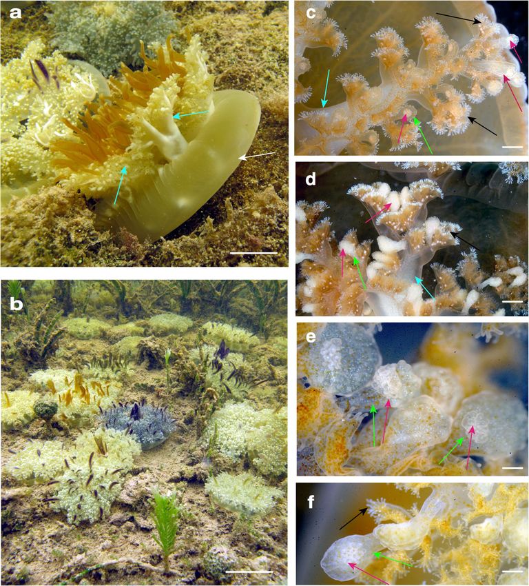
ARTICLE                                                              COMMUNICATIONS BIOLOGY | https://doi.org/10.1038/s42003-020-0777-8

J
     ellyfish, along with corals, anemones, hydroids, and myx-              that released upon contact, to which he attributed a role in
     ozoans, belong to the phylum Cnidaria, the earliest diverging         defense. The sum of these reports suggests that an investigation of
     venomous animal lineage1,2. These diploblastic animals have           the contents of Cassiopea mucus is needed to test the hypothesis
two so-called epithelial layers, outer ectoderm and inner endo-            that undeployed nematocysts and/or another nematocyst-bearing
derm, separated by a gelatinous extracellular matrix called                structure(s) present within the mucus of the upside-down jellyfish
mesoglea3,4. Despite their seemingly simple morphology, cni-               together are responsible for the phenomenon of stinging water
darians have adapted globally to most saltwater habitats and some          experienced by humans in the vicinity of Cassiopea medusae.
freshwater environments1,5. As such, cnidarians have evolved a                In this study, we used a combination of microscopy, micro-
remarkable envenomation mechanism that involves the deploy-                fluidic devices, molecular biology techniques, mass spectrometry-
ment of subcellular stinging capsules called nematocysts from              based proteomics, and other experimental assays to provide the
cnidarian-specific cells called nematocytes, which vary in size,            first detailed description, to our knowledge, of the contents of the
morphology, and bioactive contents6–8. Sea anemones possess                mucus liberated from lab-reared Cassiopea xamachana medusae.
unique nematocyte-rich structures (e.g., acrorhagi, acontia)9,10           Released within the mucus, we discovered three types of unde-
and employ strategies such as tentacle and column contraction              ployed nematocysts, as well as microscopic, motile, cellular
and expansion to enhance nematocyst deployment for prey                    masses composed of nematocytes that we formally call cassio-
capture and protection, while in medusae (i.e., jellyfish) the first         somes. While cassiosomes bear some resemblance to another
line of defense is their extendable nematocyte-laden tentacles that        cnidarian structure originating in mesenteries of the starlet sea
envenomate prey and predators they encounter in the water                  anemone Nematostella called nematosomes31, the unique traits of
column1, as well as humans participating in marine recreation. In          cassiosomes in C. xamachana include their release into the water
addition to direct stings caused by jellyfish, indirect stings have         column within mucus, the ability to trap and kill prey as mobile
also been reported. Some possible explanations for indirect jel-           grenades outside of the medusa, their organization as an outer
lyfish stings are contact with tentacle fragments in the water (e.g.,       epithelial layer surrounding a mostly empty core (rather than a
jellyfish stings in offshore fishers11), envenomation by juvenile            solid ball of cells), and the presence of centrally-located endo-
venomous jellyfish (e.g., Irukandji-like syndrome in United States          symbiotic Symbiodinium dinoflagellates. We document the pre-
Military combat divers12) or Sea Bathers Eruption caused by                sence of cassiosomes in five species spanning four families of the
microscopic jellyfish life forms (e.g., Linuche unguiculata13).             order Rhizostomeae, while also confirming their absence in the
   Another indirect stinging mechanism is through mucus, such              moon jellyfish Aurelia (Semaeostomeae), a representative of the
as in medusae of the upside-down mangrove jellyfish Cassiopea               sister lineage, and discuss the possibility of a single evolutionary
xamachana Bigelow 1892 (Class Scyphozoa; Order Rhizosto-                   event behind this envenomation strategy which, to our knowl-
meae), an emerging cnidarian model for its relevance to the study          edge, is unique. Despite the growing body of work on C. xama-
of coevolution as well as symbiosis-driven development (reviewed           chana from an organismal biology perspective14,26, this study is
in ref. 14). Additionally, the ubiquity of Cassiopea medusae in            the first to directly investigate stinging properties of the mucus of
healthy mangroves has earned the upside-down jellyfish status as            this jellyfish and the potential ecological and evolutionary
a potential bioindicator species for coastal management and                relevance.
conservation efforts15,16. Cassiopea is known to release large
amounts of mucus into the water column17–24, which has been
                                                                           Results
referred to as toxic mucus due to reports of nematocysts found
                                                                           Study overview. Observations were made on lab-reared Cassiopea
freely suspended in the vicious substance17,20,25. For instance,
                                                                           xamachana (see the “Methods” section) and on medusae in their
Cassiopea mucus is known to kill certain species of fish on con-
                                                                           natural habitat in waters of Florida Keys mangrove forests
tact24. Cassiopea is an exception to the iconic image of a jellyfish
                                                                           (Fig. 1a, b). In both cases, medusae were observed releasing
in that it lacks marginal tentacles and, instead of swimming in the
                                                                           copious amounts of mucus into the water when surrounding
water column, lies apex-down on the substrate in mangrove
                                                                           water was disturbed (by jellyfish aquarists and/or snorkelers), or
forests, seagrass beds or other coastal waters with its relatively
                                                                           when prey items were provided (e.g., Artemia nauplii in
short oral arms facing upward (reviewed in ref. 26). Despite this
                                                                           aquarium-reared medusae). Stinging water phenomenon was
benthic lifestyle, warnings have been published alerting sea
                                                                           experienced by the authors while handling lab-reared and/or wild
bathers of the stinging water or toxic mucus phenomena blamed
                                                                           C. xamachana and other rhizostome jellyfish examined in this
on unidentified potent little grenades in the water column sur-
                                                                           study (species list provided below).
rounding Cassiopea medusae20,21. In general, Cassiopea stings are
categorized as mild to moderate in humans, but crude venom
extracted from the nematocysts displays hemolytic, cardiotoxic             Cassiopea xamachana overview. Life cycle and endosymbiosis
and dermonecrotic properties27–30, suggesting that excessive               C. xamachana medusae start out like most scyphozoan jellyfish,
exposure may be detrimental for humans.                                    as an asexual microscopic polyp that metamorphosizes into a
   During the course of this study, a review of the old literature on      sexually reproducing medusa via a process known as strobila-
Cassiopea revealed a probable explanation for the grenades                 tion19. However, they differ from most jellyfish in that they host
reported in stinging water. First, Perkins17 discovered in the             endosymbiotic dinoflagellates (also called zooxanthellae)26,32.
mucus of Cassiopea undeployed nematocysts and ciliated innu-               Colonization of polyps by algal endosymbionts is the most
merable minute spherical bodies, the latter of which were dis-             common type of intracellular mutualism among cnidarians of the
missed as non-coelenterate (i.e., non-cnidarian) in nature. Next, a        class Anthozoa (e.g., corals and anemones), and although it is
brief description was published by Smith40 of peculiar structures          less common in jellyfish species33, endosymbiosis triggers the
found in the oral vesicles (i.e., vesicular appendages) of the oral        start of C. xamachana polyp strobilation18,19. During the sessile
arms of C. xamachana and conspecific C. frondosa medusae that               life stage, these polyps engulf dinoflagellates (unicellular algae
were ‘shot’ at prey, which he dubbed small bags of mesoglea and            called Symbiodinium) via the manubrium (feeding tube), which
nematocysts and suggested might play a role in predation. Finally,         are then phagocytosed by endodermal cells18,33. Bound by a
Larson43 reported polygonal-shaped bodies on the flatted sides of           membrane complex that combines host and infecting cell mem-
the oral vesicles (i.e., vesicular appendages) of the oral arms of C.      branes (called a symbiosome33), Symbiodinium spp. migrate to
xamachana and C. frondosa corresponding to nematocyst clusters             the polyp mesoglea and remain there housed in endodermal cells,

2                         COMMUNICATIONS BIOLOGY | (2020)3:67 | https://doi.org/10.1038/s42003-020-0777-8 | www.nature.com/commsbio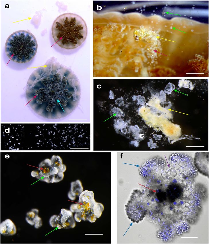
COMMUNICATIONS BIOLOGY | https://doi.org/10.1038/s42003-020-0777-8 ARTICLE
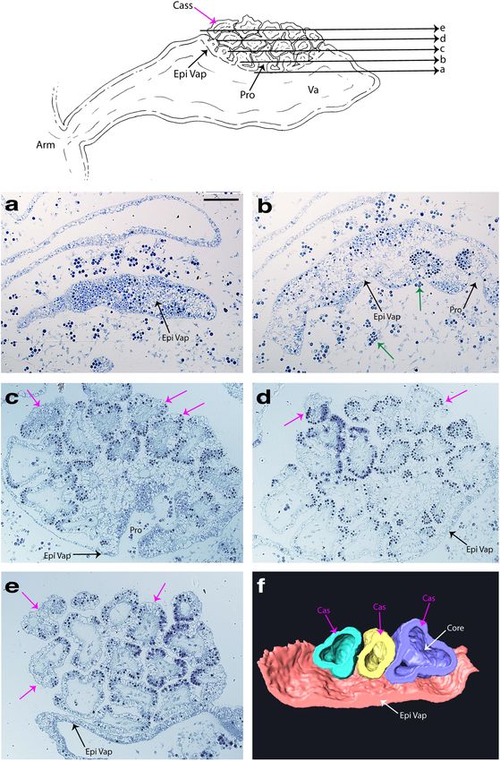
Fig. 1 Medusae of the upside-down mangrove jellyfish Cassiopea xamachana. a, b C. xamachana medusae (5–12 cm diameter) resting on umbrella apex
(white arrow) with oral arms (cyan arrows) facing up, observed by authors in the natural mangrove habitat in Key Largo, Florida (USA). Images courtesy of
A. Morandini. c–f Cassiosome nests (pink arrows) observed as white bulging spots at the termini of vesicular appendages (green arrows) off-branching
from areas of frilly digitate cirri (black arrows) on medusa oral arms (cyan arrows). Some cassiosome nests appear less full than others. Scale bars:
a = 2 cm; b = 5 cm; c, d = 1 mm; e, f = 0.5 mm.
transformed into amoebocytes34. Shortly after infection with Cassiosomes overview. Cassiosomes morphology: Numerous,
endosymbionts, C. xamachana polyps undergo strobilation, and the motile cellular structures, which we call cassiosomes, were
apical portion metamorphosis into an ephyra (juvenile medusa) observed suspended within mucus released by C. xamachana
which then develops into a sexually mature male or female medusa, medusae (3.0–8.8 cm umbrella diameter) (Fig. 2a–f) in response
with multiple color variants based on endosymbionts19,26 (Figs. 1a, to feeding or mild disruption with short bursts of seawater from a
b, 2a). Symbiodinium-generated photosynthates support the jellyfish pipette. Herein, we describe cassiosomes in C. xamachana as
host metabolism, growth, reproduction and survival. This promotes microscopic (100–550 μm in diameter), irregularly-shaped cel-
conservation and recycling of essential nutrients, given their stra- lular masses whose peripheral cell layer is primarily composed of
tegic presence amidst downwelling light, which is of unrivaled nematocytes and other irregular ectodermal cells that surrounds a
ecological importance for coral reefs and Cassiopea populations space containing amoebocytes —some hosting Symbiodinium and
alike26,32,33. others lacking them—among presumptive mesoglea (Fig. 2f).
Cassiosome motility: When multiple C. xamachana medusae
were placed together and agitated by directing water at their oral
Medusa feeding. Feeding studies on Cassiopea medusae show arms using a glass pipette, they consistently released cassiosome-
that prey capture occurs as a result of perpetual medusa pulsation laden mucus within 5–10 min (Fig. 2a–c) for periods lasting
that carries the prey into the subumbrellar space and then onto several hours. When collected mucus was transferred to a small
the oral arms where they are held by nematocyst-rich digitate glass dish, cassiosomes moved around within the mucus for about
fringed lips and vesicular appendages (i.e., small oral vesicles) 15 min and then descended to the bottom of the dish, leaving the
(Fig. 1a–f), eventually being reduced to fragments. Finally, food neutrally buoyant mucus. This permitted efficient isolation of
particles are then forced into the oral ostia of secondary mouths, numerous cassiosomes (Fig. 2b–f), which remained in constant
and ingested via ciliary action. Cassiopea are opportunistic pre- motion by rotating and displacing in various directions along the
dators, feeding on a broad range of prey items (e.g., crustaceans, bottom, but never elevating from the bottom of the dish
nematodes, eggs) in the field (Larson43), while in the lab polyps (Supplementary Movie 1). Isolated cassiosomes remained motile
and medusae are fed Artemia salina (1–3 days old, lab reared). for up to 10 days, gradually losing their corrugated appearance
COMMUNICATIONS BIOLOGY | (2020)3:67 | https://doi.org/10.1038/s42003-020-0777-8 | www.nature.com/commsbio 3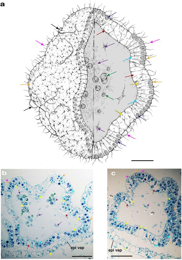
ARTICLE                                                                COMMUNICATIONS BIOLOGY | https://doi.org/10.1038/s42003-020-0777-8

Fig. 2 Observations of mucus and cassiosome release in Cassiopea xamachana medusae. a C. xamachana releasing mucus (yellow arrows) following
collection in the field by authors (Bonaire, The Netherlands). Cassiosome nests (pink arrows) appear as light bulging spots at the termini of vesicular
appendages (cyan arrow). b Mucus (yellow arrow) released into the water by C. xamachana in the lab—small white flecks correspond to live cassiosomes
(green arrows). c Live cassiosomes (green arrows) suspended in mucus (yellow arrow) harvested after release from C. xamachana. d Multiple motile
cassiosomes isolated from C. xamachana mucus. e Live cassiosome close-up (green arrows), showing irregular shape and centralized Symbiodinium
dinoflagellates as amber spheres (red arrows). f Confocal image of highly motile cassiosome immobilized on MatTek glass bottom dishes coated with Cell
Tak adhesive (Corning); image collected with ×60 objective (oil) reveals organization of the peripheral cell layer: NucBlue-Hoescht 33342 (1,100)
(ThermoFisher) stains nuclei (blue) of nematocytes with peripheral nematocytes bearing O-isorhiza nematocysts (blue arrows, DIC) and non-nematocyte
ectodermal cells (purple). DIC shows centrally Symbiodinium (dark spheres, red arrows) occupy presumptive C. xamachana amoebocytes in an otherwise
acellular core. Scale bars: a = 3 cm; b = 3 mm, c = 1 mm;, d = 5 mm; e = 300 μm, f = 50 μm.

after 5 or 6 days and shrinking in size to a smooth spherical shape          When isolated cassiosomes were added to dishes (150 ml)
until movement ceased and cassiosomes disintegrated. Addition-               containing abundant Artemia nauplii (Supplementary Movie 2),
ally, custom-designed microfluidic devices with channels equal to             cassiosomes that encountered the underside of the nauplii carapace
or slightly bigger than the cassiosomes35 (Supplementary Fig. 1d)            immediately immobilized and killed the prey items (Fig. 3a–c). In
were used to observe cassiosomes of C. xamachana to gain a                   cases where Artemia nauplii came into contact only briefly with
better understanding of their motility, and three-dimensional                cassiosomes, prey were able to escape rapid immobilization and
irregular, popcorn-shaped structure. These microscopic observa-              death (Supplementary Movie 5). Furthermore, during discharge
tions revealed motile cilia extending from the periphery that                assays when either FASW (Filtered Artificial Seawater) or mucus
propel cassiosomes.                                                          containing no cassiosomes, following manual removal, was added
   Cassiosomes kill prey: We conducted assays to determine if                (see the “Methods” section), Artemia were not affected and
cassiosomes were capable of trapping Artemia nauplii provided as             continued to swim around in the dish (Supplementary Movies 4
food in aquarium-reared medusae since so-called non-penetrant                and 3).
O-isorhiza nematocysts are the only type found in cassiosomes of                Cassiosome ultrastructure: In order to better understand their
C. xamachana. For this purpose, microfluidic chambers35 provided              organization, live cassiosomes were isolated from C. xamachana
an arena in which to document immobilization and rapid trapping              mucus. After fixing, dehydrated specimens were examined using
of Artemia, which were killed almost immediately upon substantial            scanning electron microscopy (SEM) (Fig. 4a–f). The cassiosome
contact with cassiosomes (Fig. 3a–c) (Supplementary Movie 5).                perimeter was found to be lined with nematocyst capsules and

4                           COMMUNICATIONS BIOLOGY | (2020)3:67 | https://doi.org/10.1038/s42003-020-0777-8 | www.nature.com/commsbio
COMMUNICATIONS BIOLOGY | https://doi.org/10.1038/s42003-020-0777-8 ARTICLE
Fig. 3 Cassiosomes are capable of killing brine shrimp. a Dead 2-day old Artemia nauplii (orange arrow) with cassiosome (green arrow) attached to
carapace, imaged within a microfluidic chamber. b Cassiosomes (green arrow) lodged into two different 1-day old Artemia nauplii (orange arrow). c 1-day
old Artemia nauplii (orange arrow), immobilized following cassiosome (green arrow) attachment to the carapace with visibly discharged nematocysts
(fuchsia arrows). Scale bars = 200 μm.
numerous long, spiny tubules (Fig. 4a–f) extruded from abundant mucus; and cassiosomes (isolated from mucus) (Fig. 6a–d).
O-isorhiza nematocysts in the periphery following spontaneous Measurements of undischarged nematocysts of each type in the
deployment (likely during the dehydration process (see the corresponding subsample revealed that O-isorhizas nematocysts
“Methods” section)). More abundant on the surface, however, are absent in polyps, but appear in medusae from the onset of
were much thinner filaments corresponding to abundant cilia ephyra development during strobilation (Fig. 6; Supplementary
connected to ectodermal cells (Fig. 4a–c). Close observation of Table 1). We also observed penetrant nematocytes, birhopaloids
several cassiosomes via SEM revealed along the outer layer and euryteles, which cannot be distinguished in the undeployed
emptied out regions appearing as collapsed cell membrane state (intact) within C. xamachana tissue using light microscopy.
remnants of deployed nematocysts (Fig. 4a–f), underneath which Therefore in this study, these two nematocyst types were analyzed
could be seen an amorphous thick central extracellular matrix-like together as rhopaloids (Fig. 6a–d) (as per ref. 36 in C. andromeda);
substance (Fig. 4e, f). Confocal microscopy on fixed cassiosomes hence, rhopaloids account for a larger proportion of the cnidome
with labeled nuclei, nematocysts and cilia (see the “Methods” in the medusa and mucus than distinct isorhiza types. An
section) (Fig. 5a–d) corroborated these findings of an organized assessment of the inventory of nematocysts freely suspended
cell mass. Cassiosomes are composed of a peripheral layer of within the mucus yielded a similar nematocyst profile to that of the
nematocytes bearing O-isorhiza nematocysts (Fig. 5a) patterned medusa (Fig. 6a), albeit with a proportionately higher number of
with presumptive ectoderm cells that lack nematocysts, from rhopaloids, which are implicated in envenomation28,29. Conver-
which numerous cilia protrude (Fig. 5a–d). This outer layer sely, isolated cassiosomes of C. xamachana contain exclusively
surrounds centralized clusters of Symbiodinium endosymbionts O-isorhiza nematocysts which are a ubiquitous type in jellyfish
(i.e., hosted by amoebocytes) within an otherwise apparently tentacles, functioning in prey capture and predation.
acellular region (Fig. 4e, f).
C. xamachana toxin proteins in cassiosomes. Over a century
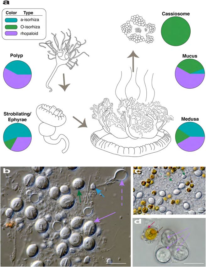
Cnidome of C. xamachana. The term cnidome refers to the ago, Perkins17 documented that disturbed C. xamachana medu-
dynamic repertoire of nematocyst types in a cnidarian species7,8. sae produced mucus containing ciliated structures as innumerable
The cnidome, a species-specific trait, often changes throughout minute spherical bodies containing unicellular zooxanthellae
the life cycle of the jellyfish as it undergoes metamorphosis from a within the interior, which he considered to be parasitic larvae,
sessile polyp, to strobila, and then to juvenile and sexually mature and claimed it was “impossible to regard [these structures] as of
medusa. Given reports implicating nematocysts, or tiny little coelenterate [Cnidaria] affinities”. These details suggest that
grenades, within Cassiopea mucus as the cause of stinging water, Perkins observed what we have herein identified as cassiosomes,
we sought to characterize the cnidome of this species at several but mistook them for entirely unique, non-cnidarian organisms.
life stages, and within the cassiosomes and contents of the mucus. In order to test this theory, and properly classify cassiosomes as
Nematocyst measurements were plotted for the following life belonging to C. xamachana, rather than being unknown organ-
stages of C. xamachana: polyps (n = 3); strobilating/released ism, we used real-time PCR (qPCR) assays to target species-
ephyrae (n = 3) and medusae (2.4–8.8 cm in diameter (n = 3)); specific cnidarian toxins, employing three custom-designed
COMMUNICATIONS BIOLOGY | (2020)3:67 | https://doi.org/10.1038/s42003-020-0777-8 | www.nature.com/commsbio 5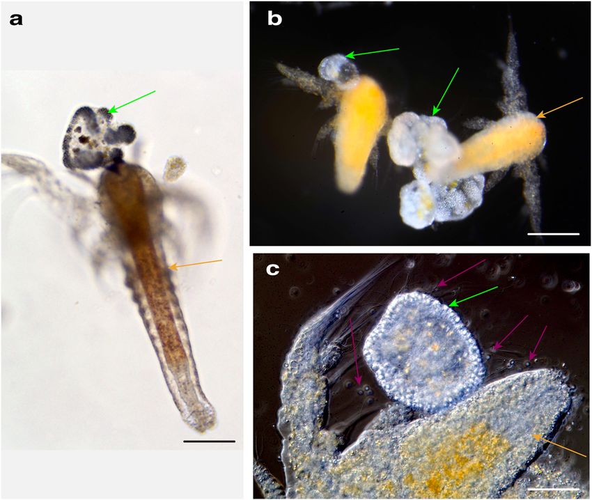
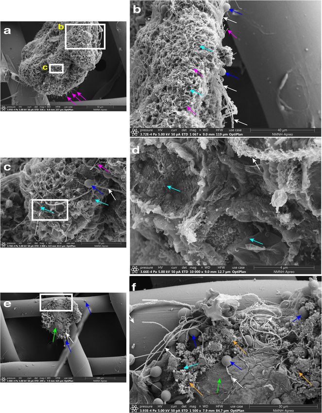
ARTICLE                                                                 COMMUNICATIONS BIOLOGY | https://doi.org/10.1038/s42003-020-0777-8

Fig. 4 Cassiosome organization revealed via SEM. a An individual cassiosome poised in a 100-μm mesh opening, revealing the irregular ‘popcorn’ shape
of the cassiosome bearing numerous cilia (pink arrows) protruding from the peripheral layer. White rectangles correspond to magnified region shown in
(b) and (c). b, c Close up of the cassiosome reveals cilia (pink arrows), and thicker tubules (white arrows) of discharged O-isorhiza nematocyst capsules
(blue arrows) in the periphery which is lined with collapsed nematocytes (cyan arrows). Spiny nematocyst tubules (white arrows) are outnumbered by the
abundant cilia (thinner filaments) (pink arrows). d Depressions fringed by deflated cell membranes outlining nematocytes (cyan arrows in c and d)
following deployment of O-isorhiza nematocysts (blue arrows in c) along the cassiosome surface. e A different cassiosome from that in (a–d). All cellular
components were lost (possibly in dehydration stage of methods) but for discharged O-isorhiza nematocyst capsules (blue arrows) and corresponding
spiny tubules (white arrows) in the cassiosome periphery lined with collapsed nematocytes (cyan arrows). White rectangle corresponds to magnified
region shown in (f) revealing the dense central, fibrous extra-cellular matrix (green arrow). Unidentified microscopic particles also present (orange
arrows). Scale bars as indicated in SEM images.

primer pairs (Supplementary Fig. 1e–g) that we designed from the              and A. alata14,39) validates the specificity of our primers to a
publicly available C. xamachana genome (see Data Availability                 C. xamachana-derived gene target, indicating that cassiosomes
section).                                                                     originate in Cassiopea medusae.
   We targeted a cnidarian-restricted CrTX/CaTX family                           To validate the potential for envenomation by cassiosomes,
toxin37,38 gene in DNA extracted separately from C. xamachana                 rather than solely by suspended intact nematocysts in the mucus
medusa tissue and isolated cassiosomes, and also from tissue of               released by medusae, we used LC-MS/MS analyses to confirm
the jellyfish Aurelia sp. (Class Scyphozoa) and a more divergent               the presence of the same CrTX/CaTX toxin proteins in two
jellyfish species Alatina alata (Class Cubozoa) for comparison.                C. xamachana sample types: cassiosomes isolated from mucus
Amplification of the qPCR gene target was observed for both                    released from ~20 medusae over a 7-h period, and several
C. xamachana tissue and cassiosome samples using primers for                  vesicular appendages containing cassiosome nests, dissected from
the CrTX/CaTX gene, herein named CassTX-C (see Data                           multiple medusae (e.g., Fig. 1c–f). A shotgun proteomic analysis
Availability section below). Conversely, failure to amplify the               identified three isoforms of the target toxin family encoded in the
target in non-Cassiopea medusozoans used in this study (despite               C. xamachana genome14, which we call CassTX-A, CassTX-B and
reports of CrTX/CaTX genes documented in both Aurelia sp                      CassTX-C (Fig. 7). Each toxin protein was identified with

6                            COMMUNICATIONS BIOLOGY | (2020)3:67 | https://doi.org/10.1038/s42003-020-0777-8 | www.nature.com/commsbio
COMMUNICATIONS BIOLOGY | https://doi.org/10.1038/s42003-020-0777-8                                                                    ARTICLE

Fig. 5 Characterization of cassiosome ultrastructure. a–f Individual cassiosome fixed and labeled with Tubulin Antibody, ActinGreenTM and NucBlueTM,
and mounted in 80% glycerol in PBS on glass slides for imaging. Imaging was performed with both DIC and confocal laser scanning with lines at 405, 488,
561, and 640 nm, and collected with a Plan Apo ×100 objective. a DIC reveals peripheral layer of nematocytes bearing spherical O-isorhiza nematocysts
(lavender arrows). Tubulin (red) reveals cnidocils (short filaments marked by white arrows) extending from apex of nematocytes, and motile cilia (long
filaments) originating from non-nematocytes ectoderm cells organized in patches along the peripheral layer among nematocyte-rich areas. NucBlue (blue)
reveals nuclei (yellow arrows) of peripheral epithelial layer – nematocytes and other ciliated ectoderm cells. Actin (green) reveals actin basket (pink
arrows) formed around the apex of nematocysts. b 3-D construction of Z-stack magnified confocal images corresponding to (a) and (c–f) shows tubulin
(red) of cnidocils (short filaments marked by white arrows) extending from around apex of nematocytes and motile cilia (long filaments) originating from
non-nematocytes putative ectoderm cells organized in patches along the peripheral layer among nematocyte-rich areas. NucBlue (blue) reveals nuclei of
peripheral epithelial layer—nematocytes and other ciliated ectoderm cells. Actin (green) reveals actin basket forming around the apex of nematocysts.
Scale bars = 10 μm.

multiple unique peptides and ≥17.0% protein coverage (Fig. 7;                 the vesicular appendages (Fig. 8a–g) (Supplementary Fig. 2c, d)
Supplementary Fig. 3; Supplementary Table 2), with the exception              which are formed of ectoderm, endoderm and mesoglea; the
of CassTX-C, which in the cassiosomes was not assigned with                   cavity of these appendages communicates with the canals of the
sufficient confidence (based on Mascot Score). Peptides identified               oral arms40. Vesicular appendages are capable of independent
and aligned to the three Cassiopea toxin homologs are shown in                movement, and during feeding of lab-reared C. xamachana
Supplementary Fig. 3, along with the protein gel (Supplementary               medusae, when Artemia nauplii approach the oral arms, the
Fig. 4).                                                                      vesicular appendage bends to cover the shrimp, thereby trapping
                                                                              the prey item; this trapping method was also reported in the
                                                                              conspecific C. frondosa (see ref. 32). Clusters of cassiosomes (i.e.,
Cassiosome provenance and development. A visual inspection                    30–100 individuals) line the surface of the numerous, variably
of C. xamachana oral arms during mucus release revealed that                  sized vesicular appendages present in C. xamachana (Figs. 1, 8).
cassiosomes occur as warty clusters within a shallow pocket on                Bigelow (1900) called these appendages nettle batteries, referring

COMMUNICATIONS BIOLOGY | (2020)3:67 | https://doi.org/10.1038/s42003-020-0777-8 | www.nature.com/commsbio                                            7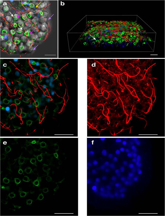
ARTICLE                                                                 COMMUNICATIONS BIOLOGY | https://doi.org/10.1038/s42003-020-0777-8

Fig. 6 Nematocyte type proportion (cnidome) varies within different life stages and structures of C. xamachana. a Figure displaying life cycle stages of
C. xamachana (observed in this study) and associated cassiosome-laden mucus release by the medusae. Pie charts indicate proportion of nematocyte types
for polyps (n = 3 distinct polyps), strobila/ephyrae (n = 3 distinct ephyrae), medusa (n = 3 distinct medusae), mucus (from n = 4 distinct medusae), and
cassiosomes (from n = 4 distinct medusae), based on measurements of multiples of each nematocyst type per life stage (see details in Supplementary
Table 1). b, c Different nematocyst types isolated from C. xamachana medusae oral-arm filaments corresponding to colors in pie charts in (a): a-isorhiza
intact (light blue arrow), O-isorhiza intact (green arrows) and deployed (dashed green arrow), and rhopaloid intact (lavender arrow) and deployed (dashed
lavender arrow); and Symbidinium (brown arrows). d Mucus contents of C. xamachana containing a triplet of rhopaloid nematocysts (lavender arrows)
intact within nematocytes, and Symbiodinium (brown arrow) disassociated from jellyfish tissue but still within amoebocytes (pink arrows). Scale bar:
b, d = 10 μm; c = 20 μm.

to their functional role in subduing prey, and possibly also in               only loosely attached to the pocket and neighboring cassiosomes
defense.                                                                      (Figs. 8, 9). This development process results in irregular popcorn-
   Images of semithin sections of five separate vesicular appen-               shaped cassiosomes, as shown in the 3-D reconstruction of their
dages revealed that cassiosomes develop within a depression                   organization within the vesicular appendages, based on semithin
externally on one side of a vesicular appendage, but occasionally             images (Fig. 8).
on both sides (Figs. 8, 9). During development, cassiosomes                      The peripheral layer (nematocytes and ectoderm) surrounds a
originate proximally as protrusions of the epithelium (ectoderm)              central space containing clusters of amoebocytes often hosting
of the vesicular appendage, and then spread out distally as they              Symbiodinium, randomly interspersed among clear empty
develop, incorporating presumptive amoebocytes (endoderm cells                patches that exhibit substantially different refractive index
that have migrated into the mesoglea), some of which host                     properties (as seen in DIC) (Figs. 5, 8, 9) reminiscent of the
Symbiodinium (Figs. 8, 9). Early developing cassiosome protru-                small bags of mesoglea and nematocysts witnessed being released
sions are connected peripherally to the pocket surface of the                 in conspecific C. frondosa by Smith (see ref. 40). These findings
vesicular appendage by their shared ectoderm epithelial layer,                corroborate those of our SEM and confocal analyses, and suggest
whereas fully developed cassiosomes awaiting deployment are                   the central region of cassiosomes is amorphous, containing only

8                            COMMUNICATIONS BIOLOGY | (2020)3:67 | https://doi.org/10.1038/s42003-020-0777-8 | www.nature.com/commsbio
COMMUNICATIONS BIOLOGY | https://doi.org/10.1038/s42003-020-0777-8 ARTICLE
Fig. 7 LC-MS/MS identification of jellyfish toxin proteins in cassiosomes. a Alignment of peptides (black boxes) identified in two sample types (C =
cassiosomes; VA = vesicular appendages) by shotgun proteomics to the full-length toxin homologs (CassTX-A, -B and -C; black bars) of the cnidarian-
restricted CrTX/CaTX toxin family identified in C. xamachana. Each protein was identified with a confidence score > 0.05 and with at least two unique
peptides in each sample, except for CassTX-C in the cassiosome sample. b–e Representative mass and annotated tandem mass spectra of CassTX-A
tryptic peptides: (b) and (c) peptide sequence VAPIPESGLEEGK; (d) and (e) peptide sequence TEIELSTDLLGDVK.
some loose cells—likely amoebocytes—many of which host setouchianum (see ref. 41), their motility, the irregular shape they
Symbiodinium (Figs. 8, 9). possess (Fig. 10k, l) when released from the oral arms, and
the eventual loss of bumpiness and disappearance after several
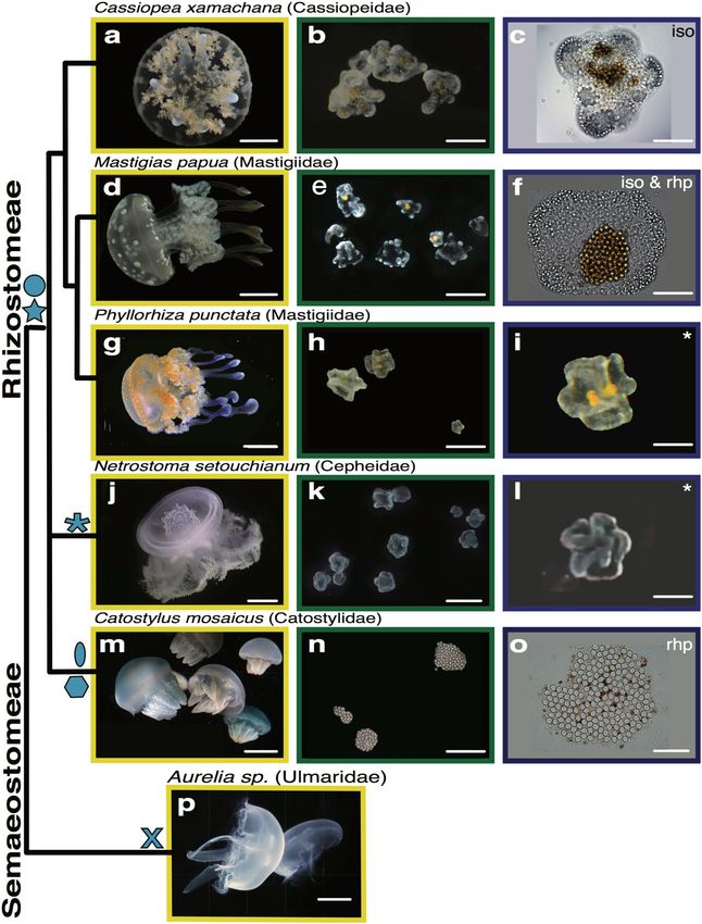
Cassiosomes in other rhizostome jellyfish species. Jellyfish of days matches the general description of cassiosomes we first
the taxonomic order Rhizostomeae, including Cassiopea xama- discovered in C. xamachana. Cassiosomes of M. papua and
chana, all lack marginal tentacles, possessing instead oral arms P. punctata medusae are highly motile, and share the same fun-
covered with minute vesicular appendages. Although the main damental structure, albeit exhibiting slight variations with respect
focus of this study is to provide a detailed description of cassio- to nematocyst types present within the peripheral nematocyte
somes in C. xamachana, in an effort to ascertain if cassiosome layer of each type (Fig. 10d–f, g–i). Superficially, N. setouchianum
production is a possible apomorphy of the rhizostome jellyfish cassiosomes (Fig. 10k, l) appear to match the morphology of the
clade, we examined the mucus of additional rhizostome jellyfish two aforementioned species, however, as we were not able to
taxa and documented cassiosomes in a total of four rhizostome examine them directly using microscopy (solely via video), the
jellyfish lineages (five different species) (Fig. 10). Cassiosomes presence of associated dinoflagellates could not be confirmed.
from all six species are classified into two main types: motile, Conversely, cassiosomes in Catostylus mosaicus (Fig. 10n, o)
bearing cilia that propel them in the water column and non- exhibit several differences in that neither motility nor centralize
motile, bearing no apparent motile cilia. Mucus was directly Symbiodinium was observed but, rather, unidentified microalgae
examined (using light microscopy) from three additional rhi- are distributed homogenously throughout the cell mass. No cas-
zostomes Mastigias papua, Phyllorhiza punctata, Catostylus siosomes were found in the mucus of the semaeostome Aurelia
mosaicus (Fig. 10d, g, m), and a single Semeastomeae (sister sp. which lacks both vesicular appendages and endosymbiotic
group) Aurelia sp. (Fig. 10p), all reared at the National Aquarium algae (Fig. 10p).
(Baltimore, USA). Additionally, we obtained a video from the
author of a citizen scientist blog post showing abundant motile Discussion
particles reportedly released by another rhizostome Netrostoma Jellyfish are remarkable aquatic animals that diverged over
setouchianum, collected in Japan41 (Fig. 10j). Although we were 600 mya and have in spite of, or possibly because of, their diplo-
unable to directly examine these cellular masses from N. blastic nature, evolved a remarkable envenomation system in the
COMMUNICATIONS BIOLOGY | (2020)3:67 | https://doi.org/10.1038/s42003-020-0777-8 | www.nature.com/commsbio 9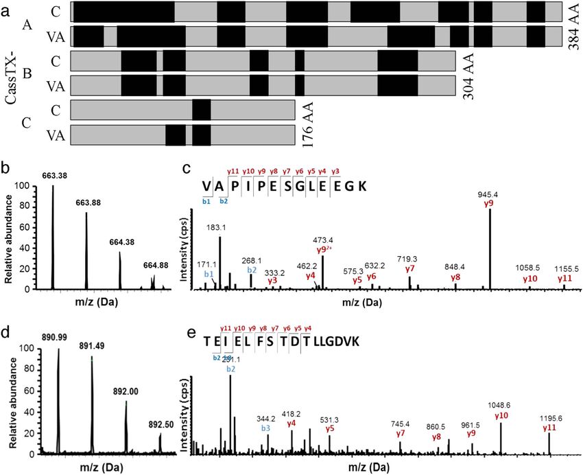
ARTICLE                                                                  COMMUNICATIONS BIOLOGY | https://doi.org/10.1038/s42003-020-0777-8

Fig. 8 Characterization of the ultrastructure of the vesicular appendages during cassiosome production and development in Cassiopea xamachana. Line
drawing of vesicular appendage demonstrates how early developing cassiosome protrusions (pro) are connected peripherally to the pocket surface of the
vesicular appendage by their shared epithelium (epi vap), whereas fully developed cassiosomes (cass) awaiting deployment are only loosely attached to
the pocket and neighboring cassiosomes. a–e Semithin sections (~1 µm) of resin-embedded vesicular appendages corresponding to arrows labeled a–e in
the line drawing of the vesicular appendages (va) extending from the oral arms (arm) of the medusae. Clusters of cassiosomes (pink arrows) developing
from protrusions (pro) in the epithelium of the concave vesicular appendage pocket (epi vap) give rise to the cassiosome peripheral layer comprising
nematocytes bearing O-isorhiza nematocysts (dark spheres stained with 1% toluidine blue) interspersed with other ectodermal cells. Clusters of
amoebocytes hosting Symbiodinium (green arrows) move into the cassiosome core at protrusions points. Cassiosome core containing presumptive
mesoglea indicated by difference in diffractive index with DIC. f Partial 3-D reconstruction showing protrusions developing from epithelium of the vesicular
appendage pocket (epi vap) into popcorn-shaped cassiosomes. Reconstruction based on sections from a different vesicular appendage than seen above but
corresponds to the region between sections a–d, revealing the empty core (core) of cassiosomes (cass) (3-D image orientation is vertical with respect to
cross sections in the line drawing). arm = medusa oral arm, cass = cassiosomes(s), core = presumptive mesoglea; pro = protrusion(s); va = vesicular
appendage(s), epi vap = epithelial layer of the vesicular appendage pocket. Scale bar = 250 µm.

form of stinging cells—nematocytes—for prey capture and defense.               on these findings, we hypothesize that cassiosomes evolved within
In this work, we reported the findings of an extensive investigation            a single lineage of jellyfish, Rhizostomeae, to further weaponize the
into the provenance, development and ultrastructure of cassio-                 jellyfish by sequestering nematocytes (and other cells) into
somes, a newly described cnidarian stinging-cell structure. Based              grenade-like structures that are freely released into the water within

10                           COMMUNICATIONS BIOLOGY | (2020)3:67 | https://doi.org/10.1038/s42003-020-0777-8 | www.nature.com/commsbio
COMMUNICATIONS BIOLOGY | https://doi.org/10.1038/s42003-020-0777-8                                                                         ARTICLE

Fig. 9 Characterization of the ultrastructure of mature cassiosomes in Cassiopea xamachana. a Line drawing, and b, c thin sections of fully developed
popcorn-shaped cassiosomes from semithin sections (~1 µm) of resin-embedded vesicular appendage (see Fig. 8). Cassiosome peripheral layer comprising
nematocytes (cyan arrows) bearing O-isorhiza nematocysts (as peripheral dark spheres stained blue with Richardson’s stain in (b) and (c)) interspersed
with patches of oddly shaped ectoderm cells (red arrows), and motile cilia (pink arrows); blue-stained nuclei (yellow arrows) visible below the base of large
nematocysts capsule in nematocytes, and also in non-nematocyte ectoderm cells. Cassiosome core containing presumptive mesoglea (gray central region
in (a), gray arrow in (b and c)), speckled with amoebocytes (purple arrows)—hosting Symbiodinium (green arrows) or empty. Rigid stereocillia/cnidocil
complex (orange arrows) visible as a point at the nematocyst apical portion, and deployed tubules (black arrows in (a)) on surface present as long, thick
spiny threads. epi vap = epithelial layer of the vesicular appendage pocket. Scale bars = 50 µm.

exuded mucus. Our findings strongly implicate cassiosomes as a                   Symbiodinium while at least one bears microalgae instead.
major contributor to the stinging water phenomenon reported by                  However, the fundamental trait distinguishing cassiosomes from
sea bathers and aquarists when interacting with rhizostome jelly-               nematosomes, analogous cell masses deriving from the mesen-
fish species.                                                                    teries of the sea anemone Nematostella31, is that the structure of
   In this inaugural study on cassiosomes, we used extensive                    cassiosomes is organized into a distinct outer epithelial layer
microscopy techniques, video-documentation and microfluidics                     surrounding a central, mostly empty, core. Further studies are
to describe these cnidarian innovations first in C. xamachana,                   needed to elucidate the role of these photosynthetic endo-
and then in taxa belonging to four additional Rhizostomeae jel-                 symbionts in cassiosomes. The complete absence of cassiosomes
lyfish families. Our preliminary findings suggest there are motile                in the mucus released by the semeastome Aurelia sp. supports our
and non-motile types of cassiosomes among the rhizostomes we                    theory that cassiosomes are a rhizostome evolutionary novelty.
examined in this study, and that some host endosymbiotic algae                  However, a comparative examination of the mucus contents

COMMUNICATIONS BIOLOGY | (2020)3:67 | https://doi.org/10.1038/s42003-020-0777-8 | www.nature.com/commsbio                                                 11
ARTICLE                                                                COMMUNICATIONS BIOLOGY | https://doi.org/10.1038/s42003-020-0777-8

Fig. 10 Cassiosomes observed in jellyfish species of the order Rhizostomeae. Cladogram of species examined in this study from two orders
Rhizostomeae: a–c Cassiopea xamachana, d–f Mastigias papua, g–i Phyllorhiza punctata, j–l Netrostoma setouchianum, and m–o Catostylus mosaicus, and
p Semaeostomeae Aurelia sp., and their respective cassiosome structures, when present. Abbreviations: iso = isorhiza nematocysts; rhp = rhopaloid
nematocysts; * = could not confirm type of nematocysts. Blue symbols: star = motile via cilary movement; hexagon = non-motile; circle = endosymbiotic
dinoflagellates within cassiosomes confirmed; oval = microalgae on the surface of cassiosomes confirmed; asterix = could not confirm presence or
absence of algal symbioints; X = no cassiosomes witnessed within the mucus. Scale bar: Scale bar: a = 1.5 cm; d, g, j, m, p = 2.5 cm; b, e, h, k, n, o =
300 µm; c, i, l = 200 µm; f, o = 100 µm.

across all rhizostome lineages, including the eight nominal Cas-             appendages releasing gray bodies (putative cassiosomes) (Sup-
siopea species26,42 is needed to test this hypothesis.                       plementary Fig. 2a, b). Conversely, in this study on C. xama-
   Furthermore, we identified the provenance of cassiosome                    chana, upon disturbance, cassiosomes spontaneously detached
production and release from oral arm vesicular appendages,                   from the surface of vesicular appendage pockets, which lack a
corroborating earlier works suggesting these vesicles (as oral               terminal aperture (Supplementary Fig. 2c, d).
vesicles17,40,43) function in defense and predation. These pre-                 All jellyfish have envenomation capabilities due to bioactive
vious works noted similar structures in the oral vesicles of Cas-            proteins comprising the venom cocktail of the cnidome (i.e.,
siopea species, dubbed either gray bodies, bags of nematocysts               repertoire of nematocysts types). The cnidarian-specific pore-
and mucous cells, minute spherical bodies, or grenades, that shot            forming CrTX/CaTX toxin family is one of the most potent toxin
when contacted. Although those reports fell short of providing               groups, and represents the main proteinaceous component of the
an adequate description, we are confident that the structures                 venom of cubozoans (box jellyfish), a clade that includes species
mentioned therein correspond to what we described herein as                  whose sting results in a deadly cardiovascular condition37. In this
cassiosomes. The mechanism of cassiosome deployment may                      study, the presence of C. xamachana-specific CrTX/CaTX toxin
vary across different rhizostome taxa, or even between con-                  family homologs was confirmed in cassiosomes at the DNA and
specifics, as according to Smith40 when prey was provided to                  protein level (i.e., CassTX), validating their expression in cassio-
Cassiopea frondosa, an aperture opened at the tip of the vesicular           somes and their contribution to the stinging water phenomenon.

12                          COMMUNICATIONS BIOLOGY | (2020)3:67 | https://doi.org/10.1038/s42003-020-0777-8 | www.nature.com/commsbio
COMMUNICATIONS BIOLOGY | https://doi.org/10.1038/s42003-020-0777-8                                                                                        ARTICLE

Although C. xamachana in Florida waters is considered a mild                             Discharge assays. To observe the effects of cassiosomes on potential prey items in
stinger, reports exist of painful human envenomation resulting in                        relatively confined spaces, discharge assays were conducted within a microfluidic
                                                                                         device into which Artemia nauplii (1–2 days old) were introduced along with
rash, vomiting, painful joints, swelling and irritation for this                         cassiosomes either isolated from or still within the mucus in FASW. This
broadly distributed species, in addition to documentation of                             6-chambered microfluidic device, with chambers 7-mm wide × 400-μm tall × 3-mm
hemolytic and cytolytic activity in crude venom of C. xamachana                          long, modified from a version originally designed as a behavioral microarena for
and conspecifics29. Following a characterization of the cnidome in                        imaging Hydra, was used to immobilize large cassiosomes (~400 μm diameter) or
C. xamachana in this study, we discovered a large proportion of                          to slow down the movement of smaller cassiosomes (
ARTICLE COMMUNICATIONS BIOLOGY | https://doi.org/10.1038/s42003-020-0777-8
gene target was identified by BLAST+ (https://blast.ncbi.nlm.nih.gov/) homology 2. Sunagar, K. & Moran, Y. The rise and fall of an evolutionary innovation:
search against the publicly available C. xamachana genome (see Data Availability contrasting strategies of venom evolution in ancient and young animals. PLoS
section below). Approximately 2 ng of extracted DNA for each sample was used to Genet. 11, 1–20 (2015).
conduct triplicate 20 μl qPCR amplification assays (KAPA SYBR® FAST qPCR 3. Huxley, T. H. On the anatomy and the affinities of the family of the Medusae
Master Mix (2×) Kit) using a ViiA 7 Real-Time PCR System (ThermoFisher) at the Thomas Henry Huxley. Philos. Trans. R. Soc. Lond. 139, 413–434 (1849).
Smithsonian Laboratories of Analytical Biology (Washington, D.C.). The following 4. Seipel, K. & Schmid, V. Evolution of striated muscle: jellyfish and the origin of
cycling conditions were used: enzyme activation 95 °C (3 min), denaturation 95 °C triploblasty. Dev. Biol. 282, 14–26 (2005).
(2 s) and annealing extension/data acquisition 60 °C (1 min) for 35 cycles, followed 5. Daly, M. et al. The Phylum Cnidaria: a review of phylogenetic patterns and
by a 60 °C (30 s) final post-read stage. diversity 300 years after Linnaeus *. Zootaxa 182, 127–182 (2007).
6. Mariscal, R. N. in Coelenterate biology: reviews and new perspectives (eds
LC-MS/MS analysis. Isolated cassiosomes and vesicular appendages in FASW (33 Muscatine, L. & Lenoff, H.) 129–178 (Academic Press, 1974).
ppt) were pelleted (4000 rpm for 10 min) and resuspended in 20 μl total 1X 7. Östman, C. A guideline to nematocyst nomenclature and classification, and
NuPAGE LDS sample buffer (Thermo Fisher Scientific, Carlsbad, CA) and 10 mM some notes on the systematic value of nematocysts. Sci. Mar. 64, 31–46 (2000).
dithiothreitol (DTT). Exclusively LCMS grade reagents were used. After sonication 8. Weill, R. Contribution a l’etude des cnidaires et de leurs nematocystes II.
with a handheld biovortex (30 s, maximum bursts for 3 min) to break down Valeur taxonomique du cnidome. Trav. la Stn. Zool. Wimereux 11, 351–355
the nematocysts capsule minicollagen double wall, and then in a sonication bath (1934). 632–701.
(10 min) to remove bubbles, samples were heated to 70 °C for 10 min and separated 9. Hessinger, D. A. & Lenhoff, H. M. Biology of Nematocysts. (Academic Press
on a 10% NuPAGE Bis-Tris mini gel for 35 min at constant voltage (200 V). Inc., 1988).
Proteins were stained with BioSafe Coomassie (Bio-Rad, Hercules, CA) and de- 10. Macrander, J., Brugler, M. R. & Daly, M. A RNA-seq approach to identify
stained overnight with water. Ten bands were excised from the gel according to putative toxins from acrorhagi in aggressive and non-aggressive Anthopleura
molecular weight (gel image available as Supplementary Fig. 4). Proteins in all elegantissima polyps. BMC Genomics 16, 221 (2015).
bands were reduced and alkylated, and then digested in gel with sequencing grade 11. Al-Rubiay, K. K., Al-Musaoi, H. A., Alrubaiy, L. & Al-Freje, M. G. Skin and
trypsin (Promega, Madison, WI). Peptides were extracted from gel by two treat- systemic manifestations of jellyfish stings in Iraqi fishermen. Libyan J. Med. 4,
ments with 2% formic acid in 50% acetonitrile and a final wash with 100% acet- 75–77 (2009).
onitrile. All extraction fractions were concentrated by speed-vac, and then 12. Grady, J. D. & Burnett, J. W. Irukandji-Like Syndrome in South Florida
resuspended in 20 μl of 0.1% formic acid. One μl of each sample was injected into Divers. Ann. Emerg. Med. 42, 763–766 (2003).
an LC-MS/MS system (U3000 LC coupled to Orbitrap Fusion Lumos mass spec- 13. Rossetto, A. L., Dellatorre, G., Silveira, F. Lda & Haddad Júnior, V. Seabather’s
trometer (Thermo Scientific, Waltham, MA)) for proteomic analysis. Resulting eruption: a clinical and epidemiological study of 38 cases in Santa Catarina
spectra were extracted, merged, and searched by Mascot (Matrix Science Inc., State, Brazil. Rev. Inst. Med. Trop. Sao Paulo 51, 169–175 (2009).
London, UK) against a database containing common standards and contaminants, 14. Ohdera, A. et al. Box, stalked, and upside-down? Draft genomes from diverse
i.e., trypsin, keratin, etc. (190 protein sequences). A searchable database was also
jellyfish (Cnidaria, Acraspeda) lineages: Alatina alata (Cubozoa), Calvadosia
created in-house of amino acid sequences corresponding to predicted open reading
cruxmelitensis (Staurozoa), and Cassiopea xamachana (Scyphozoa).
frames (ORFs), created using TransDecoder69 (v.5.5.0), for the three cnidarian
Gigascience 8, 1–24 (2019).
toxin proteins (CassTX-A, CassTX-B and CassTX-C) (Supplementary Fig. 3)
15. Todd, B. D., Thornhill, D. J. & Fitt, W. K. Patterns of inorganic phosphate
expressed in the C. xamachana transcriptome (see Data Availability section).
uptake in Cassiopea xamachana: A bioindicator species. Mar. Pollut. Bull. 52,
515–521 (2006).
Statistics and reproducibility. The mean length and width, and range, of mul- 16. Ames, C. L. et al. Field-forward eDNA sequencing with Oxford Nanopore
tiples of each nematocyst type comprising the cnidome of Cassiopea xamachana, Technology – a strategy to establish the upside-down jellyfish Cassiopea
were determined in triplicate for three separate medusae (or other specified life xamachana as a bioindicator. https://nanoporetech.com/resource-centre/field-
stage). Additionally, thousands of cassiosomes were extracted from the mucus of at forward-sequencing-oxford-nanopore-technology-strategy-establish-upside-
least 20 individual medusae over the course of this project, and imaged using the down. (2019)
various microscopy methods described herein. Harvested mucus and/or isolated 17. Perkins, H. F. Notes on medusae of the western Atlantic. Pap. from Tortugas
cassiosomes from multiple medusae (more than five biological replicates) were Lab. Carnegie Inst. Washingt. 1, 135–156 (1908).
combined for LC-MS/MS proteomics, and tissue subsamples were taken from three 18. Fitt, W. K. & Trench, R. K. Endocytosis of the symbiotic dinoflagellate
different medusae for genetic molecular analysis (PCR and qPCR). Five separate Symbiodinium microadriaticum Freudenthal by endodermal cells of the
vesicular appendages were analyzed with histological methods. Detailed explana-
scyphistomae of Cassiopeia xamachana and resistance of algae to host
tions provided within the Methods section are sufficient to ensure reproducibility
digestion. J. Cell Sci. 64, 195–212 (1983).
of the experiments conducted herein.
19. Hofmann, D. K., Fitt, W. K. & Fleck, J. Checkpoints in the life-cycle of
Cassiopea spp.: Control of metagenesis and metamorphosis in a tropical
Reporting summary. Further information on research design is available in jellyfish. Int. J. Dev. Biol. 40, 331–338 (1996).
the Nature Research Reporting Summary linked to this article. 20. Berryman, M. Marine Invertebrates of Bermuda. Cnidaria Mar. Invertebr.
Bermuda. James B Wood Eds. (2005).
21. Sterrer, W. Bermuda’s Marine Life. (Bermuda Natural History Museum and
Data availability
Bermuda Zoological, 1992), https://doi.org/10.1017/CBO9781107415324.004
The authors declare that all relevant data supporting the findings of this study are
(1992).
available within the manuscript and its Supplementary materials. Additional data are
available from the corresponding authors upon request. The following datasets are
22. Verde, E. A. & McCloskey, L. R. Production, respiration, and photophysiology
of the mangrove jellyfish Cassiopea xamachana symbiotic with zooxanthellae:
publicly available:
Effect of jellyfish size and season. Mar. Ecol. Prog. Ser. 168, 147–162 (1998).
(1) Cassiopea xamachana transcriptome (PMCID: PMC5932825)
23. Niggl, W., Naumann, M. S., Struck, U., Manasrah, R. & Wild, C. Organic
http://ryanlab.whitney.ufl.edu/downloads/Cnidaria_transcriptomes/
(2) Cassiopea xamachana genome (NCBI Accession: OLMO00000000.1)
matter release by the benthic upside-down jellyfish Cassiopea sp. fuels pelagic
food webs in coral reefs. J. Exp. Mar. Biol. Ecol. 384, 99–106 (2010).
(3) Cassiopea xamachana MS proteomics data (Fig. 6; Supplementary Figs. 3, 4,
24. Stoner, E. W., Yeager, L. A., Sweatman, J. L., Sebilian, S. S. & Layman, C. A.
Supplementary Table 2) (PXD012177 and https://doi.org/10.6019/PXD012177):
Modification of a seagrass community by benthic jellyfish blooms and nutrient
ProteomeXchange Consortium via the PRIDE partner repository70.
(4) Cassiopea xamachana toxin protein GenBank accession numbers (Fig. 5;
enrichment. J. Exp. Mar. Biol. Ecol. 461, 185–192 (2014).
25. Zeher, S. Cassiopeia, the Upside down jellyfish. Island Jane. https://
Supplementary Fig. 3). Nucleotide sequence data reported are available in the Third Party
islandjanemagazine.com/blogs/cassiopeia-the-upside-down-jellyfish/. (2015).
Annotation Section of the DDBJ/ENA/GenBank databases under the accession numbers
26. Ohdera, A. H. et al. Upside-down but headed in the right direction: review of
TPA: BK010718, BK010719, BK010720.
the highly versatile Cassiopea xamachana system. Front. Ecol. Evol. 6, 1–15
(2018).
Received: 6 March 2019; Accepted: 8 January 2020; 27. Radwan, F. F. Y., Gershwin, L. A. & Burnett, J. W. Toxinological studies on the
nematocyst venom of Chrysaora achlyos. Toxicon 38, 1581–1591 (2000).
28. Radwan, F. F. Y. et al. A comparison of the toxinological characteristics of two
Cassiopea and Aurelia species. Toxicon 39, 245–257 (2000).
29. Torres, M. et al. Electrophysiological and hemolytic activity elicited by the
venom of the jellyfish Cassiopea xamachana. Toxicon 39, 1297–1307 (2001).
References 30. Nabipour, I. et al. The cardiotoxicity of crude tentacle-only extract from the
1. Lewis Ames, C. Medusa: a review of an ancient cnidarian body form. Results Persian Gulf jellyfish “Cassiopea sp.” in isolated rat heart. Egypt. J. Aquat. Res.
Prob. Cell Differ. 65, 105–136 (2018). 43, 177–183 (2017).
14 COMMUNICATIONS BIOLOGY | (2020)3:67 | https://doi.org/10.1038/s42003-020-0777-8 | www.nature.com/commsbioCOMMUNICATIONS BIOLOGY | https://doi.org/10.1038/s42003-020-0777-8 ARTICLE
31. Babonis, L. S., Martindale, M. Q. & Ryan, J. F. Do novel genes drive Grant for Research in Marine Science from the American Museum of Natural History and
morphological novelty? An investigation of the nematosomes in the sea a Graduate Studies Summer Scholarship from the University of Kansas, as well as a
anemone Nematostella vectensis. BMC Evol. Biol. 16, 1–22 (2016). Smithsonian Research Fellowship at the NMNH. This research was developed with
32. Bigelow, R. P. The Anatomy and Development of Cassiopea xamachana. funding from the Defense Advanced Research Projects Agency (DARPA) (J.T.R. and
(Boston Society of Natural History, 1900). G.J.V.) and the National Science Foundation Enabling Discovery through Genomics Tools
33. Davy, S. K., Allemand, D. & Weis, V. M. Cell Biology of Cnidarian- program (J.T.R.). The views, opinions and findings expressed are those of the authors and
Dinoflagellate Symbiosis. Microbiol. Mol. Biol. Rev. 76, 229–261 (2012). should not be interpreted as representing the official views or policies of the U.S. Navy,
34. Colley, N. J. & Trench, R. K. Cellular events in the reestablishment of a Department of Defense or the U.S. Government.
symbiosis between a marine dinoflagellate and a coelenterate. Cell Tissue Res.
239, 93–103 (1985).
35. Badhiwala, K. N., Gonzales, D. L., Vercosa, D. G., Avants, B. W. & Robinson,
Author contributions
K.M., M.K. and C.L.A. made initial observations on cassiosomes, conceived of the
J. T. Microfluidics for electrophysiology, imaging, and behavioral analysis of
experiments and drafted the initial manuscript. Experimental design for this final study,
Hydra. Lab Chip. 18, 2523–2539 (2018).
including epifluorescence microscopy, qPCR, data collection and analysis were conducted
36. Heins, A., Glatzel, T. & Holst, S. Revised descriptions of the nematocysts and
by A.M.L.K., K.M. and C.L.A.; samples were prepared for LC-MS/MS by J.S. and C.L.A.
the asexual reproduction modes of the scyphozoan jellyfish Cassiopea
with substantial instruction by D.L.; J.S. conducted LC-MS/MS analysis with supervision
andromeda (Forskål, 1775). Zoomorphology 134, 351–366 (2015).
by D.L.; microfluidic devices were designed and constructed by K.B., with substantial
37. Brinkman, D. L. et al. Transcriptome and venom proteome of the box jellyfish
instruction by JR, who also provided training on their use for A.M.L.K., K.M. and C.L.A.;
Chironex fleckeri. BMC Genomics 16, 407 (2015).
SEM preparation and imaging was carried out by A.R. and C.L.A.; fixation, histology, and
38. Lawley, J. W. et al. Box jellyfish Alatina alata has a circumtropical
imaging of semithins and 3-D reconstructions were done by A.R.; illustrations of vesicular
distribution. Biol. Bull. 231, 152–169 (2016).
appendages and cassiosomes were conducted by N.B. and of C. xamachana life stages by
39. Lewis Ames, C., Ryan, J. F., Bely, A. E., Cartwright, P. & Collins, A. G. A new
A.M.L.K.; confocal microscopy was performed by L.D.F., M.M. and C.L.A.; J.D.J. provided
transcriptome and transcriptome profiling of adult and larval tissue in the box
lab-reared Cassiopea polyps, which were reared to medusa stage by K.M., M.K., A.M.L.K.,
jellyfish Alatina alata, an emerging model for studying venom, vision and sex.
C.L.A. and A.G.C.; J.D.J. assisted with mucus extraction from additional rhizostome
BMC Genomics 17, 650 (2016).
species provided by J.D.J.; A.M.L.K. and C.L.A. drafted the manuscript and produced
40. Smith, H. G. Contribution to the anatomy and physiology of Cassiopea
figures and movies; A.G.C., P.C. and G.J.V. supervised the project from start to finish. All
frondosa. Pap. from Tortugas Lab. Carnegie Inst. Washingt. 31, 18–52
authors revised and approved the final manuscript.
(1936).
41. Yamada, T. クラゲとふわふわ2in: Jellyclub Blog (In Japanese). https://
ameblo.jp/kuragetofuwafuwa2/entry-12306871861.html (2017). Competing interests
42. Holland, B. S., Dawson, M. N., Crow, G. L. & Hofmann, D. K. Global The authors declare no competing interests.
phylogeography of Cassiopea (Scyphozoa: Rhizostomeae): molecular evidence
for cryptic species and multiple invasions of the Hawaiian Islands. Mar. Biol.
145, 1119–1128 (2004). Additional information
43. Larson, R. J. Feeding behavior of Caribbean Scyphomedusae: Cassiopea Supplementary information is available for this paper at https://doi.org/10.1038/s42003-
frondosa (Pallas) and Cassiopea xamachana (Bigelow). Stud. Nat. Hist. Caribb. 020-0777-8.
Reg. 73, 43–54 (1997).
44. Shanks, A. L. & Graham, W. M. Chemical defense in a scyphomedusa. Mar. Correspondence and requests for materials should be addressed to C.L.A. or G.J.V.
Ecol. Prog. Ser. 45, 81–86 (1998).
45. Liu, W. et al. Stress-induced mucus secretion and its composition by a
combination of proteomics and metabolomics of the jellyfish Aurelia coerulea. Reprints and permission information is available at http://www.nature.com/reprints
https://doi.org/10.3390/md16090341 (2018).
46. Rocha, J., Peixe, L., Gomes, N. C. M. & Calado, R. Cnidarians as a source of Publisher’s note Springer Nature remains neutral with regard to jurisdictional claims in
new marine bioactive compounds - An overview of the last decade and future published maps and institutional affiliations.
steps for bioprospecting. Mar. Drugs 9, 1860–1886 (2011).
47. Badré, S. Bioactive toxins from stinging jellyfish. Toxicon 91, 114–125 (2014).
48. Ruthensteiner, B. Soft Part 3D visualization by serial sectioning and computer
reconstruction. Zoosymposia 1, 63–100 (2008).
49. Galigher A. E. & E. N. Kozloff. Essentials of Practical Microtechnique. (Lea & Open Access This article is licensed under a Creative Commons
Febiger, 1971). Attribution 4.0 International License, which permits use, sharing,
adaptation, distribution and reproduction in any medium or format, as long as you give
appropriate credit to the original author(s) and the source, provide a link to the Creative
Acknowledgements Commons license, and indicate if changes were made. The images or other third party
We thank the team that reared the medusae in National Aquarium and the Aquaroom of material in this article are included in the article’s Creative Commons license, unless
the Department of Invertebrate Zoology (NMNH) and S. Whittaker, Manager of the indicated otherwise in a credit line to the material. If material is not included in the
Imaging Lab, National Museum of Natural History, Smithsonian Institution, Washington article’s Creative Commons license and your intended use is not permitted by statutory
DC. We thank A. Morandini and A. Migotto who kindly provided images respectively for regulation or exceeds the permitted use, you will need to obtain permission directly from
Fig. 1a,b and Fig. 10c (Cnidarian Tree of Life Project), and T. Yamada for granting us the copyright holder. To view a copy of this license, visit http://creativecommons.org/
permission to publish frame grabs from his N. setouchianum cassiosomes videos as licenses/by/4.0/.
Fig. 10j–l). The research was initiated by CLA with funds from the Animal Science and
Research Committee (National Aquarium). Additionally, C.L.A., L.D.F., J.N.S., and M.M.
acknowledge postdoctoral fellowships from the National Research Council’s Research This is a U.S. government work and not under copyright protection in the U.S.; foreign
Associateships Program. A.M.L.K. acknowledges funding from a Lerner-Gray Memorial copyright protection may apply 2020
COMMUNICATIONS BIOLOGY | (2020)3:67 | https://doi.org/10.1038/s42003-020-0777-8 | www.nature.com/commsbio 15You can also read