SIX2 and SIX3 coordinately regulate functional maturity and fate of human pancreatic β cells
←
→
Page content transcription
If your browser does not render page correctly, please read the page content below
Downloaded from genesdev.cshlp.org on October 11, 2021 - Published by Cold Spring Harbor Laboratory Press

SIX2 and SIX3 coordinately regulate
functional maturity and fate
of human pancreatic β cells
Romina J. Bevacqua,1 Jonathan Y. Lam,1 Heshan Peiris,1 Robert L. Whitener,1 Seokho Kim,1
Xueying Gu,1 Mollie S.H. Friedlander,1 and Seung K. Kim1,2,3
1
 Department of Developmental Biology, Stanford University School of Medicine, Stanford, California 94305, USA; 2Department of
Medicine (Endocrinology), Stanford University School of Medicine, Stanford, California 94305, USA; 3Stanford Diabetes Research
Center, Stanford University School of Medicine, Stanford, California 94305, USA

The physiological functions of many vital tissues and organs continue to mature after birth, but the genetic
mechanisms governing this postnatal maturation remain an unsolved mystery. Human pancreatic β cells produce
and secrete insulin in response to physiological cues like glucose, and these hallmark functions improve in the years
after birth. This coincides with expression of the transcription factors SIX2 and SIX3, whose functions in native
human β cells remain unknown. Here, we show that shRNA-mediated SIX2 or SIX3 suppression in human pan-
creatic adult islets impairs insulin secretion. However, transcriptome studies revealed that SIX2 and SIX3 regulate
distinct targets. Loss of SIX2 markedly impaired expression of genes governing β-cell insulin processing and output,
glucose sensing, and electrophysiology, while SIX3 loss led to inappropriate expression of genes normally expressed
in fetal β cells, adult α cells, and other non-β cells. Chromatin accessibility studies identified genes directly regulated
by SIX2. Moreover, β cells from diabetic humans with impaired insulin secretion also had reduced SIX2 transcript
levels. Revealing how SIX2 and SIX3 govern functional maturation and maintain developmental fate in native hu-
man β cells should advance β-cell replacement and other therapeutic strategies for diabetes.
[Keywords: β cells; islet; transcription factor; diabetes mellitus; pancreas]
Supplemental material is available for this article.
Received July 7, 2020; revised version accepted December 8, 2020.

Development of vital organs, like the brain and pancreas,                      2016). The genetic and molecular mechanisms governing
includes a prenatal stage when the embryonic organ anla-                       this age-dependent β-cell functional maturation are in-
ge, specification, and expansion of major differentiated                       tensely sought. Prior studies in mice suggest that tran-
cell types, organ morphology, and anatomic position are                        scription factors (TFs) regulate functional maturation of
established, followed by a postnatal stage when differenti-                    β cells (Aguayo-Mazzucato et al. 2011; Lantz et al. 2004;
ated cell types acquire mature physiological functions and                     Schaffer et al. 2013; Yoshihara et al. 2016). However,
refine their cellular interactions (Reinert et al. 2014; Kim                   less is known about the role of TFs in human β-cell matu-
et al. 2020). Improved understanding of the mechanisms                         ration, reflecting the challenges of studying postnatal de-
underlying postnatal “functional maturation” in cells                          velopment in native human β cells. Mutations in genes
like pancreatic islet cells could accelerate efforts to im-                    encoding TFs including PDX1, NEUROD1, MAFA,
prove therapies for diabetes, including production of re-                      GATA6, and RFX6 have been linked to monogenic forms
placement human β cells from renewable sources (Arda                           of diabetes mellitus and impaired β-cell function in hu-
et al. 2016, 2018; Bakken et al. 2016; Sneddon et al. 2018).                   mans, suggesting roles in β-cell maturation (for reviews,
   During prenatal and neonatal stages, islet β cells tran-                    see Sellick et al. 2004; Allen et al. 2012; Patel et al.
siently proliferate and expand. In childhood and thereaf-                      2017; Iacovazzo et al. 2018; Urakami 2019). Thus, TFs
ter, β cells reduce proliferation (Teta et al. 2005; Meier                     may represent a class of factors governing age-dependent
et al. 2008; Wang et al. 2016a), increase insulin produc-                      postnatal β-cell functional maturation in humans.
tion, and enhance glucose-dependent insulin secretion,
all hallmarks of mature β-cell function (Artner et al.
2007, 2010; Aguayo-Mazzucato et al. 2011; Arda et al.
                                                                               © 2021 Bevacqua et al. This article is distributed exclusively by Cold
                                                                               Spring Harbor Laboratory Press for the first six months after the full-issue
                                                                               publication date (see http://genesdev.cshlp.org/site/misc/terms.xhtml).
Corresponding author: seungkim@stanford.edu                                    After six months, it is available under a Creative Commons License (Attri-
Article published online ahead of print. Article and publcation date are on-   bution-NonCommercial 4.0 International), as described at http://creative-
line at http://www.genesdev.org/cgi/doi/10.1101/gad.342378.120.                commons.org/licenses/by-nc/4.0/.

                  GENES & DEVELOPMENT 35:1–16 Published by Cold Spring Harbor Laboratory Press; ISSN 0890-9369/21; www.genesdev.org                      1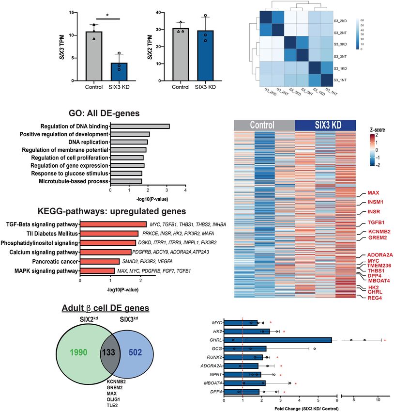
Downloaded from genesdev.cshlp.org on October 11, 2021 - Published by Cold Spring Harbor Laboratory Press

Bevacqua et al.

   We and others have found that SIX2 and SIX3, members                these studies demonstrate that SIX2 and SIX3 coordinate-
of the sine oculis homeobox family of TFs, are first ex-               ly regulate distinct genetic programs in human β cells, in-
pressed in the β cells of children by 9–10 yr of age (Blodgett         cluding expression of target genes governing mature β-cell
et al. 2015; Arda et al. 2016; Wang et al. 2016b; Cyphert              function and maintaining β-cell fate. Moreover, we show
et al. 2019), followed by increased expression in adult-               that SIX2 expression is reduced in β cells purified from hu-
hood. Moreover, neither SIX2 nor SIX3 mRNA are detect-                 man donors with T2D and impaired islet insulin secre-
able in human α cells (Blodgett et al. 2015; Arda et al. 2016;         tion, suggesting roles for SIX2 in the pathogenesis of
Wang et al. 2016b). SIX2 and SIX3 are encoded by linked                β-cell defects in T2D.
genes on human chromosome 2 (OMIM: 604994 and
603714). While they show high homology in their N-ter-
minal SIX domain and their central homeodomain (HD),                   Results
other portions of SIX2 and SIX3 differ (Kawakami et al.
                                                                       Reduced SIX2 or SIX3 expression impairs regulated
2000) and could mediate distinct interactions. For exam-
                                                                       insulin secretion by human islets
ple, SIX3, but not SIX2, interacts with members of the
groucho family of corepressors (Kobayashi et al. 2001; Ló-             We used RNAi-based strategies to reduce SIX2 or SIX3
pez-Ríos et al. 2003). In addition, SIX2 has roles in devel-           mRNA levels in primary human adult islets. After disper-
opment of kidneys, skull, stomach, and other organs                    sion of primary islets, lentiviral transduction was used to
(Kobayashi et al. 2008; Self et al. 2009; He et al. 2010;              achieve shRNA-mediated suppression of SIX2 or SIX3
Park et al. 2012), while SIX3 has roles in forebrain and               (hereafter, SIX2 kd or SIX3 kd) and to simultaneously ex-
eye development (Jeong et al. 2008; Liu et al. 2010).                  press a GFP transgene (Peiris et al. 2018), followed by reag-
   SIX2 and SIX3 have largely nonoverlapping tissue ex-                gregation into pseudoislets (Fig. 1A,B; Materials and
pression patterns that correspond with their distinct roles            Methods). We used immunostaining to detect β cells (in-
in human organogenesis. In the pancreas, however, both                 sulin [INS]), α cells (glucagon [GCG]), and δ cells (somatos-
SIX2 and SIX3 are expressed in human β cells, with coin-               tatin [SST]) in pseudoislets, and verified reaggregation of
cident onset of postnatal expression and a shared cis-regu-            these principal islet cell types in appropriate proportions
latory element that coregulates islet SIX2 and SIX3                    (Supplemental Fig. S1). Five days after lentiviral infection,
expression (Spracklen et al. 2018). This element encom-                we confirmed significant reduction of SIX2 or SIX3 in
passes single nucleotide polymorphisms (SNPs) associat-                pseudoislets by qRT-PCR (Fig. 1C,D; Materials and Meth-
ed with type 2 diabetes (T2D) and fasting hyperglycemia                ods). In contrast, we did not detect altered SIX3 mRNA
(Kim et al. 2011; Spracklen et al. 2020). Nevertheless, it re-         levels after SIX2 kd or altered SIX2 mRNA levels after
mains unknown whether SIX2 and SIX3 are required                       SIX3 kd (Fig. 1E,F), providing evidence that SIX2 and
for normal adult β-cell function, what β-cell genes these              SIX3 expression are not mutually cross-regulated.
TFs regulate, and whether their expression is altered in                 We subsequently assessed glucose-stimulated insulin
diabetes.                                                              secretion (GSIS) following SIX2 kd or SIX3 kd in primary hu-
   Prior studies suggest that SIX2 or SIX3 might regulate              man pseudoislets. Like in our prior studies (Peiris et al.
genes with roles in restricting native β-cell proliferation            2018), control pseudoislets infected with lentivirus ex-
and enhancing insulin production and secretion. The on-                pressing nontargeting shRNA (“control”) showed a signif-
set of SIX2 and SIX3 expression in postnatal human β cells             icant increase in insulin secretion after a glucose step
temporally coincides with age-dependent enhancement of                 increase from 2.8 to 5.6 mM, 16.7 mM, 25 mM, or
insulin production and secretion (Blodgett et al. 2015;                25 mM glucose supplemented with the secretion potenti-
Arda et al. 2016). Moreover, misexpression of SIX3 in im-              ator IBMX (Fig. 1G,I). By comparison, insulin secretion by
mature islets from children stimulated glucose-dependent               pseudoislets after SIX2 kd was significantly blunted in 2.8
insulin secretion (Arda et al. 2016). Neither SIX2 nor SIX3            and 16.7 mM glucose and trended toward reduction at 5.6
is expressed in mouse islets (Benner et al. 2014; Baron                mM (Fig 1G). Likewise, after SIX3 kd, there was significant
et al. 2016); thus, loss-of-function studies that clearly              blunting of insulin secretion in 2.8, 5.6, 16.7, and 25 mM
identify requirements for SIX2 or SIX3 in β-cell function              glucose (Fig. 1I). GSIS data were normalized to total pseu-
will require studies in native human islets or alternative             doislet insulin content, which was not significantly
experimental systems. Attempts to generate insulin-se-                 altered after SIX2 kd (Fig. 1H) or SIX3 kd (Fig. 1J). This sug-
creting β cells from human stem cells have only produced               gests that reduced insulin release from islets after SIX2 kd
immature progeny that express SIX2, often at low levels,               or SIX3 kd reflects impaired secretion. Together, our find-
and fail to express SIX3 (Sneddon et al. 2018; Nair et al.             ings provide index evidence that reduced SIX2 or SIX3
2019; Veres et al. 2019; Velazco-Cruz et al. 2020). To over-           function impairs human adult islet β-cell function.
come these limitations, here we investigated SIX2 and
SIX3 in adult human islets, using recently described ge-
                                                                       Elucidating the SIX2-dependent adult β-cell
netic methods permitting targeted loss of function in pri-
                                                                       transcriptome
mary human islets (Peiris et al. 2018). Controlled islet cell
dispersion and reaggregation to develop human “pseu-                   SIX2 is an established transcriptional regulator (Kobayashi
doislets” (Scharp et al. 1980; Tze and Tai 1982; Arda                  et al. 2008; Self et al. 2009; He et al. 2010; Park et al. 2012),
et al. 2016) allowed us to achieve shRNA-mediated SIX2                 but SIX2-dependent genetic targets in adult islet β cells are
or SIX3 suppression in human adult β cells. Collectively,              unknown. To identify genes regulated by SIX2, we purified

2      GENES & DEVELOPMENT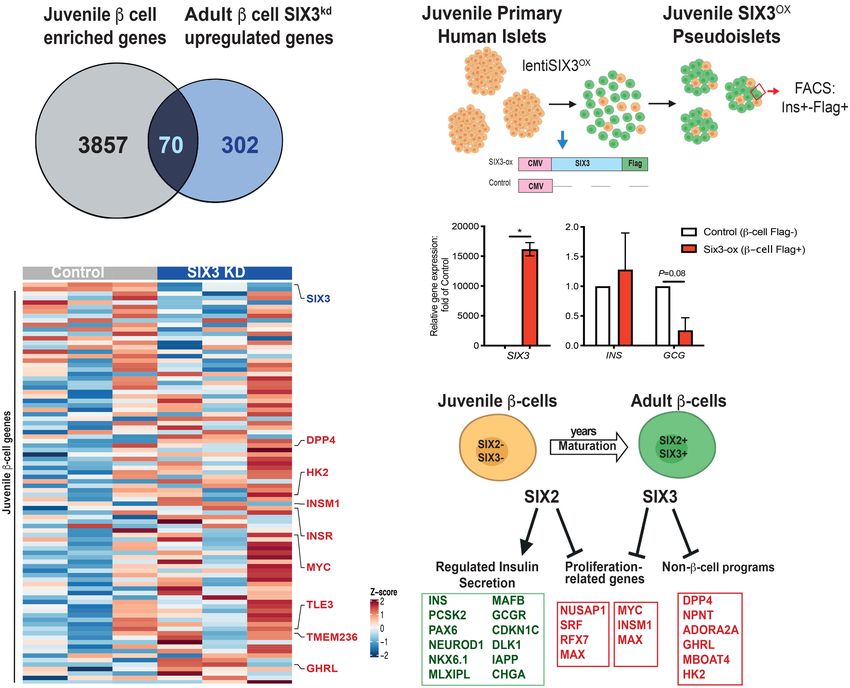
Downloaded from genesdev.cshlp.org on October 11, 2021 - Published by Cold Spring Harbor Laboratory Press

                                                                                                    SIX2 and SIX3 regulate human β-cell maturity

          A                                            B

                  C                            D                             E                              F

     G                                               H                        I                                             J

Figure 1. shRNA-mediated suppression of SIX2 and SIX3 in primary human islets results in impaired glucose-stimulated insulin secre-
tion. (A) Schematics of the lentiviral constructs coding for a short hairpin RNA (shRNA) and GFP. (B) Schematic detailing the pseudoislet
technique. (C ) SIX2 mRNA expression in primary human islets. (Gray bar) Control, (green bar) SIX2 kd. n = 9 independent donor repeti-
tions. (D) SIX3 mRNA expression in primary human islets. (Gray bar) Control, (blue bar) SIX3 kd. n = 11 independent donor repetitions.
SIX3 mRNA were not altered by SIX2 kd (n = 8 independent donor repetitions) (E), and SIX2 mRNA levels were not affected by SIX3 kd
(n = 11 independent donor repetitions) (F ). (G,I ) In vitro glucose-stimulated insulin secretion from human pseudoislets control, SIX2 kd
(n = 9 independent donor repetitions) (G), or SIX3 kd (n = 12 independent donor repetitions) (I). Secreted insulin normalized to insulin con-
tent. (Black lines) Significant differences within the control, (red lines) significant differences within the KD groups, (green lines) signifi-
cant differences between control and KD conditions. (H,J) Total insulin from human pseudoislets after transduction with SIX2 kd (n = 9
independent donor repetitions) (H) or SIX3 kd (n = 12 independent donor repetitions) (J). Data presented as mean; error bars represent
the standard error. Two-tailed t-tests used to generate P-values: (∗ ) P < 0.05, (∗∗ ) P < 0.01, (∗∗∗ ) P < 0.0001.

β cells from pseudoislets after SIX2 kd (Fig. 2A–C; Materials              the expected inter-donor variability we and others have
and Methods). Intracellular labeling with antibodies                       previously reported (Arda et al. 2016; Segerstolpe et al.
against insulin and glucagon followed by flow cytometry                    2016; Enge et al. 2017; Peiris et al. 2018). We used the
and GFP gating (Fig. 2C; Supplemental Fig. S2A; Peiris                     DE-Seq2 algorithm (Love et al. 2014) to identify differen-
et al. 2018) enriched for INS+ GFP+ β cells; qRT-PCR anal-                 tially expressed (DE) genes following SIX2 kd. Expression
ysis of this cell subset confirmed enrichment of mRNA en-                  of 1242 genes, including SIX2 itself, was significantly de-
coding INS, and depletion of GCG, or of the acinar and                     creased after SIX2 kd (Fig. 2D,G; Supplemental Table S2),
ductal cell markers, CPA1 and KRT19 (Supplemental                          whereas expression of 928 genes was significantly in-
Fig. S2B). We verified efficient shRNA-mediated suppres-                   creased (P < 0.05) (Fig. 2I; Supplemental Table S3). In con-
sion of SIX2 in INS+ GFP+ β cells compared with control                    trast, we did not detect changes in expression of β-cell
INS+ GFP+ β cells (Supplemental Fig. S2C). We then pro-                    SIX3 upon SIX2 kd (Fig. 2E). Gene ontology (GO) term anal-
duced and sequenced RNA-seq libraries from SIX2 kd and                     ysis (Fig. 2F) suggested unifying molecular functions in
control β cells (n = 4 independent donors) (Materials and                  these enriched gene sets, including terms related to adult
Methods). Pearson correlation and hierarchical clustering                  β-cell function, proliferation, and cell cycle regulation.
analysis revealed clustering of SIX2 kd and control samples                   Genes with decreased expression in SIX2 kd β cells in-
from the same donor (Supplemental Fig. S2D,E), reflecting                  cluded those encoding cardinal β-cell factors like INS,

                                                                                                                GENES & DEVELOPMENT           3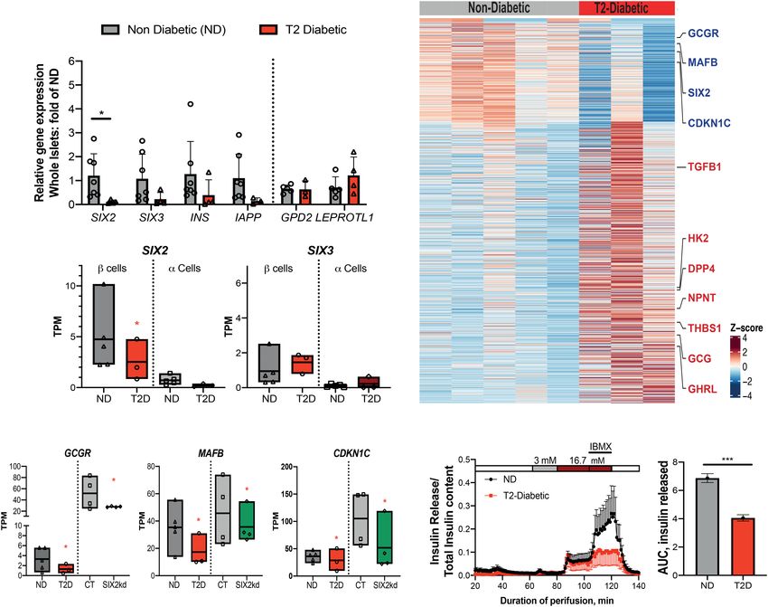
Downloaded from genesdev.cshlp.org on October 11, 2021 - Published by Cold Spring Harbor Laboratory Press

Bevacqua et al.

           A                        C                                                     D                   E

           B

                                                                           G
           F

     H

                  I

Figure 2. RNA-seq of SIX2 kd β cells reveals genes regulated by SIX2 in primary human islets. (A,B) SIX2 kd human pseudoislets. (A) Bright
field. (B) Blue light (488 nm). Scale bars, 500 µm. (C) FACS scheme used to sort GFP+ β cells. (D,E) Normalized transcript levels of SIX2 (D)
and SIX3 (E) in GFP+ β−cells. (Gray bar) Control, (green bar) SIX2 kd. n = 4. (F ) GO term enrichment in genes deregulated in β cells post-
SIX2 kd. (G,H) KEGG pathway enrichment in genes up-regulated (G) or down-regulated (H) in β cells post-SIX2 kd (n = 4). (I) Heat map show-
ing all differentially expressed (DE) genes in β cells post-SIX2 kd. The data are presented as mean; error bars represent the standard error.
(∗ ) P < 0.05

CHGA, CHGB, and IAPP; insulin processing enzymes                         that SIX2 expression may enforce β-cell cycle arrest and
like CPE and PCSK2; and transcription factors like                       studies linking β-cell cycle exit to enhanced function (Hel-
PAX6, NEUROD1, NKX6.1, MLXIPL, TCF7L2, ESRRG,                            man et al. 2016). GO and KEGG pathway analysis of DE
and MAFB (Fig. 2G,H; Supplemental Table S2). In addi-                    genes (Materials and Methods) revealed significant en-
tion, we noted severe reduction of mRNAs encoding glu-                   richment of terms including regulation of hormone secre-
cokinase (GCK), the principal sensor of glucose flux in                  tion, glucose homeostasis, calcium signaling, and insulin
β cells (Matschinsky et al. 1993); glucagon receptor                     signaling (Fig. 2F,H,I). Thus, these data suggest that SIX2
(GCGR); regulators of glycolysis and β-cell stimulus secre-              is required to maintain hallmark adult β-cell functions in-
tion coupling encoded by TPI1, ALDH2, ALDOA, ENO2,                       volved with insulin production and processing, glucose
PGK1, and FBP1; and CAMK1D, a postulated type 2 diabe-                   sensing, and proliferation, and support our finding of im-
tes risk gene (Fig. 2G; Thurner et al. 2018; Miguel-Esca-                paired GSIS after SIX2 kd in adult human islets.
lada et al. 2019). In contrast, we observed increased                       To validate our findings further, we assessed whether
levels of mRNAs encoding regulators of DNA replication                   genes regulated by SIX2 kd were enriched in gene sets
and cell cycle factors in SIX2 kd β cells, including                     whose expression changed in β cells expressing SIX2 (see
NUSAP1, MAX, and SRF (Fig. 2G,I). This is consistent                     the Materials and Methods; Blodgett et al. 2015; Arda
with prior findings (Arda et al. 2016) providing evidence                et al. 2016). For example, we asked whether the set of

4        GENES & DEVELOPMENT
Downloaded from genesdev.cshlp.org on October 11, 2021 - Published by Cold Spring Harbor Laboratory Press

                                                                                          SIX2 and SIX3 regulate human β-cell maturity

genes with decreased expression after SIX2 kd overlapped             this data set with DE transcriptomic data recently reported
with DE genes whose mRNA normally increased with ad-                 after SIX2 loss in β-like cells (“SC-β”) generated from a hu-
vancing age. In this case, we identified 287 genes with de-          man embryonic stem cell line (Velazco-Cruz et al. 2020).
creased expression after SIX2 kd in β cells, whose                   We found a general lack of concordance in DE genes (Mate-
expression was significantly increased in SIX2+ adult                rials and Methods): Specifically, we observed 30% (377/
β cells compared with fetal or juvenile SIX2neg β cells              1242) overlap of genes with reduced expression and 14%
(P < 3.873 × 10⁻³⁰) (Fig. 3A,B; Supplemental Table S7).              (132/928) overlap of genes with increased expression after
These included CDKN1C and CDKN1A, which encode in-                   SIX2 kd (Supplemental Fig. S3A,B; Supplemental Table
hibitors of cell proliferation; CACNA1C and CACNA1D,                 S12). Many mRNAs that decreased after SIX2kd in native
which encode calcium channels; GCGR, which encodes a                 β cells, like MAFB, GIPR, and GREM1, did not change after
receptor for glucagon and the incretin GLP-1 (Svendsen               SIX2 kd in SC-β cells. Other mRNAs that decreased after
et al. 2018); and MLXIPL (also known as ChREBP), a glu-              SIX2 kd in adult β cells, like CDKN1C, GCGR, TCF7L2,
cose-activated transcription factor that regulates GCGR              and NKX6.1, were found increased after SIX2 kd in SC-β
(Iizuka et al. 2012), GREM1, IAPP, and DLK1 (Fig. 3A,B;              cells (Velazco-Cruz et al. 2020). These findings indicate
Supplemental Table S7). Based on similar logic, we identi-           that genetic programs in native human β cells and SC-β
fied 146 genes with significantly increased expression after         cells are distinct and demonstrate advantages of investi-
SIX2 kd, whose expression is normally decreased in SIX2+             gating SIX2-dependent gene expression in primary adult
adult β cells (P < 5.2 × 10⁻⁵) (Fig. 3B; Supplemental Table          β cells.
S7; Blodgett et al. 2015; Arda et al. 2016). These included
NUSAP1 and SRF, postulated regulators of islet cell prolif-
                                                                     Identifying direct genetic targets of SIX2 regulation
eration (Zeng et al. 2017), and RFX7, a marker of pancreatic
                                                                     in human β cells
progenitor cells (Kim-Muller et al. 2016). We also compared
                                                                     To identify direct genetic targets of SIX2 in primary hu-
                                                                     man islet cells, we performed cleavage under targets and
                                                                     release using nuclease (CUT&RUN) (Skene and Henikoff
A                                                                    2017; Hainer et al. 2019), which enables the sensitive
                                                                     detection of genomic loci bound by TFs (Fig. 4A; Materials
                                                                     and Methods). Because antibodies that detected native is-
                                                                     let SIX2 for CUT&RUN were not available, we misex-
                                                                     pressed in pseudoislets a transgene encoding human
                                                                     SIX2 tagged with the FLAG immuno-epitope (SIX2-
                                                                     FLAG) from a rat insulin promoter element (RIP) (Karls-
                                                                     son et al. 1987), then sequenced DNA bound by the
B
                                                                     SIX2-FLAG protein with an anti-FLAG antibody (Supple-
                                                                     mental Fig. S4A). Thus, interpretation of CUT&RUN
                                                                     here is qualified by the possibility that SIX2-FLAG may
                                                                     bind sites not bound by native SIX2. We used the HOMER
                                                                     algorithm (Heinz et al. 2010) to identify genomic regions
                                                                     that were bound by SIX2-FLAG in samples from three islet
                                                                     donors (P ≤ 0.01 compared with IgG controls). Heat map
                                                                     visualization of independent peaks (Fig. 4B) as well as his-
                                                                     togram plotting of averaged reads (Supplemental Fig. S4B)
                                                                     showed enrichment of read densities in the peak centers
                                                                     for the SIX2-FLAG libraries, whereas IgG controls showed
                                                                     minimal enrichment at these sites (Fig. 4B; Supplemental
                                                                     Fig. S4B). As further validation of the specificity of
                                                                     CUT&RUN, we found that SIX2-FLAG-bound genomic
                                                                     peaks were significantly enriched for the SIX2 DNA-bind-
                                                                     ing motif, as well as the SIX1 DNA-binding motif, and mo-
                                                                     tifs of other β-cell-enriched TFs, like MAFB (Fig. 4C). We
                                                                     used the GREAT algorithm (McLean et al. 2010) to associ-
                                                                     ate SIX2-FLAG-bound genomic regions to 10,270 genes
                                                                     (Fig. 4D). Inadequate yields precluded analogous SIX3-
                                                                     FLAG studies (Materials and Methods).
                                                                        To nominate candidate genes that might be directly reg-
Figure 3. SIX2 kd in primary β cells results in down-regulation of   ulated by SIX2, we then identified genes that (1) neigh-
genes enriched in adult β-cell. (A) Venn diagram showing adult       bored SIX2-FLAG binding sites and (2) had differential
β-cell genes down-regulated post-SIX2 kd. (B) Heat map of adult      expression after SIX2 kd. After intersection of CUT&RUN
down-regulated genes and juvenile up-regulated genes in adult        with 2170 DE genes after SIX2 kd, we identified 1186 “over-
β cells post-SIX2 kd.                                                lapping genes” (Fig. 4D; Supplemental Table S4). Sixty-

                                                                                                    GENES & DEVELOPMENT             5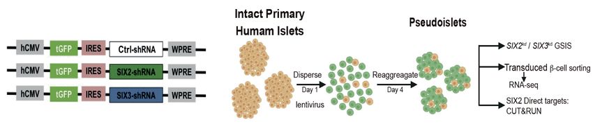
Downloaded from genesdev.cshlp.org on October 11, 2021 - Published by Cold Spring Harbor Laboratory Press
Bevacqua et al.
A B C
D E F
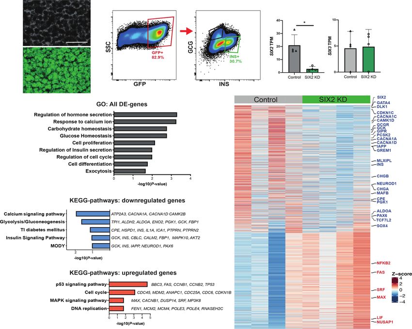
Figure 4. Identification of presumptive SIX2 genetic targets in primary human β cells using CUT&RUN. (A) Schematic of the
CUT&RUN approach: Pseudoislets overexpressing SIX2-FLAG under the RIP promoter were used for CUT&RUN with anti-FLAG anti-
body (n = 3 independent donors). (B) Heat map showing enrichment of peak read densities at the center of the peak for the CUT&RUN
libraries generated with FLAG antibody, but not for IgG. Peaks were called using HOMER. (C ) Enriched motifs in the differential peaks
were identified by HOMER. (D) Overlap of the SIX2-associated genomic regions and the SIX2 kd DE genes. (E,F) Tracks showing SIX2-
FLAG genomic regions associated with GCGR (E) or CHGA (F ). Accessible chromatin regions in human islets are shown by ATAC-
seq, H3K4me3, and H3K27ac ChiP-seq tracks. SIX2-FLAG CUT&RUN peaks are shown in pink boxes (note: for GCGR, two peaks are
shown), and regulated genes are highlighted in green boxes.
four percent of these genes (754/1186) had reduced mRNA cells, we used flow cytometry to purify β cells from human
levels after SIX2 kd, consistent with a role for SIX2 in acti- pseudoislets after SIX3 kd (Supplemental Fig. S5A–C; Ma-
vating β-cell gene expression. Presumptive direct SIX2 tar- terials and Methods). Like in our SIX2 kd studies, we con-
gets identified by this approach included PAX6, IAPP, firmed enrichment of INS+ GFP+ β cells—and depletion
MAFB, CDKN1C, DLK1, PCSK2, GCGR, MLXIPL, and of non-β cells—with qRT-PCR analysis (Supplemental
CHGA. As expected, SIX2-FLAG-bound genomic regions Fig. S5D), then generated, sequenced, and analyzed
in presumptive SIX2 target genes were found in accessible RNA-seq libraries (n = 3 independent donors). We
chromatin and colocalized with activation-associated achieved 50% reduction of SIX3 mRNA in purified INS+
H3K4me3 and H3K27ac histone marks identified by prior GFP+ β cells (Fig. 5A; Supplemental Fig. S5E). In contrast,
islet ATAC-seq and ChIP-seq studies (Mularoni et al. SIX2 mRNA levels were not detectably changed (Fig. 5A).
2017). This alignment further supports the conclusion As expected, analysis of RNA-seq libraries with Pearson
that SIX2 binds active genomic regulatory elements gov- correlation analysis and hierarchical clustering revealed
erning hallmark β-cell genes (Fig. 4E,F; Supplemental Fig. close clustering of SIX3 kd and control β-cell samples
S4C–E; Supplemental Table S4). Thus, our targeted nucle- from the same donor (Fig. 5B; Supplemental Fig. S5F).
ase-based analysis revealed hundreds of SIX2-associated With the DE-Seq2 algorithm (Materials and Methods),
candidate regulatory elements, including many that likely we identified 263 genes with significantly decreased
regulate expression of hallmark β-cell factors. mRNA levels (P < 0.05) (Supplemental Table S5), includ-
ing SIX3 itself (Fig. 5A), and 372 genes with significantly
increased mRNA levels in SIX3 kd β cells (P < 0.05) (Fig.
SIX3 and SIX2 regulate distinct gene sets in adult human
5E; Supplemental Table S6). Thus, the number of DE
β cells
genes with increased mRNA outnumbered those with de-
Gain-of-function studies have linked SIX3 to adult human creased mRNA after SIX3 kd, unlike DE genes after SIX2 kd.
β-cell functional maturation (Arda et al. 2016). To identify Gene ontology (GO) analysis suggested molecular func-
genes whose expression is regulated by SIX3 in human β tions of the enriched gene sets, including regulation of
6 GENES & DEVELOPMENT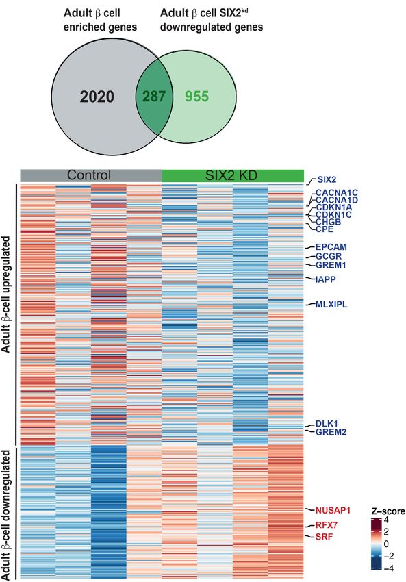
Downloaded from genesdev.cshlp.org on October 11, 2021 - Published by Cold Spring Harbor Laboratory Press

                                                                                                      SIX2 and SIX3 regulate human β-cell maturity

                         A                                                              B

                C                                                                E

                D

                        F                                                 G

Figure 5. RNA-seq of SIX3 kd β cells reveals a distinct gene set regulated by SIX3. (A) Normalized transcript levels of SIX3 and SIX2 in
GFP-expressing β cells. (Gray bar) Control, (blue bar) SIX3 kd . n = 3. (B) Heat map of the sample-to-sample distances for all the samples used
in this experiment. (C) GO term enrichment in genes deregulated in β cells post-SIX3 kd. (D) KEGG pathways enriched in genes up-regu-
lated in β cells post-SIX3 kd (n = 3). (E) Heat map showing all up-regulated genes upon SIX3 kd in β cells. (F ) Overlapped DE genes in adult
β cells post SIX2 kd and SIX3 kd. (G) Fold transcript levels of non-β-cell genes significantly altered in β cells post-SIX3 kd (n = 3). The data are
presented as mean; error bars represent the standard error. (∗ ) P < 0.05.

DNA binding, response to glucose stimulus, and DNA                            Table S8; Supplemental Fig. S6A,B). Moreover, among
replication (Fig. 5C; Supplemental Fig. S5G). Up-regulated                    these 133 DE genes, 112/133 (84%) changed in the oppo-
DE genes associated with the latter term included estab-                      site direction after SIX2 kd compared with SIX3 kd, such
lished regulators of β-cell replication, like MYC, MAX,                       as KCNMB2, GREM2, OLIG1, and TLE2. After SIX3 kd
and INSM1 (Fig. 5C,E). Pathway analysis of the DE up-reg-                     there was also a significant increase of mRNAs encoding
ulated genes (Materials and Methods) included TGF-β sig-                      genes not usually expressed in adult human β cells. This
naling, type 2 diabetes mellitus, and calcium signaling                       included genes encoding factors enriched or exclusively
pathway (Fig. 5D).                                                            expressed in islet α cells or ε cells, like DPP4, NPNT,
   The DE genes after SIX3 kd were largely distinct from DE                   TMEM236, ADORA2A, and GHRL, which encodes the
genes after SIX2 kd; only 133/2805 DE genes (
Downloaded from genesdev.cshlp.org on October 11, 2021 - Published by Cold Spring Harbor Laboratory Press

Bevacqua et al.

reaching statistical significance, we also detected an aver-             donors (ages 1.5 and 3 yr) (Fig. 6C; Supplemental Table
age 2.5-fold increase of mRNA encoding GCG in SIX3 kd β                  S1). Flow cytometry and western blotting verified and
cells (Fig. 5G). In SIX3 kd β cells, we also identified in-              quantified expression of transgenic SIX3-FLAG (Supple-
creased average levels of 70 mRNAs that are typically ex-                mental Fig. S7A–C). qRT-PCR analysis of purified β cells
pressed highly in fetal β cells, but attenuated or                       showed a reduction in GCG levels and an average increase
extinguished in adult β cells (Fig. 6A,B; Supplemental Ta-               of INS mRNA levels following SIX3 misexpression (Fig.
ble S9; Blodgett et al. 2015; Arda et al. 2016). This latter             6D), as previously reported (Arda et al. 2016). RNA-seq
group of genes included MYC (Puri et al. 2018) and a “dis-               of purified SIX3-FLAG+ INS+ β cells (Materials and Meth-
allowed” gene (Pullen et al. 2010) that encodes hexokinase               ods) confirmed reduced mRNAs encoding GCG, HK2,
(HK2) (for review, see Lemaire et al. 2016). Moreover, none              MYC, TMEM236, MBOAT4, RUNX2, GREM2, and
of these mRNAs increased in β cells after SIX2 kd. Togeth-               NPNT, mRNAs found increased after SIX3 kd in primary
er, our findings support the view that SIX2 and SIX3 regu-               β cells (Supplemental Fig. S7D; Supplemental Table
late distinct gene sets in human β cells.                                S10). Thus, SIX3 gain- and loss-of-function studies here
   We previously showed that SIX3 expression in pseu-                    produced reciprocal changes in expression of multiple
doislets from juvenile human donors aged 0.5–2 yr (which                 genes, supporting the view that SIX3 suppresses adult β-
lack SIX3 expression) was sufficient to stimulate insulin                cell expression of gene sets expressed abundantly in fetal
secretion in vitro (Arda et al. 2016); however, transcrip-               or neonatal β cells, or in adult α and ε cells. We conclude
tome studies were not performed. To assess changes of                    that SIX3 reinforces mature β-cell function, in part, by
gene expression stimulated by SIX3, we misexpressed a                    suppressing fetal gene expression programs and alterna-
SIX3-FLAG transgene in pseudoislets from two juvenile                    tive islet cell fates (Fig. 6E).

            A                                                     C

                                                                  D
            B

                                                                  E

Figure 6. SIX3 represses non-β-cell programs in the adult β-cell. (A) Venn diagram showing juvenile β-cell genes up-regulated post-SIX3 kd
in adult β cells. (B) Heat map of adult down-regulated and juvenile up-regulated genes in adult β cells post-SIX3 kd. (C) Schematic detailing
the juvenile pseudoislet technique used to overexpress SIX3-FLAG (SIX3-ox) in juvenile pseudoislets (n = 2) and of the constructs used to
overexpress SIX3 in juvenile pseudoislets: FACS was used to sort FLAG+ β cells. (D) SIX3, INS, and GCG mRNA expression in FLAG+-ex-
pressing β cells post FACS. (White bar) Control , (red bar) SIX3-ox. n = 2. (E) Schematics showing proposed coordinated regulation of mat-
uration by SIX2 and SIX3 in the β-cell. The data are presented as mean; error bars represent the standard error. (∗ ) P < 0.05.

8      GENES & DEVELOPMENT
Downloaded from genesdev.cshlp.org on October 11, 2021 - Published by Cold Spring Harbor Laboratory Press
SIX2 and SIX3 regulate human β-cell maturity
Human β-cell expression of SIX2 is reduced in islets in purified β cells from T2D islets compared with β cells
from T2D donors from control islets (P ≤ 0.01) (Fig. 7B–C; Supplemental Ta-
ble S11), while SIX3 mRNA levels were not significantly
It remains unknown whether the expression of SIX2 or changed (Fig. 7D). Thus, SIX2 expression was reduced in
SIX3 changes in islet β cells obtained from T2D patients. β cells in islets from cadaveric T2D donors. Moreover, β-
We found that both SIX2 and SIX3 mRNA appeared to be cell regulation of SIX2 and SIX3 in these T2D islets was
reduced in purified whole islets from subjects with estab- uncoupled. We also detected little to no expression of
lished T2D (n = 4), compared with islets from nondiabetic SIX2 and SIX3 in purified α cells from control or T2D is-
controls (n = 7) (Fig. 7A; for donor information, see Supple- lets (Fig. 7C,D). In addition to reduced SIX2 expression
mental Table S1). In contrast, expression of GPD2 and in β cells, we also noted impaired expression of SIX2 target
LEPROTL2, previously found up-regulated in prior stud- genes, including MAFB, GREM1, GCGR, and CDKN1C, a
ies (Segerstolpe et al. 2016), was unaltered in T2D islets candidate T2D risk gene (Fig. 7B,E–G; Supplemental Ta-
(Fig. 7A). ble S11). Consistent with our studies of impaired insulin
Bulk RNA-seq from FACS-purified α and β cells was secretion after SIX2 kd (Fig. 1G), glucose-stimulated insu-
used to assess SIX2 and SIX3 expression in β cells (Fig. lin secretion studies revealed impaired insulin secretion
7B–D; see the Materials and Methods). Transcriptome by islets from subjects with T2D compared with nondia-
analysis confirmed significant reduction of SIX2 mRNA betic controls (Fig. 7H,I; see the Materials and Methods).
A B
C D
E F G H I
Figure 7. SIX2 expression is reduced in β cells from T2D donors. (A) Gene expression levels in whole islets from nondiabetic (ND; gray
bars) (n = 5) or type 2 diabetic (T2D; red bar) (n = 3). (B) Heat map showing DE genes in nondiabetic versus T2 diabetic β cells. (C,D) Box plots
displaying TPM counts of SIX2 (C ) and SIX3 (D) in nondiabetic (ND) (n = 5) β cells (dark-gray bars) and α cells (light-gray bars) or type 2
diabetic (T2D) β cells (red bars) and α cells (dark-red bars) (n = 3). (E–G) Box plots displaying TPM GCGR (E), MAFB (F ), and CDKN1C
(G) in β-cells of ND (n = 5) and T2D (n = 3), the expression of which is DE in adult β cells post-SIX2 kd. (Gray bars) Çontrol, (green bars)
SIX2 kd. (H) Insulin secretion of ND (n = 5) versus T2D (n = 3; see the Materials and Methods). (I ) Plot of the total area under the curve
of the released insulin. The data in A are presented as mean; error bars represent the standard error. Box plots show the mean. (Red ∗ )
P ≤ 0.01, (black ∗ ) P < 0.05.
GENES & DEVELOPMENT 9Downloaded from genesdev.cshlp.org on October 11, 2021 - Published by Cold Spring Harbor Laboratory Press
Bevacqua et al.
Although SIX3 mRNA levels were not detectably like MKI67 (Supplemental Tables S2–S5). Moreover, we
changed in our sampling of T2D β cells, a subset of SIX3 showed in our prior work that misexpression of either
targets was differentially expressed in T2D β cells, includ- SIX2 or SIX3 was sufficient to suppress proliferation of
ing DPP4, NPNT, and GHRL; TGF-β signaling factors en- the human β-cell line EndoCβH1 (Arda et al. 2016). En-
coded by TGFB1 and THBS1; and the disallowed gene forcement of the post-mitotic state in β cells has been
HK2 (Fig. 7B). This raises the possibility that factors col- linked to attainment of mature function (Helman et al.
laborating with SIX3 to regulate these genes might be 2016; Puri et al. 2018; Mandelbaum et al. 2019).
changed in T2D β cells. The majority of differentially expressed genes after
Together, these findings suggest that dysregulation of SIX2 kd showed reduced expression, including those en-
SIX2- and SIX3-dependent genetic programs could con- coding crucial human β-cell factors like insulin and gluco-
tribute to impaired islet β-cell fate and function in T2D. kinase, and essential TFs that coordinate pancreatic islet
These findings also support prior genome-wide associa- development and β-cell function in humans. Consistent
tion studies linking the locus encoding SIX2 and SIX3 to with this, we observed reduced insulin secretion after
risk for T2D and diabetes-related traits (Kim et al. 2011; SIX2 kd. In contrast, after SIX3 kd the majority of DE genes
Hachiya et al. 2017; Varshney et al. 2017; Spracklen showed increased expression. These included transcripts
et al. 2018, 2020), raising the possibility that genetic influ- encoding factors not normally expressed in healthy β cells,
ences might additionally modulate β-cell expression of like the ε-cell hormone ghrelin, the α-cell-enriched prote-
SIX2 or SIX3. ase DPP4, and the disallowed factor HK2 (Dhawan et al.
2015). These SIX3 kd findings are consistent with previous
studies suggesting that SIX3 can function as a transcrip-
Discussion tional repressor (Kobayashi et al. 2001). Thus, our loss-
of-function studies revealed SIX2- and SIX3-dependent
Here, we overcame inherent challenges facing postnatal mechanisms that regulate native maturation and fate of
human developmental studies to investigate the roles of human β cells. Here, the degree of SIX2 mRNA reduction
SIX2 and SIX3 in human pancreatic β-cell maturation. Us- after SIX2 kd was greater than the degree of SIX3 mRNA
ing genetic approaches, we show that SIX2 is required for loss after SIX3 kd; thus, future studies that achieve more
expression of multiple hallmark genes in human β cells. In complete SIX2 or SIX3 loss of function could identify ad-
addition to regulation of genes governing β-cell function, ditional β-cell genetic targets. Our study was also limited
we show that SIX3—unlike SIX2—suppresses expression by the inherent variability of cadaveric human islet do-
of genes typically expressed in α cells or other non-β cells. nors, as we and others have previously reported (Arda
Thus, our studies provide index evidence that SIX2 and et al. 2016; Segerstolpe et al. 2016; Enge et al. 2017; Peiris
SIX3 regulate distinct sets of genetic targets in adult hu- et al. 2018). While SIX3 expression is restricted to the β
man β cells. shRNA-mediated suppression of either SIX2 cell, SIX2 is also expressed in islet δ cells (Baron et al.
or SIX3 expression in primary human islets impaired reg- 2016; Muraro et al. 2016). Thus, changes observed after
ulated insulin secretion by β cells. Supporting these find- SIX2 kd could reflect both β-cell autonomous and nonau-
ings, we found evidence of reduced expression of SIX2 tonomous mechanisms. Studies here also revealed that
and downstream targets in islet β cells from human sub- SIX2 and SIX3 expression in β cells can be genetically un-
jects with T2D, which coincided with significantly re- coupled and correspond well with our data showing that
duced insulin secretion by these islets. In sum, our SIX2 and SIX3 regulate distinct β-cell gene sets. Thus, dis-
study unveils a requirement for SIX2 and SIX3 in estab- tinct mechanisms likely govern expression and activity of
lishing and maintaining adult human β-cell function and SIX2 and SIX3 in β cells from healthy and diabetic cadav-
fate (Fig. 6E). eric donors.
Prior to our study, it was unclear what roles SIX2 and Elucidating how SIX2 and SIX3 expression are regulated
SIX3 had in adult human β-cell function. SIX2 and SIX3 should be aided by our identification of their targets in hu-
are coexpressed in adult human β cells, and developmen- man β cells. Prior studies have shown that transcription
tal studies of human islet cells have revealed coincident factors like SIX2 and SIX3 expressed in human β cells
increases of SIX2 and SIX3 expression after the first dec- show increased expression with age (Aguayo-Mazzucato
ade of life (Arda et al. 2016, 2018; Blodgett et al. 2015). et al. 2011; Blodgett et al. 2015; Arda et al. 2016; Wang
Moreover, studies of putative SIX2 and SIX3 cis-regulato- et al. 2016b). For shRNA-based studies here we used islets
ry elements in humans and other systems have suggested from donors >22 yr of age, when adult levels of SIX2 and
these genes may be coregulated (Spracklen et al. 2018; Suh SIX3 have been established. Intense interest in SIX2 and
et al. 2010). A common set of SIX2 and SIX3 targets iden- SIX3 regulation also stems from association of the locus
tified here includes regulators of cell cycle progression. encoding these factors to T2D and related traits like fast-
However, the onset of SIX2 and SIX3 in human β cells oc- ing hyperglycemia (Kim et al. 2011; Hachiya et al. 2017;
curs well after the period of neonatal expansion (Blodgett Varshney et al. 2017; Spracklen et al. 2018, 2020). Howev-
et al. 2015; Arda et al. 2016), suggesting that the post-mi- er, studies of whole islet RNA, or prior single islet cell
totic state of β cells is established by other factors, and RNA-seq investigations by us and others (Segerstolpe
then reinforced by SIX2 and SIX3. Consistent with this et al. 2016; Camunas-Soler et al. 2020) were not sufficient-
possibility, we observed that reduction of SIX2 alone or ly sensitive to detect changes of SIX2 or SIX3 mRNA in β
SIX3 alone did not increase markers of β-cell S-phase cells isolated from donors with T2D. Studies here provide
10 GENES & DEVELOPMENTDownloaded from genesdev.cshlp.org on October 11, 2021 - Published by Cold Spring Harbor Laboratory Press
SIX2 and SIX3 regulate human β-cell maturity
index evidence that (1) expression of SIX2, and a subset of position. Our studies further suggest that simultaneous
SIX2-dependent genes like GCGR, are significantly re- expression of both SIX2 and SIX3 may be required to pro-
duced in β cells from T2D donors, and (2) islets from duce consummately functional replacement β cells from
T2D donors with reduced β-cell expression of SIX2 had renewable sources, like human stem cell lines.
impaired insulin secretion (Fig. 7). While we did not detect In summary, this study unveils SIX2 and SIX3 functions
changes in SIX3 expression in T2D islets here, additional crucial for postnatal human islet and β-cell development
studies are required to exclude the possibility that β-cell and maturation and reveals how β-cell dysfunction might
SIX3 dysregulation is a feature of T2D. develop in diabetes. Our work demonstrates that SIX2 and
A recent report described phenotypes after SIX2 loss in SIX3 coordinately govern distinct genetic programs that
hPSC-derived β-like cells (SC-β cells) (Velazco-Cruz et al. increase insulin production and enhance mature β-cell
2020). While 25% of these insulin+ SC-β cells express physiological functions, enforce β-cell fate by suppressing
SIX2 mRNA, they lack other markers of mature β cells alternative genetic programs, and suppress proliferation.
like MAFB or SIX3, thus precluding studies of SIX3 loss Findings here also provide a unique developmental “road-
of function. After shRNA-mediated suppression of SIX2, map” for achieving human β-cell replacement.
Velazco-Cruz et al. (2020) reported reduced insulin pro-
tein content, loss of glucose-stimulated insulin secretion
without effects on “basal” insulin secretion at low glu- Materials and methods
cose concentration (2 mM), and significant changes in ex-
pression of >10,000 genes, assessed by RNA-seq of Human islet procurement
unsorted hESC progeny, with enrichment of gene sets re- Deidentified human islets were obtained from healthy, nondia-
lated to insulin secretion and calcium signaling. This in- betic organ donors or type 2 diabetic donors procured through
cluded significant increases of multiple transcripts the Integrated Islet Distribution Network (IIDP), National Diabe-
encoding islet α cell or δ cell products, like SST, DPP4, tes Research Institute (NDRI), International Institute for the Ad-
MBOAT4, and FSTL1 (Velazco-Cruz et al. 2020). These vancement of Medicine (IIAM) and the Alberta Diabetes Institute
findings support the conclusion that SIX2 is required for Islet Core. For T2D studies, data from the Human Pancreas Anal-
SC-β cells derived from hESCs to acquire some features ysis Program (HPAP-RRID: SCR_016202) Database (https://hpap
.pmacs.upenn.edu), a Human Islet Research Network (RRID:
of native human β cells.
SCR_014393) consortium (UC4-DK-112217 and UC4-DK-
In our study, we assessed the effects of SIX2 or SIX3 loss 112232) was used. See Supplemental Table S1 for details.
in native human β cells. After SIX2 kd, we observed reduc-
tion of both basal and glucose-stimulated insulin secre-
tion, without reduction of islet insulin content. FACS Constructs and lentivirus production
purification of Insulin+ β cells (and elimination of SIX2+
Lentiviral constructs coding for shRNAs targeting human SIX3 or
δ cells) and RNA-seq revealed significant changes in
SIX2 were obtained from Dharmacon. plenti-CMV-SIX3-cMyc-
mRNA levels of 2100 genes, with gene set analysis reveal- DDK was used in juvenile islet experiments (Origine). plenti-
ing enrichment of terms related to proliferation, insulin RIP-SIX2-cMyc-DDK was generated by replacing the CMV pro-
secretion, calcium signaling, carbohydrate metabolism, moter of plenti-CMV-SIX2-cMyc-DDK (Origine) with the rat in-
and exocytosis. While some of these gene sets overlapped sulin promoter (RIP). Lentiviruses were produced by
with those in SC-β cells (Velazco-Cruz et al. 2020), there transfection of HEK293T cells with lentiviral constructs,
was an overall lack of concordance between gene sets pMD2.G (Addgene 12259) and psPAX2 (Addgene 12260) packag-
(Supplemental Fig. S3; Supplemental Table S12). For ex- ing constructs. Supernatants were collected and concentrated
ample, after SIX2 kd we observed reduced mRNA encoding by PEG-it (System Biosciences).
the calcium channel subunits CACNA1A and CAC-
NA1D (“calcium signaling”), incretin receptors GIPR
Human pseudoislet generation and transduction
and GCGR (“insulin secretion”), and transcription factors
with established roles in native β-cell regulation like Human islets were dispersed into single cells by enzymatic diges-
MAFB, NEUROD1, NKX6.1, and TCF7L2; these changes tion (Accumax, Invitrogen) and transduced with 1 × 10⁹ viral
were not noted in SC-β cells. Moreover, we did not observe units/1 mL lentivirus. Transduced islet cells were cultured in ul-
increased expression of non-β-cell markers like DPP4, tra-low attachment well plates for 5 d prior to further analysis.
MBOAT4, and FSTL1 after SIX2 kd. Instead, we observed
increased expression of these genes, and other non-β-cell
RNA extraction and quantitative RT-PCR
or disallowed genes after SIX3 kd. These contrasts raise
the possibility that SIX2 activity in SC-β cells includes ec- RNA was isolated from whole pseudoislets using the PicoPure
topic functions normally fulfilled by SIX3 or other factors. RNA isolation kit (Life Technologies). For sorted β and α cells,
Together, our findings clarify the importance of investi- RNA was isolated using the RecoverALL isolation kit (Invitrogen
by Thermo Fisher Scientific). cDNA was synthesized using the
gating SIX2 and SIX3 functions in bona fide adult β cells.
Maxima first strand cDNA synthesis kit (Thermo Scientific),
While models of human β-cell development, like stem and gene expression was assessed by PCR using TaqMan gene ex-
cell-derived insulin+ cells and immortalized β-cell lines pression mix (Thermo Scientific) and the following probes AC-
have value (Sneddon et al. 2018), to date they remain fun- TIN-B, Hs4352667_m1; SIX2, Hs00232731_m1; SIX3, Hs00193
damentally different from genuine pancreatic islet cells in 667_m1; insulin, Hs00355773_m1; glucagon, Hs00174967_m1;
gene regulation, function, proliferation, and cellular com- CPA-1, Hs00156992_m1; and KRT19, Hs01051611_gH.
GENES & DEVELOPMENT 11Downloaded from genesdev.cshlp.org on October 11, 2021 - Published by Cold Spring Harbor Laboratory Press
Bevacqua et al.
In vitro insulin secretion assays the two experimental conditions. The Database for Annotation,
Visualization, and Integrated Discovery (DAVID) v6.7 was used
Batches of 25 pseudoislets were used for in vitro secretion assays
(Huang et al. 2009) for gene set enrichment analysis. RNA-seq
as previously described (Peiris et al. 2018). Briefly, pseudoislets
data sets of genes enriched in adult β cells versus juvenile β cells
were incubated at 2.8, 5.6, 16.7, 25, and 25 mM + IBMX glucose
were obtained from Arda et al. (2016) (GEO: GSE79469) and
concentrations for 60 min each, and supernatants were collected.
Blodgett et al. (2015) (GEO: GSE67543). The probability of finding
Secreted human insulin in the supernatants and pseudoislet ly-
x overlapping genes was calculated using the hypergeometric
sates were quantified using a human insulin ELISA kit (Merco-
probability formula that considers the total number of genes in
dia). Secreted insulin levels are presented as a percentage of
the genome. RNA-seq data of sorted β and α cells of T2D versus
total insulin content. Perifusion data of T2 diabetic versus nondi-
nondiabetic donors (HPAP-RRID:SCR_016202) and of SIX2 kd
abetic samples were acquired from the Human Pancreas Analysis
SC-β cells (GEO: GSE147737) (Velazco-Cruz et al. 2020) were an-
Program (HPAP-RRID: SCR_016202).
alyzed per our data using DESeq2 R package (Love et al. 2014).
Immunohistochemistry
Human pseudoislets were fixed for 1 h at 4°C and embedded in CUT&RUN and library preparation
collagen (Wako Chemicals) and OCT before sectioning and stain-
ing as previously described (Arda et al. 2016). Primary antibodies Six-hundred-thousand redispersed islet cells were used as input
used were guinea pig anti-Insulin (1:1000; DAKO A0564), mouse material for each CUT&RUN, which was performed from three
anti-glucagon (1:500; Sigma), and mouse anti-SST (1:500). Sec- donors, as described (Skene and Henikoff 2017; Hainer et al.
ondary antibodies were incubated for 2 h at room temperature. 2019). Briefly, nuclei were extracted with nuclear extraction buff-
Images were obtained using a Leica SP2 confocal microscope. er and added to concanavalin A bead slurries (Polysciences). After
blocking, the nuclei/beads were washed in wash buffer and resus-
pended with rabbit anti-FLAG (Sigma-Millipore F7425) or IgG
Intracellular staining and FACS sorting of human islet cells (Millipore) antibodies overnight at 4°C. Protein A-micrococcal
nuclease (pA-MN; EpiCypher donation) was added to a concen-
Detailed protocol can be found in Peiris et al. (2018). Briefly, pseu-
tration of 1:400 to nuclei. Cleavage was induced by 100 mM
doislets were dispersed into single cells and stained with LIVE/
CaCl2 for 30 min at 0°C. DNA fragments were released for
DEAD Fixable Near-IR dead cell stain kit (Life Technologies) pri-
20 min at 37°C and purified using phenol/chloroform/isoamyl al-
or to fixation with 4% paraformaldehyde. After permeabilization,
cohol followed by chloroform extraction and precipitated with
cells were stained with the following antibodies: guinea pig anti-
glycogen and ethanol. DNA was resuspended in 0.1× TE and
insulin (1:100; Dako) followed by anti-guinea pig Alexa Fluor 555
used for library construction with NEBnext Ultra II library kit. Li-
(1:100; Sigma) and mouse anti-glucagon antibody Alexa Fluor 647
braries were sequenced as 2 × 75 on HiSeq4000.
(1:100; Santa Cruz Biotechnology). Juvenile islet cells from SIX3-
FLAG pseudoislets were stained with anti-FLAG antibody-555
(1:100; Biolegend). Labeled cells were sorted on a special order
five-laser FACS Aria II (BD Biosciences) using a 100-µm nozzle, CUT&RUN data analysis
with appropriate compensation controls and doublet removal.
Sorted cells were collected into low retention tubes containing Paired-end reads were trimmed and aligned as per CUT&RUN-
50 µL of FACS buffer. tools (Zhu et al. 2019). Briefly, Trimmomatic (Bolger et al. 2014)
was used for trimming and Bowtie2 (Langmead and Salzberg
2012) for alignment. HOMER (Heinz et al. 2010) was used for
RNA isolation and preparation of RNA-seq libraries peak calling. Genome browser tracks were generated from
A total of 20,000 sorted, fixed β cells were used for each RNA-seq mapped reads using the “makeUCSCfile” command. Peaks
library construction of RNA with RIN number >7. SMART-seq were called using the “findPeaks” command. The GREAT algo-
v4 Ultra Low input RNA kit (Clontech) was used to amplify rithm was used for gene annotation (McLean et al. 2010). Motifs
cDNA, which was subsequently sheared, resulting in 200- to were identified using the “findMotifs” command. P-values for
500-bp fragments. RNA-seq libraries were generated using the motif enrichment were performed by HOMER software, using a
Low Input library preparation kit v2 (Clontech). Barcoded librar- binomial test.
ies were then multiplexed and sequenced as paired-end 150-bp
reads on the Illumina HiSeq4000 platform. A total of eight librar-
ies were generated from four different donors used for the SIX2 kd Statistical analysis
(four libraries) and the respective control β cells (four libraries),
while six libraries were generated from three different donor β For qRT-PCR and GSIS, the number of biological or technical rep-
cells used for SIX3 kd and their respective controls. licates (n), measure of central tendency (e.g., mean), standard
deviation, and statistical analysis is detailed in each figure legend.
Graphs and statistical analysis were produced and performed us-
Bioinformatic and statistical analysis ing GraphPad Prism (version 8) software.
RNA-seq analysis was performed on SIX2 kd and control β-cell li-
braries from four donors and on SIX3 kd and control β-cell libraries
from three donors. FastQC v0.11.4 was used for quality control.
Data visualization
All libraries had >75 million reads, and barcodes were trimmed
using Trimgalore_0.5.0. Reads were aligned to the human ge- Cytometery data were analyzed and graphed using FlowJo soft-
nome index (hg19) using STAR v2.6.1d (Dobin et al. 2013). Tran- ware (version 10.8, Beckton, Dickinson, and Company).
scripts per million (TPM) were quantified using RSEM v1.3.0 (Li Heat maps were made with ComplexHeatmap. Browser tracks
and Dewey 2011). Differentially expressed genes with fold change were made with the UCSC genome browser. The graphics were
were detected using the DESeq2 R package (Love et al. 2014) for made with BioRender.
12 GENES & DEVELOPMENTDownloaded from genesdev.cshlp.org on October 11, 2021 - Published by Cold Spring Harbor Laboratory Press
SIX2 and SIX3 regulate human β-cell maturity
Data availability Allen HL, Flanagan SE, Shaw-Smith C, De Franco E, Akerman I,
The data discussed in this publication have been deposited in Caswell R, Ferrer J, Hattersley AT, Ellard S, The International
NCBI’s GeneExpression Omnibus (Edgar et al. 2002) and are ac- Pancreatic Agenesis Consortium. 2012. GATA6 haploinsuffi-
cessible under accession number GSE164628. ciency causes pancreatic agenesis in humans. Nat Genet 44:
20–22. doi:10.1038/ng.1035
Arda HE, Li L, Tsai J, Torre EA, Rosli Y, Peiris H, Spitale RC, Dai
Acknowledgments C, Gu X, Qu K, et al. 2016. Age-dependent pancreatic gene reg-
ulation reveals mechanisms governing human β cell function.
We thank past and current members of the Kim group for advice Cell Metab 23: 909–920. doi:10.1016/j.cmet.2016.04.002
and encouragement, especially Dr. Y. Hang, Dr. S. Park, and Dr. Arda HE, Tsai J, Rosli YR, Giresi P, Bottino R, Greenleaf WJ,
A. Ibarra Urizar for technical guidance and advice; Dr. Chang HY, Kim SK. 2018. A chromatin basis for cell lineage
C. Chang, Dr. Y. Hang (Kim group), and Dr. R. Bottino (Allegheny and disease risk in the human pancreas. Cell Syst 7: 310–
Health Network) for assistance in tissue procurement; 322.e4. doi:10.1016/j.cels.2018.07.007
Dr. M. Angulo (K. Chua group, Stanford) for help with pull-downs Artner I, Blanchi B, Raum JC, Guo M, Kaneko T, Cordes S, Sie-
and the CUT&RUN protocol; Dr. G. Oliver (Northwestern Uni- weke M, Stein R. 2007. Mafb is required for islet β cell matu-
versity Feinberg School of Medicine) for initial discussions about ration. Proc Natl Acad Sci 104: 3853–3858. doi:10.1073/pnas
SIX2 and SIX3; N. Koska for help with antibody testing; and mem- .0700013104
bers of the Kim laboratory for comments on the manuscript. We Artner I, Hang Y, Mazur M, Yamamoto T, Guo M, Lindner J, Mag-
thank Dr. R. Nair (Diabetes Genomics Analysis Core, Stanford nuson MA, Stein R. 2010. MafA and MafB regulate genes crit-
Diabetes Research Center) for help with bioinformatics and pro- ical to β-cells in a unique temporal manner. Diabetes 59:
gramming. We thank Professor K. Loh and Professor A. Gloyn 2530–2539. doi:10.2337/db10-0190
for advice and encouragement. We gratefully acknowledge organ
Bakken TE, Miller JA, Ding SL, Sunkin SM, Smith KA, Ng L, Sza-
donors and their families, and islet procurement through the Al-
fer A, Dalley RA, Royall JJ, Lemon T, et al. 2016. A compre-
berta Diabetes Institute Islet Core, Integrated Islet Distribution
hensive transcriptional map of primate brain development.
Program (National Institutes of Health UC4 DK098085), the Na-
Nature 535: 367–375. doi:10.1038/nature18637
tional Disease Research Interchange, and the International Insti-
Baron M, Veres A, Wolock SL, Faust AL, Gaujoux R, Vetere A,
tute for the Advancement of Medicine. R.J.B. was supported by a
Ryu JH, Wagner BK, Shen-Orr SS, Klein AM, et al. 2016. A sin-
postdoctoral fellowship from JDRF (3-PDF-2018-584-A-N) and is
on leave from Consejo Nacional de Investigaciones Científicas gle-cell transcriptomic map of the human and mouse pancreas
y Técnicas (CONICET)-Universidad de Buenos Aires, Instituto reveals inter- and intra-cell population structure. Cell Syst 3:
de Investigaciones en Producción Animal (INPA), Buenos Aires, 346–360.e4. doi:10.1016/j.cels.2016.08.011
Argentina. H.P. was supported by fellowships from the Maternal Benner C, van der Meulen T, Cacéres E, Tigyi K, Donaldson CJ,
and Child Health Research Institute (School of Medicine, Stan- Huising MO. 2014. The transcriptional landscape of mouse
ford University, UL1TR001085), the American Diabetes Associa- β cells compared to human β cells reveals notable species dif-
tion (1-16-PDF-086), and a Young Investigator Award from the ferences in long non-coding RNA and protein-coding gene ex-
Stanford Institute for Immunity, Transplantation, and Infection; pression. BMC Genomics 15: 620. doi:10.1186/1471-2164-15-
R.L.W. was supported by fellowships from the Division of Endo- 620
crinology National Institutes of Health T32 training grant in Blodgett DM, Nowosielska A, Afik S, Pechhold S, Cura AJ, Ken-
the Department of Medicine, Stanford University (DK007217- nedy NJ, Kim S, Kucukural A, Davis RJ, Kent SC, et al.
41, to A. Hoffman and F. Kraemer) and JDRF (3-PDF-2020-931- 2015. Novel observations from next-generation RNA se-
A-N); and S.K. was supported by a fellowship from the Larry quencing of highly purified human adult and fetal islet cell
L. Hillblom Foundation (2017-D-008-FEL). Work in the Kim lab- subsets. Diabetes 64: 3172–3181. doi:10.2337/db15-0039
oratory was supported by the National Institutes of Health (R01 Bolger AM, Lohse M, Usadel B. 2014. Trimmomatic: a flexible
DK107507, R01 DK108817, and U01 DK123743 to S.K.K., and trimmer for Illumina sequence data. Bioinformatics 30:
U01 DK123716 to S.K.K. [MPI] and A. Powers [contact PI]), and 2114–2120. doi:10.1093/bioinformatics/btu170
JDRF Northern California Center of Excellence (to S.K.K. and Camunas-Soler J, Dai XQ, Hang Y, Bautista A, Lyon J, Suzuki K,
M. Hebrok). Work here was also supported by National Institutes Kim SK, Quake SR, MacDonald PE. 2020. Patch-seq links sin-
of Health grant P30 DK116074 (to S.K.K.), and by the Stanford Is- gle-cell transcriptomes to human islet dysfunction in diabe-
let Research Core, and Diabetes Genomics and Analysis Core of tes. Cell Metab 31: 1017–1031.e4. doi:10.1016/j.cmet.2020
the Stanford Diabetes Research Center. .04.005.
Author contributions: R.J.B. and S.K.K. conceptualized the Cyphert HA, Walker EM, Hang Y, Dhawan S, Haliyur R, Bonata-
study and directed the work. R.J.B. and S.K.K. were responsible kis L, Avrahami D, Brissova M, Kaestner KH, Bhushan A, et al.
for the methodology. R.J.B., J.Y.L., H.P., R.L.W., S.K., M.S.H.F., 2019. Examining How the MAFB transcription factor affects
and X.G. performed the investigations. R.J.B. and S.K.K. wrote islet β-cell function postnatally. Diabetes 68: 337–348.
the manuscript with input from all coauthors. S.K.K. supervised
doi:10.2337/db18-0903
the study. R.J.B, R.L.W., H.P., S.K., M.S.H.F., and S.K.K. acquired
Dhawan S, Tschen SI, Zeng C, Guo T, Hebrok M, Matveyenko A,
the funding.
Bhushan A. 2015. DNA methylation directs functional matu-
ration of pancreatic β cells. J Clin Invest 125: 2851–2860.
doi:10.1172/JCI79956
References
Dobin A, Davis CA, Schlesinger F, Drenkow J, Zaleski C, Jha S,
Aguayo-Mazzucato C, Koh A, El Khattabi I, Li W-C, Toschi E, Jer- Batut P, Chaisson M, Gingeras TR. 2013. STAR: ultrafast uni-
mendy A, Juhl K, Mao K, Weir GC, Sharma A, et al. 2011. Mafa versal RNA-seq aligner. Bioinformatics 29: 15–21. doi:10
expression enhances glucose-responsive insulin secretion in .1093/bioinformatics/bts635
neonatal rat β cells. Diabetologia 54: 583–593. doi:10.1007/ Campbell JE, Ussher JR, Mulvihill EE, Kolic J, Baggio LL, Cao X,
s00125-010-2026-z Liu Y, Lamont BJ, Morii T, Streutker CJ, et al. 2016. TCF1
GENES & DEVELOPMENT 13You can also read