Determining how biotic and abiotic variables affect the shell condition and parameters of Heliconoides inflatus pteropods from a sediment trap in ...
←
→
Page content transcription
If your browser does not render page correctly, please read the page content below
Biogeosciences, 17, 1975–1990, 2020
https://doi.org/10.5194/bg-17-1975-2020
© Author(s) 2020. This work is distributed under
the Creative Commons Attribution 4.0 License.

Determining how biotic and abiotic variables affect the shell
condition and parameters of Heliconoides inflatus pteropods
from a sediment trap in the Cariaco Basin
Rosie L. Oakes and Jocelyn A. Sessa
Academy of Natural Sciences of Drexel University, Philadelphia, PA 19103, USA

Correspondence: Rosie L. Oakes (roakes@drexel.edu)

Received: 30 September 2019 – Discussion started: 10 October 2019
Revised: 12 February 2020 – Accepted: 13 February 2020 – Published: 14 April 2020

Abstract. Pteropods have been nicknamed the “canary in the         in the sediment trap. Light microscope and scanning electron
coal mine” for ocean acidification because they are predicted      microscope (SEM) images show the majority of alteration
to be among the first organisms to be affected by changing         happened to dead pteropods while in the water column as-
ocean chemistry. This is due to their fragile, aragonitic shells   sociated with the decay of organic matter. The most altered
and high abundances in polar and subpolar regions where            shells occurred in samples collected in September and Oc-
the impacts of ocean acidification are most pronounced. For        tober when water temperatures were warmest and when the
pteropods to be used most effectively as indicators of ocean       amount of organic matter degradation, both within the shells
acidification, the biotic and abiotic factors influencing their    of dead specimens and in the water column, was likely to
shell formation and dissolution in the modern ocean need to        have been the greatest.
be quantified and understood. Here, we measured the shell             The hydrographic and chemical properties of the Cariaco
condition (i.e., the degree to which a shell has dissolved) and    Basin vary seasonally due to the movement of the Intertrop-
shell characteristics, including size, number of whorls, shell     ical Convergence Zone (ITCZ). Shells of H. inflatus varied
thickness, and shell volume (i.e., amount of shell material)       in size, number of whorls, and thickness throughout the year.
of nearly 50 specimens of the pteropod species Heliconoides        There was not a strong correlation between the number of
inflatus sampled from a sediment trap in the Cariaco Basin,        whorls and the shell diameter, suggesting that shell growth
Venezuela, over an 11-month period. The shell condition of         is plastic. H. inflatus formed shells that were 40 % thicker
pteropods from sediment traps has the potential to be altered      and 20 % larger in diameter during nutrient-rich, upwelling
at three stages: (1) when the organisms are live in the wa-        times when food supply was abundant, indicating that shell
ter column associated with ocean acidification, (2) when or-       growth in this aragonite-supersaturated basin is controlled by
ganisms are dead in the water column associated with biotic        food availability. This study produces a baseline dataset of
decay of organic matter and/or abiotic dissolution associated      the variability in shell characteristics of H. inflatus pteropods
with ocean acidification, and (3) when organisms are in the        in the Cariaco Basin and documents the controls on alteration
closed sediment trap cup associated with abiotic alteration by     of specimens captured via sediment traps. The methodology
the preservation solution. Shell condition was assessed using      outlined for assessing shell parameters establishes a protocol
two methods: the Limacina Dissolution Index (LDX) and the          for generating similar baseline records for pteropod popula-
opacity method. The opacity method was found to capture            tions globally.
changes in shell condition only in the early stages of dissolu-
tion, whereas the LDX recorded dissolution changes over a
much larger range. Because the water in the Cariaco Basin is
supersaturated with respect to aragonite year-round, we as-
sume no dissolution occurred during life, and there is no evi-
dence that shell condition deteriorated with the length of time

Published by Copernicus Publications on behalf of the European Geosciences Union.
1976                                    R. L. Oakes and J. A. Sessa: Variables affecting pteropod shells in the Cariaco Basin

1     Introduction                                                 web, feeding on phytoplankton and small zooplankton, such
                                                                   as diatoms, dinoflagellates, and tintinnids (Gilmer and Har-
The global ocean has absorbed over a third of anthro-              bison, 1986, 1991; Lalli and Gilmer, 1989), and being con-
pogenic carbon dioxide emissions since the industrial revolu-      sumed by zooplankton, krill, fish, and seabirds (Doubleday
tion (Gruber et al., 2009; Sabine et al., 2004). This has caused   and Hopcroft, 2014; Foster and Montgomery, 1993; Hunt et
the chemistry of the oceans to change, decreasing both the         al., 2008; Karnovsky et al., 2008; Pakhomov et al., 1996;
pH and the concentration of carbonate ions in seawater. The        Willette et al., 2001).
impact of this decrease in carbonate ion concentration on             Because of their sensitivity to ocean acidification, there
mineral formation can be expressed using the saturation state      has been a significant increase in research on this group over
equation of Broecker and Peng (1982):                              the past decades, including incubation experiments, stud-
            [Ca2+ ]SW × [CO2−                                      ies on natural CO2 gradients, and descriptions of the ge-
                           3 ]SW                                   netic variability within natural populations (see Manno et
=                                            ,             (1)
       [Ca2+ ]saturation × [CO2−
                              3 ]saturation                        al., 2017). The impact of predicted future conditions on live
where  is the calculated saturation state, [Ca2+ ] is the con-    specimens has been assessed using wide variety of parame-
centration of calcium ions, [CO2−                                  ters, including calcification (Comeau et al., 2009, 2010; Maas
                                    3 ] is the concentration of
carbonate ions, and SW is seawater. At  values greater than       et al., 2018; Moya et al., 2016), shell degradation (Bednaršek
one, seawater is supersaturated with respect to the mineral,       et al., 2012b; Bergan et al., 2017; Lischka and Riebesell,
and at values less than one, seawater is undersaturated with       2012), metabolic rates (Lischka and Riebesell, 2017; Maas
respect to the mineral, causing it to be chemically unstable.      et al., 2011; Seibel et al., 2012), respiration (Comeau et al.,
   Recent studies have proposed that biological indicators         2010; Maas et al., 2018; Moya et al., 2016), and gene ex-
of carbonate undersaturated waters can be used to monitor          pression patterns (Koh et al., 2015; Maas et al., 2015, 2018;
future changes in ocean chemistry (Bednaršek et al., 2017,         Moya et al., 2016; Thabet et al., 2017). Generally, previous
2019; Gaylord et al., 2018; Marshall et al., 2019). Estab-         studies have found that as the aragonite saturation state de-
lishing biological indicators is complicated because organ-        creases, pteropod calcification rates decrease (Comeau et al.,
isms are exposed to a multitude of variabilities in oceanic        2010, 2009; Lischka and Riebesell, 2012). This decreased
conditions, from temperature and salinity to carbonate sat-        calcification may be manifested in the formation of smaller,
uration levels and nutrient concentrations, on diurnal, sea-       thinner, or more porous shells (Bednaršek et al., 2017, 2019;
sonal, and annual timescales. All of these variables have been     Roger et al., 2012).
shown to impact shell growth in calcareous organisms (e.g.,           Although much has been learned about the response of
Comeau et al., 2009, 2010; Hettinger et al., 2013; Hiebenthal      pteropods to acidification, there are still fundamental pro-
et al., 2011; Joubert et al., 2014; Meinecke and Wefer, 1990;      cesses that remain incompletely understood, including how
Melzner et al., 2011), and it is therefore crucial that the nat-   shell characteristics, such as shell thickness or shell diameter,
ural variability of organisms’ shell parameters in response to     change through ontogeny, and whether these parameters are
environmental fluctuations is understood prior to their use as     affected by ocean chemistry. This work is hampered because
indicators of changes in ocean chemistry.                          pteropods are difficult to culture (Howes et al., 2014), with
                                                                   only one study reporting successfully rearing a captive gen-
1.1    Understanding natural pteropod variability                  eration (Thabet et al., 2015). Understanding how shell shape
                                                                   and size change through ontogeny is instead based on mea-
Pteropods are a group of pelagic molluscs that have been pro-      surements from repetitive sampling of natural populations
posed as biological indicators of ocean acidification (Bed-        (Hsiao, 1939; Redfield, 1939; Wells, 1976b) and on the di-
naršek et al., 2014a, 2017, 2019). They form their thin (10–       versity of shells in the sedimentary record (Janssen, 1990).
15 µm) shells from the mineral aragonite, a more soluble              Pteropods can be collected using plankton nets, which
form of calcium carbonate (Mucci, 1983), and therefore are         sample a living population, or using sediment traps, which
at a greater risk from ocean acidification than organisms with     captures marine detritus falling through the water column.
calcitic shells (Fabry, 2008; Orr et al., 2005). Pteropods are     Although net catches have the advantage of sampling ptero-
protandric hermaphrodites, meaning they transition from ju-        pod populations at the time of collection, they only represent
veniles to mature males to females during ontogeny (Lalli          a snapshot in time. Pteropods have patchy distributions (Bed-
and Wells, 1978). Their lifespans are thought to be between        naršek et al., 2012a; Thibodeau and Steinberg, 2018; Wang
0.5 and 2 years (Gannefors et al., 2005; Hunt et al., 2008;        et al., 2017), and therefore pteropod yields in net samples are
Kobayashi, 1974; Wang et al., 2017; Wells, 1976a). Iso-            highly variable. Sediment traps use a large, upward-facing
topic studies have found that pteropods calcify between 50         cone to collect the flux of organic and inorganic particles
and 650 m depth (Fabry and Deuser, 1992; Juranek et al.,           that sink through the water column into collection cups con-
2003; Keul et al., 2017), suggesting they are exposed to a         taining preservative. These collection cups are automatically
wide range of water chemistries during their diurnal migra-        closed and switched out on a regular basis (i.e., every 2 weeks
tion. Pteropods are also key components of the marine food         or every month), which enables the flux of particles in the

Biogeosciences, 17, 1975–1990, 2020                                                     www.biogeosciences.net/17/1975/2020/
R. L. Oakes and J. A. Sessa: Variables affecting pteropod shells in the Cariaco Basin 1977
water column, including dead plankton, to be continuously
sampled over a longer period than is possible via net catches.
Organisms falling through the water column may decay en
route to the sediment trap, which can cause dissolution in cal-
careous organisms (Lohmann, 1995; Milliman et al., 1999).
In pteropod shells specifically, Oakes et al. (2019a) found
that the majority of the postmortem dissolution was associ-
ated with the biotic decay of organic material on the inside
of the shell, and therefore specimens from sediment traps
do not perfectly capture in-life shell conditions. A further
complication of sediment trap data is that interpretation can
be skewed by the presence of “swimmers”, i.e., specimens
that were alive when they entered the trap (Harbison and Figure 1. Bathymetric map of the Cariaco Basin. The location of the
Gilmer, 1986). This is a particular concern with pteropods sediment trap (10◦ 30.00 N, 64◦ 38.50 W) is marked with a triangle
as they sink to avoid predation (Harbison and Gilmer, 1986) (modified from Marshall et al., 2013).
and therefore may enter into the trap while still alive. Addi-
tionally, sediment trap samples can be subjected to alteration
in the sediment trap cup, due to decay of the organic mat- hydrographic and nutrient changes during times of upwelling
ter and degradation associated with the preservation solution. (Romero et al., 2009).
For example, a study by Oakes et al. (2019b) found that when The CARIACO (Carbon Retention In A Colored Ocean)
left in mercuric chloride or formalin, the most common solu- project was a time-series study that ran from 1995 to 2017
tions used in sediment trap studies (e.g., Collier et al., 2000; to measure the relationships among physical and biological
Manno et al., 2007; Meinecke and Wefer, 1990; Mohan et al., processes in the Cariaco Basin, Venezuela. The CARIACO
2006; Singh and Conan, 2008), pteropod shells underwent time series coupled sediment trap samples every 2 weeks
dissolution over the study period of 15 months. The condition with monthly oceanographic cruises to measure hydrogra-
of shells from sediment traps must, therefore, be interpreted phy, nutrient concentrations, and biogeochemical parameters
in the context of water column properties when the individ- (see Muller-Karger et al., 2019). There have been numerous
uals are alive, postmortem decay before specimens reach the studies of planktic foraminifera from the CARIACO sedi-
sediment trap, and potential breakdown during the time they ment trap samples, investigating their flux, the variability
are in the sediment trap cup. of assemblages both seasonally and interannually, and their
ability to record changes in the oxygen isotopic composition
and carbonate chemistry of seawater (e.g., Marshall et al.,
1.2 The CARIACO time series
2013, 2015; McConnell et al., 2009; Tedesco et al., 2007;
Tedesco and Thunell, 2003). Despite this focus on calcare-
The Cariaco Basin is a tectonic depression on the Venezuelan ous plankton, there have not been any studies on pteropods
shelf (Fig. 1) separated from the Caribbean Sea by a shallow from the CARIACO sediment trap records.
sill (∼ 140 m) that keeps the deep waters of the basin per- The wealth of data collected during the CARIACO time
manently anoxic (Muller-Karger et al., 2001). The surface series, and the seasonal variability in water column prop-
water conditions in the Cariaco Basin vary seasonally with erties, makes the Cariaco Basin an ideal place to study the
the migration of the Intertropical Convergence Zone (ITCZ). abiotic and biotic controls on the shell characteristics of He-
During the winter and spring (December–April), the ITCZ liconoides inflatus pteropods. Temperature, salinity, nutrient
moves south, the easterly trade winds are strong (>6 m s−1 ), concentrations, and carbonate chemistry of the water column
and Ekman transport causes coastal upwelling, bringing cold, were collected as part of the CARIACO time series. To de-
high-salinity water to the surface (Astor et al., 2003, 2013). termine how changes in these water column properties affect
During the summer and fall (August–November), the ITCZ the shells of pteropods, we assessed 50 specimens from eight
moves north, causing winds to weaken and rainy conditions sediment trap samples over an 11-month period, using a com-
to become pervasive; there is no upwelling, and surface wa- bination of light microscopy, scanning electron microscopy,
ters are warm, oligotrophic, and lower salinity relative to the and CT (computed tomography) scanning.
upwelling season (Astor et al., 2013; Muller-Karger et al., Shell diameter was used as a metric for size, and shell
2019). Organic carbon fluxes in the basin vary in response thickness and amount of shell material were used as metrics
to these hydrographic changes, with one study reporting a for calcification. Shell thickness has been used as a metric
tripling of primary productivity in response to upwelling for calcification in previous studies, initially calculated from
(Thunell et al., 2000). Diatoms, a known food source for point measurements on the shell aperture from scanning elec-
pteropods (Lalli and Gilmer, 1989), contribute to over 50 % tron microscope images (Bednaršek et al., 2014b; Roger et
of this organic carbon flux, with their blooms coinciding with al., 2012), and later measured across entire shells from CT re-
www.biogeosciences.net/17/1975/2020/ Biogeosciences, 17, 1975–1990, 20201978 R. L. Oakes and J. A. Sessa: Variables affecting pteropod shells in the Cariaco Basin
constructions (Howes et al., 2017; Oakes et al., 2019a; Peck 2.3 Light microscopy
et al., 2018). Here we use modal shell thickness to compare
calcification among samples following the methods of Oakes Forty-nine of the 50 H. inflatus shells were imaged under
et al. (2019a). Although this method analyzes shell thickness the light microscope in order to assess shell condition (i.e.,
across the entire shell, the final, or body, whorl, composed the degree to which shells have undergone dissolution); one
of the most recently calcified material, is the largest portion specimen broke after CT scanning and therefore was not im-
of the shell in Heliconoides inflatus pteropods (Fig. S1 in aged via light microscopy. Thirty-eight of these 49 shells
the Supplement) (Fabry and Deuser, 1992; Keul et al., 2017). were imaged on a Zeiss Stemi 2000-C microscope with a
This final whorl therefore comprises the majority of the shell Canon G9 camera in SCN mode, in the Paleoceanography
volume and hence will dominate the modal shell thickness Lab at the Pennsylvania State University; 11 shells were im-
measurement. aged on a Leica S8APO microscope with a Leica DFC HD
camera at the ANSP. All images are available in the Supple-
ment (Fig. S1).
2 Materials and methods
2.1 Sediment trap collections and water column 2.3.1 Assessment of shell condition
properties
Dissolution visibly affects the shells of pteropods, altering
The samples for this study come from the CARIACO time them from glassy and transparent when pristine to milky
series trap deployed at 150 m water depth (also known as the white and then to white and opaque as they dissolve (Almogi-
Z trap) (10◦ 30.00 N, 64◦ 38.50 W) (Fig. 1). Sediments were Labin et al., 1986). These visible changes in pteropod shells
collected continuously for 2-week intervals in collection have been used as a metric of dissolution. Here we assess the
cups that were filled with a borate-buffered formalin solu- amount of dissolution the pteropod shells have undergone,
tion prior to trap deployment to preserve the sample (Thunell hereafter referred to as the shell condition, using two meth-
et al., 2000). There were 13 cups in the trap and the trap ods: the Limacina Dissolution Index and the opacity method.
was retrieved and redeployed every 6 months (Thunell et al., The Limacina Dissolution Index was designed to assess the
2000). On recovery, the contents of the sediment trap cups extent of dissolution in pteropods from the fossil record us-
were washed and split as described in Thunell et al. (2000) ing a scale from 0 (pristine shell) to 5 (highly dissolved shell)
and Tedesco and Thunell (2003). A quarter split was washed based on observations made using a light microscope (Ger-
over a 150 µm sieve with deionized water. Calcareous plank- hardt et al., 2000; Gerhardt and Henrich, 2001). The opac-
ton were wet-picked, and left to dry in a 40 ◦ C oven for 24 h, ity method (Bergan et al., 2017) was designed to quantify
before being stored for faunal analysis (Eric Tappa, personal small changes in shell dissolution by measuring the greyscale
communication, 2020). values of light microscope images of a shell relative to a
black background to determine how much light is able to
2.2 Specimen selection
pass through the shell. A pristine shell will have a low opac-
We analyzed 50 specimens of Heliconoides inflatus (Mol- ity (∼ 0–0.25), as the background will be visible through the
lusca, Gastropoda, Euthecosomata, Limacinidae) from eight transparent shell, and a highly altered shell will have a high
collection cup samples spanning March 2013 through Febru- opacity score (∼ 0.5–0.7), as the opaque shell will block light
ary 2014 (Table 1). All pteropod specimens were picked from traveling through the shell. The shells in this study were
analyzed by Oakes using both the LDX and opacity methods.
from the washed and dried faunal samples by Brittney Mar-
shall and Catherine Davis (University of South Carolina).
Light microscope images were used to assess shell condi- 2.4 Scanning electron microscopy
tion (Fig. S1); scanning electron microscopy was used to as-
sess the extent to which dissolution had occurred on the inter- A subset of seven specimens, spanning pristine to highly al-
nal and external areas of the shell; and CT scans were used tered shell conditions, were imaged using a scanning electron
to determine shell diameter, number of whorls, shell thick- microscope (SEM) to determine the extent of internal and ex-
ness, and total shell volume. Because H. inflatus shells are ternal shell dissolution. Specimens were imaged using an FEI
fragile, they often break along the aperture during collec- Quanta 600 ESEM at the Nanoscale Characterization Facil-
tion and processing. Although shell diameter and number of ity at the Singh Center for Nanotechnology at the University
whorls were measured on all CT-scanned specimens, a sub- of Pennsylvania, Philadelphia, USA. Samples were mounted
set of 29 shells with complete apertures was further analyzed on carbon tape and were imaged uncoated. All specimens
(Table S1). Specimens are deposited in the Malacology Col- were imaged at 200-, 500-, 50 000-, and 100 000-times mag-
lection at the Academy of Natural Sciences of Drexel Uni- nification on the external wall and 50 000- and 100 000-times
versity, Philadelphia, PA (ANSP). Catalogue numbers can be magnification on the internal wall where possible (Figs. 2 and
found with sample information in Table 1. S2).
Biogeosciences, 17, 1975–1990, 2020 www.biogeosciences.net/17/1975/2020/R. L. Oakes and J. A. Sessa: Variables affecting pteropod shells in the Cariaco Basin 1979
Table 1. Number of specimens imaged and CT scanned from each sediment trap cup, with corresponding Academy of Natural Sciences of
Drexel University (ANSP) lot catalogue numbers.
Sample Trap date Light microscope CT ANSP catalogue
imaged scanned no.
CAR34Z#10 21 March 2013 5 5 477912
CAR35Z#04 20 June 2013 8 7 477913
CAR35Z#07 1 August 2013 9 9 477914
CAR35Z#11 26 September 2013 5 3 477915
CAR35Z#13 24 October 2013 11 9 477916
CAR36Z#03 8 December 2013 5 4 477917
CAR36#06 19 January 2014 2 2 477918
CAR36#08 16 February 2014 4 5 477919
49 44
(Table S1). Thirty-one specimens were scanned at General
Electric Inspection Technologies, Lewistown, PA, using a
GE Phoenix v|tome|x m micro-CT system (General Electric,
Fairfield, CT, USA). Specimens were scanned at a resolution
of 1–2 µm per voxel using the 180 kV nanofocus tube with a
diamond target and a beam energy of 65 kV and 230 µA. X-
ray radiographs were collected with 500 ms exposure times,
and five radiographs were collected and averaged (average
5, skip 1) at 1000 projections around the specimen, yield-
ing an overall scan time of 50 min. Because of the closure
of the GE facility, the remaining 13 specimens were scanned
at the Microscopy and Imaging Facility and the American
Museum of Natural History (AMNH), New York, NY, us-
ing a GE Phoenix v|tome|x s 240 dual-tube 240/180 kV sys-
tem (General Electric, Fairfield, CT, USA). Specimens were
scanned at a resolution of 1–2 µm per voxel using the 180 kV
nanofocus tube with a diamond target and a beam energy
Figure 2. Light microscope (a, e, i), and scanning electron micro-
scope images of the external (b, c, f, g, j, k) and internal (d, h, l) of 65 kV and 230 µA. X-ray radiographs were collected with
walls of H. inflatus shells from the Cariaco Basin. Selected shells 400 ms exposure times, and three radiographs were collected
represent three examples of shell condition, ranked using the Li- and averaged (average 3, skip 1) at 1500 projections around
macina Dissolution Index (LDX). Light microscope images show the specimen yielding an overall scan time of 40 min.
the shell changing from pristine and glassy (LDX = 0) to opaque Ideally, all scans would have been conducted with the
and white (LDX = 2.5) with increasing dissolution. This change is same equipment and parameters, but the GE facility closure,
accompanied by an increase in pocking on the external shell wall to and limited scan time availability at the AMNH, resulted in a
reveal the edges of the prismatic crystals (c, g) and then the whole reduction of the total scan time from 50 min at the GE facility
prismatic layer (k). The topography on the internal shell wall is due to 40 min at the AMNH in order to scan the greatest number
to the terminations of the cross-lamellar crystals (d, h). These be-
of shells possible. To assess the impact of using both differ-
come more distinct as dissolution increases the porosity of the in-
ent scanners and different scan parameters on the calculated
ternal shell wall (l).
modal shell thickness, a key measurement used in this study,
one specimen was scanned four times: (1) original scan at
GE, (2) scan at AMNH, (3) rescan at AMNH, and (4) res-
2.5 CT scanning can at AMNH using scan parameters from GE (Table S2;
see Supplement materials for further details). Although there
2.5.1 CT data collection were minor variations among scans (Fig. S3), the modal shell
thickness calculated for all four scans was 0.008 mm. This
Forty-four of the 50 H. inflatus specimens were CT scanned demonstrates that modal shell thickness is a robust metric
(Table 1). The remaining six shells fragmented or broke com- and was not impacted by the different scanners, scan param-
pletely prior to CT scanning. CT scanning was conducted eters, or scan times used in this study.
using two different CT scanners due to scanner availability
www.biogeosciences.net/17/1975/2020/ Biogeosciences, 17, 1975–1990, 20201980 R. L. Oakes and J. A. Sessa: Variables affecting pteropod shells in the Cariaco Basin
2.6 Seawater chemistry
Water chemistry was analyzed monthly as part of the Cari-
aco Basin ocean time series program. These data are pub-
licly available at http://imars.marine.usf.edu/WebPageData_
CARIACO/Master_Hydrography/ (last access: 29 Septem-
ber 2019). Water samples were collected at discrete depth
intervals to measure nutrient concentrations and carbonate
chemistry parameters, the details of which can be found in
Astor et al. (2011). There are 12 water sampling datasets that
span the duration of this study (March 2013–February 2014).
Aragonite saturation (arag ) was calculated indirectly from
the pH and total alkalinity (TA) data from the time series
using CO2SYS (Pierrot et al., 2006). Carbonate dissociation
constants were used from Mehrbach et al. (1973) as refitted
by Dickson and Millero (1987).
2.7 Statistical analyses
Figure 3. Schematic diagram of a pteropod shell demonstrating how
shell diameter (the metric used for size) was measured and how the
Relationships among shell parameters (whorls, diameter,
number of whorls was counted. Following the methods outlined in
amount of shell material, and shell condition via LDX)
Janssen (2007), a straight line is drawn across the shell separating
the semicircular nucleus (center) from the rest of the shell. Whorls were examined relative to each other using a simple linear
are then counted as 360◦ rotation from the straight line, marked in model in the computing language R, version 3.6.0 (R Core
progressively darker shades of grey, until the aperture of the shell Team, 2019), using the RStudio interface (RStudio Team,
is reached. The number of whorls is recorded with an accuracy of 2016). To account for running multiple comparisons, p val-
an eighth of a whorl. The shell in the schematic diagram has 3 3/8 ues were corrected using both the more conservative Bon-
whorls. ferroni correction and the less conservative false discov-
ery rate (FDR) (Benjamini and Hochberg, 1995). The R 2 ,
Bonferroni-adjusted p value (p Bon.), and FDR-adjusted
2.5.2 CT data processing p value (p FDR) are reported for each comparison in the
text and in Table S5.
All CT data were reconstructed using datos|x v. 2 (General
Electric, Wunstorf, Germany) and analyzed using VGStudio
MAX v. 3.1 (Volume Graphics, Heidelberg, Germany). Shell 3 Results
material was differentiated from background using the auto-
matic surface determination module. Some shells were filled Pteropod shell condition varied throughout the course of the
with other materials, such as foraminifera tests or sediment. experiment, with LDX rankings ranging from 0 (pristine,
To ensure that only the shell of the pteropod was analyzed, transparent, and lustrous shell) to 4 (highly altered, opaque-
a region of interest (ROI) was created from the surface, and white, and lusterless shell with surface layer dissolution) and
non-pteropod shell material was manually removed from the shell opacity values ranging between 0.17 (pristine, transpar-
ROI. The resulting surface was exported as a ∗ .DICOM im- ent shell) and 0.74 (highly altered opaque, white shell) (Ta-
age stack. The volume, or amount of pteropod shell material, ble S3). Scanning electron microscopy showed that, on spec-
was calculated using the properties tool in VGStudio MAX imens with LDX rankings of 2.0 and lower, the majority of
v. 3.1. the dissolution was concentrated on the outer shell wall. At
higher values, both internal and external walls display ev-
2.5.3 Quantifying shell parameters idence of dissolution, and in some cases, the external sur-
face has dissolved completely, revealing the prismatic shell
Data were visualized and measured in Avizo v. 9.4.1. The layer (Figs. 2 and S2). The impact of preservation method
shell diameter was measured at the widest part of the shell on pteropod shell condition in this study was determined by
following the methods of Lischka et al. (2011) using the comparing the time spent in the sediment trap with the condi-
caliper tool in Avizo v. 9.4.1 (Fig. 3). The number of whorls tion of the shells (Fig. 4). Shell condition did not deteriorate
were counted to the nearest eighth of a whorl following the with the amount of time spent in the trap (Fig. 4). Although
method of Janssen (2007) (Fig. 3). Shell thickness was mea- there was a statistically significant relationship (R 2 = 0.357,
sured using the BoneJ plugin (Doube et al., 2010; Hildebrand p Bon = 5.17 × 10⁻⁵ , p FDR = 1.29 × 10⁻⁵ ) between time
and Rüegsegger, 1997) in ImageJ (Schneider et al., 2012) fol- in trap and shell condition, the trend suggests shell condi-
lowing the methods of Oakes et al. (2019a). tion improves with time in the trap (Fig. 4), which is oppo-
Biogeosciences, 17, 1975–1990, 2020 www.biogeosciences.net/17/1975/2020/R. L. Oakes and J. A. Sessa: Variables affecting pteropod shells in the Cariaco Basin 1981
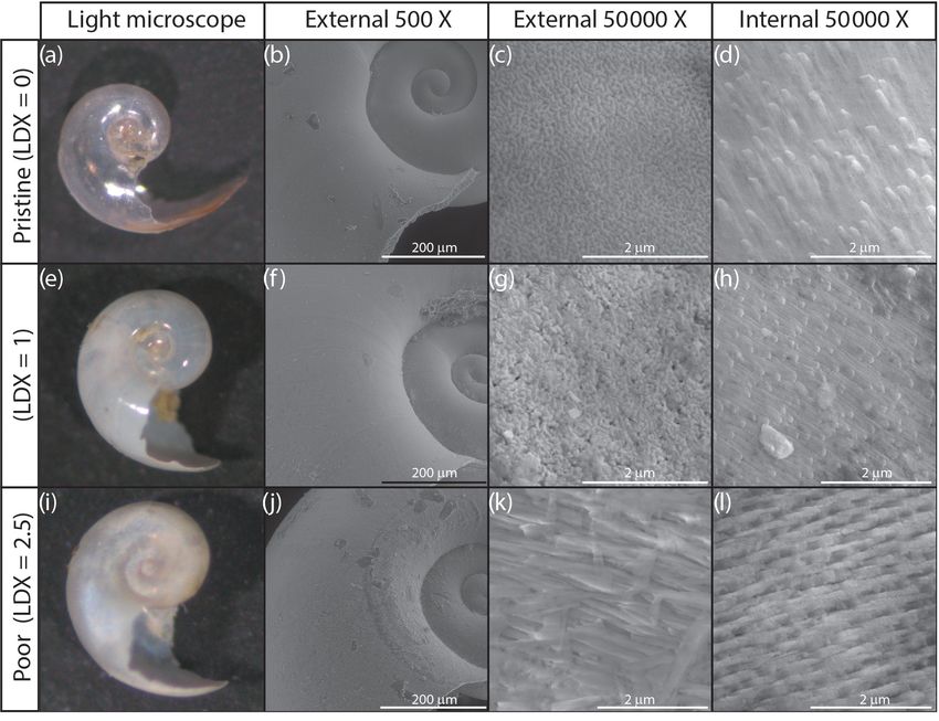
Figure 5. Shell condition of Heliconoides inflatus, ranked using the
Limacina Dissolution Index (LDX) scale, over the study period. The
Figure 4. Shell condition of Heliconoides inflatus, ranked on the samples with the poorest preservation are from September and Oc-
Limacina Dissolution Index (LDX) scale, plotted against the maxi- tober 2013, when water temperatures were the highest. The size of
mum amount of time specimens spent in the sediment trap (i.e., the the circles corresponds to n, the number of specimens plotted at a
number of weeks from the trap opening time). The size of the sym- given point, and the color of the circles corresponds to the maxi-
bols corresponds to n, the number of specimens plotted at a given mum number of weeks specimens were in the trap.
point.
pled in September 2013, with an average modal shell thick-
site from the expectation that more time in the trap would ness of 0.009 mm (Fig. 6d). There was a weak but statisti-
result in more degradation. The least well-preserved speci- cally significant correlation between shell diameter and the
mens came from the September and October 2013 samples number of whorls, which remains when analyzing the subset
(Fig. 5) and had spent a maximum of 2–6 weeks in the sed- of complete shells (Table S1) (whole dataset: R2 = 0.074,
iment trap cup (Fig. 4). The most well-preserved specimens p Bon. = 0.415, p FDR = 0.057; subset dataset (Table S1,
came from the June and December 2013 samples and had Fig. S4): R2 = 0.101, p Bon. = 0.513, p FDR = 0.057). As
spent a maximum of 20–22 weeks in the sediment trap cup shell diameter, thickness, and amount of shell material are
(Figs. 4, 5). related to size, unsurprisingly, there were significant corre-
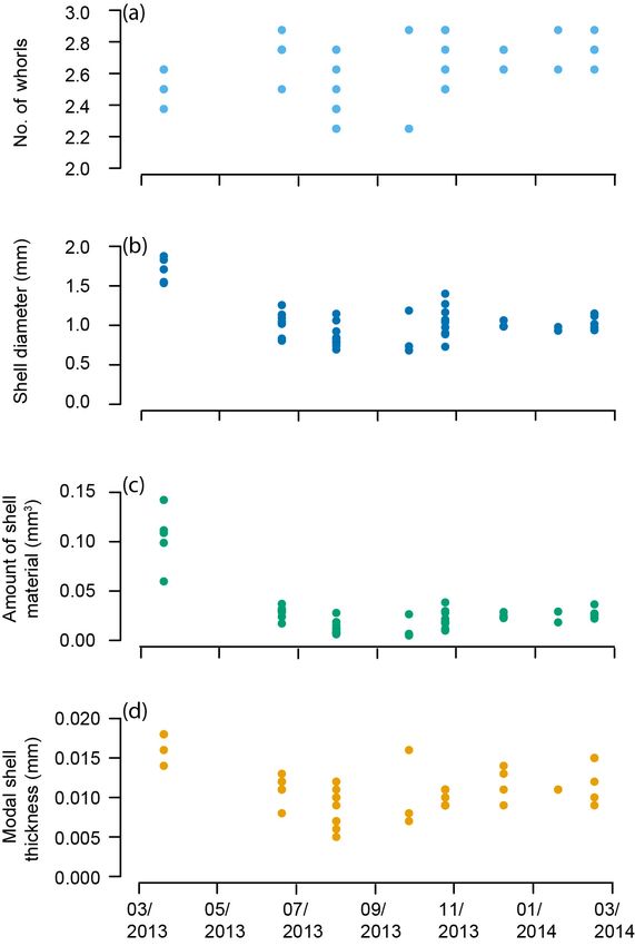
The pteropod shells varied in number of whorls, diame- lations between shell diameter and amount of shell mate-
ter, amount of shell material, and modal shell thickness both rial (R 2 = 0.819, p Bon. = 2.20 × 10⁻¹⁵ , p FDR = 2.20 ×
within and among samples throughout the year in the Cari- 10−15 ), shell diameter and shell thickness (R 2 = 0.582, p
aco Basin (Fig. 6, Table S1). The number of whorls varied Bon. = 1.79 × 10⁻⁸ , p FDR = 5.97 × 10⁻⁹ ), and shell thick-
between 2 1/4 and 2 7/8 and displayed no overall trend ness and amount of shell material (R2 = 0.680, p Bon. =
through the 11-month study (Fig. 6a; Table S1). Shell di- 6.09 × 10⁻¹¹ , p FDR = 3.05 × 10⁻¹¹ ). These results high-
ameter varied in samples collected through the year: speci- light that larger shells are generally thicker and contain more
mens from March 2013 had the greatest shell diameters (av- shell material.
erage 1.70 mm), and shells in the rest of the study period The modal shell thickness of the specimens, used in this
(June 2013–February 2014) ranged from 0.68 to 1.40 mm in study as a calcification metric, was analyzed with respect
diameter with an average of 0.98 mm (Fig. 6b). The amount to the water column properties in the Cariaco Basin (Fig. 7;
of shell material followed a similar pattern to shell diame- Table S4). Water chemistry measurements from 55 m depth
ter, with specimens from March 2013 containing the greatest were used because this was the closest water sample to
amount of shell material (0.104 mm3 ) and specimens from the most recent H. inflatus calcification depth estimate of
June 2013–February 2014 ranging from 0.005 to 0.038 mm3 , 75 m (Keul et al., 2017). The Cariaco Basin was supersatu-
with an average amount of 0.021 mm3 (Fig. 6c). The modal rated with respect to aragonite throughout the studied interval
thickness of the shells of Heliconoides inflatus also varied (arag range 2.28–3.59), and the thickest shells formed when
through the year (Fig. 6d; Table S1). The thickest shells the aragonite saturation was the lowest (March 2013, De-
were sampled in March 2013, with an average modal shell cember 2013–February 2014; average arag 2.49) (Fig. 7c;
thickness of 0.018 mm, and the thinnest shells were sam- Table S4). Specimens collected during the upwelling season
www.biogeosciences.net/17/1975/2020/ Biogeosciences, 17, 1975–1990, 20201982 R. L. Oakes and J. A. Sessa: Variables affecting pteropod shells in the Cariaco Basin
Figure 6. Heliconoides inflatus: (a) number of whorls, (b) shell di-
ameter, (c) amount of shell material, and (d) modal shell thickness
throughout the year in the Cariaco Basin. Each point represents an
individual specimen.
(December–April) were compared to those from the rainy
season when there was no upwelling (August–November),
using a Welch t test, because the two groups had different
variances and unequal sample sizes, prohibiting the use of
a Student t test (Revelle, 2018). Pteropod shells were 40 %
thicker during the upwelling season, when water tempera- Figure 7. Shell thickness and water column properties plotted over
tures were lower and nutrient concentrations were higher, the study period: (a) Heliconoides inflatus modal shell thickness,
than during the rainy season, when oligotrophic conditions (b) seawater temperature, (c) arag , (d) PO4 , and (e) NO2 and NO3 .
prevailed (Welch’s t test: p = 4.41×10⁻⁴ ; Table S6; Figs. 7, Nutrient concentrations (d, e) are plotted as proxies for upwelling
8). Pteropod shell diameters were also 20 % larger during and food availability (Romero et al., 2009; Thunell et al., 2000).
the upwelling season than during the rainy season (Welch’s All water column measurements (b–e) are from 55 m depth because
t test: p = 0.0080; Table S6). this is the water sample closest to the predicted calcification depth
Because shell diameter and shell thickness are related to of Heliconoides inflatus (Keul et al., 2017). The upwelling season
is indicated by a red box, and the rainy season, when there is no
the overall size of a specimen, the influence of shell diameter
upwelling, is indicated by a grey box.
on shell thickness was removed using a simple linear regres-
sion model of thickness as a function of diameter. Analysis
of the residuals of this model, hereafter referred to as “resid-
ual thickness”, found that specimens sampled during the up- welling season had significantly higher residual thicknesses
Biogeosciences, 17, 1975–1990, 2020 www.biogeosciences.net/17/1975/2020/R. L. Oakes and J. A. Sessa: Variables affecting pteropod shells in the Cariaco Basin 1983
Figure 9. Shell condition of Heliconoides inflatus, ranked on the Li-
macina Dissolution Index (LDX), plotted against shell condition of
Figure 8. Modal shell thicknesses of specimens from times of the same shells quantified using the opacity scale. LDX and opac-
upwelling (red) and times of no upwelling (grey) in the Cariaco ity are positively correlated until LDX scores of 2, at which point
Basin. Specimens collected during times of upwelling are signifi- there is no correlation between LDX and opacity. This breakdown
cantly thicker than those which formed at times with no upwelling is likely due to the changes in surface texture of the pteropod shell
(Welch’s t test: p = 4.4 × 10⁻⁴ ). from shiny to matte. The texture change linked to dissolution is a
factor when assigning values on the LDX scale, but as the color and
opacity do not change, it is not detected by the opacity scale.
than those sampled during the rainy season (Welch’s t test:
p = 0.0260; Fig. S5; Table S6), indicating that water column
properties impact calcification regardless of shell size.
texture, all shell condition analyses are based on LDX mea-
surements.
4 Discussion The shell condition of specimens from sediment trap sam-
ples has the potential to be altered via three mechanisms:
4.1 Shell condition (1) dissolution in the water column when the organism is
alive, (2) dissolution in the water column when the organism
This study focuses on how the interplay of biotic and abiotic is dead, and (3) alteration in the sediment trap cup associated
factors impacts the shell characteristics of the pteropod He- with the preservative. The water in the Cariaco Basin was su-
liconoides inflatus in the Cariaco Basin. The specimens used persaturated with respect to aragonite throughout the study.
in this study were collected using a sediment trap, adding a The thin, aragonitic shells of the pteropods would therefore
third variable, taphonomy. Pteropod shell condition was as- have been chemically stable in the water column, and thus
sessed using both the LDX (Gerhardt et al., 2000; Gerhardt it is unlikely that they underwent in-life dissolution. Further-
and Henrich, 2001) and opacity (Bergan et al., 2017) meth- more, there is no evidence of patchy dissolution on the exter-
ods. By comparing the results from these two methods, we nal walls of either pristine or altered shells (Figs. 2, S1, and
found that the opacity scale lacked sensitivity to changes in S2), such as has been observed in pteropod shells undergoing
shell condition at LDX values of 2 (opaque white shells with in-life dissolution in naturally undersaturated environments
lustrous surface) and higher (Fig. 9). When pteropod shells (Peck et al., 2016, 2018).
dissolve, the shell transparency changes first, from transpar- Once a pteropod dies, the degradation of the organic body
ent to milky white to opaque white, followed by the sur- and associated acid production has been found to cause
face texture (Gerhardt and Henrich, 2001). Because the opac- significant dissolution on the internal walls of the ptero-
ity method is based on greyscale values of light microscope pod shell, even in an aragonite-supersaturated water column
images, it quantifies the change in shell color but not tex- (Oakes et al., 2019a). Dissolution can occur on the exter-
ture, meaning this method is only sensitive to shell condition nal shell wall from the breakdown of free-floating organic
changes in the early stages of dissolution (LDX stages 0– matter in the water column creating aragonite-undersaturated
2; Fig. 9). Since the opacity method was designed to assess microenvironments in an otherwise aragonite-supersaturated
pteropods from an incubation experiment, it was intended water column (Milliman, 1999). LDX rankings show the
to capture the earliest stages of dissolution (Bergan et al., greatest amount of shell alteration occurred in specimens
2017). Because of the wide range of shell conditions of the from the September–October 2013 samples (Fig. 5). Scan-
specimens in this study, spanning both changes in color and ning electron microscopy reveals that the majority of this
www.biogeosciences.net/17/1975/2020/ Biogeosciences, 17, 1975–1990, 20201984 R. L. Oakes and J. A. Sessa: Variables affecting pteropod shells in the Cariaco Basin
dissolution occurred on the outside of the shells (Figs. 2 and (Fig. 6a, b), H. inflatus displays considerable plasticity dur-
S2). During September and October, water temperatures at ing growth. These measurements support the observations of
55 m were at their highest (Fig. 7b). These warm tempera- Janssen (1990), who found that both the number and diame-
tures would have increased the rate of microbial breakdown ter of the whorls of H. inflatus increase irregularly. There are
of the organic body within the shell (Oakes et al., 2019a) no patterns in the overall trend of H. inflatus shell diameter
and in the free-floating decaying organic matter in the wa- through the year in the Cariaco Basin (Fig. 6b), which sug-
ter column (Lohmann, 1995; Milliman et al., 1999; Schiebel gests there are no cohorts. Another low-latitude study found
et al., 2007). The shells of the organisms that died dur- that H. inflatus collected off the coast of Barbados repro-
ing the warmer months likely encountered more aragonite- duced throughout the year (Wells, 1976a), although H. infla-
undersaturated microenvironments associated with this or- tus from off Bermuda in the Sargasso Sea have been shown
ganic matter breakdown as they fell through the water col- to spawn in the spring (Almogi-Labin et al., 1988). Both van
umn and into the trap, increasing the rate of dissolution of der Spoel (1967) and Janssen (2004) have described variabil-
these shells relative to those trapped during cooler months. ity in the shape and position of the aperture tooth in H. infla-
These results could have been further complicated by the tus, which could be attributed to intraspecific or interspecific
presence of swimmers, which would have entered the trap variations. As there has not been any genetic work conducted
live and therefore would not have undergone any dissolution on H. inflatus from the Caribbean, we cannot be sure that the
associated with microbial breakdown in the water column. variability we see in shell shape cannot be attributed to two
If there was an increase in swimmers entering the traps at or more genetically defined species.
one time of year relative to another, it could be interpreted
as less water column breakdown during these months. The 4.3 Pteropod growth and water column properties
most pristine shells in this study entered the trap in June and
December, suggesting that there was not a seasonal pattern Because of their shell chemistry, pteropods have been pro-
to swimmer frequency. We therefore assume that the number posed as biological indicators of aragonite saturation (Bed-
of swimmers entering the sediment trap is constant through- naršek et al., 2017, 2019). In this study we used shell thick-
out the year and does not affect the seasonal trends reported ness as a metric of calcification. In the Cariaco Basin, the
above. water is permanently supersaturated with respect to aragonite
The borate-buffered formalin solution used to preserve (i.e., arag >1). In this aragonite-supersaturated setting, the
sediment trap samples has been shown to influence the con- thickness of pteropod shells does not correlate with aragonite
dition of pteropod shells (Oakes et al., 2019b). We found that saturation, and the thinnest shells were found when the arag-
shell condition did not deteriorate with time spent in the sed- onite saturation was the highest (August–November 2013 –
iment trap cups (Fig. 4). Preservation-associated dissolution average arag 3.26) (Fig. 7c). Instead, the shell thickness of
would have affected both the internal and external walls of H. inflatus varies with the physical oceanographic conditions
the shell. SEM images reveal that the internal shell walls in the Cariaco Basin, with median shell thickness increasing
were only impacted by dissolution at LDX values of 2.5 and by 40 % during times of upwelling (Fig. 8), when nutrient-
higher (Fig. 2d, h, l), indicating that the preservative did not rich waters are brought to the surface, relative to shells form-
cause dissolution. Specimens that had undergone the most ing during the rainy season when there is no upwelling and
dissolution were sampled during the warmest months, which oligotrophic conditions prevail (Figs. 7, 8; Table S6) (Muller-
happened to coincide with the shortest amount of time in the Karger et al., 2001, 2019). These upwelling-related nutrient
trap. This produced an apparent trend of improving shell con- changes in the Cariaco Basin have been shown to correspond
dition with time in the trap (Figs. 4, 5). This suggests that the with increases in organic carbon flux and diatom blooms
preservative in the sediment trap collection cups effectively (Thunell et al., 2000; Romero et al., 2009), indicating that
minimized post-collection sample degradation and that any pteropod food supply (Lalli and Gilmer, 1989) increases dur-
sediment trap-associated changes in shell condition likely oc- ing upwelling conditions. The diameters of pteropod shells
curred on timescales of 2 weeks or less, the amount of time sampled during times of upwelling were 20 % larger than
the specimens were in the final sediment trap collection cup those formed during the rainy season (Table S6), and the
before trap recovery. trend of increased shell thickness during times of upwelling
still holds once the influence of shell diameter on shell thick-
4.2 Pteropod development ness is removed (Fig. S5, Table S6). The observed changes
in H. inflatus modal shell thickness and diameter are likely
Assessing the number of whorls, shell diameter, amount of linked to changes in nutrients, and therefore food supply, in
shell material, and shell thickness provides an integrated the Cariaco Basin through the year.
view of H. inflatus shell growth in the Cariaco Basin. The link between food availability and shell growth has
The number of whorls varies both within and among sam- been proposed for another species of pteropod in the same
ples throughout the year. Although there is a weak rela- superfamily as H. inflatus, Limacina retroversa, which was
tionship between the number of whorls and shell diameter found to form smaller shells when food resources were lim-
Biogeosciences, 17, 1975–1990, 2020 www.biogeosciences.net/17/1975/2020/R. L. Oakes and J. A. Sessa: Variables affecting pteropod shells in the Cariaco Basin 1985
ited (Meinecke and Wefer, 1990). Furthermore, the avail- rate with increased time in the sediment trap cup. The most
ability of food has been found to offset, or even negate, poorly preserved specimens came from sediment trap sam-
the negative effects of increased pCO2 levels or low pH in ples collected when seawater temperatures were the highest,
other groups of marine calcifiers such as mussels, oysters, suggesting that dead specimens were affected by dissolution
and corals (Heinemann et al., 2012; Hettinger et al., 2013; from increased rates of microbial breakdown of organic mat-
Kroeker et al., 2016; Ramajo et al., 2016; Thomsen et al., ter, both in the water column and within the pteropod shell.
2013; Towle et al., 2015), presumably because organisms re- The size, number of whorls, thickness, and amount of shell
quire energy for biomineralization (Palmer, 1992). Feeding material in the shells of H. inflatus vary throughout the year,
rates in calcifiers can also be affected by acidified conditions, and therefore are likely to be influenced by external factors.
with responses varying according to phylum, feeding style, Water chemistry in the Cariaco Basin is controlled by the
life stage, and exposure time. Suspension-feeding molluscs movement of the ITCZ and has two distinct phases: an up-
were found to be particularly susceptible to decreased feed- welling phase and a non-upwelling, oligotrophic phase. We
ing rates with increased CO2 (Clements and Darrow, 2018). find that H. inflatus produces larger, thicker shells during
There have not been any studies conducted on the response times of upwelling, when food availability is greater. The
of pteropods to varying acidification and food availability Cariaco Basin was supersaturated with respect to aragonite
conditions; however, we assume that, as in other groups of throughout the study period (i.e., arag >1), and shell thick-
marine calcifiers, food availability plays an important role in ness does not correlate with arag . This demonstrates that, in
calcification. This body of research supports the findings of this aragonite-supersaturated setting, the availability of food
this study: when seawater is supersaturated with respect to has a greater control on shell formation than aragonite sat-
aragonite, such as in the Cariaco Basin, food availability is uration. This pattern has been seen in other groups of mol-
the main control of H. inflatus shell growth. luscs, such as oysters and mussels, and underlines the ne-
cessity of assessing pteropod shell parameters and dissolu-
4.4 Further work tion in the context of multiple biotic and abiotic factors, not
just aragonite saturation. We hope that the baseline dataset of
Micro-CT scanning enables pteropod shells to be digitized pteropod shell parameters presented in this study is the first
in three dimensions, creating the opportunity for more com- of many focused regional studies around the world. These
plex quantitative analyses of shell shape and parameters than datasets will enable the quantification of the response of this
presented in this study. Despite their geometrically simple sentinel group to ocean acidification.
shapes, gastropod shells are particularly challenging to ana-
lyze via geometric morphometric analyses because of their
lack of fixed landmark points (Liew et al., 2016). There has Data availability. The data which support the conclusions in this
been recent progress in the field of gastropod 3D geomet- paper are available in the tables, figures, references, and supple-
ric morphometrics, to understand variability in shell form mental materials. CT data are available on MorphoSource (https://
(Liew et al., 2016) and changes in shell calcification asso- www.morphosource.org/Detail/ProjectDetail/Show/project_id/908,
ciated with ocean acidification (Harvey et al., 2018). Such Oakes and Sessa, 2020).
analyses are beyond the scope of this study; however, the
CT scans of the specimens analyzed in this study are avail-
Sample availability. Specimens have been deposited in the Mala-
able to researchers on MorphoSource and can be down-
cology Collection at the Academy of Natural Sciences of Drexel
loaded via this link: https://www.morphosource.org/Detail/
University, Philadelphia, PA, USA (ANSP). A sample list, includ-
ProjectDetail/Show/project_id/908 (23 March 2020). ing the ANSP catalogue numbers, can be found in Table 1.
5 Conclusions Supplement. The supplement related to this article is available on-
line at: https://doi.org/10.5194/bg-17-1975-2020-supplement.
In this study, we analyzed the shell diameter, number of
whorls, thickness, amount of shell material, and shell condi-
tion of Heliconoides inflatus, a species of pteropod from the Author contributions. RLO conceptualized the project, developed
Cariaco Basin, over an 11-month period. Because specimens the methodology, performed the data analysis, created the figures,
in this study came from a sediment trap, the impact of time and wrote the first draft of the paper. RLO and JAS curated the data,
in the sediment trap on shell condition was analyzed. Shells performed statistical analyses, and edited the manuscript.
were assessed using both the LDX and opacity methods;
however, the opacity method was only sensitive to changes
in shell condition at LDX scores of 2 or lower, and there- Competing interests. The authors declare that they have no conflict
of interest.
fore LDX was used for all analyses. Although all shells had
undergone some alteration, shell condition did not deterio-
www.biogeosciences.net/17/1975/2020/ Biogeosciences, 17, 1975–1990, 20201986 R. L. Oakes and J. A. Sessa: Variables affecting pteropod shells in the Cariaco Basin
Acknowledgements. The authors would like to thank Brittney Mar- Bednaršek, N., Možina, J., Vogt, M., O’Brien, C., and Tarling, G.
shall for picking the first batch of specimens while in the midst of A.: The global distribution of pteropods and their contribution to
finishing her PhD and Catherine Davis for picking the second set of carbonate and carbon biomass in the modern ocean, Earth Syst.
samples and for helpful discussions about sample processing. Light Sci. Data, 4, 167–186, https://doi.org/10.5194/essd-4-167-2012,
microscopy was performed in the Paleobotany Lab at the Pennsyl- 2012a.
vania State University thanks to Peter Wilf and at the Academy of Bednaršek, N., Tarling, G. A., Bakker, D. C., Fielding, S., Co-
Natural Sciences thanks to Roger Thomas and Chris Vito. Scanning hen, A., Kuzirian, A., McCorkle, D., Lézé, B., and Mon-
electron microscopy was performed at the Nanoscale Characteriza- tagna, R.: Description and quantification of pteropod shell dis-
tion Facility at the Singh Center for Nanotechnology at the Univer- solution: a sensitive bioindicator of ocean acidification, Glob.
sity of Pennsylvania, Philadelphia, thanks to Jamie Ford. CT scan- Chang. Biol., 18, 2378–2388, https://doi.org/10.1111/j.1365-
ning was performed at GE in Lewistown thanks to Jeff Urbanksi 2486.2012.02668.x, 2012b.
and Whitney Yetter and the American Museum of Natural History Bednaršek, N., Tarling, G. A., Bakker, D. C. E., Field-
in New York thanks to Morgan Hill Chase and Mark Siddall. We ing, S., and Feely, R. A.: Dissolution dominating cal-
thank Tom Woodger, Jordan Foster, and Otto Woodger for logistical cification process in polar pteropods close to the point
support. We thank Eric Tappa for information about the CARIACO of aragonite undersaturation, PLoS One, 9, e109183,
sediment trap and for providing a map, as well as Tim Bralower, https://doi.org/10.1371/journal.pone.0109183, 2014a.
Maria Potapova, and Gary Rosenberg for discussions that helped Bednaršek, N., Feely, R. A., Reum, J. C. P., Peterson, B., Menkel, J.,
to shape this paper. We thank Katsunori Kimoto, Ahuva Almogi- Alin, S. R., and Hales, B.: Limacina helicina shell dissolution as
Labin, and an anonymous reviewer for their thoughtful, constructive an indicator of declining habitat suitability owing to ocean acid-
comments which helped to improve this paper. ification in the California Current Ecosystem, Proc. R. Soc. B,
281, 20140123, https://doi.org/10.1098/rspb.2014.0123, 2014b.
Bednaršek, N., Klinger, T., Harvey, C. J., Weisberg, S., McCabe, R.
Financial support. This research has been supported by the Col- M., Feely, R. A., Newton, J., and Tolimieri, N.: New ocean, new
lege of Earth and Mineral Sciences, Pennsylvania State University needs: Application of pteropod shell dissolution as a biological
(Deike Research Grant); and the Academy of Natural Sciences of indicator for marine resource management, Ecol. Indic., 76, 240–
Drexel University (John J. & Anna H. Gallagher Fellowship). 244, https://doi.org/10.1016/j.ecolind.2017.01.025, 2017.
Bednaršek, N., Feely, R. A., Howes, E. L., Hunt, B. P. V.,
Kessouri, F., León, P., Lischka, S., Maas, A. E., McLaugh-
Review statement. This paper was edited by Jean-Pierre Gattuso lin, K., Nezlin, N. P., Sutula, M., and Weisberg, S. B.: Sys-
and reviewed by K. Kimoto and one anonymous referee. tematic review and meta-analysis toward synthesis of thresh-
olds of ocean acidification impacts on calcifying pteropods
and interactions with warming, Front. Mar. Sci., 6, 1–16,
https://doi.org/10.3389/fmars.2019.00227, 2019.
Benjamini, Y. and Hochberg, Y.: Controlling the False Discovery
References Rate: A Practical and Powerful Approach to Multiple Testing, J.
R. Stat. Soc. Ser. B, 57, 289–300, https://doi.org/10.1111/j.2517-
Almogi-Labin, A., Luz, B., and Duplessy, J.-C.: Quaternary paleo- 6161.1995.tb02031.x, 1995.
oceanography, pteropod preservation and stable-isotope record Bergan, A. J., Lawson, G. L., Maas, A. E., and Wang, Z. A.: The
of the Red Sea, Palaeogeogr. Palaeocl., 57, 195–211, 1986. effect of elevated carbon dioxide on the sinking and swimming
Almogi-Labin, A., Hemleben, C., and Deuser, W. G.: Seasonal vari- of the shelled pteropod Limacina retroversa, ICES J. Mar. Sci.,
ation in the flux of euthecosomatous pteropods collected in a 1–13, https://doi.org/10.1093/icesjms/fsx008, 2017.
deep sediment trap in the Sargasso Sea, Deep-Sea Res., 35, 441– Broecker, W. S. and Peng, T.-H.: Tracers in the Sea, Lamont – Do-
464, 1988. hery Geological Observatory, Palisades, New York, 1982.
Astor, Y., Muller-Karger, F., and Scranton, M. I.: Seasonal and in- Clements, J. C. and Darrow, E. S.: Eating in an acidifying ocean: a
terannual variation in the hydrography of the Cariaco Basin: Im- quantitative review of elevated CO2 effects on the feeding rates
plications for basin ventilation, Cont. Shelf Res., 23, 125–144, of calcifying marine invertebrates, Hydrobiologia, 820, 1–21,
https://doi.org/10.1016/S0278-4343(02)00130-9, 2003. https://doi.org/10.1007/s10750-018-3665-1, 2018.
Astor, Y. M., Fanning, K., Guzman, L., Li, X., Lorenzoni, L., Collier, R., Dymond, J., Honjo, S., Manganini, S., Francois, R., and
Masserini, R., Muller-Karger, F. E., Tappa, E., and Varela, Dunbar, R.: The vertical flux of biogenic and lithogenic material
R.: Handbook of methods for the analysis of oceanographic in the Ross Sea: moored sediment trap observations 1996–1998,
parameters at the Cariaco Time-Series Station, edited by: Deep-Sea Res. Pt. II, 47, 3491–3520, 2000.
Astor, Y. M., Lorenzoni, L., and Scranton, M. I., available at: Comeau, S., Gorsky, G., Jeffree, R., Teyssié, J.-L., and Gattuso,
http://imars.marine.usf.edu/sites/default/files/project/cariaco/ J.-P.: Impact of ocean acidification on a key Arctic pelagic
publications/CARIACO_Methods_Manual.pdf (29 Septem- mollusc (Limacina helicina), Biogeosciences, 6, 1877–1882,
ber 2019), 2011. https://doi.org/10.5194/bg-6-1877-2009, 2009.
Astor, Y. M., Lorenzoni, L., Thunell, R., Varela, R., Muller- Comeau, S., Jeffree, R., Teyssié, J.-L., and Gattuso, J.-P.: Re-
Karger, F., and Troccoli, L.: Deep-Sea Research II Interan- sponse of the Arctic pteropod Limacina helicina to pro-
nual variability in sea surface temperature and f CO2 changes jected future environmental conditions, PLoS One, 5, e11362,
in the Cariaco Basin, Deep-Sea Res. Pt. II, 93, 33–43, https://doi.org/10.1371/journal.pone.0011362, 2010.
https://doi.org/10.1016/j.dsr2.2013.01.002, 2013.
Biogeosciences, 17, 1975–1990, 2020 www.biogeosciences.net/17/1975/2020/You can also read