Auditory thalamus dysfunction and pathophysiology in tinnitus: a predictive network hypothesis
←
→
Page content transcription
If your browser does not render page correctly, please read the page content below
Brain Structure and Function (2021) 226:1659–1676
https://doi.org/10.1007/s00429-021-02284-x

    REVIEW

Auditory thalamus dysfunction and pathophysiology in tinnitus:
a predictive network hypothesis
Pia Brinkmann1         · Sonja A. Kotz1,2      · Jasper V. Smit3       · Marcus L. F. Janssen4,5     · Michael Schwartze1

Received: 26 August 2020 / Accepted: 21 April 2021 / Published online: 2 May 2021
© The Author(s) 2021

Abstract
Tinnitus is the perception of a ‘ringing’ sound without an acoustic source. It is generally accepted that tinnitus develops after
peripheral hearing loss and is associated with altered auditory processing. The thalamus is a crucial relay in the underlying
pathways that actively shapes processing of auditory signals before the respective information reaches the cerebral cortex.
Here, we review animal and human evidence to define thalamic function in tinnitus. Overall increased spontaneous firing
patterns and altered coherence between the thalamic medial geniculate body (MGB) and auditory cortices is observed in
animal models of tinnitus. It is likely that the functional connectivity between the MGB and primary and secondary auditory
cortices is reduced in humans. Conversely, there are indications for increased connectivity between the MGB and several areas
in the cingulate cortex and posterior cerebellar regions, as well as variability in connectivity between the MGB and frontal
areas regarding laterality and orientation in the inferior, medial and superior frontal gyrus. We suggest that these changes
affect adaptive sensory gating of temporal and spectral sound features along the auditory pathway, reflecting dysfunction in
an extensive thalamo-cortical network implicated in predictive temporal adaptation to the auditory environment. Modulation
of temporal characteristics of input signals might hence factor into a thalamo-cortical dysrhythmia profile of tinnitus, but
could ultimately also establish new directions for treatment options for persons with tinnitus.

Keywords Tinnitus · Medial geniculate nucleus · MGB · Prediction · Temporal processing

Introduction                                                                 with the affected person’s daily life (Langguth et al. 2013;
                                                                             McCormack et al. 2016; Schlee et al. 2017). Severe forms
Tinnitus is frequently described as hearing a sound without                  of tinnitus exert a particular negative impact on the qual-
an external source or as a ringing in the ears (Baguley et al.               ity of life, with symptoms of depression, anxiety, sleep
2013). The prevalence of tinnitus ranges from 10 to 15% in                   disturbances, concentration difficulties, or reduced cogni-
the general population, and in 1–2% it severely interferes                   tive efficiency (Hallam et al. 2004; Langguth 2011). Con-
                                                                             sequently, tinnitus has direct societal impact, as reflected in
                                                                             high healthcare costs and loss of productivity (Maes et al.
* Pia Brinkmann                                                              2013). Currently, there is no curative evidence-based therapy
  p.brinkmann@maastrichtuniversity.nl
                                                                             for tinnitus, i.e., although drug targets, cognitive, behavioral,
1
     Department of Neuropsychology and Psychopharmacology,                   and neuromodulative interventions have been put forward,
     University of Maastricht, Universiteitssingel 40,                       there is a lack of randomized controlled trials confirming
     6229 Maastricht, The Netherlands                                        effective tinnitus treatment (Kleinjung and Langguth 2020).
2
     Department of Neuropsychology, Max Planck Institute                        Over the past two decades, general interest in tinnitus
     for Human Cognitive and Brain Sciences, Leipzig, Germany                has rapidly grown as part and parcel of new hypotheses
3
     Department of Ear Nose and Throat/Head and Neck Surgery,                about tinnitus pathophysiology (Moller et al. 2015; Rob-
     Zuyderland Medical Center, Sittard/Heerlen, the Netherlands             erts and Salvi 2019). Reflecting parallel advances in neu-
4
     Department of Clinical Neurophysiology, Maastricht                      roimaging methodology, the general focus shifted from
     University Medical Center, Maastricht, The Netherlands                  otology to neuronal correlates of tinnitus (Langguth et al.
5
     School for Mental Health and Neuroscience, Faculty                      2013). Although there is no strong consensus, it is gener-
     of Health, Medicine and Life Sciences, Maastricht                       ally assumed that hearing loss precedes the development
     University, Maastricht, The Netherlands

                                                                                                                                  13
                                                                                                                             Vol.:(0123456789)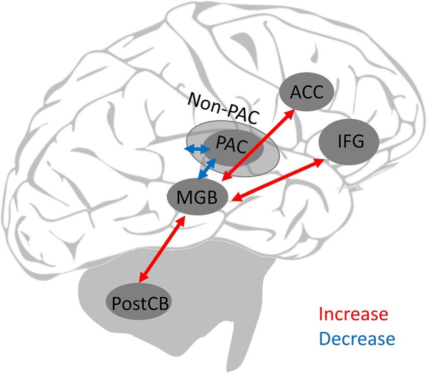
1660                                                                                         Brain Structure and Function (2021) 226:1659–1676

of tinnitus. Consequently, changes along the classical and                 relay station along the auditory pathway, its contribution to
non-classical auditory pathway, expressed in alterations                   tinnitus pathology is often disregarded.
of spontaneous firing activities, neural synchronization, or                  The MGB is part of the classical and non-classical audi-
tonotopic organization are possible key elements of tinnitus               tory pathway, mediating the thalamo-cortical network
pathogenesis (Elgoyhen et al. 2015).                                       involved in tinnitus. It actively shapes information process-
   The classical ascending auditory pathway includes projec-               ing between subcortical and cortical areas (Bartlett 2013;
tions to mainly primary auditory regions, while non-classical              De Ridder et al. 2015; Llinas et al. 1999). Animal research
auditory pathways have been described as extralemniscal,                   provides first indications of successful tinnitus treatment by
diffuse, or polysensory pathways that involve connections to               invasively stimulating the MGB in rats (van Zwieten et al.
non-primary auditory areas (Fig. 1) (Aitkin 1986; Graybiel                 2019b). The MGB should hence not only be considered a
1972; Møller 2012). Next to neural correlates of tinnitus,                 major gateway station for auditory signals transmitted to the
current theories suggest maladaptive gating, increased cen-                cerebral cortex, but also as a crucial component in devel-
tral gain or altered neural thalamo-cortical coherence as fac-             oping a better understanding of tinnitus pathology (Leaver
tors underlying the development of tinnitus (De Ridder et al.              et al. 2011; Moller 2003). Taking this perspective and start-
2015; Llinas et al. 1999; Norena 2011; Rauschecker et al.                  ing with a review of thalamic contributions to auditory pro-
2010). However, although the auditory thalamus, and in par-                cessing, we formulate a hypothesis of thalamic function-
ticular the medial geniculate body (MGB), is a mandatory                   ing in tinnitus pathology from a comparative perspective,
                                                                           integrating animal and human evidence. We propose that
                                                                           changes in thalamic functioning affect sensory gating at
                                                                           the level of the MGB, suggesting a dedicated timing and
                                                                           temporal prediction mechanism as an independent source of
                                                                           information and a potential tool for modulating the experi-
                                                                           ence of tinnitus.

                                                                           Functional neuroanatomy of the medial
                                                                           geniculate body of the thalamus

                                                                           To improve understanding of tinnitus and the role of the
                                                                           auditory thalamus in tinnitus pathophysiology, it is neces-
                                                                           sary to first consider the functional anatomy of the MGB.
                                                                           In general, the auditory pathway contains ascending and
                                                                           descending connections to auditory cortices and along its
                                                                           way, information is transformed and reorganized (Møller
                                                                           2011; Oertel and Doupe 2013). Input travels through the
                                                                           ear, the cochlea (Fig. 1), the cochlear nuclei (CN) and the
                                                                           inferior colliculus (IC) before reaching the MGB (Oertel
                                                                           and Doupe 2013).
                                                                              Originating from the IC, two ascending pathways, the
                                                                           classical and the non-classical auditory pathway innervate
                                                                           the MGB, primary (PAC) and non-primary auditory cortices
                                                                           (non-PAC), as well as limbic regions (Møller 2002; Pick-
                                                                           les 2015). The IC can be divided into three distinct nuclei,
                                                                           the central part of the IC (ICC), the dorsal cortex of the IC
                                                                           (ICD), and the external nucleus of the IC (ICX). In the clas-
                                                                           sical pathway, the ICC provides the main input to the ventral
Fig. 1  Schematic and simplified representation of the classical and
                                                                           MGB (MGV; Table 1 for an overview).
non-classical ascending auditory pathway. Ascending auditory sig-             The MGV forms the “core” subdivision of the MGB.
nal travels from the ears to primary and secondary auditory cortices,      The MGV has a pronounced tonotopic organization, narrow
while taking two different pathways. PAC primary auditory cortex,          tone frequency tuning, and exclusively responds to audi-
Non-PAC non-primary auditory cortices, CN cochlear nucleus, ICC
central inferior colliculus, ICD dorsal inferior colliculus, ICX, exter-
                                                                           tory input (Aitkin and Webster 1972; Bartlett 2013; Hackett
nal inferior colliculus, MGB medial geniculate body, MGD dorsal            et al. 2011). Fibers from the MGV primarily innervate the
MGB, MGM medial MGB, MGV ventral MGB                                       primary auditory cortex (Bartlett 2013).

13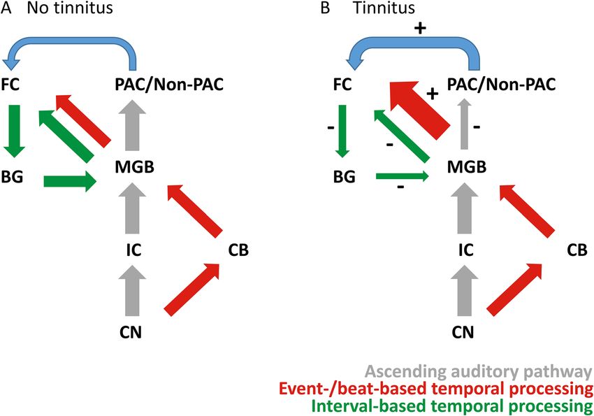
Brain Structure and Function (2021) 226:1659–1676 1661
The non-classical pathway processes input beyond the
tone latency, sharp tuning curves, habituation and
auditory domain and innervates limbic regions, such as the
Wide, multipeaked tuning curves, short and long
Dorsal IC, tegmentum, sagulum, somatosensory
Secondary auditory cortices (non-PAC, i.e., belt
amygdala, next to primary and secondary auditory corti-
areas), lateral nucleus of the amygdala, non-
ces (Bartlett 2013; Møller 2002). The ICD and the ICX
provide input to the medial and dorsal subdivisions of the
MGB in the non-classical pathway. The dorsal subdivision
of the MGB (MGD) is not tonotopically organized (Bartlett
2013) and projects to the Non-PAC, the lateral nucleus of
the amygdala and non-auditory areas (Bartlett 2013). The
stimulus adaptation
medial subdivision of the MGB (MGM) is the most hetero-
Multisensory relay
auditory areas geneous part of the MGB. The tonotopic organization of the
Non-classical
Dorsal MGB
MGM is not as pronounced as in the MGV, tone frequency
tuning is heterogeneous and neurons in the MGM respond
system
not only to auditory, but also to visual and somatosensory
NA
No
input (Bartlett 2013; Hackett et al. 2011; Rouiller et al.
1989). Projections from the MGM terminate in primary and
short tone latency, fire at stimulus onset, associa-
non-PAC), lateral nucleus of the amygdala, non-
External IC, dorsal IC, central IC, lateral tegmen-
tive learning responses (i.e., fear conditioning)
Heterogeneous tuning curves, spatial selectivity,
Primary and secondary auditory cortices (PAC,
PAC primary auditory cortex, Non-PAC secondary non-primary auditory cortex, IC inferior colliculus, MGB medial geniculate body non-primary auditory cortices as well as the amygdala (Ait-
kin 1986; Bartlett 2013; Møller 2002). Moreover, all MGB
subdivisions receive input from the reticular nucleus in the
tum, spinal cord, superior colliculus
thalamus (TRN), which influences general excitability of
neuronal activity in the MGB (Bartlett 2013; Møller 2002).
Consequently, classical and non-classical ascending
Yes (less than ventral MGB)
auditory areas, striatum
auditory pathways contribute differently to the processing
of auditory stimuli in the MGB and most likely to tinnitus
Multisensory relay
pathophysiology. Due to the different input and output struc-
tures, the three subdivisions of the MGB may form three
Non-classical
Medial MGB
separate and parallel pathways to higher cortical auditory
areas (Pickles 2015; Winer et al. 2005).
NA
Tonotopic map and sound level tuning
Ipsilateral primary auditory cortex (PAC)
Sharp tuning curves, single-peaked, short
Low-to-high ( ventrolateral-dorsomedial)
tone latency, closely connected to the
in the MGB
Ipsilateral central nucleus of the IC
As described above, the MGB is divided into different
parts, the MGD, MGM and MGV. These parts have differ-
ent neurophysiological properties and respond differently
Table 1 Overview of MGB subdivisions and their functionality
to external auditory stimuli. Frequency maps have been
created in animal models by means of electrophysiological
Primary role in functional neuroanatomy Auditory relay
Ventral MGB
studies. Frequency tuning in the MGB is sharpest in the
Classical/non-classical auditory pathway Classical
MGV in awake marmoset primates (Bartlett et al. 2011).
PAC
Similar results were obtained in rats (Bordi and LeDoux
Yes
1994). Comparable to the MGV, neurons in the MGM have
also been shown to respond in a narrow fashion in the anes-
thetized rat (Anderson and Linden 2011). An intermediate
level of frequency tuning has been described for the MGD
(Bartlett 2013). However, results are not consistent and fast
habituation to repeated stimuli using isointensity tones in
Tonotopic organization
the MGD are described in Bordi and LeDoux (1994). The
Responses to sound
picture is even more complex as recent evidence suggests
Tonotopic map
more variable tonotopic maps for neurons with multi-peaked
frequency tuning curves (Gaucher et al. 2019).
Sound level tuning on the other hand, is monotonic, i.e.,
Output
Input
the sound intensity changes, while the frequency remains
131662 Brain Structure and Function (2021) 226:1659–1676
stable. Neurons exhibiting either a progressive increase or a Discrimination of speech-like contrasts seems to occur at
progressive decrease in firing rates when intensity changes the level of the MGB, as observed in mismatch responses in
can be classified as monotonic. Non-monotonic responses the caudo-medial MGB of guinea pigs (Kraus et al. 1994).
are observed when firing rates increase with increasing tone Cai et al. (2016) investigated whether young, old, awake,
intensity until a plateau is reached, after which firing rates or anesthetized rats differentially process complex auditory
decrease. Rouiller et al. (1983) investigated sound level stimuli in the MGB. They found that MGB cells in the old
tuning in the MGB in anesthetized cats and found that the awake rat preferred regular predictable, vocalization-like
majority of units exhibited non-monotonic responses. signals, especially when increasing the difficulty in modu-
Animal studies investigating the MGB predominantly rely lation frequency (Cai et al. 2016). In young rats, however,
on invasive techniques whereas in human studies non-inva- randomly presented modulated sequences were preferred
sive methods are predominant due to self-evident ethical rea- (Cai et al. 2016). This suggests that with increasing age,
sons. This makes the identification and subsequent manipu- top–down processes may enhance the processing of expected
lations of the small (i.e., 5 × 4 × 5 mm, Winer et al. 1984) and stimuli with the same formal structure at the level of the
densely clustered MGB nuclei intrinsically difficult. Techno- MGB. Accordingly, previous research shows that the MGB
logical advances such as ultra-high field functional neuroim- is not only tonotopically organized, but that it is closely
aging (i.e., 7 T) allow creating precise tonotopic maps of the involved in the representation of complex vocalizations
human MGB (Berlot et al. 2020; Mihai et al. 2019a; Moerel across species and that its functioning may change across
et al. 2015). These functional magnetic resonance imaging the life span (i.e., preferring predictable stimuli) (Amin et al.
(fMRI) techniques allow high spatial resolution imaging, 2010; Cai et al. 2016; Huetz et al. 2009; Kraus et al. 1994).
but depend on slow changes in blood oxygenation. Thus the Human research specifically targeting MGB activity in
signal depends on the vascular morphology of the relatively response to human vocalization and speech is rare (Mihai
small MGB (Moerel et al. 2015). In humans, a low-to-high et al. 2019a, c). The MGB is active irrespective of content
tonotopic map has been identified in the MGV in a ventro- or loudness manipulations of speech sounds (von Kriegstein
lateral-dorsomedial direction (Moerel et al. 2015). Moerel et al. 2008). Mihai et al. (2019a) assessed speech recognition
et al. (2015) further observed another, dorsomedial area with abilities in the core subdivision of the auditory thalamus
a preference for low frequency stimuli, located outside the (i.e., MGV) and found behaviorally-relevant task dependent
MGV. Berlot et al. (2020) investigated the MGB tonotopy fMRI modulation of the left MGV. Furthermore, left MGV
in persons with tinnitus and healthy controls, confirming a was found to be increasingly activated when participants had
low–high–low frequency preference in the sagittal plane, to recognize speech in noise compared to intelligible speech
comparable between groups. Tonotopic organization of the (Mihai et al. 2019b). Previously, the ventral intermediate
MGV and of the pars lateralis (PL) in the MGB in anesthe- nucleus (VIM) has been reported to respond to syntactic
tized cats has been found to also range from low-to-high in and semantic components in spoken language (Wahl et al.
a latero-medial gradient (Aitkin and Webster 1972; Morel 2008). In addition, it has been proposed that the thalamus
et al. 1987). Thus, mounting evidence supports a roughly contributes to speech processing via its differential encod-
similar low-to-high tonotopic organization in animals and ing of temporal and spectro-temporal information (Kotz and
humans in the MGV, validating the comparative usage of Schwartze 2010). Taken together, evidence across several
animal models. species indicates that the MGB dynamically shapes simple
tones and complex vocalizations before auditory sensations
reach the cerebral cortex.
Representation of complex sounds
It is likely that artificially created sine tones do not entirely Information processing in the MGB—
capture the functioning of the MGB when it perceives more intrinsic cell properties
complex auditory stimuli, such as vocalizations. In awake
guinea pigs, the MGB has been shown to respond to ampli- To gain better understanding of information processing in
tude-modulated (AM) and frequency-modulated (FM) sine the auditory thalamus and how these processes transform
tones as well as to natural calls (Creutzfeldt et al. 1980). auditory information before it reaches the cortex, it is nec-
Interestingly, the MGB responded to natural calls of the essary to focus on specific electrophysiological properties
same and of other species, and its response was depicted of MGB neurons. Thalamic neurons respond to incoming
in more detail in the MGB than in cortical cells, meaning information in either a burst or a tonic mode (Sherman and
that MGB units could differentiate between high modula- Guillery 2006). Thus, questions arise as to how the two fir-
tion frequencies, while cortical cells could not (Creutzfeldt ing modes (i.e., burst and tonic mode) emerge and how they
et al. 1980). shape auditory information processing.
13Brain Structure and Function (2021) 226:1659–1676 1663
Next to classical action potentials (i.e., single-spikes), firing—single-spike pattern to a single-spike—single-spike
low threshold spikes (LTS) are important voltage depend- pattern in response to a stimulus pair occurs around an inter-
ent conductance mechanisms for thalamic relay cells (Jahn- pair-interval of 1000 ms, when applying intra-pair-intervals
sen and Llinas 1984b; Sherman and Guillery 2006). A LTS of 200–1000 ms (Bayazitov et al. 2013). These results indi-
involves the membrane depolarization of T-type voltage- cate temporal sensitivity when switching between the tonic
gated calcium channels (Jahnsen and Llinas 1984a, b), and the burst firing mode. A similar hypothesis has previ-
while classical action potentials are provoked by the open- ously been formulated by Bartlett (2013), stating that in
ing of sodium ( Na+) channels. The threshold to elicit a LTS speech, where fast-changing temporal features are common,
is approximately 10 mV lower (i.e., more hyperpolarized) burst firing may encode the rhythmic dynamic of syllabic
than for classical action potentials (Hu 1995; Jahnsen and on- and offsets, while tonic firing may help discriminating
Llinas 1984b; McCormick et al. 1991). In addition to the between finer, more faint auditory signals. In addition, it has
fact that T-type calcium channels act on more hyperpolar- been suggested that burst mode patterns are more frequently
ized membrane potentials, than sodium channels, T-type cal- encountered in MGD neurons and single-spike firing pre-
cium channels are slower and need approximately 100 ms to dominantly in MGV (Hu 1995), a pattern that has not been
switch between states of inactivation (Sherman and Guillery confirmed by Bartlett and Smith (1999). Thalamic cells thus
2006). fire in a burst or a tonic mode, which map onto non-linear
Of specific interest for subsequent information processing and linear information processing, respectively. However, it
in the MGB are the two different firing modes in response is unclear how the different firing modes and spiking pat-
to LTSs. The state of the T-type calcium channels, deter- terns may relate to tinnitus pathology.
mines the respective firing mode of the thalamic neurons
(Ramcharan et al. 2000). Irrespective of the neuron type,
thalamic neurons have been found to respond in either a The MGB in tinnitus pathology
tonic or a burst mode and also switch between these modes
(Jahnsen and Llinas 1984b; McCormick et al. 1991; Sher- Animal models of tinnitus are frequently employed to sys-
man 2001; Sherman and Guillery 2006). The tonic mode tematically investigate the pathophysiology of tinnitus and
has been described as preserving input linearity, whereas the changes it causes along the auditory pathway, including
the burst mode acts as a ‘wake-up call’ to cortical targets the MGB. These models can be broadly divided into inter-
(Ramcharan et al. 2000; Sherman 1996). Rhythmic burst rogative models and reflexive models (Brozoski and Bauer
firing has been primarily observed during sleep, potentially 2016; Galazyuk and Brozoski 2020). Interrogative models
indicating reduced transmission of sensory information evaluate voluntary behavior (i.e., performing an action to
to the cortex (Domich et al. 1986; Sherman and Guillery obtain food when hearing a sound), while reflexive models
2006). However, it has been shown that burst firing is not evaluate involuntary behavioral responses to the acoustic
limited to sleep and can be recorded from the thalamus of startle reflex (Brozoski and Bauer 2016; Galazyuk and Bro-
awake behaving macaque monkeys (Ramcharan et al. 2000). zoski 2020). Across species, the most frequently employed
Information processing in burst mode has been suggested reflexive model uses the gap–prepulse inhibition of the
to be less detailed and less noisy, but also more efficient, as acoustic startle (GPIAS) to determine the presence and
only infrequent ‘wake-up calls’ are processed (Sherman and course of tinnitus pathology (Galazyuk and Hebert 2015;
Guillery 2006). Information processing in the tonic mode, Turner et al. 2006). To induce tinnitus, animals are either
however, maintains a more detailed representation of an administered high doses of sodium salicylate (Su et al. 2012;
input signal (i.e., more linearity). Yang et al. 2007) or exposed to loud sound (Brozoski and
When T-type channels are inactivated by membrane depo- Bauer 2016). In the latter case, animals under anesthesia
larization, the tonic mode is elicited (Ramcharan et al. 2000). are unilaterally exposed to loud broad-band noise while the
To elicit burst firing, T-type calcium channels are activated contralateral ear is plugged to prevent hearing loss. In the
from a hyperpolarized condition (Ramcharan et al. 2000). In GPIAS paradigm, acoustic startle responses are reduced,
mice, it was shown that switching between burst and tonic when a silent gap (e.g., 50 ms) is inserted before the startle
firing in MGV neurons partly underlies paired-pulse depres- sound (Smit et al. 2016). However, when a sound matching
sion in thalamo-cortical neurons (Bayazitov et al. 2013). the tinnitus frequency is played and a silent gap is presented,
Bayazitov et al. (2013) employed an auditory paired-pulse the gap will not be perceived by the animal experiencing
paradigm (i.e., intra-pair-interval = 100–1000 ms, inter- tinnitus, because it has been filled-in by the tinnitus fre-
pair interval = 500–10,000 ms) and found that thalamic quency (Turner et al. 2006). Thus, animals experiencing
neurons responded to the first tone of the pair with a burst, tinnitus show increased startle responses in comparison to
followed by a single-spike action potential. Furthermore, unexposed controls (Turner et al. 2006; Yang et al. 2007).
it was found that the point of switching between the burst Advantages of the GPIAS model are that it is relatively fast
131664 Brain Structure and Function (2021) 226:1659–1676
to administer, does not require training, and motivational a role in the excitability of MGB neurons. Another study
states (i.e., frequently managed via diet restrictions) play observed reduced numbers of neurons exhibiting burst
a minor role (Brozoski and Bauer 2016). One of the disad- activity patterns, decreased spikes per burst and bursts
vantages is habituation, i.e., when repeated, unconditioned per minute in anesthetized rats who were administered an
reflexes diminish in amplitude (Lobarinas et al. 2013; Lon- acoustic noise trauma, irrespective of tinnitus presence
genecker and Galazyuk 2011). This issue was addressed (Barry et al. 2019). Another study investigating the MGB
by administering fewer trials and by randomly varying the in anesthetized rats with and without noise exposure clas-
inter-stimulus interval (van Zwieten et al. 2019a, b). Based sified four response types (i.e., fast, sustained, suppressed
on the assumption that tinnitus pathogenesis relies on mal- and no response) (van Zwieten et al. 2021). It was found
functioning of a vast network of primary auditory and non- that noise exposure resulted in an overall decrease of fast
auditory structures (Llinas et al. 1999; Rauschecker et al. responding neurons, while non-responsive increased (van
2010), it has also been criticized that the GPIAS model does Zwieten et al. 2021). In addition, spontaneous firing rates
not take hyperacusis and emotional factors such as stress into increased in sustained and suppressed neurons, while this
account (Brozoski and Bauer 2016; Kleinjung and Langguth was not the case for fast responding neurons. Acquired
2020). Thus, evaluating animal studies investigating MGB LFPs suggest suppressed thalamocortical synchronization
functioning in tinnitus pathology requires close monitoring in the beta and gamma bands, independent of noise trauma
of the paradigm choice, because even if motivational states (van Zwieten et al. 2021).
do play a minor role in the GPIAS model, stress might still Oscillatory coherence between the MGB and the pri-
influence an animal’s performance. mary auditory cortex has been investigated using local field
potentials (LFP) in anesthetized rats, while tinnitus was
Animal studies investigating the MGB in tinnitus induced by sodium salicylate (Vianney-Rodrigues et al.
2019). Results indicate that sodium salicylate decreased
Several studies investigated MGB changes in tinnitus animal theta, alpha, and beta oscillations in the MGB. Decreased
models (Table 2). coherence (i.e., the strength of a correlation between two
However, due to heterogeneous methodology and the signals as a function of frequency) between theta and alpha
overall limited number of studies, it is difficult to identify oscillations was further observed, while gamma coherence
generalizable result patterns. When focusing on changes was increased between pairs of electrodes positioned in
related to neurotransmitters, decreased GABA has been the MGB and PAC (Vianney-Rodrigues et al. 2019). Inter-
found in the MGB in rat models of tinnitus (Brozoski and estingly, when assessing the coherence (i.e., synchrony)
Odintsov 2012; Llano et al. 2012). However, contradict- between the MGB and PAC, sodium salicylate decreased
ing evidence exists (Sametsky et al. 2015). Administering coherence measures in the beta, alpha, and theta bands and
high doses of sodium salicylate decreased the excitability again, enhanced coherence for the gamma band (Vianney-
of neurons in the MGB, leading to increased hyperpolari- Rodrigues et al. 2019). Enhanced gamma coherence relates
zation of resting state potentials (Su et al. 2012; Wang to previous research, as gamma band activity was sug-
et al. 2016). Another approach to assess alterations in the gested to be a direct neural correlate of tinnitus, influencing
MGB in animals experiencing tinnitus is to investigate thalamo-cortical networks (Schlee et al. 2009; Sedley et al.
firing patterns, in vitro or in vivo in either anesthetized or 2012; van der Loo et al. 2009).
awake animals. In vitro, both healthy control animals and In awake rats, Kalappa et al. (2014) found similar results
rats with behavioral evidence of tinnitus, displayed burst as Sametsky and colleagues (2015), confirming increased
firing after a current injection to the soma (Sametsky et al. number of bursts per minute, increased mean burst duration
2015). Animals with tinnitus had an increased number of and mean spikes in a burst. Kalappa et al. (2014) showed
spikes per burst in comparison to controls and increased increased spontaneous firing in the MGD, MGM, and the
tonic GABAA currents. This suggests a shift towards MGV in a rat model of tinnitus. However, spontaneous fir-
increased tonic inhibition, which may result in abnormal ing rates in the MGB have also been found to be unaffected
bursting activity in the MGB, in turn leading to increased in rats with acoustic noise trauma or tinnitus (Barry et al.
output from the MGB to higher auditory cortices (Samet- 2019). Most importantly, enhanced behavioral evidence of
sky et al. 2015). Moreover Sametsky et al. (2015) investi- tinnitus pathology (i.e., increased z-scores of the raw-gap-
gated whether changes in LTS responses could be associ- startle in the GPIAs) was linked to higher spontaneous firing
ated to the increase of spikes per burst in rats with tinnitus. rates, irrespective of sound exposure (Kalappa et al. 2014).
The authors found no differences between tinnitus and The increases in spontaneous firing could be specified by
control animals in amplitude or area of LTS for bursts elic- increases in bursts per minute, in mean spikes per burst,
ited by injecting a hyperpolarizing current. Thus, suggest- and in overall burst duration (Kalappa et al. 2014). Taken
ing that multiple and additional mechanisms might play together, this suggests a shift towards a more spontaneous
13Table 2 Animal studies investigating the MGB in tinnitus
Study Subjects Tinnitus induction Tinnitus assessment Paradigm Method Activity
Brozoski et al. 2012 10 Ctrl, 10 Tin Unilateral NT (1 h, band- Interrogative model In vitro–spectroscopy Proton magnetic reso- Decrease GABA and glu in
limited noise) nance spectroscopy contralateral MGB
Su et al. (2012) 6 Tin Sodium salicylate Reflexive model In vitro–single cell Whole-cell patch-clamp Decreased synaptic trans-
(GPIAS) mission (hyperpolariza-
tion resting membrane
potential; decreased
firing rates)
Kalappa et al. (2014) 9 Ctrl, 6 Tin Unilateral NT(1 h, octave Reflexive model In vivo (awake) –single Tetrode microdrives Increased spontaneous
band noise) (GPIAS) cell firing in Tin, mean bursts
per minute, mean spikes
per burst and mean burst
Brain Structure and Function (2021) 226:1659–1676
duration
Sametsky et al. (2015) 10 Ctrl, 14 Tin, 4 non-Tin Unilateral NT (1 h, Reflexive model (PPI) In vitro–single cell Whole-cell patch-clamp Increase in number of
octave band noise) spikes per burst
Vianney-Rodrigues et al. 10 Tin Sodium salicylate None In vivo (anesthetized) Microelectrode arrays Decrease theta, alpha, beta,
(2019) –LFP increased coherence in
gamma
Barry et al. (2019) 12 Ctrl, 16 Tin Unilateral NT (2 h, pure Reflexive model (GPIAS, In vivo (anesthetized)– Microelectrodes No differences for
tone) PPI)a single cell spontaneous firing
rates between groups,
increased bursting pat-
terns in Tin, decreased
percentages of spikes per
burst in Tin
Van Zwieten et al. (2021) 5 Ctrl, 9 Tin Unilateral NT (1.5 h Reflexive model In vivo (anesthetized)– Microelectrode and bipo- Decrease in fast respond-
octave band noise) (GPIAS)b single cell and LFP lar electrode ing neurons, increase in
non-responsive neurons,
increased spontane-
ous firing in neurons of
sustained and suppressed
type, fast responding
neurons did not change
the spontaneous firing
rate, in both groups: DBS
suppressed thalamocorti-
cal synchronization in
beta and gamma bands
Ctrl, control animals, GABA gamma-aminobutyric acid, GPIAS gap–prepulse inhibition of the acoustic startle, Glu glutamate, LFP local field potential, non-Tin noise exposure but no tinnitus,
NT noise trauma, PPI prepulse inhibition, Tin animals experiencing tinnitus
a
Tinnitus was assessed using the GPIAS and the PPI model on a subgroup n = 9 from the n = 16 Tin rats
b
The set-up did not allow valid discrimination between non-Tin and Tin animals
13
16651666 Brain Structure and Function (2021) 226:1659–1676
hyperactive bursting pattern of MGB neurons, moreover LFP cortex, right precentral gyrus was found in persons with
studies show an altered coherence in the MGB in tinnitus. chronic tinnitus (Zhang et al. 2015). When the right thala-
mus was used as a seed region, decreased functional connec-
Human neuroimaging studies investigating tivity between the right thalamus and the left superior tem-
the MGB in tinnitus poral gyrus (STG), left amygdala, right SFG, left precentral
gyrus, and left middle occipital gyrus was observed (Zhang
To the best of our current knowledge, intracranial or single- et al. 2015). Conversely, increases in functional connectiv-
unit recordings from the MGB in humans do not exist. Para- ity were observed between the thalamus and the posterior
digms investigating functionalities of the MGB in persons cerebellum, middle, and posterior cingulate cortices (Zhang
with tinnitus therefore often rely on measures of functional et al. 2015). Taken together, these results confirm that the
and structural connectivity obtained with fMRI (Table 3). thalamus plays a central role in a wider thalamo-cortical net-
A large cross sectional population-based study conducted work implicated in tinnitus pathology. However, Zhang et al.
in Japan identified an inverse relation between cerebral (2015) and Chen et al. (2014) did not differentiate between
infarction in the thalamus and tinnitus, which could either subcomponents within the thalamus (i.e., parts of the MGB),
be interpreted as cerebral infarctions inhibiting tinnitus or and it is noteworthy that decreased functional connectiv-
as increased tinnitus symptoms being present with no or ity and increased spontaneous neural activity between the
reduced cerebral infarctions (Sugiura et al. 2008). Investi- thalamus and SFG were observed in both experiments. Lv
gations of MGB volume in persons with tinnitus is gener- et al. (2020) investigated changes in functional connectiv-
ally in favor of similar MGB sizes in persons with tinnitus ity before and after sound therapy. This study found higher
and controls (Landgrebe et al. 2009; Zhang et al. 2015), connectivity measures at baseline for the tinnitus group
but opposing findings exist (Allan et al. 2016; Muhlau et al. between the thalamus, the inferior frontal gyrus (IFG; Brod-
2006; Tae et al. 2018). Irrespective of hearing loss, diffusion man area (BA) 45), and the anterior cingulate cortex (ACC;
tensor imaging (DTI) revealed that MGB connectivity was BA 33), which were restored (i.e., decreased) after treat-
bilaterally reduced in persons with tinnitus (Gunbey et al. ment (Lv et al. 2020). Reduced tinnitus severity could be
2017). Another DTI study confirmed reduced white matter associated with decreased functional connectivity between
integrity in persons with tinnitus in the anterior thalamic the right thalamus and the right IFG. Thus, the study of
radiation (Aldhafeeri et al. 2012), but opposing evidence Lv et al. (2020) indicates increased functional connectiv-
exists, suggesting increased white matter integrity in the ity for persons with tinnitus at baseline, whereas, a differ-
anterior thalamic radiation in persons experiencing tinni- ent pattern (i.e., decrease in functional connectivity) for the
tus after noise induced hearing loss (Benson et al. 2014). A superior and middle frontal gyrus was previously suggested
task-based fMRI study investigated a group of persons with by Zhang et al. (2015). Nevertheless, Lv et al. (2020) sug-
chronic tinnitus listening to music segments (Smits et al. gests that decreased functional connectivity may represent a
2007). When participants experienced bilateral tinnitus, decrease in attention in tinnitus pathology and a reduction in
signal change in the MGB was bilateral and if participants the involvement of the noise cancellation system (i.e., sen-
experienced tinnitus in the left ear, the right thalamus had sory gating) (Rauschecker et al. 2010), which supports the
a lower activation ratio (Smits et al. 2007). The reverse pat- previously discussed findings. Another recent study focused
tern (i.e., right tinnitus percept) was not significant, which on resting-state activity in persons with tinnitus (Berlot et al.
is likely attributable to a smaller sample size. Another task- 2020). Here, the MGB seed regions were chosen based on
based fMRI study suggests reduced sound-evoked responses responses to the individual tinnitus frequency and to a con-
in the MGB in persons with tinnitus (Hofmeier et al. 2018). trol frequency, which had the farthest distance to the tinnitus
Resting-state fMRI in persons with tinnitus suggests over- pitch (i.e., using tonotopic maps from each participant-con-
all decreased functional connectivity between the MGB and trol pair), while connectivity was measured along several
cortical regions. Han et al. (2019) found increased functional centers of the auditory pathway. Results suggest reduced
connectivity strength in the thalamus in persons with tinni- connectivity measures in persons with tinnitus starting at
tus compared to controls. Amplitude low-frequency fluctua- the level of the MGB (Berlot et al. 2020). Thus, in persons
tions (ALFFs), a measure that has previously been related to with tinnitus functional connectivity between the MGB and
spontaneous neural activity (Lv et al. 2018), was bilaterally the primary auditory cortex and between the primary and
decreased in the thalamus in persons with chronic tinnitus the secondary cortices were reduced for the tinnitus and the
(Chen et al. 2014). There was a positive correlation between control frequency seed (Berlot et al. 2020). These findings
tinnitus duration and increases in ALFFs in the superior are in line with the findings reported by Zhang et al. (2015),
frontal gyrus (SFG) (Chen et al. 2014). Decreased functional suggesting reduced connectivity between the left thalamus
connectivity between the left thalamus and right middle tem- seed and the right MTG.
poral gyrus (MTG), right middle OFC, left middle frontal
13Table 3 Human studies reporting effects on the auditory thalamus in tinnitus
Study Participants Tinnitus duration Tinnitus assessment Control for Paradigm Method Results auditory thalamus
HL
Ctrl Tin
Structural MRI
Mülhau et al. (2006) 28 28 > 4.5y GHS Yesa Structural lesions MRI Increased gray matter concentra-
tion in the MGB in tinnitus
Sugiua et al. (2008) 1450 743 NA Questionnaire No Structural lesions MRI Inverse association between cer-
ebral infarction and tinnitus
Functional MRI
Smits et al. (2007) 10 7 BLTin, 22 LTin, 13 > 5y Pitch matching No Task-based fMRI Symmetrical signal change in
RTin BLTin, Ltin decreased activation
contralateral to Tin
Chen et al. (2014) 32 32 > 3.4y THQ Yes Resting-state fMRI (ALFF) Decreased activity in bilateral
Brain Structure and Function (2021) 226:1659–1676
thalamus
Zhang et al. (2015) 33 31 > 3.5y THQ Yesa Resting-state fMRI (VBM) LThal = decrease in: MTG, mOFC,
mFG,R PrecG, calcarine cortex;
increase: angular gyrus, mCC,
postCB; RThal = decrease in:
STG, amygdala, SFG, L PrecG,
mOG, increase: pCC, postCB; no
changes in thalamic volume
Allan et al. (2016) 55 73 min. 6 months THI or THQ Yesb Resting-state fMRI (VBM, SBM) Reduced white matter volume
in the right MGB in severely
affected tinnitus subgroup,
reduced gray matter volume with
increasing HL in the bilateral
MGB; when comparing the
subgroup Tin no HL vs HC, no
effects in the MGB were found
Hofmeier et al. (2018) 17 17 > 4 weeks GHS Yesa Resting-state/task- fMRI Reduced connectivity in the left
based MGB in tinnitus, reduced sound-
evoked response in the MGB in
tinnitus
Han et al. (2019) 27 27 ≥ 6 months THI Yesa Resting-state fMRI (FCS) Increased functional connectivity
strength in the thalamus tinnitus
compared to controls
Lv et al. (2020) 25 25 > 2y THI Yesa Resting-state fMRI Increased connectivity between
thalamus and IFG and ACC at
baseline
Berlot et al. (2020) 6 6 > 0.5y TQ Yes Resting-state/task- fMRI Decreased connectivity starting
based at the MGB to higher auditory
cortices
13
1667Table 3 (continued)
1668
Study Participants Tinnitus duration Tinnitus assessment Control for Paradigm Method Results auditory thalamus
HL
13
Ctrl Tin
DTI
Aldhafeeri et al. 14 14 > 6y THI Yes Resting-state DTI Decreased white matter integrity
(2012) in persons with tinnitus in the
anterior thalamic radiation
Benson et al. (2014) 13 NIHL 13 NIHL Tin min. 6 months THI Yes Resting-state DTI Four clusters in the anterior
thalamic radiation reflected
increased white matter integrity
for NIHL Tin
Gunbey et al. (2017) 20 18 TinHL, 18 Tin > 4y THI, VAS Yes Resting-state DTI Decreased connectivity in MGB in
Tin patients
ECoG
De Ridder et al. – 1 BLTin 14y VAS No Awake –resting- ECoG Tinnitus-linked gamma-theta
(2011) state coupling, hypothesized to be
influenced in thalamus
Sedley et al. (2015) – 1 BLTin Approx. 15y THI No Awake–task-based ECoG Tinnitus-linked delta oscillations,
hypothesized to be triggered in
thalamus
ACC anterior cingulate gyrus, ALFF amplitude low-frequency fluctuations, BLTin bilateral tinnitus, Ctrl, Controls, DTI diffusion tensor imaging, ECoG electrocorticography, FCS functional
connectivity strength, fMRI, functional magnetic resonance imaging, GHS Goebel–Hiller-Score tinnitus questionnaire, HC healthy controls, HL hearing loss, HQ hyperacusis questionnaire, IFG
inferior frontal gyrus, LFP local field potentials, LTin left tinnitus, mCC medial cingulate cortex, mFG middle frontal gyrus, MGB medial geniculate body, mOFC medial orbitofrontal cortex,
mOG middle occipital gyrus, MTG middle temporal gyrus, MRI magnetic resonance imaging, NIHL noise-induced hearing loss, postCB posterior cerebellum, PrecG precentral gyrus, RTin right
tinnitus, SBM surface-based morphometry, STG superior temporal gyrus, THI tinnitus handicap inventory, Tin tinnitus, TinHL tinnitus with hearing loss, THQ tinnitus handicap questionnaire,
TQ tinnitus questionnaire, VAS visual analog scale, VBM voxel-based morphometry
a
In addition, controlled for hyperacusis
b
Forming subgroups from the original sample
Brain Structure and Function (2021) 226:1659–1676Brain Structure and Function (2021) 226:1659–1676 1669
Previously the inhibitory influence of the TRN on the
MGB was incorporated in the noise-cancellation approach,
stating that in persons without tinnitus, the TRN cancels out
or filters unwanted sounds (Leaver et al. 2011; Rauschecker
et al. 2010; Zhang 2013). However, in persons with tinni-
tus, this filtering becomes distorted, leading to the percep-
tion of tinnitus (Leaver et al. 2011; Rauschecker et al. 2010;
Zhang 2013). To the best of our knowledge, there is only
one human study investigating the TRN in tinnitus (Gunbey
et al. 2017). Results by Gunbey et al. (2017) for the TRN
parallel their findings for the MGB in persons with tinnitus
this is reflected in decreased fractional anisotropy (FA) and
increased apparent diffusion coefficient (ADC) values.
From the existing evidence, it can hence be concluded
that functional connectivity between the auditory thala-
mus and auditory cortices (i.e., PAC, non-PAC) seems to
be reduced in persons with tinnitus. In addition, there is
increased connectivity between cingulate cortices, likely
Fig. 2 Summary and schematic representation of increased/decreased
the IFG and posterior cerebellum, which indicates changes
functional connectivity measures between the MGB and cortical
in the function of a widespread network due to disrupted areas. The representation is based on baseline measures of Lv et al.
thalamo-cortical functional connectivity (Fig. 2). A recent (2020), Berlot et al. (2020), and Zhang et al. (2015). Depicted are
study by Lin et al. (2020) compared topological network only areas with altered connections to the bilateral MGB. Zhang et al.
(2015) observed decreased connectivity between the left thalamus to
changes in gray matter between persons with tinnitus and
the medial frontal gyrus and the right thalamus and superior frontal
controls using a graph-theoretical approach. Their between- gyrus, contrasting with increased connectivity between the MGB
ness centrality analyses revealed exclusive hubs in the amyg- and the IFG (BA 45) by Lv et al. (2020). Zhang et al. (2015) further
dala and parahippocampus in persons with tinnitus, while observed increased connectivity between the left thalamus and the
middle cingulate cortex, and the right thalamus and the posterior cin-
hubs in the auditory cortex, insula, and the thalamus were
gulate cortex. ACC, Anterior cingulate cortex (BA 33), PAC, Primary
exclusively present in controls but not in persons with tin- auditory cortex, Non-PAC, Non-Primary auditory cortices, IFG, Infe-
nitus (Lin et al. 2020). The absence of the thalamus hub in rior frontal gyrus, MGB, Medial geniculate body, PostCB, Posterior
the tinnitus group suggests altered interactions between the cerebellum. Lv et al. (2020), Berlot et al. (2020), Zhang et al. (2015)
auditory thalamus and related auditory regions.
Currently, there are no invasive human MGB recordings the large tinnitus-driven network characterized by changes
available, but a limited amount of case studies performed in delta coherence in addition to delta, theta and alpha
intracranial cortical recordings in patients that also expe- power changes. The second is the tinnitus memory network
rienced tinnitus, which can be linked to alterations in the involved in auditory memory and mainly characterized by
MGB. Two case studies investigated persons with tinnitus, increases in alpha power. The third network is the tinnitus
while performing intracranial recordings from the (second- perception network characterized by changes in the gamma
ary) auditory cortex (i.e., electrocorticography, ECoG). and beta range (Sedley et al. 2015). Although these networks
Results from one study in a person suffering from severe do not specifically focus on the functioning of the MGB, the
tinnitus for 14 years showed increased gamma and theta authors note that the observed alterations in delta oscilla-
activity in one of the eight implanted electrode poles (De tions may be triggered by the thalamus.
Ridder et al. 2011). Interestingly, the pole reflecting the
enhanced gamma-theta coupling was located in an area
that showed maximal BOLD activity levels in response to Overarching framework: linking animal
tones in the tinnitus frequency during an fMRI session (De and human findings and implementation
Ridder et al. 2011). Lastly, intracranial measures recorded in theoretical framework of temporal
from the auditory cortex in a patient suffering from com- predictions
plex temporal lobe seizures suggest that tinnitus suppression
(measured via residual inhibition) is linked to widespread Due to fundamental methodological differences, the integra-
delta band coherence (Sedley et al. 2015). Moreover, Sedley tion of results from animal studies investigating tinnitus and
et al. (2015) observed increases in gamma (> 28 Hz) and MGB functioning in humans faces several issues (for a sum-
beta2 (20–28 Hz) bands during tinnitus suppression. The mary of animal and human studies (Tables 1, 2). It is there-
authors identified three tinnitus sub-networks. The first is fore important to evaluate the advantages and constraints
131670 Brain Structure and Function (2021) 226:1659–1676 of each method in order to draw conclusions about how to et al. 2016). Therefore, in order to bridge the gap between relate measurements at different functional levels to each the results obtained by animal models and human studies, other. Animal studies employ methods measuring single cell additional research is clearly needed to link the underlying and multi-unit activity, or LFPs. These approaches allow mechanisms to the known functional characteristics of the drawing conclusions about neurotransmission, spontaneous auditory thalamus. firing rates, or coherence. In contrast, human studies report data obtained from large populations of neurons, or even Thalamo‑cortical dysrhythmia and sensory gating whole brain analyses, with the thalamus increasingly being in tinnitus recognized as a seed region for connectivity analyses (Berlot et al. 2020; Lv et al. 2020; Zhang et al. 2015). As the MGB Several theoretical approaches attempted to explain the is a small subcortical structure, accessibility by means of development of tinnitus (for an overview see: Sedley et al. high-temporal neuroimaging methods such as EEG/MEG to (2016)). However, only a few specifically account for MGB assess neural synchrony and coherence is severely limited. function. The noise cancellation approach for instance, pro- Therefore, resting-state functional connectivity or structural poses interactions between limbic structures and the audi- measurements are most common. Next to these methodolog- tory thalamus in tinnitus pathogenesis in a top–down fash- ical constraints, variability in tinnitus pathology is another ion (Rauschecker et al. 2010; Song et al. 2015). Healthy critical factor. In animals, tinnitus is often induced using individuals engage the non-classical auditory pathway to noise trauma or by administering sodium salicylate before evaluate the emotional content of sound stimuli in parallel behaviorally testing for tinnitus using either interrogative or to auditory processing along the classical auditory pathway. reflexive models (i.e., GPIAS). While sodium salicylate was Unpleasant auditory input is normally “cancelled out” at found to reliably induce tinnitus (Day et al. 1989; Lobarinas the level of the MGB (Rauschecker et al. 2010). In persons et al. 2004; Stolzberg et al. 2012; Su et al. 2012), affective with tinnitus, however, the noise cancellation (i.e., sensory components such as anxiety or stress are typically not con- gating) mechanism is dysfunctional, leading to disinhibition sidered (Brozoski and Bauer 2016; Kleinjung and Langguth of the MGB, possibly contributing to the perception of a tin- 2020). In addition, the type of tinnitus induced with sodium nitus sound (Elgoyhen et al. 2015). Sensory gating may also salicylate is quite different when compared to the poise be conceived as an adaptive mechanism that is employed to induced model, as tinnitus experience after receiving sodium filter out irrelevant information based on spectral and tempo- salicylate is more intense and not accompanied by hearing ral information to predictively adapt and optimize auditory loss, which could occur when administering a noise trauma function (Schwartze and Kotz 2013). (Norena et al. 2010). The GPIAS model on the other hand Another approach suggests that distorted firing patterns has been criticized to not be transferable to humans, because and altered oscillatory coupling mechanisms at the level of in humans gap detection thresholds were similar for persons the MGB may induce tinnitus in a bottom–up fashion (De with tinnitus and controls (Clayton and Koops 2021; Zeng Ridder et al. 2015; Llinas et al. 1999). The thalamo-cortical et al. 2020). In humans, tinnitus pathology is heterogene- dysrhythmia hypothesis suggests aberrant neural synchrony ous as well (Cederroth et al. 2019; Kleinjung and Langguth within and between the thalamus and cortex. Decreased 2020). For instance, persons with tinnitus differ with respect auditory input leads to altered rhythmic burst firing in the to perceptual characteristics, time course, comorbidities and MGB (i.e., increased low-frequency thalamic oscillations, response to interventions (Kleinjung and Langguth 2020). triggered by LTS), which leads to increased activation in The identification of reliable tinnitus subtypes therefore higher auditory cortices in theta, delta and gamma ranges remains a major challenge (Cederroth et al. 2019; Kleinjung (De Ridder et al. 2015; Llinas et al. 1999). De Ridder et al. and Langguth 2020). In general, tinnitus is likely preceded (2015) speculate that in tinnitus with limited deafferentia- by peripheral hearing loss and the majority of persons with tion, alpha oscillations slow down and turn into theta oscil- tinnitus have abnormal audiograms. However, several issues lations, which are coupled to gamma oscillations, while remain, as for example, the majority of people experiencing gamma has been interpreted as the bottom–up transmitted hearing loss does not develop tinnitus (Roberts et al. 2006; prediction error. In severe deafferentiation, however, audi- Sedley 2019). Of note that peripheral hearing loss leads to tory information retrieval might be mediated by parahip- deafferentiation at the level of the cochlear, but that even pocampal auditory memories acting in the theta range (De without behaviorally measurable hearing loss, deafferentai- Ridder et al. 2015). Altered high frequency activity in the tion is probably still present in persons with tinnitus (Weisz dorsal ACC or pregenual anterior cingulate might represent et al. 2006). Another unresolved paradox is that the devel- allostasis processes involved in a reference resetting, indicat- opment of tinnitus is difficult to explain by either a pure ing that the new norm state might be the tinnitus state and peripheral or central model, although, even though tinnitus not the silent state (De Ridder et al. 2015). Theta is sug- is thought to be initialized by peripheral hearing loss (Sedley gested to act as a carrier frequency, needed to activate the 13
Brain Structure and Function (2021) 226:1659–1676                                                                                           1671

tinnitus network, while gamma encodes the tinnitus intensity               information (i.e., gating in) (Grunwald et al. 2003; Mar-
(De Ridder et al. 2011, 2015).                                             shall et al. 2004; Pratt et al. 2008; Schwartze and Kotz
   Support for dysfunctional sensory gating mechanisms in                  2013). Schwartze and Kotz (2013) introduced an integra-
the thalamus in persons with tinnitus was recently provided                tive subcortico-cortical network for feature-based and
by Lin et al. (2020), showing in a graph-theoretical approach              temporal predictions. Feature-based information (used to
that the thalamus hub was only present in the control group                generate “what” predictions based on the formal structure
and not in persons with tinnitus. Eliciting tinnitus-like symp-            of a dynamic input) is primarily encoded linearly (i.e.,
toms using an auditory illusion in healthy young adults with-              engaging thalamic tonic firing), whereas temporal infor-
out hearing loss, resulted in enhanced total theta power in                mation (used to generate “when” predictions based on
the parahippocampus, pregenual ACC, the ventro-medial                      salient input features such as onsets, offsets, and rising
PFC and OFC, further supporting inadequate sensory gat-                    energy contours) are encoded non-linearly (i.e., engaging
ing even in healthy participants (Mohan et al. 2020). The                  thalamic burst firing) in the MGB. The resulting dual-
concept of sensory gating allows linking the intrinsic firing              pathway neural architecture for specific temporal predic-
modes of the thalamus, the top–down noise-cancellation                     tion may provide a common framework for understanding
approach (Rauschecker et al. 2010) and the bottom–up                       how alterations in the MGB could translate to the expe-
thalamo-cortical dysrhythmia approach (De Ridder et al.                    rience of tinnitus (Fig. 3). Reduced sensory gating (i.e.,
2015; Llinas et al. 1999) into a common theoretical frame-                 reduced inhibition) at the level of the cortex, as suggested
work for predictive adaptation.                                            by the noise-cancellation approach, the thalamo-cortical
   The functional principle of sensory gating (i.e., the fil-              dysrhythmia approach and by the increases in spontane-
tering out of irrelevant information) has been associated                  ous firing rates at the level of the MGB may be key to
with reduced neural activity for predicted information                     guide understanding of the role of the MGB in tinnitus
(i.e., gating out) and increased activity for unpredicted                  pathology.

Fig. 3  Schematic representation of the neural architecture for specific   frontal cortices (blue). In tinnitus (B), connections between the MGB
temporal prediction in persons without tinnitus a and with tinnitus b.     and auditory cortices are reduced. Starting from the MGB, increased
Here, the ascending auditory pathway does not distinguish between          burst and spontaneous firing leads to an increase in event-/beat-based
the classical and the non-classical auditory pathway. The schema           temporal processing. Tonic firing is proposed to be reduced, reflected
does not depict predictive top–down modulation of the network by           by decreased interval-based temporal processing, as depicted by the
dynamic input. The MGB forms a major hub in transmitting a tim-            different arrow sizes + and – signs. In severe deafferentiation, mem-
ing signal to higher cortical areas (event-/beat-based temporal pro-       ory retrieval increasingly relies on parahippocampal and auditory
cessing (red)). This signal forms the basis for interval-based temporal    areas. PAC/Non-PAC primary and non-primary auditory cortices, BG
processing (green) in BG circuits. Parallel activation and integration     basal ganglia, CB cerebellum, CN cochlear nucleus, FC frontal cor-
of memory representations recruit connections between temporal and         tex, IC inferior colliculus, MGB medial geniculate body

                                                                                                                                     13
You can also read