Advantages and challenges of stem cell therapy for osteoarthritis (Review)
←
→
Page content transcription
If your browser does not render page correctly, please read the page content below
BIOMEDICAL REPORTS 15: 67, 2021

                            Advantages and challenges of stem cell
                             therapy for osteoarthritis (Review)
                                    STEPHANIE JYET QUAN LOO and NYET KUI WONG

                    Division of Applied Biomedical Sciences and Biotechnology, School of Health Sciences,
                                International Medical University, Kuala Lumpur 57000, Malaysia

                                       Received January 25, 2021; Accepted April 12, 2021

                                                     DOI: 10.3892/br.2021.1443

Abstract. Osteoarthritis (OA) is a degenerative disorder of the      1. Introduction
cartilage and is one of the leading causes of disability, particu‑
larly amongst the elderly, wherein patients with advanced‑stage      Arthritis is an umbrella term used to refer to diseases that
OA experience chronic pain and functional impairment of the          cause pain and inflammation of the joints, and it character‑
limbs, thus resulting in a significantly reduced quality of life.    ized by painful inflammation and stiffness of the joints (1).
The currently available treatments primarily revolve around          Osteoarthritis (OA) is one of the most commonly diagnosed
symptom management, and is thus palliative rather than cura‑         types of arthritis, and it is considered a chronic, debilitating
tive. The aim of the present review is to briefly discuss the        and prevalent joint disease, accounting for ~23% of all cases
limitations of some of the currently available treatments for        of musculoskeletal disorders, according to the Global Burden
patients with OA, and highlight the value of the potential use       Disease study 2017 (2). In brief, OA occurs due to the loss
of stem cells in cellular therapy, which is widely regarded as       of articular cartilage within the synovial joints with the
the breakthrough that can address the present unmet medical          natural propensity to occur in elderly individuals (3,4). The
needs for treatment of degenerative diseases, such as OA. The        high count of years lived with disability of patients with OA
advantages of stem cell therapy, particularly mesenchymal            makes it one of the leading causes of disability, where patients
stem cells, and the challenges involved are also discussed in        with advanced‑stage OA tend to experience chronic pain and
this review.                                                         functional impairment of the limbs, thus resulting in a poor
                                                                     quality of life (1).
                                                                         Generally, a healthy joint possesses a layer of slippery
Contents                                                             tissue known as the articular cartilage, which is comprised
                                                                     primarily of chondrocytes and extracellular matrix (ECM).
1. Introduction                                                      The ECM is predominantly made up of proteoglycan and
2. Challenges of conventional treatment methods for OA               type II collagen fiber (5). With several roles in the musculo‑
3. Stem cell therapy                                                 skeletal system, the articular cartilage lubricates the bones
4. Types of MSC therapy for the treatment of OA                      during angular movements and absorbs shock to prevent the
5.	General advantages of MSCs over other types of stem cells        bones from impacting one another. This involves the spreading
    for therapeutic purposes                                         of the load evenly across the joints during weight‑bearing
6.	MSC therapy offers analgesic, chondroprotective and              activities (such as walking and weight‑lifting), as well as
    regenerative properties when used for the treatment of OA        high‑intensity activities (such as running and jumping).
7. Challenges involved in the use of MSC therapy for OA              Additionally, the articular cartilage also acts as a reservoir
8. Conclusions                                                       that stores synovial fluid; a fluid that transports nutrients to
                                                                     the joints (1).
                                                                         Cartilage can be damaged through several factors,
                                                                     such as injuries as well as autoimmune diseases including
                                                                     rheumatoid arthritis (6). However, it can also be damaged
                                                                     from wear‑and‑tear over time. Typically, the occurrence of
                                                                     wear‑and‑tear are often counteracted by the repair and renewal
Correspondence to: Professor Nyet Kui Wong, Division of Applied
                                                                     of articular cartilage. However, the regenerative capacity is
Biomedical Sciences and Biotechnology, School of Health Sciences,
International Medical University, Kuala Lumpur 57000, Malaysia       dependent on several aspects, including genetic background,
E‑mail: wongnyetkui@imu.edu.my                                       age, sex, body weight and the level of physical activity an indi‑
                                                                     vidual partakes in (7). When the cartilage damage outweighs
Key words: osteoarthritis, stem cell therapy, mesenchymal stem       the regenerative capacity of the body, thinning of the articular
cells                                                                cartilage occurs followed by a progressive loss of articular
                                                                     cartilage. Under such circumstances, the individual will thus
                                                                     be diagnosed with OA (5).
2 LOO and WONG: STEM CELL THERAPY FOR OSTEOARTHRITIS: ADVANTAGES AND CHALLENGES
OA is also referred to as degenerative arthritis due to its of braces available for various purposes. The unloader knee
tendency to develop as a person ages, as well as the fact that brace is used specifically for patients with OA to alleviate
it is characterized by the loss of the cartilage that leads to pain and improve physical function. Other categories of braces
constant friction followed by the eventual deformation of the include the prophylactic knee brace, which is used to provide
bones (1,8). In such events, the patient will experience inflam‑ protection of the healthy knees against injuries during athletic
mation and subsequently, pain and stiffness to the joints (7,8). activities; the patellofemoral knee brace which is used for
OA can affect any joint in the body, but often occurs in anterior knee pain; and the functional knee brace is used to
the knees, small joints of the fingers, lower back, neck and improve stability of an unstable knee in ligament injuries,
hips (9). Though it was typically accepted that it occurs as part such as a torn anterior cruciate ligament (ACL) or post‑ACL
of the aging process, there are other causes of OA including reconstruction (22). Studies have reported that the use of
congenital bone deformities, joint overuse, traumatic injuries, braces has beneficial effects for patients with OA by reducing
obesity and genetic diseases, such as Paget's disease and the pain they experience, improving the physical function of
diabetes (7,10,11). the affected joint, as well as delaying the need for surgical
interventions (23,24). However, despite the benefits of these
2. Challenges of conventional treatment methods for OA braces, it is only able to provide short‑term pain relief, and
are inefficient for long‑term management (25). Furthermore,
The primary process that underlies the development of OA is the efficacy of the knee braces varies between patients with
the substantial degeneration of the structures within the artic‑ OA; as indicated in certain studies, the use of braces lacks
ular cartilage, thus causing severe pain and reduced mobility. symptomatic relief, may fit poorly, and may cause discomfort
Unfortunately, the treatment options available for patients with when wearing the braces as well as skin irritation (26,27).
OA are palliative measures rather than curative. Symptom
management, with a focus on halting or slowing the progres‑ Medication. The use of pharmacological treatments for OA
sion of OA range from physical and nonsurgical therapies to is often considered as a supplement in cases of severe OA,
surgery, including: i) Exercise programs for muscle strength‑ following failure to relieve symptoms by non‑pharmacological
ening and weight loss, and the use of supporting devices such methods (28). Some of the drugs used are analgesics, such
as braces; ii) pharmacological interventions to alleviate pain; as acetaminophen and opioids, NSAIDs and COX‑2 inhibi‑
and iii) surgical interventions (12,13). However, there are tors (29). Although the complementary usage of drugs and
certain challenges and limitations to all of these approaches as non‑pharmacological regimen has been shown to be most
discussed in the following subsections. effective for pain management of OA, there are safety concerns
regarding the adverse effects of the drugs on the human body,
Physical measures. A basic attempt to treat OA involves the such as liver toxicity as well as renal, cardiovascular and
introduction of an exercise program to strengthen the muscles gastrointestinal side effects (29).
surrounding the affected joints, and to promote weight loss if
required. With regards to knee OA, where the knees act as the Surgical intervention. Surgery is considered as the final
pillar of support to the human body, a study demonstrated the resort, when both pharmacological and non‑pharmacological
association between muscle weakness, particularly the quadri‑ regimens fail to relieve the symptoms in patients with severe
ceps, and the development of knee OA (14). In addition to weak OA, particularly when their joints have entered a state of severe
muscle strength, obesity which is suggested to be secondary damage, causing unbearable pain, as well as deterioration of
to inactivity, is well‑established to favor the development of function in the affected patient. The types of surgical treatments
knee OA through increased leverage, whereby the risk of knee for OA include arthroscopic lavage and debridement, cartilage
OA is reported to increase by 36% with every 2 units of body repair, osteotomies and joint arthroplasty (30). Arthroscopic
mass index (BMI) gained, and the likelihood of developing lavage and debridement are often performed as an initial
knee OA by 4.2x in individuals with a BMI >30 kg/m2 (15,16). surgical option that entails the removal of fragments of the
Hence, it was recommended that patients with OA increase the meniscus, loose cartilage or osteophytes, and shaving of rough
amount of exercise they do, such as weight lifting and strength cartilage, and has been shown to alleviate pain and improve
training to increase muscle strength, as this has been proven physical function (31). However, it has been demonstrated in
to reduce pain and improve physical function, as well as aid in randomized controlled trials that arthroscopic surgery was no
weight‑loss (17‑20). However, the pain and physical restrictions more effective than a placebo surgery for treating knee OA. In
that come with OA often act as hurdles that keep OA patients the early 2000's, two seminal studies by Kirkley et al (32) and
from implementing and sustaining such activities. In addition, Moseley et al (33), made an impact on the use of arthroscopy
maintaining a healthy weight is another challenge faced by for OA, in which the authors reported that patients with OA
patients with OA, as it requires (often substantial) changes to who received arthroscopy lavage and debridement did not
their lifestyle, long term determination, and commitment to show any improvements in the pain score and physical func‑
achieve noticeable results. tions compared with a placebo group who underwent a sham
surgery, a group that received optimized therapy, and a group
Use of braces. The general purpose of the braces is to provide that underwent physical therapy. The publication of these two
support, as well as align and immobilize the area of the landmark studies was followed by the update of the guidelines
affected the joint (21). They prevent and correct deformi‑ for the treatment of OA by the United Kingdom's National
ties, thereby improving function and assisting in slowing the Institute for Health and Care Excellence, which no longer
progression of the disease (22). There are several categories recommends arthroscopy as a treatment for OA (1).BIOMEDICAL REPORTS 15: 67, 2021 3
The final resort for a patient with OA who has progressed a breakthrough in relatively recent years, and has exhibited
to the most severe grade would be a total joint replacement. encouraging outcomes when used for treatment of several
However, the procedure for joint replacements may cause chronic degenerative conditions, such as degenerative disc
unbearable pain and requires a long duration for rehabilita‑ disease, Parkinson's disease and amyotrophic lateral sclerosis.
tion. The adverse outcomes observed in patients who undergo Numerous studies have demonstrated the safety and efficacy
total knee replacement surgery include myocardial infarction, of adult stem cells for the treatment of several diseases (45‑47).
infections and pulmonary embolism (34).
HSCs. HSCs are the building blocks for the production of blood
3. Stem cell therapy cells, including the erythrocyte and leukocyte lineages, as well
as platelets (48,49). HSCs are found in the bone marrow and the
The inconsistency of palliative treatments for OA highlight umbilical cord, and are now primarily used for the treatment
the need for a more reliable and curative approach that targets of the majority of disorders of blood cells, including primary
the root cause of OA; the degeneration of articular cartilage. immune deficiencies, congenital cytopenia, and storage and
Hence, the notion of stem cell therapy has galvanized intensive metabolic disorders (50,51). The transplantation of HSCs has
investigation into its potential use for treatment of OA, due to its been shown to ameliorate bone lesions, a decline in cognitive
regenerative properties. Owing to their excellent self‑renewing and central nervous function, as well as improving the survival
capacity as well the ability to differentiate into >200 cell types, of children diagnosed with Hurler's syndrome (the most severe
the use of stem cells in cellular therapy has ushered in an form of mucopolysaccharidoses) (52). Furthermore, the use
exciting new epoch for the fields of regenerative medicine with of HSCs has been deemed to be curative in the treatment of
grounds for optimism to address the present unmet medical sickle cell diseases (53). There have been attempts to assess
needs to treat a variety of degenerative diseases, including the efficacy of HSCs for the treatment of OA, wherein a study
OA (35). At present, three types of stem cells are commonly by Abdelmoaty et al (54) showed that patients who received
studied with regard to stem cell therapy: Embryonic stem cells repeated injections of autologous peripheral blood stem
(ESCs), induced pluripotent stem cells (iPSCs), and adult stem cells experienced improvements in physical function as well
cells, and these are discussed below. as improved articular cartilage quality. At present, the only
FDA‑approved stem cell products consist of hematopoietic
ESCs. ESCs are considered to be totipotent stem cells progenitor cells that are derived from umbilical cord blood,
derived from the fertilized zygote cell, wherein the embryo solely for the treatment of blood disorders involving the
is usually 4‑5 days old (35,36). The use of embryonic stem hematopoietic system (55).
cells has generated ethical concerns, particularly with regard
to how they are obtained (37). Hence, it is restricted for use MSCs. MSCs are multipotent stem cells that are found ubiq‑
in biomedical research only, and is to date, illegal for use as a uitously throughout the musculoskeletal system in the human
treatment of any diseases, as their remains a notable bone of body, and can differentiate into various cell types including,
contention over the considerable ethical issues that arise, given but not limited to, osteoblasts, chondrocytes, adipocytes,
that an embryo must be aborted to obtain the ESCs (37,38). astrocytes and cardiomyocytes (56‑61). They can be isolated
from the HSCs based on their ability to adhere readily to
iPSCs. As one of the major revolutions in stem cell research, the plastic surfaces of tissue culture plates (56). MSCs can
the identification and interest in research on iPSCs was be derived from various tissues in the body, including bone
spurred on by the ethical issues raised over the sourcing marrow, adipose tissue, synovium, umbilical cord blood, dental
of ESCs (39,40). iPSCs were first discovered in 2006 by pulp, amniotic fluid, dermis and peripheral blood (62‑67). The
Takahashi and Yamanaka (39) who successfully repro‑ characterization of MSCs are defined based on a guideline
grammed the terminally differentiated fibroblast to an iPSC proposed by the International Society for Cellular Therapy
via introduction of four transcription factors, the so called (ISCT); the minimum criteria being that cells exhibit expres‑
Yamanaka factors; Sox2, Oct3/4, Klf4 and c‑Myc (39,41). sion of specific markers, including CD44, CD90 and CD105,
Similar to ESCs, iPSCs exhibit a high degree of pluripotency, but lack expression of CD34, DC45 and CD133, as these are
with the additional benefit of circumventing the ethical markers of HSCs (68). The revelation of the intrinsic nature of
concerns regarding the use of ESCs. MSCs to regenerate and differentiate into chondrocytes has
Despite its initial promise as a potential substitute for ESCs increased interest in the investigation of MSCs, and they show
however, the transition to iPSC research for clinical applica‑ potential as an excellent alternative treatment option for OA.
tions highlighted several obstacles inherent to the use of iPSCs One study that used bone marrow aspirates in treating knee
for cellular therapy, which includes genomic instability, immu‑ OA has successfully entered clinical trial phase 4, and was
nogenicity, teratoma formation and clonal variations amongst registered in ClinicalTrials.gov (Identifier, NCT03289416).
iPSCs derived from the same donor cells, thus raising major In this clinical trial, undifferentiated cells found in the bone
concerns over the safety of their use clinically (40,42,43). marrow aspirate concentrate (BMA) were shown to promote
healing of damaged tissue, and aid in growth, repair and tissue
Adult stem cells. Adult stem cells are usually found in regeneration. Whereas the full benefits of BMA remain to
differentiated cells of specific tissues after birth, and are be elucidated, studies have shown that this treatment can relieve
further categorized into hematopoietic stem cells (HSCs) pain, and improve healing in articular cartilage and bone
and mesenchymal stem cells (MSCs) (44). The regeneration grafts (69). Although MSCs can be derived from numerous
of the damaged tissues using adult stem cells was greeted as tissues in the body as mentioned previously, the two types of4 LOO and WONG: STEM CELL THERAPY FOR OSTEOARTHRITIS: ADVANTAGES AND CHALLENGES
MSCs that are widely studied for the treatment of OA are bone preferential sources of obtaining MSCs, due to their relative
marrow‑derived MSCs (BMSCs) and adipose‑derived stem abundance in the human body, particularly in the subcutaneous
cells (ASCs) (70,71). adipose tissue (88).
4. Types of MSC therapy for the treatment of OA Multilineage differential potential. MSCs can be differenti‑
ated into various cell lineages. Over the years, in addition to
BMSCs. BMSCs are a population of fibroblast‑like cells that the production of osteocytes, chondrocytes and adipocytes
reside in the stroma of the bone marrow (66). These MSCs from MSCs, studies have also successfully induced MSCs to
were initially isolated from the bone marrow aspirate of the differentiate into oligodendrocytes (89‑91), insulin‑producing
iliac crest, before the subsequent emergence of MSCs derived cells (71,92,93) and cardiomyocytes (94), highlighting
from other tissues, such as adipose tissue, umbilical cord and their potential for the treatment of various degenerative
amniotic fluid (72,73). Currently, BMSCs are regarded as the diseases, including diabetes mellitus (95), cardiovascular
gold standard and remain the most frequently investigated diseases (96,97) and bone diseases (98).
cell type, as they are hypothesized to possess higher potential
for chondrogenic differentiation (70). There are 58 registered Lower risk of malignant transformation. MSCs are endog‑
clinical trials in Clinicaltrials.gov (as of March 2021), on the use enously programmed to exhibit limited proliferation capacity
of BMSCs for treatment of osteoarthritis using the key words in cultures, after which cells enter a state of senescence,
‘osteoarthritis’ and ‘bone marrow stem cells’. However, only preventing their ability to divide; this is termed the ‘Hayflick
a handful of studies have been completed and have published limit’ (99). Senescence is defined as a stress response that
their results (74‑81). results in the arrest of cell proliferation, thus preventing
the propagation of damaged cells and lowering the risk of
ASCs. ASCs are stem cells isolated from adipose tissues. malignant transformation in the body (100).
They were first identified as an MSC in 2001 (82), following
which, they have been widely studied for their potential Immunomodulatory properties. Another characteristic of
therapeutic value in regenerative medicine and tissue engi‑ MSCs that contributes to the advantage of using MSCs is that
neering (63,83‑85). Although ASCs exhibit similarities to the they have been shown to exhibit immunomodulatory properties,
BMSCs, there are several distinct characteristics between these wherein they secrete anti‑inflammatory cytokines to suppress
2 types of stem cells, such as their differentiation potential both the adaptive and innate immune responses, thus permitting
and the complement of cell surface markers. BMSCs express their use as universal donor cells without the need for immu‑
CD106 (a marker that is involved in MSCs‑mediated immu‑ nosuppressants (101‑103). This is due to the presence of unique
nosuppression and the binding of hematopoietic progenitor surface markers that permit the MSCs to remain undetected by
cells) which was found to be absent on ASCs (86,87) and the immune system, including the lack of expression of major
the ASCs express CD49d (α 4 integrin that is involved in histocompatibility complex class II and co‑stimulatory cluster
facilitating leukocyte migration) which was not detected on of differentiation (CD) molecules such as CD40 ligand, CD40,
BMSCs (86). Unlike BMSCs, ASCs can be obtained in high CD80 and CD86 (104‑107). This highlights the possibility for
yields from adipose tissues, which can be found abundantly the use of allogeneic MSCs to treat patients who do not meet
in the body (84). It has been estimated that MSCs account the criteria for autologous stem cell therapy.
for 0.001‑0.004% of the bone marrow aspirate cells, whereas
ASCs account for ~2% of the lipoaspirate cells (84). The isola‑ Lack of ethical issues. Unlike ESCs, MSCs can be derived
tion of ASCs can be performed via liposuction aspirates or from various tissues in the body and hence, the ethical
from subcutaneous adipose tissue fragments, and is less inva‑ concerns associated with ESCs do not apply to MSCs.
sive compared to BMSCs (63). As of March 2021, 52 registered Although ESCs have received significant interest due to their
clinical studies were found on Clinicaltrials.gov using the key high‑degree of pluripotency, the use of ESCs in clinical appli‑
words ‘osteoarthritis’ and ‘adipose stem cells’. cations remains controversial due to the safety concerns over
teratoma formation, as well as the ethical issues with regard to
5. General advantages of MSCs over other types of stem sourcing (108,109).
cells for therapeutic purposes
6. MSC therapy offers analgesic, chondroprotective and
MSCs are favored over the other types of stem cells, as they regenerative properties when used for the treatment of OA
exhibit numerous advantages for therapeutic purposes, such as
their relative abundance, ease of isolation, their multilineage Studies on MSC therapy for OA that have been performed
differential potential, lower risk of malignant transformation, globally to evaluate their safety and efficacy, ranging from
immunomodulatory properties and the lack of ethical issues. proof‑of‑concept studies to randomized controlled clinical
trials, and have yielded positive results. Additionally, there are
Abundance and ease of isolation. Previous reports have no studies showing notable side effects of the use of BMSCs
suggested that MSCs originate from the perivascular niche, and ASCs, and they have seen progressive improvements when
thus making it possible to isolate them from various tissues used to reduce pain, physical function, stabilization of cartilage
in the body such as bone marrow, adipose tissue, peripheral defects and even the thickening of articular cartilage in patients
blood, the placenta and the umbilical cord (72,73). However, with OA. Table I summarizes the randomized controlled trials
the bone marrow and subcutaneous adipose tissues remain the of BMSCs and ASCs as a treatment for OA performed betweenTable I. Summary of randomized clinical trials of BMSCs and ASCs for the treatment of knee osteoarthritis between 2010 and 2020.
Author, year MSC source/type Type of study Experimental design Intervention Measurement Outcomes (Refs.)
Lamo‑Espinosa et al, 2016 Autologous BMSC Case series (n=32). Multicenter, randomized, Administration, Single VAS, WOMAC, Pain reduction at 3 months. The group (75)
Final follow‑up, controlled, phase I/II intra‑articular injection X‑ray and MRI of patients injected with 1x10⁸ cells
12 months clinical trial of MSCs. exhibited the best response to MSC
Cell dose, 1x10⁷ cells treatment, which reduced pain and
(low‑dose), and preserved knee joint space at month 12.
1x10⁸ cells (high‑dose).
Bastos et al, 2018 Autologous BMSC Case series (n=18). Double‑blinded, Administration, Single KOOS MSCs alone or in combination with PRP (136)
Final follow up, randomized, phase I intra‑articular injection were safe and resulted in significant
12 months clinical trial of MSCs vs. MSCs/PRP. clinical improvement in patients with
Cell dose, Not mentioned knee OA for up to 12 months.
Bastos et al, 2020 Autologous BMSC Case series (n=47). Double‑blinded, Administration, Single KOOS and Knee MSCs/PRP groups showed the highest (137)
Final follow up, randomized, controlled intra‑articular injection ROM percentages of improvement for the
12 months clinical trial of MSCs vs. MSCs/PRP KOOS domains and global score.
vs. Corticosteroid.
Cell dose, 4x10⁷ cells
Emadedin et al, 2018 Autologous BMSC Case series (n=47). Triple‑blinded, Administration, Single VAS and WOMAC Significantly reduced pain in 6 months. (138)
Final follow up, randomized, intra‑articular injection
6 months placebo‑controlled of MSCs.
phase I/II clinical trial Cell dose, 4x10⁷ cells
Hernigou et al, 2018 Autologous BMSC Case series (n=30). Prospective, randomized, Administration, Single Knee society score, Reduction in pain, increase in cartilage (139)
Final follow up, controlled trial intra‑articular injection radiograph and MRI volume, and reduction in bone marrow
12 years of MSCs. lesions in patients who received MSC
BIOMEDICAL REPORTS 15: 67, 2021
Cell dose, 40 ml of treatment.
bone marrow graft
with an average of
6.5x10³ MSCs/ml.
Vega et al, 2015 Allogeneic BMSC Case series (n=30). Multicentered, Administration, Single VAS, WOMAC, Allogeneic BMSC transplantation is (77)
Final follow up, randomized, controlled intra‑articular injection Lesquesne safe and feasible with no major adverse
12 months phase I/II clinical trial. of MSCs. algofunctional outcomes; provides pain relief and
Cell dose, 4x10⁷ cells. indices, SF‑12 significantly improves cartilage quality.
and MRI
Gupta et al, 2016 Allogeneic BMSC Case series (n=60). Randomized, Administration, Single VAS, WOMAC, Maximum symptoms reduction in (140)
Final follow up, double‑blind, intra‑articular injection ICOAP, X‑ray patients treated with 25x106 cells,
12 months multicentric, of MSCs and MRI at 12 months. However, no
placebo‑controlled, Cell dose, 2.5x10⁷ cells, improvements in the morphology
phase II study 5x10⁷, 7.5x10⁷, and of cartilage were observed in any of
1.5x10⁸ cells the groups of patients.
56
Table I. Continued.
Author, year MSC source/type Type of study Experimental design Intervention Measurement Outcomes (Refs.)
Song et al, 2018 Autologous ASC Case series (n=18). Double‑blinded, Administration, Repeated WOMAC, NRS‑11, Significant reduction in pain and (141)
Final follow up, randomized, phase I/IIa intra‑articular injection SF‑36 and MRI increase in volume of cartilage at
96 weeks clinical trial of MSCs. week 48 onwards, particularly in
Phase l, 1x10⁷ cells the high dose group. Furthermore,
(low‑dose), 2x10⁷ cells the cartilage increased to a higher
(mid‑dose), and degree after the third injection.
5x10⁷ cells (high‑dose),
repeated; Phase II,
5x10⁷ cells (high‑dose)
Freitag et al, 2019 Autologous ASC Case series (n=30). Unblinded randomized Administration, Single WOMAC, KOOS, Significant reduction in pain and (85)
Final follow up, controlled trial or two intra‑articular NPRS and MRI functional improvements in all
12 months injections of MSCs. patients who received the treatment.
Cell dose, 1x10⁷ cells OA did not progress by the time of
the final follow‑up period in 89%
of the patients who received
two‑injections.
Lee et al, 2019 Autologous ASCs Case series (n=24). Double‑blinded, Administration, Single VAS, WOMAC, Reduced pain and preserved size of (142)
Final follow up, randomized, intra‑articular injections KOOS, physical cartilage defect of medial femoral
6 months placebo‑controlled of MSCs. examination condyle over 6 months.
phase IIb clinical trial Cell dose, 1x10⁸ cells and MRI
MSC, Mesenchymal stem cell; BMSC, bone marrow‑derived stem cell; ASC, adipose‑derived stem cell; PRP, platelet‑rich plasma; IL, interleukin; OA, osteoarthritis; TKA, total knee arthroplasty; MRI, magnetic resonance
imaging; VAS, Visual Analogue Scale; WOMAC, The Western Ontario and McMaster Universities osteoarthritis index; KOOS, Knee Injury and Osteoarthritis Outcome score; Knee ROM, Knee range of motion; SF‑12, 12‑item
Short Form health survey; SF‑36, 36‑item Short Form health survey; WORMS, Whole Organ Magnetic Resonance Imaging Score; NRS‑11, The 11‑point Numerical Rating Scale; ICOAP, The Measure of Intermittent and Constant
Osteoarthritis Pain; NPRS, Numeric Pain Rating Scale
LOO and WONG: STEM CELL THERAPY FOR OSTEOARTHRITIS: ADVANTAGES AND CHALLENGESBIOMEDICAL REPORTS 15: 67, 2021 7
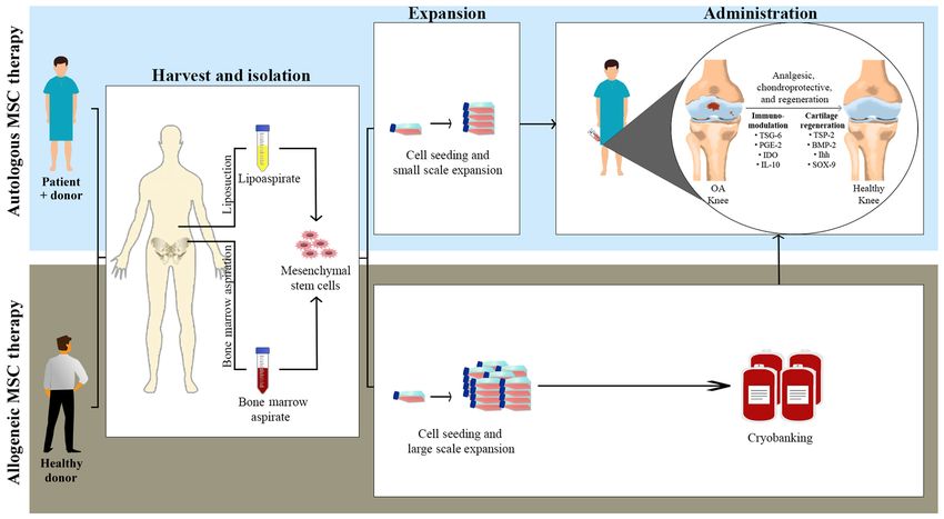
Figure 1. General procedures in using BMSCs and ASCs for the treatment of OA. Autologous MSCs are isolated from the patient's own tissue, whereas
allogeneic MSCs are isolated from a healthy donor who is not a patient. The BMSCs and ASCs are harvested via bone marrow aspiration and liposuction,
respectively, followed by the isolation of MSCs. The expansion of MSCs is performed on a small scale for autologous MSCs and large scale for allogeneic MSCs,
and the latter are stored in a cryobank, to be later administered to patients with OA when required. MSCs are administered to the patients via intra‑articular
injection, and the expected beneficial outcomes post‑treatment are analgesia, chondroprotective effects and cartilage regeneration. MSC, mesenchymal stem
cell; BMSC, bone marrow‑derived stem cells; ASC, adipose‑derived stem cells; OA, osteoarthritis.
2010 and 2020. The general procedures used are as shown in Chondrocyte regenerative effect. The regenerative effect of
Fig. 1. Collectively, the studies highlight their beneficial prop‑ MSCs on the cartilage has been shown to involve the high
erties, exhibiting analgesic, chondroprotective and anatomical expression of several genes responsible for inducing chondro‑
regenerative properties. genesis, and the subsequent development of normal cartilage.
These genes include the production of thrombospondin‑2,
Analgesic and chondroprotective effects. Although the which promotes the Notch signaling pathway; the produc‑
specific molecular mechanisms by which MSCs exert an tion of bone morphogenetic protein 2, which induces the
analgesic effect remain to be elucidated, it is hypothesized SMAD signaling pathway; and the Indian hedgehog signaling
that they revolve around its immunomodulatory effects, by pathway, which promotes the expression of SOX9 followed by
inducing the synthesis of anti‑inflammatory cytokines, such increased expression of the Col2a1 gene, thus stimulating the
as IL‑10, and downregulating the production of pro‑inflam‑ production of proteoglycans and type II collagen, all of which
matory cytokines, such as IL‑1, IL‑6, TNFα and interferon are involved in cartilage regeneration (121‑123). The likely
(IFN)‑γ (110‑114). MSCs are able to secrete numerous soluble mechanism by which MSCs exert their anti‑inflammatory and
growth factors and cytokines, including TNFα‑stimulated cartilage regenerative effects is summarized in Fig. 2.
gene/protein 6 (TSG‑6), prostaglandin E2 (PGE‑2) and Several studies have highlighted the analgesic effects
indoleamine 2,3‑dioxygenase (IDO), which contribute to their of MSCs is addition to their cartilage regenerative effects;
ability to mitigate pain (103,115‑117). It has been shown that however, follow‑up is usually limited to 6‑12 months
the presence of MSCs results in the production of TSG‑6, post‑treatment. The long‑term potential of MSC therapy may
leading to inhibition of the toll‑like receptors‑2/nuclear factor thus be underrated, as the structural changes required for a
κ‑light‑chain‑enhancer of activated B cells signaling pathway, prominent effect take at least a year to occur (79,124).
followed by the subsequent downregulation of inflammatory In general, numerous reports have shown that as little as a
mediators, such as nitric oxide, TNFα and IL‑1 (103,115). single injection of a 1x10⁶ cell dose is sufficient for initiation of
Upregulation of PGE‑2 by MSCs leads to inhibition of the their analgesic effects, although eliciting chondrocyte regen‑
IFN‑γ, inducing the differentiation of M1‑type macrophages to eration response in cartilage requires a much higher dose of at
M2‑type macrophages (116). Similarly, the production of IDO least 1x10⁸ cells (81,125). Hence, higher quality randomized,
by MSCs also promotes the conversion of M1 macrophages to controlled clinical studies with larger cohorts are required
M2 macrophages (117). to strengthen the evidence and evaluate the quality of its
The homing migratory ability of MSCs to sites of injury therapeutic outcomes. Additionally, established protocols for
and inflammation provides beneficial relief in both BMSC and consideration of the optimal dose, time of intervention, method
saline‑treated knee in patients with bilateral OA (118‑120). of delivery and safety precautions also require extensive study.8 LOO and WONG: STEM CELL THERAPY FOR OSTEOARTHRITIS: ADVANTAGES AND CHALLENGES
Figure 2. Summary of the likely mechanism by which MSCs exert their anti‑inflammatory (black) and cartilage regeneration (grey) effects. Solid arrows indicate
stimulation; dashed arrows indicate differentiation; flatheaded arrows indicate inhibition. MSC, mesenchymal stem cell; Col2a1, collagen type 2 α1 chain;
IDO, indoleamine 2,3‑dioxygenase; NF‑κ B, nuclear factor κ‑light‑chain‑enhancer of activated B cells; NO, nitric oxide; PGE‑2, prostaglandin E2; SOX9, SRY‑box
transcription factor 9; TLR‑2, toll‑like receptor‑2; TNF, tumor necrosis factor; TSG‑6, TNFα‑stimulated gene/protein 6; TSP‑2, thrombospondin‑2.
7. Challenges involved in the use of MSC therapy for OA to proliferate. This is a double‑edged characteristic, as it may
lower the risk of malignant tumors, but limit the therapeutic use
Despite the promising aspects for the use of MSCs clinically, this of stem cells. It was documented that the MSCs exhibited abnor‑
approach is considered relatively new and is surrounded by chal‑ malities in the morphology of the cells, such as enlargement,
lenges involved in its use for OA, and these are discussed below. reduced expression of specific surface markers, and finally
senescence as they reached higher passage numbers (133,134).
Effects of the donor's health condition and age on stem cell The entry into a state of senescence in MSCs was shown to
properties. The differentiation potential and proliferation affect the differential potential of the cells, the immunomodu‑
capacity of the MSCs are known to be affected by patients lation capability and their migratory ability (133). Although
requiring autologous MSC therapy. Certain metabolic cryopreservation of MSCs can be used to address this issue, the
conditions, such as diabetes and obesity, can generate microen‑ process comes with its own challenges; a decrease in viability,
vironmental cues that predispose the differentiation potential colony forming units and integrin expression were observed
of MSCs towards adipocyte differentiation rather than chon‑ after cryopreservation and thawing of the cells (135).
drocyte differentiation (126‑128). Besides, the proliferative
capacity of MSCs decreased with age (126). Technical challenges. MSC therapy requires highly skilled
professionals, as the culturing of MSCs must be performed with
Instability of chondrocyte‑like phenotypes. One of the major utmost care to prevent contamination of the cells. Additionally,
challenges accompanying the use of MSCs for treatment of considerably more research is required in order to establish a
OA is the unsustainable cellular and hyaline cartilage pheno‑ clear protocol for the isolation, expansion, differentiation and
type of differentiated chondrocytes. Previous studies have pre‑conditioning of MSCs, and to determine the appropriate
documented the possible involvement of these cells in the concentration of MSCs for use in patients with OA.
development of heterotopic ossification, a process where bone
formation occurs in non‑skeletal tissues (129‑132). These Social. While stem cell therapy may offer an attractive option
studies reported on the transient secretion of type II collagen for treatment of currently uncurable diseases, the costs are
from the MSCs, followed by the up‑regulated expression of currently considerably high, owing to the need to cover the
collagen type X, matrix metalloproteinase and alkaline phos‑ cost of the individual harvesting, isolation, and expansion of
phatase activity, thus indicating a shift from the chondrogenic cells in a sterile facility. Additionally, the increasing popu‑
to a hypertrophic phenotype that precedes osteogenesis, larity of stem cell therapy has warranted the use widespread
a phenomenon that does not normally occur amongst the biobanking, and an increased access to the various highly
chondrocytes found in the hyaline cartilage in the joints. multipotent stem cells, which could be at risk of exploitation.
Hence, policies on biobanking of stem cells must be regulated
Limited replicative lifespan. As mentioned earlier, the MSCs to address the possible issues regarding its usage, control as
will enter a state of senescence whereby they lose their ability well as patients' consent.BIOMEDICAL REPORTS 15: 67, 2021 9
8. Conclusions 3. Blagojevic M, Jinks C, Jeffery A and Jordan KP: Risk factors
for onset of osteoarthritis of the knee in older adults: A systemic
review and meta‑analysis. Osteoarthritis Cartilage 18: 24‑33,
The emergence of stem cell‑based therapies has brought 2010.
about novel avenues to address the, as of yet, unmet curative 4. Loeser RF: Molecular mechanisms of cartilage destruction:
Mechanics, inflammatory mediators, and aging collide. Arthritis
treatment for various degenerative disorders, including OA. Rheum 54: 1357‑1360, 2006.
The multipotency of the MSCs, along with its self‑renewal 5. Ng HY, Alvin Lee KX and Shen KX: Articular cartilage:
and immunomodulatory properties, availability and ease Structure, composition, injuries and repair. JSM Bone Jt Dis 1:
1010, 2017.
of isolation highlight the potential use of MSCs for cellular 6. Ostrowska M, Maśliński W, Prochorec‑Sobieszek M,
therapeutic approaches in OA, and promising results have Nieciecki M and Sudoł‑Szopińska I: Cartilage and bone damage
been demonstrated in various pre‑clinical and clinical trials. in rheumatoid arthritis. Reumatologia 56: 111‑120, 2018.
7. Mahajan A, Verma S and Tandon V: Osteoarthritis. J Assoc
However, several challenges are involved in this process, and Physicians India 53: 634‑641, 2005.
this requires standardized solutions before they can be recom‑ 8. Houard X, Goldring MB and Berenbaum F: Homeostatic
mended clinically. Efforts on investigating and establishing mechanisms in articular cartilage and role of inflammation in
osteoarthritis. Curr Rheumatol Rep 15: 375, 2013.
protocols to increase the stability of the chondrocyte‑like 9. Gignac MAM, Irvin E, Cullen K, Van Eerd D, Beaton DE,
phenotype of the MSCs is required to raise the success rates Mahood Q, McLeod C and Backman CL: Men and women's
of MSC‑based treatment in patients with OA, and to lower the occupational activities and the risk of developing osteoarthritis
of the knee, hip, or hands: A systematic review and recommen‑
cost. In addition, the appropriate concentration of stem cells dations for future research. Arthritis Care Res (Hoboken) 72:
for specific treatments, and the long‑term follow‑ups of patients 378‑396, 2020.
with OA treated with MSCs should be performed to investigate 10. Michael JW, Schlüter‑Brust KU and Eysel P: The epidemiology,
etiology, diagnosis, and treatment of osteoarthritis of the knee.
the long‑term safety and efficacy of MSC‑based therapy. Dtsch Arztebl Int 107: 152‑162, 2010.
11. Helliwell P: Osteoarthritis and Paget's disease. Br J Rheumatol 34:
Acknowledgements 1061‑1063, 1995.
12. Mora JC, Przkora R and Cruz‑Almeida Y: Knee osteoarthritis:
Pathophysiology and current treatment modalities. J Pain Res 11:
Not applicable. 2189‑2196, 2018.
13. de l'Escalopier N, Anract P and Biau D: Surgical treatments for
osteoarthritis. Ann Phys Rehabil Med 59: 227‑233, 2016.
Funding 14. Øiestad BE, Juhl CB, Eitzen I and Thorlund JB: Knee extensor
muscle weakness is a risk factor for development of knee osteo‑
No funding was received. arthritis. A systematic review and meta‑analysis. Osteoarthritis
Cartilage 23: 171‑177, 2015.
15. March LM and Bagga H: Epidemiology of osteoarthritis in
Availability of data and materials Australia. Med J Aust 180: S6‑S10, 2004.
16. Messier SP: Obesity and osteoarthritis: Disease genesis and
nonpharmacologic weight management. Rheum Dis Clin North
Not applicable. Am 34: 713‑729, 2008.
17. Vincent KR and Vincent HK: Resistance exercise for knee osteo‑
Authors' contributions arthritis. PM R 4 (Suppl 5): S45‑S52, 2012.
18. Rogers MW and Wilder FV: The effects of strength training
among persons with hand osteoarthritis: A two‑year follow‑up
SJQL and NKW both wrote and revised the article. Both study. J Hand Ther 20: 244‑249; quiz 250, 2007.
authors read and approved the final manuscript. Data sharing 19. Latham N and Liu CJ: Strength training in older adults: The
benefits for osteoarthritis. Clin Geriatr Med 26: 445‑459, 2010.
is not applicable. 20. Aguiar GC, Rocha SG, da Silva Rezende GA, do Nascimento MR
and Scalzo PL: Effects of resistance training in individuals with
Ethics approval and consent to participate knee osteoarthritis. Fisioter Mov 29: 589‑596, 2016.
21. Thoumie P, Marty M, Avouac B, Pallez A, Vaumousse A,
Pipet LPT, Monroche A, Graveleau N, Bonnin A, Amor CB and
Not applicable. Coudeyre E: Effect of unloading brace treatment on pain and
function in patients with symptomatic knee osteoarthritis: The
ROTOR randomized clinical trial. Sci Rep 8: 10519, 2018.
Patient consent for publication 22. Chew KT, Lew HL, Date E and Fredericson M: Current evidence
and clinical applications of therapeutic knee braces. Am J Phys
Not applicable. Med Rehabil 86: 678‑686, 2007.
23. Lee PY, Winfield TG, Harris SR, Storey E and Chandratreya A:
Unloading knee brace is a cost‑effective method to bridge and
Competing interests delay surgery in unicompartmental knee arthritis. BMJ Open
Sport Exerc Med 2: e000195, 2017.
24. Ostrander RV, Leddon CE, Hackel JG, O'Grady CP and Roth CA:
The authors declare that they have no competing interests. Efficacy of unloader bracing in reducing symptoms of knee
osteoarthritis. Am J Orthop (Belle Mead NJ) 45: 306‑311, 2016.
References 25. Wilson B, Rankin H and Barnes CL: Long‑term results of an
unloader brace in patients with unicompartmental knee osteoar‑
thritis. Orthopedics 34: e334‑e337, 2011.
1. National Clinical Guideline Centre (UK): Osteoarthritis‑Care 26. Yu SP, Williams M, Eyles JP, Chen JS, Makovey J and Hunter DJ:
and Μanagement in Αdults. London: National Institute for Effectiveness of knee bracing in osteoarthritis: Pragmatic trial
Health and Care Excellence (UK), 2014. in a multidisciplinary clinic. Int J Rheum Dis 19: 279‑286, 2016.
2. James SL, Abate D, Hassen Abate K, Abay SM, Abbafati C, 27. Squyer E, Stamper DL, Hamilton DT, Sabin JA and Leopold SS:
Abbasi N, Abbastabar H, Abd‑Allah F, Abdela J, Abdelalim A, et al: Unloader knee braces for osteoarthritis: Do patients actually
Global, regional, and national incidence, prevalence, and years wear them? Clin Orthop Relat Res 471: 1982‑1991, 2013.
lived with disability for 354 diseases and injuries for 195 countries 28. Nejati P, Farzinmehr A and Moradi‑Lakeh M: The effect of exer‑
and territories, 1990‑2017: A systematic analysis for the Global cise therapy on knee osteoarthritis: A randomized clinical trial.
Burden of Disease Study 2017. Lancet 392: 1789-1858, 2018. Med J Islam Repub Iran 29: 186, 2015.10 LOO and WONG: STEM CELL THERAPY FOR OSTEOARTHRITIS: ADVANTAGES AND CHALLENGES
29. Reid MC, Eccleston C and Pillemer K: Management of chronic 53. Walters MC: Update of hematopoietic cell transplantation for
pain in older adults. BMJ 350: h532, 2015. sickle cell disease. Curr Opin Hematol 22: 227‑233, 2015.
30. Rönn K, Reischl N, Gautier E and Jacobi M: Current surgical 54. Abdelmoaty N, Alattar E, Ahmed K, Ibrahim Y and Darwish B:
treatment of knee osteoarthritis. Arthritis 2011: 454873, 2011. Regenerative power of autologous peripheral blood stem cell
31. Wang X, Wanyan P, Wang JM, Tian JH, Hu L, Shen XP and injection in knee osteoarthritis by a non‑invasive approach‑MRI
Yang KH: A randomized, controlled trial to assess the efficacy Study. Ann Rheum Dis 73: 1066, 2014.
of arthroscopic debridement in combination with oral medica‑ 55. U.S Food and Drug Administration: Approved Cellular and
tion versus oral medication in patients with gouty knee arthritis. Gene Therapy Products, 2021.
Indian J Surg 77 (Suppl 2): S628‑S634, 2015. 56. Prockop DJ: Marrow stromal cells as stem cells for nonhemato‑
32. Kirkley A, Birmingham TB, Litchfield RB, Giffin JR, Willits KR, poietic tissues. Science 276: 71‑74, 1997.
Wong CJ, Feagan BG, Donner A, Griffin SH, D'Ascanio LM, et al: 57. Makino S, Fukuda K, Miyoshi S, Konishi F, Kodama H, Pan J,
A randomized trial of arthroscopic surgery for osteoarthritis of Sano M, Takahashi T, Hori S, Abe H, et al: Cardiomyocytes
the knee. N Engl J Med 359: 1097‑1107, 2008. can be generated from marrow stromal cells in vitro. J Clin
33. Moseley JB, O'Malley K, Petersen NJ, Menke TJ, Brody BA, Invest 103: 697‑705, 1999.
Kuykendall DH, Hollingsworth JC, Ashton CM and Wray NP: 58. Wakitani S, Saito T and Caplan AI: Myogenic cells derived from
A controlled trial of arthroscopic surgery for osteoarthritis of the rat bone marrow mesenchymal stem cells exposed to 5‑azacyti‑
knee. N Engl J Med 347: 81‑88, 2002. dine. Muscle Nerve 18: 1417‑1426, 1995.
34. Choi HG, Kwon BC, Kim JI and Lee JK: Total knee arthroplasty 59. Nuttall ME, Patton AJ, Olivera DL, Nadeau DP and Gowen M:
reduces the risk of mortality in osteoarthritis patients up to Human trabecular bone cells are able to express both osteoblastic
12 years: A Korean national cohort longitudinal follow‑up study. and adipocytic phenotype: Implications for osteopenic disorders.
J Orthop Surg (Hong Kong) 28: 2309499020902589, 2020. J Bone Miner Res 13: 371‑382, 1998.
35. Rajabzadeh N, Fathi E and Farahzadi R: Stem cell‑based regen‑ 60. Pittenger MF, Mackay AM, Beck SC, Jaiswal RK, Douglas R,
erative medicine. Stem Cell Investig 6: 19, 2019. Mosca JD, Moorman MA, Simonetti DW, Craig S and
36. Landry DW and Zucker HA: Embryonic death and the creation of Marshak DR: Multilineage potential of adult human mesen‑
human embryonic stem cells. J Clin Invest 114: 1184‑1186, 2004. chymal stem cells. Science 284: 143‑147, 1999.
37. Ede V and Obeagu EI: Ethical issues in human embryonic stem 61. Bianco P, Robey PG and Simmons PJ: Mesenchymal stem
cell research: A christian perspective. Int J Med Sci Dent Res 1: cells: Revisiting history, concepts, and assays. Cell Stem Cell 2:
8‑14, 2019. 313‑319, 2008.
38. Lo B and Parham L: Ethical issues in stem cell research. Endocr 62. Roufosse CA, Direkze NC, Otto WR and Wright NA: Circulating
Rev 30: 204‑213, 2009. mesenchymal stem cells. Int J Biochem Cell Biol 36: 585‑597,
39. Takahashi K and Yamanaka S: Induction of pluripotent stem 2004.
cells from mouse embryonic and adult fibroblast cultures by 63. Lindroos B, Suuronen R and Miettinen S: The potential of
defined factors. Cell 126: 663‑676, 2006. adipose stem cells in regenerative medicine. Stem Cell Rev
40. Shi Y, Inoue H, Wu JC and Yamanaka S: Induced pluripotent Rep 7: 269‑291, 2011.
stem cell technology: A decade of progress. Nat Rev Drug 64. Erices A, Conget P and Minguell JJ: Mesenchymal progenitor
Discov 16: 115‑130, 2017. cells in human umbilical cord blood. Br J Haematol 109: 235‑242,
41. Takahashi K, Tanabe K, Ohnuki M, Narita M, Ichisaka T, 2000.
Tomoda K and Yamanaka S: Induction of pluripotent stem 65. Gronthos S, Mankani M, Brahim J, Robey PG and Shi S:
cells from adult human fibroblasts by defined factors. Cell 131: Postnatal human dental pulp stem cells (DPSCs) in vitro and
861‑872, 2007. in vivo. Proc Natl Acad Sci USA 97: 13625‑13630, 2000.
42. Gutierrez‑Aranda I, Ramos‑Mejia V, Bueno C, Munoz‑Lopez M, 66. Haniffa MA, Wang XN, Holtick U, Rae M, Isaacs JD,
Real PJ, Mácia A, Sanchez L, Ligero G, Garcia‑Parez JL and Dickinson AM, Hilkens CM and Collin MP: Adult human
Menendez P: Human induced pluripotent stem cells develop fibroblasts are potent immunoregulatory cells and function‑
teratoma more efficiently and faster than human embryonic stem ally equivalent to mesenchymal stem cells. J Immunol 179:
cells regardless the site of injection. Stem Cells 28: 1568‑1570, 1595‑1604, 2007.
2010. 67. Sessarego N, Parodi A, Podesta M, Benvenuto F, Mogni M,
43. Zhang M, Wang L, An K, Cai J, Li G, Yang C, Liu H, Du F, Raviolo V, Lituania M, Kunkl A, Ferlazzo G, Bricarelli FD, et al:
Han X, Zhang Z, et al: Lower genomic stability of induced Multipotent mesenchymal stromal cells from amniotic fluid:
pluripotent stem cells reflects increased non‑homologous end Solid perspectives for clinical application. Haematologica 93:
joining. Cancer Commun (Lond) 38: 49, 2018. 339‑346, 2008.
44. Christensen R and Serakinci N: Adult Stem Cells. In: 68. Dominici M, Le Blanc K, Mueller I, Slaper‑Cortenbach I,
Encyclopedia of Cancer. Springer, Heidelberg, pp1‑5, 2015. Marini F, Krause D, Deans R, Keating A, Prockop DJ and
45. Centeno C, Markle J, Dodson E, Stemper I, Williams CJ, Hyzy M, Horwitz E: Minimal criteria for defining multipotent mesen‑
Ichim T and Freeman M: Treatment of lumbar degenerative chymal stromal cells. The International society for cellular
disc disease‑associated radicular pain with culture‑expanded therapy position statement. Cytotherapy 8: 315‑317, 2006.
autologous mesenchymal stem cells: A pilot study on safety and 69. Gianakos AL, Sun L, Patel JN, Adams DM and Liporace FA:
efficacy. J Transl Med 15: 197, 2017. Clinical application of concentrated bone marrow aspirate in
46. Berry JD, Cudkowicz ME, Windebank AJ, Staff NP, Owegi M, orthopaedics: A systematic review. World J Orthop 8: 491‑506,
Nicholson K, McKenna‑Yasek D, Levy YS, Abramov N, 2017.
Kaspi H, et al: NurOwn, phase 2, randomized, clinical trial 70. Mohamed‑Ahmed S, Fristad I, Lie SA, Suliman S, Mustafa K,
in patients with ALS: Safety, clinical, and biomarker results. Vindenes H and Idris SB: Adipose‑derived and bone marrow
Neurology 93: e2294‑e2305, 2019. mesenchymal stem cells: A donor‑matched comparison. Stem
47. Riecke J, Johns K M, Cai C, Vahidy FS, Pa rsha K, Cell Res Ther 9: 168, 2018.
Furr‑Stimming E, Schiess M and Savitz SI: A meta‑analysis of 71. Gabr MM, Zakaria MM, Refaie AF, Abdel‑Rahman EA,
mesenchymal stem cells in animal models of Parkinson's disease. Reda AM, Ali SS, Khater SM, Ashamallah SA, Ismail AM,
Stem Cells Dev 24: 2082‑2090, 2015. Ismail HEA, et al: From human mesenchymal stem cells to
48. Ng AP and Alexander WS: Haematopoietic stem cells: Past, insulin‑producing cells: Comparison between bone marrow‑ and
present and future. Cell Death Discov 3: 17002, 2017. adipose tissue‑derived cells. Biomed Res Int 2017: 3854232,
49. Yamamoto R, Wilkinson AC and Nakauchi H: Changing concepts 2017.
in hematopoietic stem cells. Science 362: 895‑896, 2018. 72. da Silva Meirelles L, Chagastelles PC and Nardi NB:
50. Domen J, Wagers A and Weissman IL: Bone marrow Mesenchymal stem cells reside in virtually all post‑natal organs
(Hematopoietic) stem cells. Stem Cell Information: The National and tissues. J Cell Sci 119: 2204‑2213, 2006.
Institutes of Health resource for stem cell research: 13‑34, 2006. 73. Crisan M, Yap S, Casteilla L, Chen CW, Corselli M, Park TS,
51. Morgan RA, Gray D, Lomova A and Kohn DB: Hematopoietic Andriolo G, Sun B, Zheng B, Zhang L, et al: A perivascular
stem cell gene therapy: Progress and lessons learned. Cell Stem origin for mesenchymal stem cells in multiple human organs.
Cell 21: 574‑590, 2017. Cell Stem Cell 3: 301‑313, 2008.
52. Taylor M, Khan S, Stapleton M, Wang J, Chen J, Wynn R, Yabe H, 74. Orozco L, Munar A, Soler R, Alberca M, Soler F, Huguet M,
Chinen Y, Boelens JJ, Mason RW, et al: Hematopoietic stem cell Sentís J, Sánchez A and García‑Sancho J: Treatment of knee
transplantation for mucopolysaccharidoses: Past, present, and osteoarthritis with autologous mesenchymal stem cells: A pilot
future. Biol Blood Marrow Transplant 25: e226‑e246, 2019. study. Transplantation 95: 1535‑1541, 2013.BIOMEDICAL REPORTS 15: 67, 2021 11
75. Lamo‑Espinosa JM, Mora G, Blanco JF, Granero‑Moltó F, 91. Steffenhagen C, Dechant FX, Oberbauer E, Furtner T,
Nuñez‑Córdoba JM, Sánchez‑Echenique C, Bondía JM, Weidner N, Küry P, Aigner L and Rivera FJ: Mesenchymal stem
Aquerreta JD, Andreu EJ, Ornilla E, et al: Intra‑articular cells prime proliferating adult neural progenitors toward an
injection of two different doses of autologous bone marrow oligodendrocyte fate. Stem Cells Dev 21: 1838‑1851, 2012.
mesenchymal stem cells versus hyaluronic acid in the treat‑ 92. Allahverdi A, Abroun S, Jafarian A, Soleimani M, Taghikhani M
ment of knee osteoarthritis: Multicenter randomized controlled and Eskandari F: Differentiation of human mesenchymal stem
clinical trial (phase I/II). J Transl Med 14: 246, 2016. cells into insulin producing cells by using a lentiviral vector
76. Garay‑Mendoza D, Villarreal‑Martínez L, Garza‑Bedolla A, carrying PDX1. Cell J 17: 231‑242, 2015.
Pérez‑Garza DM, Acosta‑Olivo C, Vilchez‑Cavazos F, 93. Chen LB, Jiang XB and Yang L: Differentiation of rat marrow
Diaz‑Hutchinson C, Gómez‑Almaguer D, Jaime‑Pérez JC and mesenchymal stem cells into pancreatic islet beta‑cells. World
Mancías‑Guerra C: The effect of intra‑articular injection of J Gastroenterol 10: 3016‑3020, 2004.
autologous bone marrow stem cells on pain and knee function 94. Singh A, Singh A and Sen D: Mesenchymal stem cells in cardiac
in patients with osteoarthritis. Int J Rheum Dis 21: 140‑147, regeneration: A detailed progress report of the last 6 years
2018. (2010‑2015). Stem Cell Res Ther 7: 82, 2016.
77. Vega A, Martín‑Ferrero MA, Del Canto F, Alberca M, García V, 95. Solis MA, Moreno Velásquez I, Correa R and Huang LLH: Stem
Munar A, Orozco L, Soler R, Fuertes JJ, Huguet M, et al: cells as a potential therapy for diabetes mellitus: A call‑to‑action
Treatment of knee osteoarthritis with allogeneic bone marrow in Latin America. Diabetol Metab Syndr 11: 20, 2019.
mesenchymal stem cells: A randomized controlled trial. 96. Sun R, Li X, Liu M, Zeng Y, Chen S and Zhang P: Advances in
Transplantation 99: 1681‑1690, 2015. stem cell therapy for cardiovascular disease (Review). Int J Mol
78. Shadmanfar S, Labibzadeh N, Emadedin M, Jaroughi N, Med 38: 23‑29, 2016.
Azimian V, Mardpour S, Kakroodi FA, Bolurieh T, Hosseini SE, 97. Terashvili M and Bosnjak ZJ: Stem cell therapies in cardiovas‑
Chehrazi M, et al: Intra‑articular knee implantation of autologous cular disease. J Cardiothorac Vasc Anesth 33: 209‑222, 2019.
bone marrow‑derived mesenchymal stromal cells in rheumatoid 98. Paspaliaris V and Kolios G: Stem cells in osteoporosis: From
arthritis patients with knee involvement: Results of a random‑ biology to new therapeutic approaches. Stem Cells Int 2019:
ized, triple‑blind, placebo‑controlled phase 1/2 clinical trial. 1730978, 2019.
Cytotherapy 20: 499‑506, 2018. 99. Mercado‑Sáenz S, Ruiz‑Gómez MJ, Morales‑Moreno F and
79. Al‑Najar M, Khalil H, Al‑Ajlouni J, Al‑Antary E, Hamdan M, Martínez‑Morillo M: Cellular aging: Theories and technological
Rahmeh R, Alhattab D, Samara O, Yasin M, Abdullah AA, et al: influence. Brazilian Arch Biol Technol 53: 1319‑1332, 2010.
Intra‑articular injection of expanded autologous bone marrow 100. Shay JW and Wright WE: Role of telomeres and telomerase in
mesenchymal cells in moderate and severe knee osteoarthritis is cancer. Semin Cancer Biol 21: 349‑353, 2011.
safe: A phase I/II study. J Orthop Surg Res 12: 190, 2017. 101. Keating A: Mesenchymal stromal cells: New directions. Cell
80. Anz AW, Hubbard R, Rendos NK, Everts PA, Andrews JR and Stem Cell 10: 709‑716, 2012.
Hackel JG: Bone marrow aspirate concentrate is equivalent 102. Le Blanc K and Mougiakakos D: Multipotent mesenchymal
to platelet‑rich plasma for the treatment of knee osteoarthritis stromal cells and the innate immune system. Nat Rev
at 1 year: A prospective, randomized trial. Orthop J Sports Immunol 12: 383‑396, 2012.
Med 2: 2325967119900958, 2020. 103. Prockop DJ and Oh JY: Mesenchymal stem/stromal cells
81. Chahal J, Gómez‑Aristizábal A, Shestopaloff K, Bhatt S, (MSCs): Role as guardians of inflammation. Mol Ther 20:
Chaboureau A, Fazio A, Chisholm J, Weston A, Chiovitti J, 14‑20, 2012.
Keating A, et al: Bone marrow mesenchymal stromal cell treat‑ 104. Ryan JM, Barry FP, Murphy JM and Mahon BP: Mesenchymal
ment in patients with osteoarthritis results in overall improvement stem cells avoid allogeneic rejection. J Inflamm (Lond) 2: 8,
in pain and symptoms and reduces synovial inflammation. Stem 2005.
Cells Transl Med 8: 746‑757, 2019. 105. Klyushnenkova E, Mosca JD, Zernetkina V, Majumdar MK,
82. Zuk PA, Zhu M, Mizuno H, Huang J, Futrell JW, Katz AJ, Beggs KJ, Simonetti DW, Deans RJ and McIntosh KR: T cell
Benhaim P, Lorenz HP and Hedrick MH: Multilineage cells responses to allogeneic human mesenchymal stem cells:
from human adipose tissue: Implications for cell‑based therapies Immunogenicity, tolerance, and suppression. J Biomed Sci 12:
2. Tissue Eng 7: 211‑228, 2001. 47‑57, 2005.
83. Zuk PA, Zhu M, Ashjian P, De Ugarte DA, Huang JI, Mizuno H, 106. Di Nicola M, Carlo‑Stella C, Magni M, Milanesi M, Longoni PD,
Alfonso ZC, Fraser JK, Benhaim P and Hedrick MH: Human Matteucci P, Grisanti S and Gianni AM: Human bone marrow
adipose tissue is a source of multipotent stem cells. Raff M (ed). stromal cells suppress T‑lymphocyte proliferation induced by
Mol Biol Cell 13: 4279‑4295, 2002. cellular or nonspecific mitogenic stimuli. Blood 99: 3838‑3843,
84. Coughlin RP, Oldweiler A, Mickelson DT and Moorman CT III: 2002.
Adipose‑derived stem cell transplant technique for degenerative 107. Tse WT, Pendleton JD, Beyer WM, Egalka MC and Guinan EC:
joint disease. Arthrosc Tech 6: e1761‑e1766, 2017. Suppression of allogeneic T‑cell proliferation by human marrow
85. Freitag J, Bates D, Wickham J, Shah K, Huguenin L, Tenen A, stromal cells: Implications in transplantation. Transplantation 75:
Paterson K and Boyd R: Adipose‑derived mesenchymal stem cell 389‑397, 2003.
therapy in the treatment of knee osteoarthritis: A randomized 108. Lee H, Shamy GA, Elkabetz Y, Schofield CM, Harrsion NL,
controlled trial. Regen Med 14: 213‑230, 2019. Panagiotakos G, Socci ND, Tabar V and Studer L: Directed
86. De Ugarte DA, Alfonso Z, Zuk PA, Elbarbary A, Zhu M, differentiation and transplantation of human embryonic stem
Ashjian P, Benhaim P, Hedrick MH and Fraser JK: Differential cell‑derived motoneurons. Stem Cells 25: 1931‑1939, 2007.
expression of stem cell mobilization‑associated molecules 109. Su W, Zhou M, Zheng Y, Fan Y, Wang L, Han Z, Kong D,
on multi‑lineage cells from adipose tissue and bone marrow. Zhao RC, Wu JC, Xiang R and Li Z: Bioluminescence reporter
Immunol Lett 89: 267‑270, 2003. gene imaging characterize human embryonic stem cell‑derived
87. Yang ZX, Han ZB, Ji YR, Wang YW, Liang L, Chi Y, Yang SG, teratoma formation. J Cell Biochem 112: 840‑848, 2011.
Li LN, Luo WF, Li JP, et al: CD106 identifies a subpopulation 110. Aggarwal S and Pittenger MF: Human mesenchymal stem
of mesenchymal stem cells with unique immunomodulatory cells modulate allogeneic immune cell responses. Blood 105:
properties. PLoS One 8: e59354, 2013. 1816‑1822, 2005.
88. Krampera M, Marconi S, Pasini A, Galiè M, Rigotti G, Mosna F, 111. Li H and Fu X: Mechanisms of action of mesenchymal stem
Tinelli M, Lovato L, Anghileri E, Andreini A, et al: Induction cells in cutaneous wound repair and regeneration. Cell Tissue
of neural‑like differentiation in human mesenchymal stem cells Res 348: 371‑377, 2012.
derived from bone marrow, fat, spleen and thymus. Bone 40: 112. Maxson S, Lopez EA, Yoo D, Danilkovitch‑Miagkova A and
382‑390, 2007. Leroux MA: Concise review: Role of mesenchymal stem cells in
89. Kennea NL, Waddington SN, Chan J, O'Donoghue K, wound repair. Stem Cells Transl Med 1: 142‑149, 2012.
Yeung D, Taylor DL, Al‑Allaf FA, Pirianov G, Themis M, 113. Williams CG, Kim TK, Taboas A, Malik A, Manson P and
Edwards AD, et al: Differentiation of human fetal mesenchymal Elisseeff J: In vitro chondrogenesis of bone marrow‑derived
stem cells into cells with an oligodendrocyte phenotype. Cell mesenchymal stem cells in a photopolymerizing hydrogel.
Cycle 8: 1069‑1079, 2009. Tissue Eng 9: 679‑688, 2003.
90. Shah S, Yin PT, Uehara TM, Chueng ST, Yang L and Lee KB: 114. Zhang W, Ge W, Li C, You S, Liao L, Han Q, Deng W and
Guiding stem cell differentiation into oligodendrocytes using Zhao RC: Effects of mesenchymal stem cells on differentiation,
graphene‑nanofiber hybrid scaffolds. Adv Mater 26: 3673‑3680, maturation, and function of human monocyte‑derived dendritic
2014. cells. Stem Cells Dev 13: 263‑271, 2004.You can also read