Antinerve growth factor monoclonal antibodies for the control of pain in dogs andcats
←
→
Page content transcription
If your browser does not render page correctly, please read the page content below
Veterinary Record: first published as 10.1136/vr.104590 on 27 October 2018. Downloaded from http://veterinaryrecord.bmj.com/ on 7 November 2018 by guest. Protected by copyright.
                                                                                                                                       REVIEW

Antinerve growth factor monoclonal
antibodies for the control of pain in
dogs and cats
Masataka Enomoto,1 Patrick W Mantyh,2 Joanna Murrell,3 John F Innes,4 B Duncan X Lascelles1,5,6,7

Nerve growth factor (NGF) is essential for the survival of sensory and sympathetic neurons during development.
However, in the adult, NGF and its interaction with tropomyosin receptor kinase A receptor (TrkA) has been
found to play a critical role in nociception and nervous system plasticity in pain conditions. Thus, various
monoclonal antibody (mAb) therapies targeting this pathway have been investigated in the development of new
pharmacotherapies for chronic pain. Although none of the mAbs against NGF are yet approved for use in humans,
they look very promising for the effective control of pain in humans. Recently, species-specific anti-NGF mAbs for
the management of osteoarthritis (OA)-associated pain in dogs and cats has been developed, and early clinical
trials have been conducted. Anti-NGF therapy looks to be both very effective and very promising as a novel
therapy against chronic pain in dogs and cats. This review outlines the mechanism of action of NGF and reviews
its role of NGF in osteoarthritis, reviews research in rodent OA models and the current status of the development
of anti-NGF mAbs in humans. Furthermore, we describe and discuss the recent development of species-specific
anti-NGF mAbs for the treatment of OA-associated pain in veterinary medicine.

Introduction                                                                           Although most commonly initiated early in life by
Osteoarthritis (OA) is a slowly progressive degenerative                               developmental disease (eg, hip dysplasia), many other
joint disease characterised by whole-joint structural                                  factors play a role in its development, including diet,
changes including articular cartilage, synovium,                                       genetics, environment, obesity and age.4 6 7 In cats, hip,
subchondral bone and periarticular components,                                         stifle, hock and elbow joints are the most commonly
which can lead to pain and loss of joint function.1–4                                  affected synovial joints.3 Although much less is
It is considered to primarily affect the hip, stifle and                               known about the aetiology of OA in cats, idiopathic
elbow joints in dogs,5 although no comprehensive,                                      OA is considered common, with congenital, traumatic,
prospective studies of the prevalence of canine                                        infectious, nutritional and immune-mediated causes
OA throughout the skeleton have been performed.                                        having been documented, similar to other species.3
                                                                                       OA is a condition associated with clinical signs in a
Veterinary Record (2018)                             doi: 10.1136/vr.104590
                                                                                       large percentage of the population, with an estimated
                                                                                       minimum of 20 per cent to 30 per cent of dogs affected
1
 Translational Research in Pain,            6
                                             Center for Pain Research and              clinically and up to 40 per cent of all cats being affected
Comparative Pain Research and               Innovation, UNC School of Dentistry,       clinically (90 per cent of all cats over 12 years of
Education Centre, Department of             Chapel Hill, North Carolina, USA
Clinical Sciences, College of Veterinary    7
                                             Center for Translational Pain Research,
                                                                                       age).3 5 8 The disease is currently incurable with negative
Medicine, North Carolina State              Department of Anesthesiology, Duke         consequences related to pain, mobility impairment
University, Raleigh, North Carolina, USA    University, Durham, North Carolina,        and decreased quality of life.9 Pain results in both
2
 Cancer Center’s Cancer Biology             USA                                        local and distant deterioration of the musculoskeletal
Program, Department of Pharmacology,        E-mail for correspondence:                 system as a result of decreased and altered mobility.
College of Medicine, University of
Arizona, Tucson, Arizona, USA
                                            dxlascel@ncsu.edu                          The pathological processes of OA, such as joint capsule
3
 School of Veterinary Sciences,             Provenance and peer review Not             thickening and fibrosis, contribute to altered range of
University of Bristol, Bristol, UK          commissioned; externally peer              motion that compounds the musculoskeletal changes.
                                            reviewed.
4
 Units E&F, CVS Vets Ltd, Chester, UK                                                  Additionally, the ongoing nociceptive input into the CNS
5
 Comparative Medicine Institute,                                                       results in somatosensory system changes and central
                                            Received June 26, 2017
Department of Clinical Sciences,
College of Veterinary Medicine, North
                                            Revised July 10, 2018                      sensitisation10 11, which contributes to the perception of
                                            Accepted September 19, 2018                pain. The combined effects of pain, central sensitisation
Carolina State University, Raleigh, North
Carolina, USA                                                                          and activity impairment may have negative effects on

VET RECORD |                                                                                                                                     1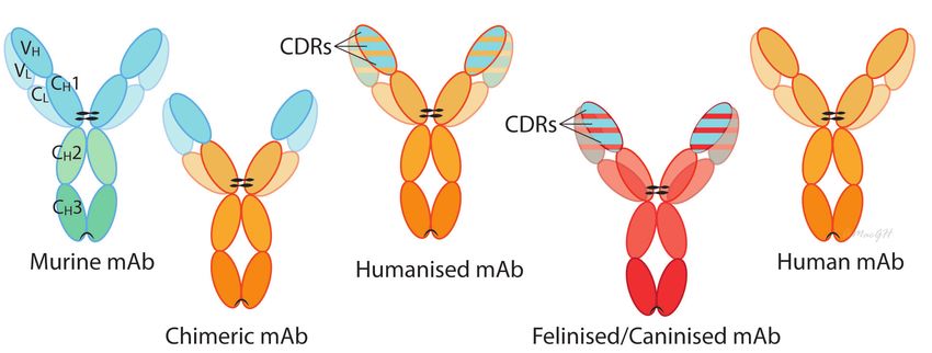
the affective state, heightening anxiety, depression, Biological agents are medical products that are isolated
Veterinary Record: first published as 10.1136/vr.104590 on 27 October 2018. Downloaded from http://veterinaryrecord.bmj.com/ on 7 November 2018 by guest. Protected by copyright.
sleep impairment12 and cognitive dysfunction as from a variety of natural sources (human, animal or
reported in humans.13 14 microorganism). This is in contrast to most drugs that
Currently, pharmacological treatment of pain are chemically synthesised. Biological agents include
centres around non-steroidal anti-inflammatory drugs a wide range of products such as vaccines, blood and
(NSAIDs). These are used to relieve pain and promote blood components, gene therapy and recombinant
functional improvement.2 Globally, several NSAIDs therapeutic proteins. They also may be produced by
are approved for use in dogs, but only two NSAIDs are biotechnology methods. In human medicine, biological
approved for use long-term in cats and only in certain products often offer the most effective means to treat
countries. Despite their widespread use and obvious a variety of medical illnesses and conditions. The
benefit in many cases, NSAIDs are not always sufficiently monoclonal antibodies (mAbs) have proven to be
effective when used as monotherapy.15 Additionally, extremely effective in a variety of diseases in humans.
there are safety and tolerability concerns with their Monoclonal antibodies are produced from single
use in both dogs and cats.16–19 Beyond cyclooxygenase- B-lymphocyte clones in mice or through recombinant
inhibiting NSAIDs and the recently approved engineering. They are monovalent antibodies that
piprant NSAID, a prostaglandin receptor antagonist, specifically bind to target molecules including cytokines,
grapiprant,20 treatment options for the control of pain receptors or cells.24 The binding results in blocking
are very limited. Furthermore, evidence for efficacy of the activity of the target. They were first generated in
so-called adjunctive analgesics is extremely limited15 21. mice in 1975 using a hybridoma technique25 involving
Additionally, there are few proven non-drug therapies, immunising a certain species against a specific epitope
and none that have been shown to provide rapid pain on an antigen and obtaining B-lymphocytes from the
relief. Therefore, OA related-pain remains a challenging animal’s lymphoid tissue. B-lymphocytes are then fused
clinical entity to treat, and indeed, OA-associated pain with an immortal myeloma cell, a type of B-cell tumour,
is one of the most common reasons for euthanasia and these hybridomas can be maintained in vitro and
in dogs.22 23 Thus, there is an urgent need to develop will continue to secrete antibodies with appropriate
effective treatments for OA-related pain in dogs and specificity. Administering purified non-human-derived
cats. antibodies to humans can cause immune reactions,26 and
so antibody engineering technology has been developed
Advent of monoclonal antibodies to lower the risk of immune reactions.27 Engineering
With greater understanding of pathogenesis of OA of antibodies by replacing mouse sequence-derived
coupled with advances in biotechnology, specific amino acids with human sequences has significantly
immunomodulatory therapies called biological reduced immunogenicity of this class of therapeutics.28
agents have been introduced for the treatment of joint Over the last 25 years, chimeric, humanised and fully
diseases, specifically immune-mediated joint diseases. human mAbs have been developed for use in humans
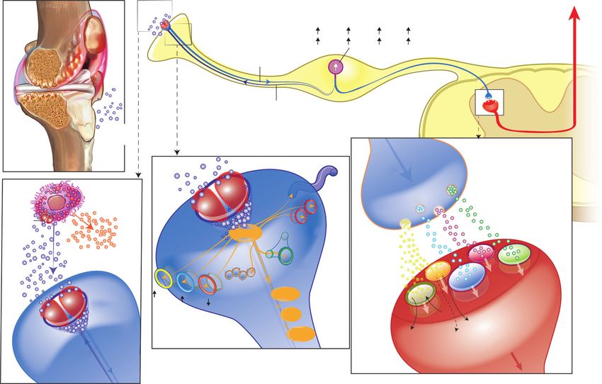
Figure 1 Antibodies are large glycoproteins typically composed of two heavy chains and two light chains, each of which contain a variable domain (variable heavy (VH)
or variable light (VL) and a constant domain (constant heavy (CH) or constant light (CL). The amino acid sequence of the variable domain varies greatly among
antibodies and six ‘hyper-variable’ complementarity-determining region (CDR) loops within the variable domains give the antibody its specificity for binding to an
antigen. In contrast, the constant domain is identical in all antibodies of the same isotype but differs in antibodies of different isotypes (IgA, IgD, IgE, IgG and IgM). The
tail region of the constant domain (Fc region: CH2 and CH3) may direct immune effector functions by binding to cell receptors expressed on immune cells or initiating
complementary-dependent cytotoxicity. Murine monoclonal antibodies (mAbs) are antibodies produced from individual cloned and immortalised mouse B cells. Most
useful therapeutic antibodies have been constructed with the gamma immunoglobulin (IgG) isotype. Chimeric mAbs are antibodies made by fusing the genes encoding
the variable region from a murine-derived mAb, with those from an immunoglobulin (Ig) constant region from a human antibody. Humanised mAbs retain only the CDRs
(part of the variable domain from the original murine-derived mAb that binds to the specific antigen). Fully human mAbs have no murine sequences. Caninised and
felinised mAbs are fully canine or feline specific. These can be made in several ways; for example, Nexvet have used a process of conversion based on alignment with
immunoglobulin complementary DNA libraries (PETization).
2 | VET RECORDto decrease immunogenicity29 30 (figure 1). Chimeric anti-NGF mAbs in humans and will also discuss the
Veterinary Record: first published as 10.1136/vr.104590 on 27 October 2018. Downloaded from http://veterinaryrecord.bmj.com/ on 7 November 2018 by guest. Protected by copyright.
mAbs are antibodies made by fusing the variable region recent development of anti-NGF mAbs for the treatment
from a murine-derived mAb, with the immunoglobulin of OA-associated pain in experimental and clinical
(Ig) constant region from a human antibody.31 32 The studies in veterinary medicine.
resulting construct is approximately three quarters
human.31 32 The next advance was the ‘humanization’ Monoclonal antibodies targeting NGF
process.33 Initially, only the complementarity- NGF and its pain pathway
determining regions (CDRs) (part of the variable NGF was originally discovered as a critical factor for
domain from the original murine-derived mAb that the development and maintenance of sensory and
binds to the specific antigen) are retained, resulting in a sympathetic neurons in the developing nervous system
construct that is approximately 95 per cent human.31 32 (reveiwed in ref 39). In the prenatal and early postnatal
Subsequently, fully human mAbs, which have no murine periods, NGF is required for survival of both sensory
sequences, have been produced through transgenic and sympathetic neurons.40 41 However, in adults,
mice and phage technologies.24 In general, the more the main role of NGF in the periphery shifts from
humanised the construct is, the less immunogenic the trophic support of sensory and sympathetic neurons
mAb. However, the immunogenicity cannot be predicted to modulation of nociceptive neuronal activity.39
based only on the amount of non-human sequence on Preclinical and clinical research over the past several
the molecule. In fact, antidrug antibodies to even fully decades has clearly demonstrated the important role of
humanised mAbs have been reported due to factors NGF in nociceptor sensitisation in a wide variety of both
such as aggregates and adjuvant-like contaminants, acute and chronic pain states including postoperative
although these issues have largely been resolved by and OA pain39 42–44 (figure 2). In this respect, NGF can
improvements in manufacturing and formulating be considered to be similar to prostaglandin E2 (PGE2),
practices.34 Moreover, other key factors that are relevant which also produces nociceptor sensitisation, and
to the immunogenicity of a compound include route of both play a role in the sensitisation of nerves following
administration (intravenous v subcutaneous), treatment injury—the fundamental protective effect of pain.
paradigm (continuous v intermittent) and concurrent NGF is produced and released by peripheral tissues in
immunosuppressive therapy. In clinical practice, these response to noxious stimuli. It functions as a soluble
factors have proven to be relevant considerations in signalling protein that mediates its activity via binding
the therapeutic use of mAbs.31 32 Immune responses to to two distinct cell surface receptors (NGFRs), the high-
therapeutic mAbs are undesirable as they can neutralise affinity NGF-specific tropomyosin receptor kinase A
the action of therapeutic mAbs, and hypersensitivity can (TrkA) and the low affinity p75 neurotrophin receptor
result in morbidity and mortality.35 Importantly, some of (p75NTR). When NGF binds to TrkA expressed on the
the mAb immunogenicity appears to be idiotypic where peripheral terminals of sensory nerve fibres, the NGF/
patients who develop antibodies after treatment with TrkA complex is internalised. The NGF/TrkA complex
1 chimeric mAb might not be expected to demonstrate is retrogradely transported to the cell body of sensory
equal reactivity to another chimeric mAb.31 32 neurons, located in the dorsal root ganglia (DRG). This
There are multiple mechanisms by which mAbs modulates and/or increases the expression of a variety
produce their effect. These include blockade of ligand– of cell surface receptors and ion channels involved in
receptor interaction or signalling pathways; altering nociception including the transient receptor potential
cell populations (by engaging effector functions vanilloid 1, acid-sensing ion channels, bradykinin
including the complement-dependent cytotoxicity, receptors, voltage-gated sodium channels, voltage-
antibody-dependent cellular cytotoxicity and antibody gated calcium channels and mechanotransducers.
dependent phagocytosis or apoptosis).29 This results in an increase the excitability of primary
The activation and sensitisation of peripheral afferent fibres (peripheral sensitisation) through
nociceptors by inflammatory and hyperalgesic mediators phenotypic alterations. NGF/TrkA signalling also leads
such as cytokines is recognised as one of the main to transcriptional changes that result in the increased
peripheral mechanisms responsible for joint pain.36 In expression of pronociceptive neurotransmitters such as
parallel with an increased understanding of the role of substance P, calcitonin gene-related peptide (CGRP) and
the cytokines, chemokines and neurotrophins in joint brain-derived neurotrophic factor. Thus, NGF induces
pathology and pain,36 there has been growing interest functional, as well as phenotypic, alterations in the
the use of mAb therapy to target these molecules.29 primary afferent fibre. In the periphery, NGF also binds
Nerve growth factor (NGF) is one of the cytokines37 that to TrkA located on mast cells and other immune cells
has received significant attention as a key regulator and elicits the release of inflammatory mediators such
involved in both inflammatory and neuropathic pain.38 as histamine, serotonin and NGF itself. Thus, NGF can
This paper will review the role of NGF in the arthritic trigger peripheral sensitisation and sensitises adjacent
joint, efficacy of anti-NGF therapy based on murine nociceptive neurons as a result of the release of these
OA models, the current status of the development of inflammatory mediators.39 42–44 In conditions where NGF
VET RECORD | 32 NGF binds to TrkA receptor
Veterinary Record: first published as 10.1136/vr.104590 on 27 October 2018. Downloaded from http://veterinaryrecord.bmj.com/ on 7 November 2018 by guest. Protected by copyright.
1
Increased transcription
3 SP TRPV1 CGRP TrkA
TO BRAIN
BDNF ASIC3 BR2 p75
Retrograde transportation
of NGF/TrkA complex
DRG
4
Anterograde transport pf
transmitters, receptors, and ion
NGF channels to peripheral
released and central terminals
t Dorsal horn synapse
4 eren
aff
Canine stifle joint with OA 3 ry
p75 + pri
ma
A
Trk SP
2 Inflammatory/
immune cell BDNF CGRP
TrkA glutamate
receptor
TrkA TRPV1
Histamines
NGF ASIC3
CGRP-R
NK-1
BR2
Nav CGRP
TrkA Cav K NMDA TrkB
receptor SP +
AMPA K Ca2+ ro n
nt eu
e Na+ nn
er BDNF Mg2+ ctio
eff roj
e
AIN
t& rp BR
n rde O
ere Local modulation of receptors/ion channnels d o T
+ aff 2n
kA
Tr
Figure 2 Schematic diagram of the involvement of nerve growth factor (NGF) in nociception and nervous system plasticity. In osteoarthritis (box 1), NGF is produced
and released by peripheral tissues (such as chondrocytes) and can bind to its receptor, TrkA located on primary afferent (sensory) fibres. In addition, NGF that is
released in the periphery also binds to TrkA located on mast cells and other immune cells and subsequently elicits the release of inflammatory mediators such as
histamine, serotonin (5HT) and NGF itself (box 2). When NGF binds to TrkA, on TrkA-positive primary afferent nerve fibres, the NGF/TrkA complex is internalised
and retrogradely transported to the cell body of the sensory neurons that are located in the DRG. This modulates and/or increases the expression of a variety of cell
surface receptors and ion channels involved in nociception including TRPV1, ASIC, BR2, Nav, Cav, K and putative mechanotransducers, which result in an increase the
excitability of primary afferent fibres (peripheral sensitisation) (box 3). NGF/TrkA signalling also leads to transcriptional changes that result in the increased expression
of pronociceptive neurotransmitters such as SP, CGRP and BDNF. When these peptidergic (TrkA-positive) primary afferent neurons are subsequently stimulated, release
of these peptides, in addition to glutamate acting on AMPA receptors, and binding to their respective receptors (SP to NK-1, CGRP to CGRP-R, BDNF to TrkB) may
cause strong depolarisation of the postsynaptic second order projection neuron (box 4). This will result in the removal of the magnesium (Mg2+) block of the NMDA
receptor, facilitating cellular windup. This increases the probability of central sensitisation and facilitated transmission through the dorsal horn synapse and then, via
third-order neurons, to the sensory cortex in the brain. Thus, NGF is involved in the processes of inflammation in the periphery, and also in the sensitisation of primary
afferent neurons through alteration of their functional phenotype. 5-HT, 5-hydroxytryptamine; AMPA, the α-amino-3-hydroxy-5-methyl-4-isoxazolepropionic acid;
ASIC, acid-sensing ion channel; BDNF, brain-derived neurotrophic factor; BR2, bradykinin receptor 2; Cav, voltage-gated calcium channel; CGRP, calcitonin gene-related
peptide; CGRP-R, calcitonin gene-related peptide receptor; DRG, dorsal root ganglia; K, delayed-rectifier potassium channel; Nav, voltage-gated sodium channel; NMDA;
the glutamatergic N-methyl-D-aspartate; NGF, nerve growth factor; NK-1, neurokinin 1 receptor; p75, neurotrophin receptor; SP, substance P; trkA, tropomyosin receptor
kinase A; trkB, tropomyosin receptor kinase B; TRPV1, transient receptor potential vanilloid 1.
is playing a pivotal role in the pronociceptive processes, restricted to the periphery, such as mAbs, modulate
an analgesic that blocks NGF/TrkA signalling may be centrally driven or maintained processes involved in
useful.44 45 chronic pain.
It should be noted that several reports suggest that
NGF and TrkA expression in the CNS may contribute to NGF and OA-associated pain and pathology
driving chronic pain.46 47 The present review is focused Evidence for a contribution of locally produced NGF
on peripherally restricted anti-NGF mAb therapies that to joint pathology, as well as pain in arthritic joints
do not readily cross the blood–brain barrier (BBB). has emerged. NGF can induce joint pain via a direct
However, previous reports using a small molecule sensitisation of nociceptors.50 Indeed, a single intra-
pan-Trk inhibitor (which readily crosses the BBB and articular (IA) injection of NGF into normal rat knees
binds to TrkA, B and C with nanomolar affinity) showed produced dose-dependent, long-lasting increases in pain
very similar efficacy as peripherally restricted anti-NGF behaviours (asymmetrical gait and allodynia at a site
mAb therapies in attenuating both non-malignant and distal to the injected joint), joint swelling and synovial
malignant skeletal pain.39 48 49 Thus, while peripherally macrophage infiltration.51 Other work has shown that
restricted anti-NGF therapies presumably blocks NGF in arthritic joints, NGF is released by damaged cells
induced sensitisation and sprouting of nociceptors,39 including synovial cells and chondrocytes, and elevated
it will be important to further define how NGF and NGF and/or its receptors are detected in synovium and
TrkA in the CNS may also contribute to the induction articular cartilage in murine models of OA.52 53 Similarly,
and maintenance of chronic pain. Additionally, it will elevated NGF levels are found in synovial fluid,
be important to define how anti-NGF therapies that are synovium, osteochondral junction and articular
4 | VET RECORDcartilage in human patients with arthritis.54–58 A recent in response to tissue and/or nerve injury, and NGF may
Veterinary Record: first published as 10.1136/vr.104590 on 27 October 2018. Downloaded from http://veterinaryrecord.bmj.com/ on 7 November 2018 by guest. Protected by copyright.
work has shown elevated levels of NGF in synovial fluid promote angiogenesis itself.39 56 Studies of tissue from
in dogs with naturally occurring OA compared with human patients with OA and rheumatoid arthritis have
healthy joints.38 Interestingly, experimental studies have showed that the NGF immunoreactivity in pathological
demonstrated that augmented NGF gene expression and joint tissue was significantly higher than normal joint
NGF immunoreactivity in the joint correlates with pain- tissues. These studies showed NGF immunoreactivity
related behavioural changes in surgically induced OA in was associated with increased sensory nerve innervation
mice.53 59 Administration of an anti-NGF/TrkA signalling and sprouting (CGRP positive neurons) in the synovium
molecule significantly decreased pain behaviours in a and at the osteochondral junction.56 57 Interestingly, data
murine model of OA.59–63 The antinociceptive effect of suggest NGF activity may be correlated with symptoms.
anti-NGF antibody appeared to be equal to the highest Investigators compared tissue from human patients
tolerated dose of indomethacin, as assessed by a with similar macroscopic chondropathy, half of whom
vocalization test in a complete Freud’s adjuvant (CFA) were symptomatic and half of whom were asymptomatic
induced arthritic model in rats.60 In another study knee chondropathy patients.53 57 In these reports,
comparing the efficacy of different analgesics, Adams the NGF immunoreactivity was significantly greater
et al61 examined the ability of different analgesics in the synovium from patients with symptomatic OA
(morphine, tramadol, NSAIDs and anti-NGF therapy) compared those with asymptomatic OA.53 57 Supporting
to attenuate gait impairment using a gait analysis this association between NGF immunoreactivity and
system in the same arthritic model. They showed symptoms, Kc et al53 reported that sensory nerve (protein
that intraperitoneal administration of anti-NGF mAb gene product 9.5 positive neurons) fibre innervation
produced a profound improvement in gait parameters was markedly increased in the synovium from patients
in a dose-dependent manner. Its effect was the same or with symptomatic OA, and this change was colocalised
greater than clinically efficacious doses of morphine with augmented NGF immunoreactivity. This may
and NSAIDs. Overall, basic science studies have shown suggest that upregulation of sensory neurons and
a role of NGF in pain and pathology in OA and shown associated NGF/TrkA signalling are better correlated
that anti-NGF therapies show robust analgesia that is with OA symptoms than are cartilage lesions. In
equal to or greater than current analgesics. an experimental study in mice, sensory (CGRP and
Peripheral and central neuronal plasticity are key neurofilament 200 positive neurons) and sympathetic
mechanisms in the development and maintenance of nerve fibres (tyrosine hydroxylase positive neurons) in
chronic pain64 and evidence points to NGF as being a synovium and periosteum were significantly increased
major determinant of plasticity in both the peripheral following the injection of CFA.70 In the same mouse
and CNS. Small doses of exogenous NGF delivered model, administration of anti-NGF mAb significantly
to intact DRG or into the intrathecal space in rats reduced the sprouting of these sensory and sympathetic
triggered a persistent hyperalgesia, indicative of nerve fibres in synovium in the OA joint and attenuated
peripheral and central sensitisation, respectively.65 66 joint pain.71 There appears to be good evidence that NGF
A recent study reported the effect of anti-NGF therapy may be responsible for pathological nerve sprouting
on peripheral and CNS plasticity and associated in arthritis, and that this, and the presence of NGF,
hyperalgesia.53 Behavioural tests showed significantly correlates well with clinical signs of pain.
reduced OA-associated pain and hyperalgesia in mice Although NGF is known to induce angiogenesis that
injected with anti-NGF mAb compared with a saline could contribute to inflammation, the effects of anti-NGF
injected control group, suggesting that anti-NGF therapy on experimentally induced synovitis are mixed.
therapy is a potentially effective analgesic treatment One study reported anti-NGF therapy significantly
for both the peripheral and the central plasticity decreased synovitis and cellular infiltration,62 while
components of chronic pain. It is worth noting that other studies have concluded that anti-NGF therapy
reducing sensitisation by NGF signalling blockade is did not alter synovitis and cellular infiltration.63 71 72 In
not anticipated to block normal, protective, nociceptive clinical orthopaedics in humans and veterinary species,
signalling unlike traditional analgesics such as there is ongoing debate as to whether OA pain treatments
opiates.67 should have anti-inflammatory effects to be optimally
One potential mechanism behind OA-associated pain effective.
is ectopic sprouting of sensory and sympathetic nerve As cartilage damage progresses in rodent models of
fibres. With the progression of OA in humans, nerve OA, gene expression and immunoreactivity of NGF in
sprouting along new blood vessels can be detected, both articular cartilage are increased.53 73 Similarly, in human
into structures that are normally not innervated (eg, patients, immunoreactivity of NGF and its receptors in
non-calcified cartilage, the osteochondral junction and articular cartilage and chondrocytes are elevated with
meniscus), as well as normally innervated structures (eg, severity of cartilage damage.53 58 Recently, an in vitro
synovium).53 56 57 68 69 NGF is considered to be one of the study suggested that NGF signalling is a contributing
main factors inducing nerve sprouting and neuromas factor in articular cartilage degeneration in OA. In this
VET RECORD | 5study, human cartilage tissue explants were harvested delivery of NGF into the damaged brain neurons and to
Veterinary Record: first published as 10.1136/vr.104590 on 27 October 2018. Downloaded from http://veterinaryrecord.bmj.com/ on 7 November 2018 by guest. Protected by copyright.
from early OA joints and cultured in serum-free medium bypass safely the BBB (eg, nose-to-brain).74 NGF may
with or without NGF for 14 days. NGF treatment affect a variety of CNS neurons, other than cholinergic
resulted in extracellular matrix catabolism indicated origin, including the visual system.80 NGF and TrkA
by increased sulfated glycosaminoglycan release and are expressed by many tissues in the eye.81 Topically
matrix metalloproteinase (MMP) levels and activity.58 applied NGF eye drops can reach the retina and the
Additionally, treatment with NGF neutralising antibody optic nerve,82 and there is interest in topically applied
inhibited increased MMP levels and enhanced the NGF for neurodegenerative diseases, such as glaucoma
level of tissue inhibitor of matrix metalloprotease-1 and neurotrophic keratitis.83 Moreover, eye NGF topical
in OA cartilage explants. However, the severity of administration enhanced tear release in humans
cartilage damage in rats administered anti-NGF mAb and bulldogs suffering of dry eye.74 Of note in these
was the same as those given saline control in a sodium discussions is the fact that anti-NGF mAbs are confined
monoiodoacetate (MIA) model of OA.63 72 Overall, to the periphery because of the BBB.
at present, it is unclear if anti-NGF therapy has any Recent data have suggested that NGF is a pleiotropic
potential protective effect on articular cartilage. factor and its actions extend beyond the nervous
Thus, given the role of NGF in nociception and system.76 NGF is produced and used by several cell types
contribution to OA disease progression, various ways of including structural (eg, epithelial cells and endotherial
preventing activation of NGF/TrkA have been developed, cells), accessory (eg, glial cells and astrocytes) and
including capturing free NGF, preventing NGF binding immune cells (eg, lymphocytes and mast cells).74 During
to TrkA or inhibition of TrkA function.39 42–44 Methods for the last two decades, evidence has been accumulated
capturing free NGF and inhibition of TrkA function have supporting the hypothesis that NGF possesses potential
been advanced into clinical trials in humans. therapeutic properties on tissue healing (cutaneous
and corneal ulcers), cardiomyopathy, and myocardial
Potential beneficial effects of NGF ischaemia.74
This review focuses on the treatment of pain via
inhibition of NGF. However, there are potential beneficial Development of anti-NGF therapy for humans
effects of NGF. NGF is essential for the development and Although none of the mAbs against NGF are yet
phenotypic maintenance of neurons in the peripheral approved for use in humans, anti-NGF mAbs are in
nervous system (PNS) and for the functional integrity of development as treatments for several pain conditions.
cholinergic neurons in the CNS.74 In the PNS, in addition Currently, three drugs that capture free NGF have been
to the role in the regulation of neurotransmitters and developed: tanezumab (humanised mAb; Pfizer, in
neuropeptides synthesis, NGF has the protective action collaboration with Eli Lilly), fulranumab (fully human
on the survival of degenerating peripheral nerve cells.39 mAb; Amgen) and fasinumab (fully human mAb;
Deprivation of NGF can lead to damage of neurons, while Regeneron Pharmaceuticals, in collaboration with
exogenous NGF administration can promote peripheral Sanofi). In studies performed thus far, they have been
nerve growth and re-establish functional activity.74 administered intravenously or subcutaneously every
Thus, NGF attracted clinicians for the potential clinical four weeks to eight weeks and have demonstrated dose-
application to neurodegenerative diseases. However, dependent efficacy in human patients with moderate to
the phase III clinical trials did not show positive results severe pain associated with symptomatic knee or hip
after administration of human recombinant NGF. OA.84–86 In a study in human patients with knee or hip
Additionally, undesired adverse events (AEs), such as OA, tanezumab reduced OA pain and improved function
the peripheral pain, were observed.75 Clinical trials more than that observed with NSAIDs or opiates.87 The
were halted.74 A specific role of NGF has been proposed most common AEs observed across the clinical trials
for the cholinergic neuron population of the CNS.76 performed so far were peripheral oedema, arthralgia,
NGF contributes to the maintenance of cell morphology extremity pain and neurosensory symptoms (primarily
and physiology of cholinergic neurons, which are paraesthesia, hypoesthesia and hyperaesthesia).43
highly dependent on NGF during both development However, overall, the AE rate has been small (1 per
and adulthood.77 Studies demonstrated that NGF was cent to 10 per cent in most studies), and anti-NGF mAb
able to promote survival of basal forebrain cholinergic therapy has been generally well tolerated by the human
neurons, known to degenerate in age-related disorders patients.43 88–91 Abnormal sensory symptoms tended
(such as in Alzheimer’s disease (AD)).78 This leads to to occur within a short time after the first dose and
the hypothesis that intracerebral administration of NGF were generally transient but tended to develop more
might reduce or prevent brain neuronal degeneration frequently with higher doses of anti-NGF mAbs.43 84
of patients with AD. However, clinical studies were Overall, symptoms were generally considered mild to
stopped due to only mild neurological improvements moderate in severity and did not generally result in early
and AEs, including systemic pain.79 In more recent exit from the study.43 Furthermore, most symptoms
years, other strategies have been applied for the were transient and resolved without permanent
6 | VET RECORDsequelae within one month.43 Thus, based on work studies, and the reason for the increased incidence
Veterinary Record: first published as 10.1136/vr.104590 on 27 October 2018. Downloaded from http://veterinaryrecord.bmj.com/ on 7 November 2018 by guest. Protected by copyright.
in rodent models and human clinical trials, anti-NGF of RPOA associated with concurrent use of anti-NGF
mAb therapy looks promising for the effective control mAb and NSAIDs has not been elucidated. It appears
of OA pain. However, the development programmes for the recommendation will be to avoid use of NSAIDs
anti-NGF mAbs were temporary put on clinical hold concurrently with anti-NGF therapy (see below).
by the Food and Drug Administration (FDA) due to an Another concern with using anti-NGF mAb was whether
increased incidence of serious joint-related AEs from this therapy could cause loss of sensory or sympathetic
2010 to 2012. The incidence of serious joint-related nerve fibres in the adult as NGF itself is known to be
AEs were initially postulated to be osteonecrosis and/ required for the normal development of sensory and
or rapidly progressive OA (RPOA) in the hip, knee and sympathetic nerve fibres in developing animals and
shoulder joints, which lead to early than expected joint humans. Experiments in both mice and monkeys showed
replacement. These AEs were reported in 83 patients that anti-NGF therapy did not induce loss of sensory or
who had either received tanezumab monotherapy or sympathetic nerve fibres in the skin or bone nor any sign
tanezumab and NSAIDs. The incidence rate was 9 and of injury or degeneration in the cell bodies of sensory
23.9 per 1000 patient-years (the sum of events divided neurons in the DRG.94 99 100 Interestingly a partial FDA
by the duration of administration of tanezumab), clinical hold was placed on all anti-NGF programmes
respectively. These AEs occurred during phase II and from 2012 to 2015 due to anatomical changes in the
III trials in the tanezumab development programme cell bodies of postganglionic neurons, which was
where the mean duration of treatment was 199 days.92 based on an observed reduction in size and neuronal
The incidence of joint destruction was higher in the count of primates receiving very high doses of anti-NGF
patients with longer exposure of anti-NGF mAbs, therapy. However, subsequent detailed toxicology
larger doses of anti-NGF mAbs and concurrent use of studies in monkeys did not demonstrate any reduction
NSAIDs.42 92 However, serious joint-related AEs were in sympathetic function or neuronal cell counts.89 101
observed in some patients following a single treatment While it did appear that primates exposed to high and
of anti-NGF mAb.91 Several cases occurred in multiple prolonged doses of anti-NGF did have a reduced size
joints and also in non-index joints.92 Following extensive of postganglionic sympathetic cell bodies, this change
adjudication of these AEs,92 less than 1 per cent of the in cell body size returned to normal upon cessation of
AEs were deemed to be due to osteonecrosis and the anti-NGF administration.100 In light of these data, a risk
majority classified as RPOA. Characteristics of RPOA minimisation plan has been incorporated subsequent
are rapid clinical deterioration (increase in pain) and anti-NGF human studies by excluding patients with
radiographic progression of joint degeneration.93 The ongoing disorders of the sympathetic nervous system.100
cause of these cases of RPOA is not currently understood After reviewing the data, the FDA advisory committee
and experimental studies found no evidence of a concluded that these serious joint AEs were probably
direct adverse effect on bone healing or joint health related to the anti-NGF treatment. However, clinical
(bone, cartilage, joint vasculature or joint innervation) trials for the development of anti-NGF mAbs in human
in animals (rodents) treated with anti-NGF mAbs at have been restarted in 2015 with the adoption of a risk
large multiples of the clinical exposure.94 95 In human mitigation strategy, which included monitoring patient
medicine, the most prominent concern around anti-NGF overuse of the skeletal using accelerometers, dosing
revolves around RPOA. Although theories have been restrictions and a recommendation against concomitant
proposed, the cause of anti-NGF related RPOA remains NSAID use in patients with OA. The decision by the FDA
unclear.96 Overloading, resulting from increased activity to allow the continued development of the drug class
and weight-bearing due to good analgesia (analgesic was made due to the potential significant benefit of
arthropathy), immune reactions and neuropathic anti-NGF therapy for a multitude of pain conditions
arthropathy (nerve damage resulting in loss of ability to and the absence of any direct link between the
feel the joint and decreases in joint stability) have been administration of anti-NGF mAbs and joint destruction.
suggested as potential factors leading to RPOA following Indeed, in 2017, tanezumab received FDA fast track
anti-NGF therapy.91 97 98 The increased incidence of designation, recognising the significant potential
RPOA associated with concurrent NSAID use is not benefit from this therapeutic. A recent search of the
understood, but it is possible that NSAIDs contribute National Institutes of Health web site (ClinicalTrials.
to RPOA through prostaglandin-dependent and gov) revealed that tanezumab is currently in phase III
prostaglandin-independent mechanisms, including studies in the patients with OA of the hip and knee and
increasing the risk of microvascular thrombotic events chronic lower back pain with the bone cancer pain trials
in bone and inhibiting the repair of subchondral being conducted outside the USA.90
microfractures.91 However, this is speculative at the Glenmark Pharmaceuticals Ltd. has developed a
moment. To the authors’ best knowledge, the influence TrkA antagonist (GBR 900) with the target indication
of coadministration of anti-NGF therapy and NSAIDs being the treatment of chronic pain. This antibody has
on joint health has not yet been evaluated in basic completed phase I enabling preclinical development
VET RECORD | 7programme102 and a phase I trial in normal volunteers are necessary, the most similar amino acid in the matrix
Veterinary Record: first published as 10.1136/vr.104590 on 27 October 2018. Downloaded from http://veterinaryrecord.bmj.com/ on 7 November 2018 by guest. Protected by copyright.
(ClinicalTrials.gov identifier: NCT02235727). In the of the target species is substituted.
Good laboratory practice (GLP) toxicity studies, no This results in 100 per cent species-specific mAb
dose-limiting toxicities were detected with GBR 900, sequences that carry a lower risk of rejection due to
even at high doses. This potentially differentiates immunoreaction, while preserving high affinity and
GBR 900 from anti-NGF antibodies that do show potent bioactivity. Nexvet successfully converted the
preclinical toxicity. Reportedly, preclinical head-to- rat anti-NGF mAb (αD11) into caninised and felinised
head comparisons with anti-NGF antibodies in animal anti-NGF mAbs with the goal of managing pain states,
models of inflammatory pain demonstrated that GBR including OA.
900’s efficacy profile compares favourably with that
of anti-NGF antibodies.102 Another TrkA inhibitor Development of anti-NGF therapy for canine OA
(GZ389988) has been developed by Sanofi, formulated Early work with Nexvet’s fully caninised anti-NGF mAb
for IA administration to control OA pain. In the rat MIA (ranevetmab) indicated a favourable PK profile (mean
model, a single IA injection of GZ389988 resulted in tissue distribution phase half-life of approximately
more normal weight-bearing for four weeks without 12 hours and a mean plasma half-life time of nine days)
any significant histopathological changes in joint and no evidence of an acute neutralising immunogenic
tissues compared with placebo.103 Interestingly, IA response in dogs.104 In the kaolin injection pain model
injection into the contralateral joint had no effect on the in dogs, efficacy was seen (reduced lameness).104
ipsilateral limb joint pain, suggesting that IA injection Currently, there are two published clinical trials that
does not result in substantial systemic exposure, which evaluate the efficacy of single intravenous injection of
may limit the risk of AEs. A proof-of-concept study to ranevetmab (0.2 mg/kg).105 106 Both studies required
assess the efficacy, tolerability and safety of GZ389988 a two-week withdrawal period of NSAIDs prior to the
in human patients with painful OA of the knee has study starting, and NSAIDs were not permitted to be
recently completed, but results are not yet available used throughout the study period, in order to best assess
(ClinicalTrials.gov identifier: NCT02845271). efficacy of the anti-NGF therapy. In a randomised and
double-blind study where all dogs received ranevetmab,
Development of anti-NGF mAbs for the dog and cat the safety and clinical effect was examined using an
Canine and feline NGF are closely homogenous to NGF owner completed questionnaire—the Canine Brief Pain
in other species, such as human and mice. However, as Inventory (CBPI) score.105 Nine dogs with OA received a
mAbs from one species often can induce an immune single injection of ranevetmab during the 10 weeks of
response when used without modification in another study period (either at the start, two or four weeks into
species, for therapeutic purposes, antibodies need to be the study), with owners blinded to the time of injection.
species specific to reduce the risk of immunoreactions They were evaluated every two weeks for six weeks
to the antibody. It is likely that several companies following injection. At other evaluation times, the dogs
have developed or are developing technology to received sterile saline, and the owners were unaware
create species-specific antibodies for the veterinary of the evaluation time at which the mAb had been
market (as evidenced by the recent approval of Zoetis’ administered. This study showed that significantly
interleukin (IL)-31 mAB (https://www.zoetisus.com/ lower CBPI scores were seen compared with baseline
products/dogs/cytopoint/)), but in the pain arena, scores until four weeks after treatment, and although
the only publicly available information at the time of values were not statistically significant, CBPI scores
writing this review was for the company Nexvet, which at six weeks after administration were still lower than
was recently purchased by Zoetis (http://news.zoetis. baseline scores. In another randomised, double-blind,
com/ press- release/ investors/ zoetis- acquire- nexvet- placebo-controlled pilot study,106 26 dogs suffering
biopharma-innovator-monoclonal-antibody-therapies- from OA pain were allocated to placebo or treatment
comp). Nexvet developed technology to create anti-NGF group based on predominant site of problem (fore
mAbs specifically for canine and feline use. In this or hind limb impairment) and CBPI score. The dogs
approach, complementary DNA libraries are used to were assessed every two weeks for four weeks using
compare the natural variations in the sequences of objective accelerometry (which measures activity
the heavy and light chain of the mAb between donor and movement) and subjective, owner-completed
species (human or rodent) and target species. This clinical metrology instruments (CMIs, CBPI, Client-
comparison enables the determination of the minimal Specific Outcome Measures (CSOM) and Liverpool
number of changes at each position in the amino acid Osteoarthritis in Dogs) for the evaluation of efficacy.
sequences that are required to convert the donor mAb The dogs that received ranevetmab had significant
variable region heavy and light chain sequences into improvement in all three CMIs compared with baseline
mAb sequences containing only amino acids identified scores throughout the study period and significantly
within target species. At sites where amino acid changes greater activity compared with placebo group during
the daytime period (09.00–17.00). Additionally, the
8 | VET RECORDdistribution of success/failure rates for the CBPI and assess the efficacy of a single dose of frunevetmab
Veterinary Record: first published as 10.1136/vr.104590 on 27 October 2018. Downloaded from http://veterinaryrecord.bmj.com/ on 7 November 2018 by guest. Protected by copyright.
CSOM was very similar to previous studies using (0.4 mg/kg or 0.8 mg/kg, subcutaneously) over a
carprofen and grapiprant.20 107 In both clinical studies, nine-week period. A two-week wash-out period
no AEs associated with treatment were reported. from NSAIDs was required prior to the study, and
Finally, although the data have not been published yet, cats were not permitted to be on NSAIDs during the
a pivotal, multicentre, placebo-controlled, randomised, study period. Outcome measures were objectively
double blind study was conducted at 12 sites (http:// measured using collar-mounted accelerometers
ir. nexvet. com/ phoenix. zhtml? c= 253841& p= irol- and CMIs.117 Objective accelerometry revealed a
newsArticle&ID=2176442: this was announced on the significant increase in activity compared with placebo
website, but it is no longer active). Two hundred and treatment from two weeks to six weeks after injection.
sixty-two dogs with OA were enrolled in this study and Furthermore, the mean activity increase over placebo
allocated to treatment or placebo on a 2:1 ratio. They over first three weeks after treatment (12.9 per
received ranevetmab subcutaneously once per month cent) was greater than the increase over placebo
for three months. The efficacy and safety of the drug were produced by daily administration of 0.035 mg/kg of
evaluated using CMIs (CBPI and CSOM) over 84 days. meloxicam (5.97 per cent) over a three-week period
Subcutaneous administration of ranevetmab appeared in an earlier study.118 Subjective owner assessments
to result in a statistically significant improvement showed a significant effect of treatment over the first
on the assessed level of pain as measured using CBPI three weeks after administration, the first time in the
improvement success/fail criteria as well as using published literature that owners have been able to
CSOM improvement success/fail criteria. Furthermore, correctly distinguish between treatment and placebo
subjective veterinarian assessment of limb function and in cats with OA-associated pain under a parallel
joint pain were also numerically superior to placebo for design, placebo-controlled study. This is particularly
each composite variable at most evaluation points and noteworthy given the very high caregiver placebo effect
included a statistically significant overall treatment seen in feline chronic pain studies.119 The anti-NGF
effect for lameness, particularly relevant given the mAb was well tolerated, with a single injection of
large placebo effect seen in veterinarian lameness frunevetmab producing positive treatment effects
assessments.108 with a duration of up to six weeks. Although the data
Basic science studies have evaluated the IA route have not been published yet, multicentre, placebo-
of administration, but as yet, there are no reports of controlled, randomised, double-blind pilot field study
studies looking at IA injection in the dog. Although there that enrolled 126 cats with OA was conducted over
are no other publications on anti-NGF mAbs and dogs, three months to examine the efficacy of intravenous
a recent search revealed that Zoetis LLC109 and Abbott and subcutaneous monthly administration of
Laboratories110 as well as Nexvet Biopharma111–114hold frunevetmab (http://ir.nexvet.com/phoenix.zhtml?c=
patents for anti-NGF mAb in dogs. 253841&p=irol-newsArticle&ID=2176442: this was
announced on the website, but it is no longer active).
Development of anti-NGF therapy for feline OA It appeared that both routes of administration were
Currently, to our knowledge, Abbott Laboratories110 and effective and, when they were combined for analysis,
Nexvet Biopharma115 hold a patent for anti-NGF mAbs frunevetmab demonstrated statistically significant
in cats. However, the only published data in cats are for efficacy over placebo at multiple time points using
the Nexvet feline anti-NGF mAb. CMIs (CSOM and Feline Musculoskeletal Pain Index).
Initial pharmacokinetic and efficacy evaluation No significant adverse safety signals were observed
of frunevetmab, Nexvet’s fully felinised anti-NGF in this study. In December 2016, Nexvet announced
mAb, were performed by investigators using eight initiation of pivotal efficacy study of frunevetmab in
cats administered four different doses (from 2.0 mg/ cats: a placebo-controlled, randomised, double blind
kg to 28 mg/kg) subcutaneously with plasma study with a target enrolment of 250 cats with OA
concentrations measured over a 42-day period at approximately 20 clinical sites around the USA.
following injection.116 Frunevetmab had a peak Enrolled cats will be randomly assigned to receive
plasma concentration of approximately three days frunevetmab or placebo at a 2:1 ratio. Each cat will
and a mean plasma half-life time of nine days. It was receive three doses, with each dose given 28 days
well tolerated at dosages up to 28 mg/kg. In placebo- apart. There are no updates on this at the time of
controlled, unblinded work, using the kaolin injection publication.
pain model of cats showed that a single dose of 2 mg/ Generally, anti-NGF mAb exposure is limited to
kg significantly decreased subjective lameness scores peripheral tissues, because mAbs do not cross the
compared with placebo treatment.116 No AEs were BBB,120 although recent work has been performed
seen. A single clinical trial has been published.117 developing techniques to allow mAbs to cross the
This was a blinded, placebo-controlled pilot study BBB.121 However, anti-NGF mAbs can pass through
conducted in 34 cats with OA-associated pain to the placental blood barrier and also be excreted in
VET RECORD | 9milk,122 and their use should be avoided in pregnant or It is likely that if approved, anti-NGF therapy will be
Veterinary Record: first published as 10.1136/vr.104590 on 27 October 2018. Downloaded from http://veterinaryrecord.bmj.com/ on 7 November 2018 by guest. Protected by copyright.
lactating animals. Anti-NGF mAbs from the maternal used in joint diseases of differing aetiopathogenesis,
circulation cause fetal abnormalities in rodents, and in and there is much to learn about the role of NGF and
pregnant non-human primates, they caused increased the effects of anti-NGF in relation to the differing
rates of stillbirth, and increased postpartum infant pathologies, such as immune-mediated joint disease
mortality and morbidity, decreased infant growth, versus non-inflammatory OA.
sensory and sympathetic nerve system changes and One final consideration is that if the pain relief
decreased infant primary antibody responses.123 is indeed greater than seen with NSAIDs, it may be
prudent to control the increase in activity seen after
Current limitations of our knowledge of anti-NGF therapy administration of the anti-NGF mAb in order to prevent
for canine and feline OA musculoskeletal damage due to overuse of a poorly
Overall, in both dogs and cats, significant improvement conditioned body. This is speculative but something
has been seen in both subjective and objective that should be considered as clinical use begins.
(activity) measures following administration of
anti-NGF mAbs, suggesting a positive analgesic effect Potential use of anti-NGF in other pain conditions
of the same magnitude or greater than that expected Endogenous levels of NGF are increased in a wide
with NSAIDs. However, the current assessment of range of painful disorders such as inflammatory
the response to anti-NGF mAbs has not included gait arthritis, degenerative intervertebral disc disease,
analysis, which was the mainstay of assessment of prostatitis and cancer. Administration of anti-NGF
NSAIDs until recently. Gait analysis is most suited to mAb has been shown to provide effective analgesia
dogs with asymmetrical gait, and the dogs recruited to in a number of animal models of human disease
the studies performed thus far had multiple joint OA, including inflammatory arthritis, fracture pain, joint
better reflecting the majority of dogs with OA.106 This surgery, cancer pain and pancreatic pain.71 94 128–130
is why gait analysis was not used, and an alternative Interestingly, in nearly all of conditions, anti-NGF on
objective measure, activity monitoring, was used. average reduced pain by 30 per cent to 50 per cent and
However, it would be interesting to know how anti-NGF this anti-NGF reduction of pain does not appear to
therapy compares with NSAIDs using gait analysis dissipate with time of treatment.
in dogs, as well as in cats.124–126 Although not yet
performed, Quantitative Sensory Testing10 11 127 could Cancer pain
be used to more completely assess sensory function An estimated 30 per cent to 50 per cent of human
changes, as the most common AEs in human use is patients with cancer experience moderate to severe
abnormal peripheral sensation.43 cancer-related pain, and in advanced or metastatic
While no anti-NGF therapy-related AEs in dogs and cancer, 75 per cent to 95 per cent of human patients
cats have been seen, the safety of long-term exposure report life-altering cancer induced pain.131 Although
over years, possibly starting early in life, needs to pain arises from numerous causes, bone metastasis is
be determined. Currently, the longest follow-up time the most common cause of cancer pain. This occurs in
after anti-NGF mAb therapy in dogs and cats is three 60 per cent to 84 per cent of patients.132 133 However,
months, compared with the mean trial duration therapeutics such as opioids, which are commonly
of 199 days in humans. Additionally, the safety used in these patients, are not fully effective in many
of concomitant use of NSAIDs with anti-NGF mAb patients and often have significant side effects. In
therapy has not been elucidated. This will be important addition, the use of opioids is contributing to the
information because in addition to higher dose and opioid epidemic.134 Thus, in humans, a significant
longer exposure to anti-NGF mAbs, concurrent use unmet need remains for development of novel
of NSAIDs was a risk factor for the development of agents for cancer pain treatments. Preclinical and
RPOA in the human trials.42 92 Interestingly, RPOA is clinical research over the past several decades have
not a recognised or described phenomenon in dogs suggested that anti-NGF mAb therapy might have an
or cats, anecdotally or otherwise. Additionally, the impact on the management of cancer pain.90 135 136
normal rates of progression of radiographic OA in Almost all sensory innervation of bone is provided by
veterinary species has not been defined, leaving no nociceptors that express CGRP and TrkA, which are
‘benchmark’ against which to evaluate potential cases susceptible to the blockade by anti-NGF therapy.128
of RPOA. Such a benchmark will be important in order Additionally, cancers express TrkA and p75NTR and are
to distinguish clinicians’ focus on the progression of stimulated by NGF, which is thought to contribute to
OA in cases that receive an anti-NGF mAb from true cancer progression.137
RPOA. Currently, there are no data to inform whether Rodent models suggest significant therapeutic
NSAIDs can be used with anti-NGF mAbs, nor what potential in cancer pain in humans, and the same is
sort of withdrawal period (if any) is needed for NSAIDs likely true of companion animals. To our knowledge,
prior to animals receiving anti-NGF mAbs. no efficacy studies with anti-NGF mAb have been
10 | VET RECORDYou can also read