Two Sides of the Same Coin: The Roles of KLF6 in Physiology and Pathophysiology
←
→
Page content transcription
If your browser does not render page correctly, please read the page content below
biomolecules

Review
Two Sides of the Same Coin: The Roles of KLF6 in
Physiology and Pathophysiology
Saiful E. Syafruddin 1, * , M. Aiman Mohtar 1 , Wan Fahmi Wan Mohamad Nazarie 2
and Teck Yew Low 1
 1   UKM Medical Molecular Biology Institute (UMBI), Universiti Kebangsaan Malaysia, Cheras,
     Kuala Lumpur 56000, Malaysia; m.aimanmohtar@ppukm.ukm.edu.my (M.A.M.);
     lowteckyew@ppukm.ukm.edu.my (T.Y.L.)
 2   Biotechnology Programme, Faculty of Science and Natural Resources, Universiti Malaysia Sabah,
     Kota Kinabalu 88400, Malaysia; wanfahmi@ums.edu.my
 *   Correspondence: effendisy@ppukm.ukm.edu.my; Tel.: +60-3-9145-9040

 Received: 7 September 2020; Accepted: 26 September 2020; Published: 28 September 2020

 Abstract: The Krüppel-like factors (KLFs) family of proteins control several key biological processes
 that include proliferation, differentiation, metabolism, apoptosis and inflammation. Dysregulation
 of KLF functions have been shown to disrupt cellular homeostasis and contribute to disease
 development. KLF6 is a relevant example; a range of functional and expression assays suggested that
 the dysregulation of KLF6 contributes to the onset of cancer, inflammation-associated diseases as
 well as cardiovascular diseases. KLF6 expression is either suppressed or elevated depending on the
 disease, and this is largely due to alternative splicing events producing KLF6 isoforms with specialised
 functions. Hence, the aim of this review is to discuss the known aspects of KLF6 biology that covers
 the gene and protein architecture, gene regulation, post-translational modifications and functions of
 KLF6 in health and diseases. We put special emphasis on the equivocal roles of its full-length and
 spliced variants. We also deliberate on the therapeutic strategies of KLF6 and its associated signalling
 pathways. Finally, we provide compelling basic and clinical questions to enhance the knowledge and
 research on elucidating the roles of KLF6 in physiological and pathophysiological processes.

 Keywords: transcription factor; gene regulation; developmental process; inflammation; cancer; KLFs

1. Introduction
     The human KLF genes encode Krüppel-like factors, a family of zinc finger DNA-binding proteins.
These KLFs are the mammalian homologs of Krüppel, a D. melanogaster gene that is evolutionarily
conserved across species [1]. In fruit flies, mutations in the Krüppel gene were reported to disrupt the
anterior-posterior segmentation pattern during the early stage of embryogenesis [2]. These seminal
findings by Nüsslein-Volhard and Wieschaus, and their subsequent works in understanding the genetic
determinants of early embryonic development, led them to the 1995 Nobel Prize in Physiology, along
with Edward B. Lewis. Within the KLF family, EKLF or KLF1, which is an important transcription
factor that regulates erythroid development and maturation [3], was the first member of the family
discovered in humans. The deletion of KLF1 in mice has been demonstrated to lead to anaemia and
β-globin deficiency and is therefore embryonically lethal [4]. The discovery of this erythroid-specific
KLF1 transcriptional regulator was ensued by the identification of 17 additional members of this family,
designated as KLF2-KLF18. Nonetheless, KLF18, which is the latest member of this family, is classified
as a pseudogene that has arisen from gene duplication or retro-transposition events [5].
     The human KLFs family has been shown to be involved in regulating a myriad of cellular
processes, including proliferation, differentiation, metabolism and pluripotency maintenance, as well

Biomolecules 2020, 10, 1378; doi:10.3390/biom10101378                     www.mdpi.com/journal/biomolecules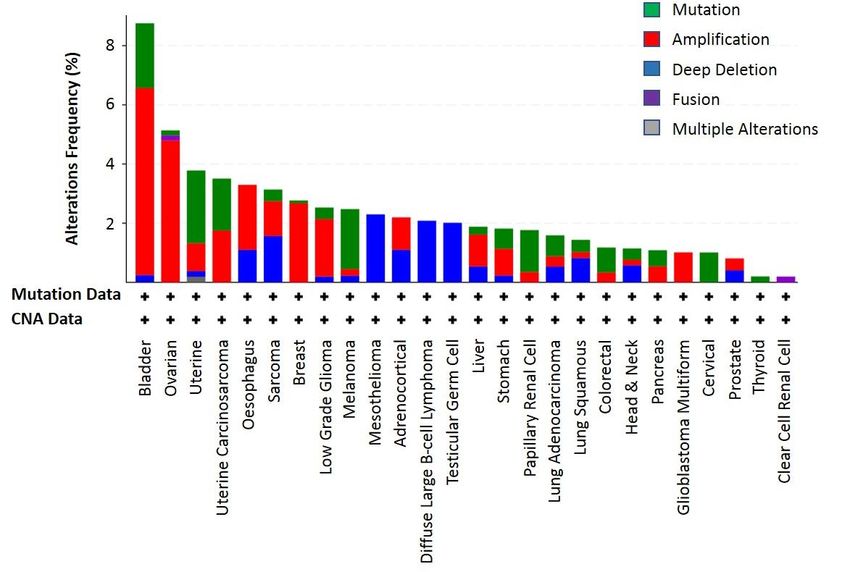
Biomolecules 2020, 10, 1378                                                                                           2 of 22

as inflammation and injury responses [1]. Therefore, the dysregulation in KLF functions is expected to
disrupt cellular homeostasis and can consequently result in the development of diseases [1]. In this
review, we focus on KLF6. The review first starts with the current knowledge of the KLF6 gene and
the protein structures. Then, we comprehensively discuss the roles of KLF6 in regulating normal
physiological processes, as well as its involvement in driving the pathogenesis of several diseases,
with specific emphasis on the opposing roles of full-length KLF6 and KLF6-SV1. In addition, we
also posit KLF6 therapeutic potentials. Last, we provide several compelling fundamental and clinical
questions that need to be addressed to improve our overall understanding of KLF6 functions in health
and diseases.

2. KLF6 Gene Structure and Post-Transcriptional Modifications
      The human KLF6 gene is located on the short arm of chromosome 10 (10p15). Comprising
four exons, the KLF6 gene can be transcribed into seven different transcripts. However, only three
transcripts are suggested to be translated into proteins: KLF6-204, KLF6-206 and KLF6-207, as shown
in Figure 1A. KLF6-206 (referred to as KLF6 hereinafter) is annotated as the primary transcript, which
gives rise to a protein that is 283 amino acids in length. KLF6 was originally identified as a core
promoter-binding protein (CPBP) that was highly enriched in the placenta to regulate the expression
of pregnancy-specific glycoprotein genes [6,7]. Other than the placenta, the KLF6 gene was also
detected and cloned from the liver [8] and leukocytes [9]. In addition to CPBP, several aliases have been
assigned to KLF6, including ZF9, the core promoter element-binding protein (COPEB), B-cell-derived
protein 1 (BCD1) and the suppressor of tumorigenicity 12 (ST12). KLF6 is involved in modulating the
expression of cellular injury response and tissue repair-related genes such as transforming growth
factor beta 1 (TGFβ1) and transforming growth factor beta receptor (TGFβR) [10], collagen type 1
alpha 1 (COL1A1)
Biomolecules 2020, 10, x [11], plasminogen activator urokinase (PLAU) [12] and endoglin (ENG) [13].3 of 22

     Figure
     Figure 1.
             1. KLF6
                KLF6 gene
                        gene structure
                             structure and
                                         and the
                                              the associated
                                                   associated post-transcriptional
                                                                post-transcriptional modifications.
                                                                                      modifications. (A)
                                                                                                      (A) The
                                                                                                           The KLF6
                                                                                                                 KLF6
     gene
     gene can
           can be
                be transcribed
                    transcribed toto produce
                                     produce 77 different
                                                  different transcripts,
                                                            transcripts, of
                                                                          of which
                                                                             which only
                                                                                     only KLF6-204,
                                                                                          KLF6-204, KLF6-206
                                                                                                     KLF6-206 and  and
     KLF6-207
     KLF6-207 are
                aretranslated
                     translated into
                                 into proteins.
                                      proteins. (B)
                                                 (B)The
                                                     Theprimary
                                                          primaryKLF6
                                                                   KLF6transcript,
                                                                          transcript,KLF6-206,
                                                                                      KLF6-206,contains
                                                                                                containsfour
                                                                                                          fourexons
                                                                                                                 exons
     whereby
     whereby germline
                germline mutation
                           mutation IVSIVS 1-27
                                            1-27 GG >>AA can
                                                          can induce
                                                               induce alternative
                                                                       alternativesplicing
                                                                                   splicingthat
                                                                                            thatgives
                                                                                                 gives rise
                                                                                                        rise to
                                                                                                              to three
                                                                                                                  three
     additional  KLF6   spliced  variants: KLF6-SV1,    KLF6-SV2    and
     additional KLF6 spliced variants: KLF6-SV1, KLF6-SV2 and KLF6-SV3.  KLF6-SV3.

3. KLF6 Protein Structures
    All human KLF proteins contain three repeats of Cys2-His2 (C2H2) that constitute a high-
conserved, DNA-binding zinc finger in the C-terminus. These repeats preferentially bind to the “GC-
box” or “CACCC” motifs that normally exist in promoter regions [16]. On the other hand, the N-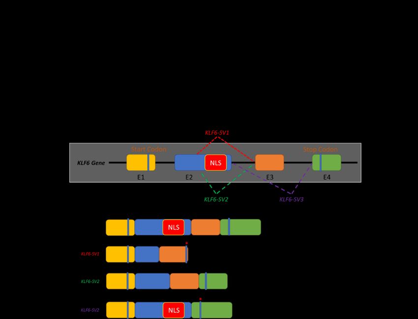
Biomolecules 2020, 10, 1378                                                                              3 of 22

      Meanwhile, a KLF6 G > A germline mutation has been reported to occur at the intervening
sequence 1-27 (IVS 1-27 G > A). This single nucleotide polymorphism (SNP) generates de novo splicing
sites, resulting in the emergence of three additional KLF6 spliced variants: KLF6-SV1, KLF6-SV2 and
KLF6-SV3 [14]. These KLF6 spliced variants differ in length and structure due to the utilisation of
different splicing donor and/or splicing acceptor sites (Figure 1B). Directed by the nuclear localisation
sequences (NLS) located at the end of exon 2, the full-length KLF6 localises in the nucleus, corroborating
its roles as a transcriptional regulator. However, due to alternative splicing, this NLS domain is lost
in KLF6-SV1 and KLF6-SV2. Hence, rather than being transported into the nucleus, KLF6-SV1 and
KLF6-SV2 are localised in the cytoplasm and are, therefore, unable to regulate the gene transcription
process [14]. KLF6-SV3, on the other hand, retains the NLS domain but loses the whole exon 3. Among
these three spliced variants, KLF6-SV1 is widely studied, especially in cancer, whereas the roles of the
other two isoforms remain elusive [14,15].

3. KLF6 Protein Structures
      All human KLF proteins contain three repeats of Cys2-His2 (C2H2) that constitute a high-conserved,
DNA-binding zinc finger in the C-terminus. These repeats preferentially bind to the “GC-box” or
“CACCC” motifs that normally exist in promoter regions [16]. On the other hand, the N-terminus
domain for KLF proteins allow them to interact with different client proteins, including transcription
factors, coactivators and corepressors, as well as the chromatin-modifying enzymes [16]. Therefore, the
functional diversity of KLFs is determined by their N-terminal domain. The phylogenetic classification
of the human KLFs separate these proteins into three distinct subgroups (Figure 2). Group 1 consists
of KLF3, KLF8 and KLF12, which function as transcriptional repressors by interacting with the
C-terminal-binding proteins (CtBP1/2). KLF1, KLF2, KLF4, KLF5, KLF6 and KLF7, on the other hand,
belong to group 2, and they predominantly function as transcriptional activators. Finally, KLF9, KLF10,
KLF11, KLF13, KLF14 and KLF16 are in group 3, and they are classified as transcriptional repressors
due  to their2020,
 Biomolecules  interactions
                   10, x    with corepressor Sin3a [1].                                            4 of 22

      Figure 2. A human KLF family phylogenetic tree generated using the Clustal Omega tool (https:
       Figure 2. A human KLF family phylogenetic tree generated using the Clustal Omega tool
      //www.ebi.ac.uk/Tools/msa/clustalo/). There are 17 members of the human KLF family, which can be
       (https://www.ebi.ac.uk/Tools/msa/clustalo/). There are 17 members of the human KLF family, which
      further classified into three subgroups. KLF7 is the closest to KLF6 in the phylogeny.
       can be further classified into three subgroups. KLF7 is the closest to KLF6 in the phylogeny.
     Due to their close structural and functional characteristics within the human Krüppel-like family,
KLF6 and KLF7 are classified together in the same phylogenetic group (Figure 2), whereby a common
progenitor has been traced to a gene named LUNA in Drosophila [17,18]. The phylogenetic tree of KLF6
orthologs in several other species is shown in Figure 3. Structurally, KLF6 possesses three functional

       Figure 3. The phylogenetic relationship among the five selected KLF6 orthologs from D. melanogaster,
       D. rerio, X. laevis, M. musculus and H. sapiens. The phylogram was generated using the Clustal Omega
       tool (https://www.ebi.ac.uk/Tools/msa/clustalo/).
Biomolecules 2020, 10, 1378 4 of 22
domains that endow them the ability to bind to specific DNA sequences, localise into the nucleus
and regulate the transcription of its downstream targets (Figure 4). The N-terminal acidic domain
of KLF6 stretches from Met1 to Ser112. This domain is responsible for recruiting and interacting
with other transcription factors and cofactors, such as Sp1, KLF4, p53, hypoxia-inducible factor 1
alpha (HIF1α), runt related transcription factor 1 (RUNX1), E2F1, GTF3C1 and histone deacetylase 3
(HDAC3),
Figureto2.either activateKLF
A human or repress
family the transcription
phylogenetic treeprocess in a context-dependent
generated using the Clustal Omega mannertool[19–25].
It isFigure
noteworthy to point out that sequences
2. A human KLF family phylogenetic
(https://www.ebi.ac.uk/Tools/msa/clustalo/). in this domain
tree
There are
aregenerated
17 membershighly
using variable
of thethe among
Clustal
human the
KLFOmega KLF
toolfamily
family, which
members,
can beproviding another
intolayer
three of diversity
(https://www.ebi.ac.uk/Tools/msa/clustalo/).
further classified There
subgroups. that may
areis17
KLF7 contribute
members
the closest the to
ofKLF6
to ina the
human wide
KLFrange
family,ofwhich
phylogeny. regulatory
activities
can be [17,26].
further classified into three subgroups. KLF7 is the closest to KLF6 in the phylogeny.
Figure 3.
Figure 3. The
The phylogenetic relationship among
phylogenetic relationship among the
the five
five selected
selected KLF6
KLF6 orthologs
orthologs from D. melanogaster,
from D. melanogaster,
Figure
D. 3. The
D. rerio,
rerio, X. phylogenetic
X. laevis,
laevis, M. relationship
M. musculus
musculus and H.
and amongThe
H. sapiens.
sapiens. the five
The selectedwas
phylogram
phylogram KLF6
was orthologs
generated
generated fromthe
using
using D.Clustal
the melanogaster,
Clustal Omega
Omega
D.tool
rerio,
tool X. laevis, M. musculus and H. sapiens.
(https://www.ebi.ac.uk/Tools/msa/clustalo/).
(https://www.ebi.ac.uk/Tools/msa/clustalo/).The phylogram was generated using the Clustal Omega
tool (https://www.ebi.ac.uk/Tools/msa/clustalo/).
Figure 4.4.KLF6
Figure KLF6protein
protein structure
structureandandthe associated post-translational
the associated modifications.
post-translational The KLF6
modifications. Theprotein
KLF6
consists
Figure
protein of three
4. consists domains:
KLF6 proteinof three (I) N-terminal
structure
domains: and acidic
(I)the domain,
associated
N-terminal (II) serine/threonine-rich
post-translational
acidic central domain
modifications. The KLF6
domain, (II) serine/threonine-rich and
central
(III)
protein
domainC-terminal
consists
and (III) DNA-binding
of C-terminal
three domains: domain. The protein
(I) N-terminal
DNA-binding domain. structure
acidic
The domain, is adapted from Andreoli
(II) serine/threonine-rich
protein structure et al. [17].
central
is adapted from Andreoli
According
domain and to PhosphositePlus
(III) C-terminal (https://www.phosphosite.org/),
DNA-binding domain. The protein the KLF6
structure
et al. [17]. According to PhosphositePlus (https://www.phosphosite.org/), the KLF6 protein protein
is undergoes
adapted from three types
Andreoli
etof post-translational
al. [17]. three
undergoes According modifications
types to (PTMs). Themodifications
PhosphositePlus
of post-translational sole ubiquitylation siteThe
(https://www.phosphosite.org/),
(PTMs). for KLF6 isthe
located
KLF6
sole ubiquitylationat Lys66-Ub.
protein
site for
Phosphorylation
undergoes is
three types
KLF6 is located the best-documented
of post-translational
at Lys66-Ub. PhosphorylationKLF6 PTM,
modificationsencompassing T147-p,
(PTMs). The sole
is the best-documented KLF6 S150-p, S151-p,
ubiquitylation S171-p,
site
PTM, encompassing for
S192-p
KLF6 is and S233-p.Lys66-Ub.
located Finally, four lysine acetylation sitesbest-documented
have been documented PTM, for the C-terminal, i.e.,
T147-p, S150-p,atS151-p, Phosphorylation
S171-p, S192-p and S233-p.is the Finally, four lysineKLF6 acetylation encompassing
sites have been
S209-Ac,
T147-p, S213-Ac,
S150-p, S218-Ac
S151-p, and S192-p
S171-p, S228-Ac. and S233-p. Finally, fourand
lysine acetylation sites have been
documented for the C-terminal, i.e., S209-Ac, S213-Ac, S218-Ac S228-Ac.
documented for the C-terminal, i.e., S209-Ac, S213-Ac, S218-Ac and S228-Ac.
On the other hand, the central domain of KLF6 starts at Ser113 and continues up to Arg208.
On the other hand, the central domain of KLF6 starts at Ser113 and continues up to Arg208. This
ThisOncentral domain istheparticularly enriched in Ser and Thr residues, renderingtothis domain
This a
centralthe other
domain hand,
is central
particularly domain
enriched inofSer
KLF6andstarts
Thr at Ser113
residues, and continues
rendering thisup Arg208.
domain a potential
potential
central target for post-translational modifications, especially phosphorylation. Indeed, it has been
targetdomain is particularly enriched
for post-translational in Ser and
modifications, Thr residues,
especially rendering thisIndeed,
phosphorylation. domain ita potential
has been
demonstrated
target earlier by Slavin
for post-translational et al. that KLF6
modifications, is constitutively
especially phosphorylated
phosphorylation. in vivo [27].
Indeed, in A
it vivo number
has [27].
beenA
demonstrated earlier by Slavin et al. that KLF6 is constitutively phosphorylated
demonstrated earlier by Slavin et al. that KLF6 is constitutively phosphorylated in vivo [27].exact
of kinases have been demonstrated to increase the level of phosphorylation of KLF6, although the A
sites of phosphorylation have not been confirmed. For example, Lang et al. reported the increased
phosphorylation of KLF6 based on in-vitro and in-vivo 32 P incorporation assays upon the transient
transfection of glycogen synthase kinase 3 beta (GSK3β) and the subsequent transactivation of a
p21 [28]. Besides, the phosphorylation of KLF6 by ribosomal protein S6 kinase beta-1 (S6K1) has also
been linked to the induction of TGFB gene transcription [29].
The C-terminal domain, Lys209 to Leu283, contains three C2H2 zinc finger-type structures.
The NLS precede these C2H2 zinc fingers. The C2H2 zinc fingers are among the ancient and common
domains found in the transcription factors of higher eukaryotes [30]. Typically, each C2H2 zinc
finger motif is comprised of 25–30 amino acid residues [31]. In the case of KLF6, as well as otherBiomolecules 2020, 10, 1378 5 of 22
KLFs, the first two zinc finger motifs consist of 23 amino acids, and the third zinc finger is 21 amino
acids long. These motifs bind the DNA, especially to the GC box or the 50 -CACCC-30 regions [31,32].
Meanwhile, the three zinc finger motifs are connected by a linker that is highly conserved with a
sequence motif (TGE(R/K)(P/k/r)(F/y)X)) and is capable of binding certain other zinc finger proteins
with high affinity [32]. Although the exact 3D structure of the KLF6 protein has not been solved, the
three C2H2 motifs in the KLF family generally form β-hairpin starting from the N-terminal, followed
by α-helix, which form a left-handed ββα structure. Apart from that, the zinc-finger structure is
stabilised by the coordination of a zinc atom with two conserved cysteine residues at one end of the β
sheet and with two conserved His residues at the α-helix C-terminus. The C2H2 motifs constitute
a defining feature not only for the 17 human KLF family members but, also, the related specificity
proteins (SPs) transcription factors, whereby more than 65% sequence identity has been demonstrated
among this family members [26]. In fact, the specificity proteins (SPs) and KLFs are members of the
same higher-rank family of transcription factors, the so-called Sp/KLF family, that share conserved
zinc finger domains. These transcription factors are expressed in a tissue-specific manner, and so are
their activities and functions. It has been demonstrated that the Sp/KLF family regulates the growth,
development, differentiation, proliferation and embryogenesis
4. KLF6 Post-Translational Modifications
Post-translational modifications (PTMs) refer to covalent moieties that are added on the amino
acid residues to modify, fine-tune and diversify the inherent biological functions of a protein. The KLF6
amino acid residues that undergo PTMs are shown in Figure 4. According to PhosphositePlus, one of
the most comprehensive repository for PTMs, a total of six phosphorylation sites have been reported
for KLF6 so far, whereby five of them: Thr147-p, Ser150-p, Ser151-p, Ser171-p and Ser192-p, except for
Ser233-p, are located at the Ser- and Thr-rich central domain [33,34]. Since all these phosphorylation
sites were derived from large-scale phosphoproteomics studies, the precise functional implications for
each site has not been systematically investigated, despite that a selected number of kinases that are
associated with KLF6 functions have been reported [29,35].
Moreover, there are four Lys acetylation sites reported for KLF6: Lys209-Ac, Lys213-Ac, Lys218-Ac
and Lys228-Ac [34,36]. Among these four sites, the first three were discovered by Li et al. by incubating
four synthetic peptide fragments that covered the majority of the lysine residues of KLF6 with
CREB-binding protein (CBP) and P300/CBP-associated factor (PCAF), followed by an in vitro histone
acetyltransferase assay using 3H-labeled acetyl-CoA. As a result, CBP, but not PCAF, was found to
acetylate Lys209, Lys213 and Lys218 [36]. The authors further demonstrated that, when Lys209 was
mutated to arginine, acetylation at this site was abrogated, leading to the loss of ability of KLF6
to transactivate p21. Interestingly, only one ubiquitylation site has been reported for KLF6 to date,
Lys66-Ub, albeit this site was reported by three independent large-scale ubiquitome studies [37–39].
This is consistent with the study of Banck et al., who demonstrated that KLF6 could be subjected
to proteasome-mediated degradation [40]. Recently, SCFFbxw7 was found to ubiquitylate KLF7 for
proteasomal degradation in a manner that was dependent on GSK3β-mediated phosphorylation [41].
Despite KLF7 being the closet paralog of KLF6, the authors’ results showed that only KLF7, but not
KLF6, was a bona fide substrate of SCFFbxw7.
5. KLF6 Roles in Normal Physiological Processes
5.1. Cellular Differentiation and Proliferation
Eukaryotic development is a finely tuned process, orchestrated by the spatiotemporal expression
and interactions of developmental-associated transcription factors [42]. Much insight on the roles of
KLFs in the regulation of human developmental processes have been contributed by studies conducted
on model organisms such as mice, zebrafish and flies. To be specific, KLF6 is expressed throughout
embryonic and tissue development, including the development of kidneys [43], eyes [44], prostate [45]Biomolecules 2020, 10, 1378 6 of 22
and several other tissue types [46], thus signifying its importance in developmental process regulation.
Indeed, the homozygous mutant Klf6−/− mice died at E12.5 due to defects in haematopoiesis and
yolk sac vascularisation [47]. It was further demonstrated that KLF6 bi-allelic deletion in embryonic
stem (ES) cells impaired the cell differentiation and proliferation capability [47]. Furthermore, KLF6 is
involved in the expansion and maintenance of hematopoietic stem cells and progenitor cells, as well as
in the development of endoderm-derived organs, including the liver [48,49].
There are also other studies that further supported the KLF6 key roles in regulating cellular
differentiation. For example, Racca et al. demonstrated that KLF6 regulates trophoblast differentiation, a
precursor step for placenta development, by transactivating the expression of β-chorionic gonadotropin
and pregnancy-specific glycoprotein genes in this cell type [50]. Additionally, KLF6 has been shown to
induce the differentiation of preadipocytes into adipocyte by repressing the expression of delta-like
1 (Dlk1) via its interaction with HDAC3 [25,51]. In the central nervous system, KLF6 acts as
the downstream effector for the GP130-STAT3 axis to transactivate the expression of importin-a5,
which is required for oligodendrocyte progenitor cells differentiation and neurons myelination [52].
KLF6 facilitates the adaptation of pancreatic β-cells to metabolic stress both by promoting and
preventing β-cell proliferation and insulin resistance-induced dedifferentiation/transdifferentiation
into glucagon-producing α-cells, respectively [53]. During skeletal myogenesis, KLF6 is a downstream
target of the Smad3-dependent TGFβ signalling pathway in supporting myoblast proliferation and
survival [54].
5.2. Immune and Inflammatory Responses
The macrophage is a part of the innate immune system that plays critical roles in modulating
early immune and inflammatory responses during infections or tissue injuries [55]. The polarisation
of macrophages either into M1 (proinflammatory) or M2 phenotypes (anti-inflammatory) rely on
cues from the environments. These signals will be transduced intracellularly to the downstream
effectors—normally, the transcription factors—which will subsequently regulate the expression of
appropriate immune and inflammation responses-associated genes [56]. In-line with the key roles
of transcription factors in modulating this process, a high expression of KLF6 was found in human
and murine macrophages [57]. Functionally, KLF6 regulates macrophage polarisation to the M1
phenotype and the expression of proinflammatory genes by cooperating with NF-κβ and suppressing
the expression peroxisome proliferator-activated receptor gamma (PPARγ) and B-cell lymphoma 6
(BCL6) [57,58]. In support of these findings, an independent study by Zhang et al. reported that KLF6
acted as a NF-κβ coactivator to regulate the expression of several NF-κβ downstream targets, including
the proinflammatory cytokines monocyte chemoattractant protein-1 (MCP1), chemokine C-X-C motif
ligand 2 (CXCL2) and interleukin 8 (IL-8) [59].
Interestingly, a recent study has unravelled a novel KLF6 role in regulating HIF1α expression
and the hypoxic-response transcriptional program in macrophages [60]. The activation of this
hypoxia-associated gene expression program would allow the macrophage to adapt and survive at
the potentially hypoxic site of infection and injury and the subsequent elicitation of proinflammatory
responses. Besides, some reports showed a direct interaction between KLF6 and miRNAs in
regulating macrophage polarisation. Kim et al. demonstrated that KLF6 was able to upregulate
the expression of proinflammatory genes in the macrophage by repressing the expression of
anti-inflammatory miR-223 [61]. Additionally, an independent study by Bi et al. showed that the
miR-181a-mediated suppression of KLF6 and CCAAT-enhancer-binding proteins (C/EBPα) promoted
macrophage polarisation towards the anti-inflammatory M2 phenotype [62]. Collectively, these
findings highlight the KLF6 involvement in modulating macrophage polarisation and the elicitation of
proinflammatory responses.
Nitric oxide (NO), which is a free radical that is produced upon L-arginine oxidation by nitric oxide
synthase (NOS), also contributes to the innate immunity in combating the invading pathogens [63].
Various cell types are able to release NO, including macrophages, neutrophils, vascular endothelialBiomolecules 2020, 10, 1378 7 of 22
cells, neuronal cells and lung epithelial cells [63]. As an immune response-associated transcriptional
regulator, KLF6 is also involved in regulating NO production by directly transactivating the expression
of inducible nitric oxide synthase (iNOS) in response to environmental stimulants or challenges [64].
Two other NOS isoforms exist: i.e., neuronal NOS (nNOS) and endothelial NOS (eNOS). Subsequent
findings showed that KLF6 was required for nitric oxide-mediated apoptosis of the infected cells
during flu and respiratory syncytial virus infections [65,66]. It is important to highlight that excess
NO production would lead to hyperinflammation and profound tissue damage, thus aggravating the
severity of the infection and disease. Therefore, the activation of iNOS and downstream NO production
must be precisely regulated by their upstream regulator—which, in this case, is the KLF6—in conferring
protections against the invaders.
5.3. Tissue Injury and Wound Healing
The responses towards tissue injury and the promotion of wound healing involve coordinated
crosstalk between the injured tissue and microenvironment that includes the immune cells, endothelial
cells and extracellular matrix [67]. These processes are modulated by the pertinent growth factors and
gene expression programs that are activated within the injured tissue, as well as its surroundings, thus
ensuring that the appropriate post-injury cascades are undertaken [68]. TGFβ is among the growth
factors that plays an essential role in modulating tissue repair and wound-healing processes [69].
TGFβ expression during tissue injury was shown to be directly transactivated by KLF6 [10]. Along
with TGFβ, KLF6 also regulates the expression of several other members of the TGFβ pathway,
including COL1A1 [11], PLAU [12] and ENG [13], further highlighting KLF60 s roles in governing the
wound-healing processes. For example, KLF6 expression was upregulated in hepatic stellate cells
(HSCs) during acute/chronic liver injury and was found to transcriptionally regulate the expression of
COL1A1, an extracellular matrix protein that participates in the wound-healing process [11]. Of note,
HSCs are a population of mesenchymal cells residing within the liver. Upon activation by signals from
the damaged cells, HSCs will secrete extracellular matrix proteins and other factors that will facilitate
the repair wound-healing processes [70]. Besides, KLF6 expression was also found to be elevated in
acute liver failure patients and mice models. Functionally, KLF6 mediated the autophagy process,
which was to clear the damaged cells prior to liver generation, via the direct transcriptional activation
of autophagy-associated genes ATG7 and BECLIN1 [71].
The roles of KLF6 in tissue repair and remodelling are not only limited in hepatocytes but, also,
other cells types. KLF6 mediated the transactivation of vascular injury and repair-related genes such
as PLAU, ENG, activin-receptor like kinase 1 (ALK1) and membrane metalloproteinase 14 (MMP14),
of which ENG and ALK1 are members of the TGFβ signalling pathway [12,13,72,73]. In-line with
these findings, the marked upregulation of KLF6 and TGFβ were also observed at the early phase of
kidney ischemic reperfusion (I/R) injury, and KLF6 targeting impaired the apoptotic process following
the ATP-induced ischemia [74]. Whilst KLF6 upregulation might induce apoptosis during I/R injury,
KLF6 was found to be involved in promoting podocyte survival during injury. The deletion of
podocyte-specific Klf6 in mice increased the susceptibility to Adriamycin-induced glomerulosclerosis
and tubulointerstitial injury, followed by podocytes apoptosis [75]. It was demonstrated that KLF6
modulated the mitochondrial functions by preventing the activation of the cytochrome C-mediated
intrinsic apoptotic pathway [75]. A subsequent study revealed that KLF6 was also involved in
protecting the mitochondria from injury and apoptosis under diabetic conditions [76]. In the central
nervous system (CNS), KLF6 interacted with STAT3 to coregulate the expression of genes involved
in axon and corticospinal tract neurons regeneration following injury [77]. Overall, these findings
denote KLF6 functions as the early responder to tissue injury and the mediator of tissue repair and
wound-healing processes, in part by transcriptionally activating the expression of TGFβ and several
other key components of the TGFβ signalling pathway. The roles of KLF6 in normal physiological
processes are simplified in Figure 5.Biomolecules 2020, 10, x 8 of 22
[77]. Overall, these findings denote KLF6 functions as the early responder to tissue injury and the
mediator of tissue repair and wound-healing processes, in part by transcriptionally activating the
expression
Biomolecules 2020,of10,TGFβ
1378 and several other key components of the TGFβ signalling pathway. The roles8of
of 22
KLF6 in normal physiological processes are simplified in Figure 5.
Figure
Figure 5. 5.Diagram
Diagramsummarising
summarising the
the roles
roles of
of KLF6
KLF6 in
in normal
normalphysiological
physiologicalprocesses.
processes.
6. KLF6 Implication
6. KLF6 ImplicationininHuman
HumanDiseases
Diseases
TheThe perturbationofofnormal
perturbation normal physiological
physiological processes
processes and
and homeostasis
homeostasiscan cancontribute
contribute to to
thethe
development
development ofofdiseases.
diseases.As
Asdiscussed
discussed above,
above, to
to prevent
preventthese
theseprocesses
processesfrom
fromgoing
going awry,
awry,they
theyareare
tightly
tightly regulated
regulated by by coordinated
coordinated interactions
interactions of respective
of the the respective regulatory
regulatory networks
networks [78]. [78]. In review,
In this this
review, we will specifically focus on the roles of KLF6 in driving cancer and inflammatory-associated
we will specifically focus on the roles of KLF6 in driving cancer and inflammatory-associated disease
disease pathogenesis.
pathogenesis. Cancer is Cancer is a group
a group of diseases
of diseases causedcaused by uncontrolled
by uncontrolled cellcell growth,manifested
growth, manifestedby
theby the cells’
cells’ capability
capability to proliferate
to proliferate indefinitely,
indefinitely, evadeimmunosurveillance
evade immunosurveillance and andresist
resistapoptosis
apoptosis [79].
[79].
Besides, inflammation also promotes cancer emergence and progression [79]. Since KLF6 plays a
Besides, inflammation also promotes cancer emergence and progression [79]. Since KLF6 plays a
central role in modulating these processes, genetic alterations and/or the aberrant expression of KLF6
central role in modulating these processes, genetic alterations and/or the aberrant expression of KLF6
have been reported to support the formation and progression of many cancer types [17]. Furthermore,
have been reported to support the formation and progression of many cancer types [17]. Furthermore,
KLF6 has also been implicated in the pathogenesis of several inflammatory-associated diseases due
KLF6 has also been implicated in the pathogenesis of several inflammatory-associated diseases due
to its key function as the mediator for proinflammatory and tissue injury responses. Thus, even a
to its key function as the mediator for proinflammatory and tissue injury responses. Thus, even
slight dysregulation in KLF6 functions could potentially perturb these processes, resulting in hyper-
a slight dysregulation
inflammation and tissuein damage.
KLF6 functions could potentially perturb these processes, resulting in
hyper-inflammation and tissue damage.
6.1. Cancer
6.1. Cancer
6.1.1. Full-Length KLF6
6.1.1. Full-Length KLF6
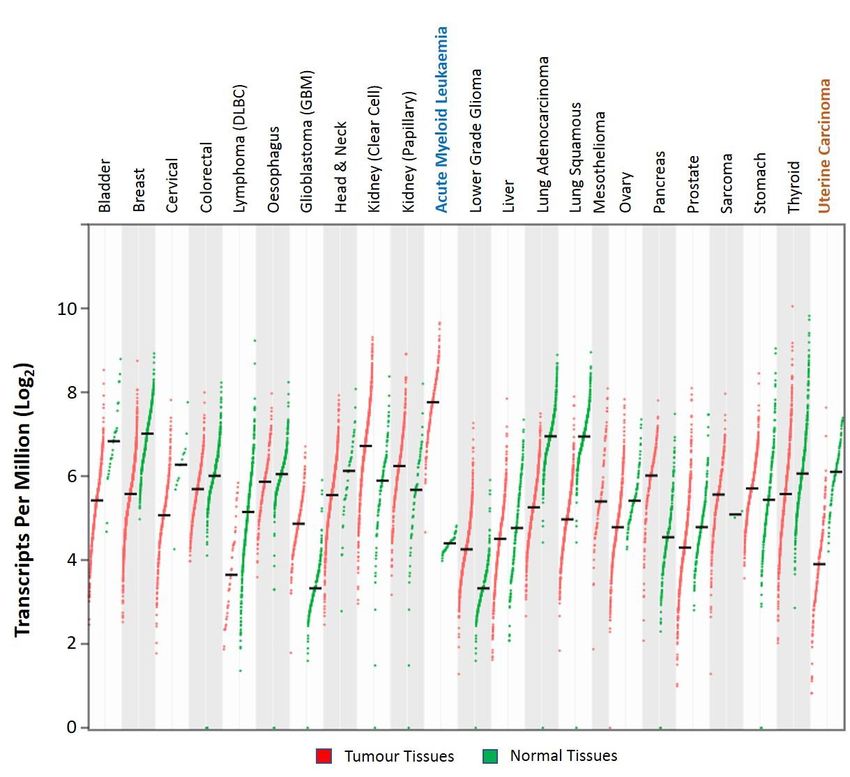
Analysis of the publicly available large-scale The Cancer Genome Atlas (TCGA) RNA-Seq data
Analysis
showed thatof
thethe publicly of
expression available
the KLF6 large-scale
gene varied The Cancer
among Genome
cancer types Atlas
(Figure (TCGA)
6). KLF6RNA-Seq
was highlydata
showed that the expression of the KLF6 gene varied among cancer types (Figure 6).
expressed in acute myeloid leukaemia, renal cancer of clear cell and papillary subtypes, glioblastoma KLF6 was highly
expressed
multiformein acute myeloid leukaemia,
and pancreatic cancer [80].renal
Apartcancer
fromofthese,
clearthe
cellexpression
and papillary subtypes,
of KLF6 glioblastoma
was found to be
multiforme and pancreatic cancer [80]. Apart from these, the expression of
downregulated in other cancer types as compared to their respective normal tissues [80]. Early KLF6 was found to be
downregulated in other cancer types as compared to their respective normal tissues
reports showed that KLF6 was frequently inactivated in sporadic prostate cancer cases. Loss-of- [80]. Early reports
showed that KLF6(LOH)
heterozygosity was frequently inactivated
analysis revealed that in sporadic
KLF6 prostate
inactivation cancer cases.
conformed to theLoss-of-heterozygosity
Knudson’s two-hit
(LOH) analysis
hypothesis, thusrevealed thatKLF6
classifying KLF6 as inactivation conformed
a tumour suppressor [81].to the Knudson’s
Stemming from thistwo-hit
prostatehypothesis,
finding,
thus classifying KLF6 as a tumour suppressor [81]. Stemming from this prostate finding, studied
the KLF6 mutational status, expression level and tumour-suppressive roles were extensively the KLF6
in variousstatus,
mutational cancerexpression
types. Consistent
level andwith the reports by Narla
tumour-suppressive et al.
roles [81],extensively
were KLF6 was also found
studied to be
in various
eithertypes.
cancer inactivated or downregulated
Consistent in a significant
with the reports by Narlafraction of colorectal
et al. [81], KLF6 was cancer
also[82], non-small
found cell
to be either
lung cancer [83], ovarian cancer [15], gliomas [84] and hepatocellular carcinoma
inactivated or downregulated in a significant fraction of colorectal cancer [82], non-small cell lung [85] cases. In
cancer [83], ovarian cancer [15], gliomas [84] and hepatocellular carcinoma [85] cases. In addition,
promoter hypermethylation and suppression by the noncoding RNAs and microenvironment factors
could also result in KLF6 downregulation in cancer [86–90]. KLF6 functional interrogations revealed
that KLF6 overexpression had negative effects on tumour growth and progression, whereby KLF6
silencing resulted in increased tumorigenicity [83,91–94]. Mechanistically, it has been demonstratedBiomolecules 2020, 10, x 9 of 22
addition, promoter hypermethylation and suppression by the noncoding RNAs and
microenvironment factors could also result in KLF6 downregulation in cancer [86–90]. KLF6
functional interrogations revealed that KLF6 overexpression had negative effects on tumour growth
Biomolecules
and 2020, 10, 1378
progression, whereby KLF6 silencing resulted in increased tumorigenicity [83,91–94]. 9 of 22
Mechanistically, it has been demonstrated that KLF6 exerts its cell growth suppressive functions by
modulating the expression
that KLF6 exerts of genes
its cell growth involved
suppressive in the cell
functions cycle, apoptosis
by modulating and senescence
the expression regulation
of genes involved
[81,95–99].
in the cell cycle, apoptosis and senescence regulation [81,95–99].
Figure 6.
Figure Dotplots
6. Dot plotsrepresenting
representingthe
theKLF6
KLF6expression
expressionRNA-Seq
RNA-Seqdatadatainin tumour
tumour and
and normal
normal tissue
tissue
samples from
samples fromThe
TheCancer
CancerGenome
GenomeAtlas (TCGA)
Atlas and The
(TCGA) andGenotype-Tissue Expression
The Genotype-Tissue (GTEx) databases,
Expression (GTEx)
respectively. KLF6 was found to be significantly upregulated and downregulated in acute
databases, respectively. KLF6 was found to be significantly upregulated and downregulated in acute myeloid
leukaemia
myeloid and uterine
leukaemia and carcinoma, respectively
uterine carcinoma, |2|; p-value
(Log2 FC(Log
respectively < 0.05). The data were analysed
2FC |2|; p-value < 0.05). The data were
and visualised using the GEPIA2 web application [80].
analysed and visualised using the GEPIA2 web application [80].
Nonetheless, the KLF6 mutational status in cancer has been a subject of debate. Several independent
Nonetheless, the KLF6 mutational status in cancer has been a subject of debate. Several
studies have reported the absence of KLF6 genetic alterations in colorectal, liver, brain and prostate
independent studies have reported the absence of KLF6 genetic alterations in colorectal, liver, brain
cancers [100–106], contradicting earlier studies that showed the frequent KLF6 inactivation in these
and prostate cancers [100–106], contradicting earlier studies that showed the frequent KLF6
cancer types. It has been pointed out that PCR amplification artefacts due to the use of archived
inactivation in these cancer types. It has been pointed out that PCR amplification artefacts due to the
cancer tissues or other technical issues might contribute to these discrepancies in the KLF6 mutational
use of archived cancer tissues or other technical issues might contribute to these discrepancies in the
status [107]. Moreover, analyses of the more recent TCGA genomic data revealed that KLF6 was rarely
KLF6 mutational status [107]. Moreover, analyses of the more recent TCGA genomic data revealed
deleted/inactivated in the previously studied cancers [108,109]. Collectively, the percentage of cancer
that KLF6 was rarely deleted/inactivated in the previously studied cancers [108,109]. Collectively, the
cases that had the KLF6 gene alteration were quite low (Figure 7). Among the analysed cancer types,
percentage of cancer cases that had the KLF6 gene alteration were quite low (Figure 7). Among the
bladder cancer had the highest frequency of KLF6 genetic alterations—36 out of 411 cases—of which
analysed cancer types, bladder cancer had the highest frequency of KLF6 genetic alterations—36 out
2.19% were mutations, 6.33% were amplifications and 0.24% were deep deletions. These data indicate
of 411 cases—of which 2.19% were mutations, 6.33% were amplifications and 0.24% were deep
that KLF6 was not a common target for genetic alterations, and its deregulation in cancer could be
deletions. These data indicate that KLF6 was not a common target for genetic alterations, and its
caused by other mechanisms, potentially at the level of transcriptional or translational regulations.Biomolecules 2020, 10, x 10 of 22
Biomolecules 2020, 10, 1378 10 of 22
deregulation in cancer could be caused by other mechanisms, potentially at the level of
transcriptional or translational regulations.
Figure 7. The
Figure 7. The frequency
frequency ofof KLF6
KLF6 genetic
genetic alterations
alterations inin twenty-six
twenty-six different
different cancer
cancer types
types that were
that were
molecularly characterisedininthe
molecularly characterised thelarge
large scale
scale TCGA
TCGA project.
project. These
These KLF6
KLF6 genetic
genetic alterations
alterations frequency
frequency data
data queried
were were queried and extracted
and extracted using cBioPortal
using cBioPortal (https://www.cbioportal.org/)
(https://www.cbioportal.org/) [108,109]. [108,109]. The
The identified
identified genetic alterations
genetic alterations in KLF6mutation,
in KLF6 include include mutation, amplification,
amplification, deep deletion,
deep deletion, fusion and fusion and
multiple
multiple alterations.
alterations. The “+” The
sign“+” signofoneach
on top top cancer
of eachtype
cancer type indicates
indicates that the mutation
that the mutation and copyand copy
number
number
alterationalteration (CNA)
(CNA) data are data are available
available for thesefor thesetypes.
cancer cancer types.
Contrary to its widely reported role as a tumour suppressor, there were several studies that
reported the growth-promoting functions of KLF6 KLF6 inin cancer.
cancer. The pro-oncogenic fusion protein
RUNX1-ETO upregulated
upregulated the the expression
expression of ofKLF6
KLF6in inacute
acutemyeloid
myeloidleukaemia
leukaemia[21]. [21].KLF6,
KLF6, which
which is
is highly
highly expressed
expressed in AML,
in AML, cooperated
cooperated withwith RUNX1-ETO
RUNX1-ETO to drive
to drive the expression
the expression of thisprotein
of this fusion fusion
protein
downstreamdownstream
of targetsof targets in promoting
in promoting leukaemia
leukaemia development
development [21]. [21].
TwoTwo independent
independent studies
studies by
by Sirach
Sirach etand
et al. al. and D’Astolfo
D’Astolfo et al. et al. demonstrated
demonstrated that protected
that KLF6 KLF6 protected hepatocellular
hepatocellular carcinoma carcinoma
(HCC)
(HCC) cells
cells from from apoptosis
apoptosis to promoteto promote HCC progression
HCC progression [110,111]. [110,111].
Meanwhile, Meanwhile, in ductal
in ductal breast breast
carcinoma,
carcinoma,
KLF6 KLF6 colocalised
colocalised with the ERBB2 with the ERBB2 oncoprotein
oncoprotein in the nucleus,in thewhereby
nucleus,itswhereby
expressionits expression
positively
positively
correlated correlated with thereceptor
with the estrogen estrogen receptor
alpha alpha expression
expression [112]. KLF6
[112]. Targeting Targeting
in theKLF6
breast incancer
the breast
cell
cancer
line cell linethe
reduced reduced the cells’ proliferative
cells’ proliferative capacity, capacity,
denotingdenoting KLF6 pro-oncogenic
KLF6 pro-oncogenic roles in roles
breastin cancer
breast
cancerA[112].
[112]. recent A recent
study study discovered
discovered that KLF6 that KLF6 expression
expression in clear cell in clear
renal cellcell renal cell
carcinoma carcinoma
(ccRCC) was
(ccRCC)
driven bywas driven by a super-enhancer
a super-enhancer that resultedthat resulted
in its in its highin
high expression expression
ccRCC tissuesin ccRCC
and tissues
cell linesand cell
[113].
lines [113]. CRISPR-mediated
CRISPR-mediated KLF6 targeting KLF6impaired
targetingccRCC
impairedcellccRCC
growth cellboth
growth bothand
in vitro in vitro and in
in vivo due vivo
to
due to perturbation
perturbation in the mTORC1
in the mTORC1 signalling
signalling pathway pathway andhomeostasis
and lipid lipid homeostasis
[113]. [113].
TheseThese
findingsfindings
were
were consistent
consistent with with the growing
the growing evidence
evidence demonstrating
demonstrating that cancer
that cancer cellscells are highly
are highly dependent
dependent on
on the
super-enhancer-driven
the super-enhancer-driven genes to support
genes their
to support development
their development andandprogression
progression [114,115].
[114,115].
6.1.2. KLF6
6.1.2. KLF6 Spliced
Spliced Variants
Variants
The contradicting
The observations on
contradicting observations on the
the KLF6
KLF6 mutational
mutational status
status and
and role
role in
in cancers
cancers have
have been
been
further confounded by the discovery of KLF6 spliced variants and their involvement in
further confounded by the discovery of KLF6 spliced variants and their involvement in regulating regulating
tumorigenesis, particularly
tumorigenesis, particularly KLF6-SV1.
KLF6-SV1. This
This KLF6
KLF6 alternative
alternative splicing
splicing was
was induced
induced byby the G>
the G A
A
germline mutation at the intervening sequence 1-27, in which this mutation was first identified
germline mutation at the intervening sequence 1-27, in which this mutation was first identified in in
prostate
prostate cancer
cancer cases,
cases, which
which lead
lead to
to alternative
alternative splicing.
splicing. This
This single
single nucleotide
nucleotide polymorphism
polymorphism was wasBiomolecules 2020, 10, 1378 11 of 22
associated with an increased risk of developing prostate cancer [14]. The hepatic growth factor
and RAS signalling pathway have also been linked to enhancing the KLF6 alternative splicing
event in cancer [116,117]. Whilst the full-length KLF6 is established as a tumour suppressor in
many cancer types, KLF6-SV1 is deemed to be pro-oncogenic by antagonising the full-length KLF6
functions in a dominant-negative manner. KLF6-SV1 has been reported to promote the progression
and associate with a poor prognosis of the following cancer types: prostate [118,119], breast [120],
ovarian [121] and lung cancer cells [122,123]. Intriguingly, KLF6-SV2, which also lacks the NLS
domain, was downregulated in colorectal and liver cancers and possessed the tumour suppressive
roles. The overexpression of KLF6-SV2 in colorectal and liver cancer cells reduced the cell proliferation
whilst inducing apoptosis [124,125]. Even though the role of KLF6 and its SV1 and SV2 counterparts
have garnered significant interests, KLF6-SV3 functions are yet to be elucidated. The functional roles
of full-length KLF6 and the three KLF6 spliced variants in cancer are summarised in Table 1.
Table 1. The functional roles of full-length KLF6 and KLF6 spliced variants in cancer.
Isoforms Functions
• Downregulated expression in cancers, except in acute myeloid
leukaemia, kidney (clear cell and papillary subtypes), glioblastoma
multiforme and pancreatic cancer (TCGA RNA-Seq).
Full-length KLF6 • Widely regarded as a tumour suppressor [91–99], despite the
contradicting findings showing full-length KLF6 has a
growth-promoting role [21,110–113].
• Pro-oncogenic by promoting the progression and association with
poor prognosis in prostate [118,119], breast [120], ovarian [121] and
KLF6-SV1 lung cancers [122,123].
• Antagonising the growth-suppressive properties of KLF6 in a
dominant-negative manner.
• Downregulated in liver [124] and colorectal cancers [125].
KLF6-SV2 • Has antiproliferative and proapoptotic functions in colorectal and
liver cancers.
KLF6-SV3 • Functions are yet to be elucidated.
Nonetheless, the presence of the IVS 1-27 G > A germline mutation has also been controversial
due to several opposing findings. This variant was detected at a significantly lower frequency in
Ashkenazi prostate cancer patients as compared to the control subjects [126]. Similarly, a genome-wide
association study in the Italian lung cancer patients cohort revealed that IVS 1-27 G > A was significantly
associated with a reduced risk of lung cancer, whilst the validation study using the Norwegian lung
cancer patients cohort showed no significant association between this polymorphism and the risk of
developing lung cancer [127]. Similarly, there was no significant association between IVS 1-27 G > A
and risk of developing benign prostate hyperplasia and prostate cancer in the Finnish population [128].
Hence, the functional impact of this polymorphism towards cancer susceptibility could vary between
different populations or ethnic groups.
6.2. Metabolic and Inflammatory Diseases
Liver serves as the hub organ that governs and responds to the body’s metabolic and energy
demands. Glucose, lipid and protein metabolisms take place in the liver, tightly controlled by
metabolic-associated transcriptional networks in response to the hormonal cues, nutrient status andBiomolecules 2020, 10, 1378 12 of 22
body-fed state [129]. Dysregulation in liver metabolic functions could induce the development of
insulin resistance and dyslipidaemia, for instance, which subsequently promotes the pathogenesis of
metabolic disorders such as non-alcoholic fatty liver disease (NAFLD), hypercholesterolemia and type II
diabetes [129]. Specifically, NAFLD could advance to non-alcoholic liver steatohepatitis (NASH), which
is marked by chronic liver inflammation and advanced fibrosis. Patients with NASH could develop
life-threatening complications, including liver failure, liver cancer and cardiovascular diseases [130].
KLF6 has been shown as one of the key regulators in liver development and functions, particularly
in response to injury and NAFLD pathogenesis. Functionally, KLF6 was shown to directly transactivate
the expression of glucokinase, a glucose sensor that functions to catalyse the conversion of glucose to
glucose-6-phosphate [131]. Targeting KLF6 reduced the glucokinase expression that consequently led
to an increased hepatic insulin resistance and the development of NAFLD [131]. Additionally, KLF6
deletion, either hepatocyte-specific or in all tissues, improved the insulin sensitivity and rendered
protection against high-fat diet-induced steatosis via the post-transcriptional regulation of PPARα and
its downstream targets [132]. Interestingly, full-length KLF6 and KLF6-SV1 also had opposing roles
in driving the progression of NAFLD, similar to those observed in cancer. A liver biopsy analysis of
patients with NAFLD revealed that the elevated expression of full-length KLF6 was associated with
advanced NAFLD, characterised by increased steatosis and fibrosis [133]. On the contrary, patients
with KLF6 IVS 1-27 G > A polymorphism reported lower numbers of liver fibrosis, as well as a milder
form of NAFLD [133]. The expressions of KLF6 and TGFβ were found to be augmented during
the progression of NAFLD to NASH, thereby corroborating the roles of the KLF6-TGFβ axis in the
development and progression of liver fibrosis and steatosis [134].
Epithelial-to-mesenchymal transition (EMT) plays a pivotal role in tissue repair and the
development of fibrosis. Two independent studies identified the TGFβ-KLF6 axis as playing a
central role in driving renal and pulmonary fibrosis in the in-vitro and in-vivo models of diabetic
nephropathy [135,136]. The anti-inflammatory protein PPARγ was found to be one of the downstream
targets of the TGFβ-KLF6 pathway during renal inflammation and the progression of diabetic
nephropathy. KLF6 negatively regulated the expression of PPARγ that, consequently, led to the
de-repression of proinflammatory macrophage inflammatory protein-3 alpha (MIP-3), a PPARγ target
gene [137,138]. In addition to its role as the mediator of diabetic-associated complications, KLF6 has
also been implicated in promoting the pathogenesis of mesangial proliferation glomerulonephritis
(MsPGN). It has been demonstrated that the lysine acetyltransferase 7 (KAT7)-mediated acetylation of
KLF6 directly transactivated the expression of chemokines MCP1 and RANTES, subsequently leading
to the proliferation and inflammation of glomerular mesangial cells, extracellular matrix accumulation
and development of proteinuria [139].
An earlier finding by Tarabishi and colleagues showed that KLF6 and TGFβ were among the
initial genes to be upregulated following ischemic reperfusion injury [74]. A recent study by Zhang et
al. found that KLF6 was involved in coactivating the NF-κβ-mediated inflammatory response that, in
turn, exacerbated the severity of the ischemic-reperfusion injury [140]. Meanwhile, they also reported
that miR-181d-5p conferred a protective role against renal ischemic injury by reducing inflammation
and cells death via its inhibitory role on KLF6 expression [140]. Besides the aforementioned diseases
and complications, KLF6 has also been shown to contribute to the pathogenesis of inflammatory
bowel (IBD), pulmonary vascular and muscle degenerative diseases [141–143]. In regard to IBD, KF6
expression was significantly upregulated in the IBD clinical samples and animal model, characterised
by chronic intestinal inflammation due to macrophage polarisation from inflammatory anergic towards
the pathogenic M1 phenotype [141]. Consistent with previous reports, KLF6 promoted IBD progression
by cooperating with the proinflammatory-associated NF-κβ signalling pathway whilst suppressing
the STAT3 anti-inflammatory signalling pathway in macrophages [141].Biomolecules 2020, 10, 1378 13 of 22
7. KLF6 Therapeutic Potential
The roles of KLF6 in cancer—particularly, the full-length KLF6—has been the subject of active
investigations and discussions. This is due to the contradicting reports on the full-length KLF6 roles
and mutations status in a myriad of studies, as well as variations in the KLF6 gene expression level in
different cancer types. Nonetheless, shreds of evidence from functional studies have demonstrated
that full-length KLF6 possesses growth-suppressive roles in several cancers, such as prostate, ovarian,
colorectal and liver. Since KLF6 was found to be either downregulated or inactivated in these cancer
types, direct KLF6 targeting for cancer therapeutic purposes might not be a viable option. Thus, one
possibility is to reintroduce or upregulate the expression of tumour-suppressive full-length KLF6 in
these cancer types. This could potentially be achieved by targeting the specific upstream regulators
that mediate the full-length KLF6 repression or inactivation in cancers such as the miRNAs and long
noncoding RNAs [87,88,144]. For example, KLF6 is the downstream target of miR-342-p in pancreatic
cancer cells, whereby stable expression of KLF6 reduced the cancer cells’ viability and increased the
sensitivity to gemcitabine [144]. Besides, the treatment with lovastatin, a hypercholesterolemia drug,
was also able to induce the expression of KLF6 in cisplatin-resistant HCP4 cervical cancer and PCDP5
prostate cancer cells, leading to cell cycle arrest and apoptosis [145].
On the other hand, direct KLF6 targeting might be more relevant and applicable to the cancer
types where KLF6 functions as pro-oncogene. Even though transcription factors, if not all, are deemed
to be undruggable due to the lack of domains that can be chemically targeted, there has been a
line of evidence demonstrating that transcription factors can indeed be targeted by small molecule
inhibitors. One example of this is the successful development of small molecule inhibitors targeting
hypoxia inducible factor 2 alpha (HIF2α) in renal cell carcinoma [146,147]. Alternatively, the efforts to
develop novel anticancer therapy could be centred on targeting the pro-oncogenic KLF6-SV1 isoform.
In cancers where pro-oncogenic KLF6-SV1 supports tumorigenesis, primarily by antagonising the
tumour-suppressor full-length KLF6, depleting this KLF6-SV1 isoform seems an attractive option.
Indeed, several studies have shown that inhibiting this cytoplasmic-localised KLF6-SV1 was effective
in suppressing tumour growth and progression both in vitro and in vivo [121,122,148]. Furthermore,
there are functional modulators that can inhibit alternative splicing [149]. Therefore, it is interesting to
explore whether this alternative splicing inhibitor could reduce the expression of the pro-oncogenic
KLF6-SV1 whilst maintaining the expression of full-length KLF6 [149].
Due to the KLF6 key roles in promoting the pathogenesis of several metabolic and inflammatory
diseases, KLF6 could also be a therapeutic target candidate for these diseases. However, direct KLF6
targeting KLF6 might be complicated, because KLF6 also participates in the regulation of normal
physiological processes [52,57,72,75]. If novel therapeutic strategies for these diseases are to be
developed around KLF6, the ideal avenue forward would be to target the KLF6-regulated downstream
pathways. Despite the above speculations on the KLF6 therapeutic potential, it might still be a
while before KLF6 could eventually be developed into an efficient therapeutic target and reach the
bedside. This is largely due to the complexity and limited understanding on the biology and roles
of this transcription factor in both physiological and pathophysiological settings. Hence, it is vital
to first increase our understanding on the KLF6 biology and address the possible reasons behind the
contradicting observations on KLF6 functional roles, in which these should be the main priority for the
future KLF6 studies.
8. Future Perspectives and Concluding Remarks
In sum, KLF6 plays key roles in regulating cellular proliferation and differentiation and modulating
inflammation and immune responses, as well as promoting tissue repair and wound healing. It is
important to highlight that KLF6 may have different functional roles in different tissue types or
physiological processes. Several factors might contribute to KLF6 functional diversities. First, KLF6
may co-opt or interact differently with the existing regulators and gene expression programs of a
particular tissue type or biological processes. Next, the tissue-type or context-specific epigeneticYou can also read