PET Evaluation of Lung Cancer
←
→
Page content transcription
If your browser does not render page correctly, please read the page content below
CONTINUING            EDUCATION

PET Evaluation of Lung Cancer*
Tira Bunyaviroch, MD1; and R. Edward Coleman, MD2
1BostonUniversity School of Medicine and Boston Medical Center, Boston, Massachusetts; and 2Duke University Medical Center,
Durham, North Carolina

I  n the last hundred years, lung cancer has risen from a
reportable disease to the most common cause of death from
                                                                             diagnosis of recurrent disease. Furthermore, many in-
                                                                             stitutions have found significant value in 18F-FDG PET
                                                                             for treatment monitoring (6–8).
cancer in both men and women in developed countries (1).
When descriptions of lung cancer were published in 1912,                     CONVENTIONAL IMAGING OF LUNG NODULES
there were only 374 reported cases (2). In the 1950s, little                    The definition of a solitary pulmonary nodule is an opac-
more than the chest radiograph and sputum cytologic anal-                    ity in the lung parenchyma that measures up to 3 cm and
ysis were available for lung cancer screening. Since then,                   that has no associated mediastinal adenopathy or atelecta-
the mortality from lung cancer has decreased, but the 5-y                    sis. Lesions measuring greater than 3 cm are classified as
cure rates have barely improved (1). The annual number of                    masses (9). Lung nodules can be benign or malignant and
deaths from lung cancer is greater than the numbers of                       can have a multitude of causes, ranging from inflammatory
deaths from breast, colon, and prostate cancers combined.                    and infectious etiologies to malignancies. The morphologic
More than 150,000 patients died of lung cancer in 2004.                      characteristics revealed by chest radiographs and CT pro-
The 5-y survival rates currently are 16% in the United                       vide much information to aid in the diagnosis of a nodule.
States and 5% in the United Kingdom. The association of                      18F-FDG PET provides complementary information on the

lung cancer with tobacco smoking was initially reported in                   metabolic activity of a nodule that cannot be obtained by
the 1950s (3) and subsequently led to the determination by                   radiographic methods and that otherwise can be inferred
the U.S. Surgeon General that smoking is harmful to one’s                    only over time.
health (4). Further investigation has led to the discovery                      The evaluation of a solitary pulmonary nodule often begins
that this association is related to the type and amount of                   when it is discovered incidentally on a chest radiograph,
tobacco product used, the age at initiation, and the duration                prompting further workup. Additional evaluation may re-
of use.                                                                      veal characteristics that indicate benignity or that warrant
   Lung cancer often presents as a solitary pulmonary nodule                 follow-up or biopsy. A nodule newly discovered on a chest
on chest radiographs. Chest radiographs usually are per-                     radiograph should be analyzed for benign characteristics. A
formed for patients as a preoperative or physical examina-                   uniformly and densely calcified rounded nodule on a chest
tion screening test, often in the absence of symptoms. Few                   radiograph is classified easily as benign. Few nodules can
signs and symptoms are present at an early stage, leading to                 be determined to be benign on the basis of chest radio-
more advanced disease when patients present to their phy-                    graphic findings, and most cases are referred for CT evalu-
sicians. One third of lung nodules in patients more than                     ation. Radiographs obtained before CT are invaluable for
35 y old are found to be malignant. Over 50% of the radio-                   determining the time course of the development of a nod-
graphically indeterminate nodules resected at thoracoscopy                   ule. Subtle changes are not well evaluated on chest radio-
are benign (5). It is clear that there is a need for the accurate            graphs, but finding little change in appearance over 2 y or,
diagnosis of these lesions. The use of PET has much                          preferably, longer would be more convincing of benignity.
promise as an aid to the noninvasive evaluation of lung                         Before the advent of PET, an indeterminate nodule on a
cancer. 18F-FDG PET currently is indicated for the char-                     chest radiograph was best evaluated initially with CT (10,11).
acterization of lung lesions, staging of non–small cell lung                 CT remains an integral part of the evaluation of solitary
carcinoma (NSCLC), detection of distant metastases, and                      pulmonary nodules; however, more options are now avail-
                                                                             able to clinicians for evaluating such nodules. CT is used to
  Received Nov. 1, 2005; revision accepted Jan. 20, 2006.                    evaluate the shapes, borders, and densities of nodules. CT
  For correspondence or reprints contact: R. Edward Coleman, MD, Nuclear
Medicine Section, Department of Radiology, Duke University Medical Center,
                                                                             densitometry has been used to detect calcifications within
Durham, NC 27710.                                                            nodules. Although internal calcifications in general are
  E-mail: colem010@mc.duke.edu
  *NOTE: FOR CE CREDIT, YOU CAN ACCESS THIS ACTIVITY THROUGH THE
                                                                             frequently associated with benignity, calcified lung nodules
SNM WEB SITE (http://www.snm.org/ce_online) THROUGH MARCH 2007.              also may result from metastasis from primary bone tumors,

                                                        PET EVALUATION        OF   LUNG CANCER • Bunyaviroch and Coleman              451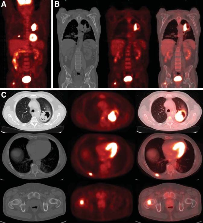
soft-tissue sarcomas, and mucin-producing adenocarcinomas.
In addition, internal hemorrhage, such as that which occurs
within choriocarcinoma and melanoma metastases, can sim-
ulate the increased density of calcifications. Diffuse calci-
fications measuring greater than 300 Hounsfield units (HU)
throughout a nodule are indicative of a benign nodule. A
well-circumscribed nodule with central or lamellar calcifi-
cations also is indicative of benignity (9). The diagnosis of
a benign nodule is presumed only when a majority of the
lesion demonstrates attenuation consistent with calcium.
The calcifications must be located in the center of the lesion
to be considered benign. Other patterns include popcorn or
chondroid calcifications, which, in conjunction with fat, are
characteristic of hamartomas. Figures 1 and 2 demonstrate
shapes, borders, and patterns of calcification in pulmonary
nodules. In addition, the pattern of contrast enhancement
can indicate benignity. A nodule that enhances at less than
15 HU in its central portion is considered benign. A nodule
with enhancement at greater than 25 HU is considered
malignant (12,13). The use of contrast enhancement to char-
                                                                 FIGURE 2. Patterns of calcification in pulmonary nodules.
acterize pulmonary nodules as benign or malignant has not        Nodules 1 and 2 have central calcifications, a benign pattern.
gained widespread acceptance.                                    Nodules 3 and 4 have eccentric calcifications, which cannot be
   Ground-glass opacities also can have a nodular appear-        classified as benign. (Reprinted with permission of (9).)
ance. Ground-glass nodules are less dense than solid nod-
ules and the surrounding pulmonary vasculature and do not
obscure the lung parenchyma (Fig. 3). These nodules also are     According to the Early Lung Cancer Action Program
referred to as subsolid nodules and can be purely ground-        (ELCAP) study, 20% of pulmonary nodules on baseline
glass in appearance or can have mixed solid and ground-          screening are ground-glass or subsolid (14). That study
glass components. Ground-glass opacities continue to be a        demonstrated that the overall frequency of malignancy is
dilemma, as the morphologic characteristics of a benign or       much higher in ground-glass and mixed nodules than in
malignant ground-glass nodule are less well described.           solid nodules. The cell types of malignancies within these
                                                                 nodules also are different from those within solid nodules.
                                                                 The cell types typically included pure bronchioalveolar
                                                                 cells or adenocarcinomas with bronchioalveolar features.
                                                                 Solid nodules are typically invasive subtypes of adenocar-
                                                                 cinoma. There are few data on the evaluation of ground-
                                                                 glass nodules by 18F-FDG PET. One source reported a
                                                                 sensitivity of 10% and a specificity of 20% for ground-glass
                                                                 nodules on 18F-FDG PET (15). Further investigation is
                                                                 necessary; however, the pathology findings of the ELCAP
                                                                 study suggest that there will be little utility in the diagnosis
                                                                 or follow-up of ground-glass nodules by 18F-FDG PET
                                                                 because of the small size of the nodules and the potential
                                                                 for false-negative findings in focal bronchioalveolar cell
                                                                 carcinoma.
                                                                    Certain morphologic characteristics of pulmonary nod-
                                                                 ules are considered indicative of malignancy; these include
                                                                 a spiculated outer margin (Fig. 1), a hazy and indistinct
                                                                 margin, endobronchial extension, extension to pulmonary
                                                                 veins, and focal retraction of the adjacent pleura. Hetero-
                                                                 geneous internal composition and associated necrosis are
                                                                 indicative of malignancy. Malignant lesions also can sim-
                                                                 ulate benign conditions by creating air bronchograms that
FIGURE 1. Schematic diagram of pulmonary nodules. Nodule
1 has smooth, well-defined border. Nodule 2 has lobulated        are commonly associated with pneumonia. Entities such as
border. Nodule 3 has spiculated border. (Reprinted with per-     bronchioalveolar cell carcinoma and lymphoma can mas-
mission of (9).)                                                 querade as benign lung lesions.

452     THE JOURNAL    OF   NUCLEAR MEDICINE • Vol. 47 • No. 3 • March 2006
FIGURE 3.     Ground-glass opacity in peripheral right lung. Mild   18F-FDG   activity is associated with this lesion.

   Malignant nodules are not always easily distinguished           ability is greater than 60% (19,20). About half of the pa-
from benign nodules. Furthermore, 25%239% of malig-                tients undergoing surgical biopsy of an indeterminate
nant nodules are inaccurately classified as benign after ra-       pulmonary nodule have benign disease (5,21).
diologic assessment of morphologic characteristics, including
                                                                   18F-FDG
                                                                         PET FOR EVALUATION OF SOLITARY
size, margins, contour, and internal characteristics (16).
Morphologic stability over 2 y is considered a reliable sign       PULMONARY NODULES
of benignity. The doubling time of the volume of a nodule             The development of 18F-FDG PET has taken the evalu-
is a commonly used marker of the growth of the nodule. For         ation of solitary pulmonary nodules beyond morphologic
malignant nodules, the doubling time is usually 30–400 d.          and predictive analyses to functional and metabolic ana-
Benign nodules demonstrate doubling times outside this             lyses of disease. PET alone has been described as a better
range, both higher and lower. Because a doubling in volume         predictor of malignancy than clinical and morphologic cri-
amounts to a 26% increase in nodule diameter (17), a sig-          teria combined (22,23). A prospective study of 87 patients
nificant change in nodule size may be difficult to appreciate,     examined whether preferential 18F-FDG uptake in malig-
especially for a small nodule. Furthermore, the predictive         nant nodules could differentiate these from benign pulmo-
value of stability in size may be only 65% (18).                   nary nodules (24). The investigators found that when a
   Clinical information often is useful in the assessment of       mean standardized uptake value (SUV) of greater than or
pulmonary nodules. Important features of the patient’s his-        equal to 2.5 was used for detecting malignancy, the sensi-
tory can be combined with imaging findings to calculate a          tivity, specificity, and accuracy were 97%, 82%, and 92%,
likelihood ratio for malignant disease (Table 1). This type        respectively (Fig. 4). In addition, they also determined that
of Bayesian analysis can be used to stratify the patient’s         there was a significant correlation between the doubling
risk of malignancy and to guide management. In this                time of tumor volume and the SUV. Subsequent studies
scheme, patients are monitored if the probability of cancer        demonstrated a sensitivity of 90%2100% and specificity of
is less than 5%, the lesion is biopsied if the probability is      69%295% for PET (15,25,26). Although the SUV is a
between 5% and 60%, and nodules are resected if the prob-          useful tool, it has been shown to be equivalent to the visual
                                                                   estimate of metabolic activity by experienced physicians
                                                                   (27,28).
                          TABLE 1
   Likelihood Ratios for Lung Cancer, as Determined by
                                                                      Studies that favor 18F-FDG PET for the diagnostic
         Morphologic and Demographic Information                   workup of solitary pulmonary nodules to reduce inappro-
                                                                   priate invasive diagnostic investigation and subsequent
   Feature or characteristic                  Likelihood ratio     complications are emerging. A study performed in Italy
   Spiculated margin                               5.54            compared the traditional workup of a solitary pulmonary
   Lesion . 3 cm                                   5.23            nodule with CT, fine-needle aspiration, and thoracoscopic
   Subject . 70 y old                              4.16            biopsy with a diagnostic workup including 18F-FDG PET
   Malignant growth rate                           3.40            (29). That study demonstrated a cost reduction of approx-
   Subject smokes tobacco                          2.27
                                                                   imately E50 (;$60) per patient when PET was added to the
   Upper-lobe nodule                               1.22
   Lesion , 1 cm                                   0.52            traditional workup. A recent study in France compared the
   Smooth margins                                  0.30            cost-effectiveness ratios of 3 management scenarios for soli-
   Subject 30–39 y old                             0.24            tary pulmonary nodules: wait and watch with periodic CT,
   Subject never smoked                            0.19            PET, and CT plus PET (30). For their typical patient, a 65-
   Subject 20–29 y old                             0.05
                                                                   y-old male smoker with a 2-cm solitary pulmonary nodule
   Benign calcification                            0.01
   Benign growth rate                              0.01            and an associated high risk of malignancy of 43%, the wait-
                                                                   and-watch scenario was the least effective strategy. CT plus
                                                                   PET was the most effective strategy and had a lower incre-
  Reprinted with permission of (16).
                                                                   mental cost-effectiveness ratio. Their conclusion was that

                                                 PET EVALUATION     OF   LUNG CANCER • Bunyaviroch and Coleman                    453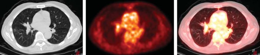
FIGURE 4. Solitary pulmonary nodule with spiculated borders in left upper lobe. No mediastinal adenopathy was present on
additional images. Hypermetabolism is present within this nodule. Maximum SUV measures 6.7 g/mL. Findings are consistent with
malignancy.

CT plus PET was the most cost-effective strategy for pa-         malignancy; the authors determined that the sensitivity and
tients with a risk of malignancy of 5.7%287%. The wait-          specificity of the tests were 80% and 94% (single) and
and-watch scenario was most cost-effective for patients          100% and 89% (dual), respectively (36). Pathophysiolog-
with a risk of 0.3%25%.                                          ically, the differences in levels of glucose-6-phosphatase
   The minimum size of a pulmonary nodule has been an            and hexokinase within benign and malignant cells have
issue with regard to accurate diagnostic evaluation, follow-     been postulated as the reason for this effect (37). Although
up, and even biopsy. The NY-ELCAP study monitored 378            these studies appear promising, the use of dual-time-point
patients with pulmonary nodules determined by CT to be           imaging remains controversial. Further data are needed
less than 5 mm in diameter. None of these nodules was di-        before widespread use can be recommended.
agnosed as pathologically malignant, leading the researchers        18F-FDG PET is known to show little uptake in malig-

to suggest limiting further workup to nodules that were          nancies with low metabolic activity. Focal bronchioalveolar
5 mm or larger (31). A group in Spain investigated the utility   cell carcinoma has been shown to have less proliferative
of PET in evaluating nodules of 5–10 mm in diameter and          potential and a longer mean doubling time than NSCLC
greater than 10 mm in diameter; the sensitivity for detecting    (38,39). Further investigation has shown that different sub-
malignancy in all nodules was fairly low, at 69%, whereas        types of bronchioalveolar cell carcinoma exhibit different
the sensitivity for detecting malignancy in nodules of           rates of metabolic activity. Focal or pure bronchioalveolar
greater than 10 mm was 95% (32). The authors noted that          cell carcinoma appears as a peripheral nodule or localized
the apparent uptake in nodules decreased when the diam-          ground-glass attenuation and may show false-negative re-
eter was less than twice the spatial resolution of the system    sults on 18F-FDG PET (40). In contrast, the multifocal form
(approximately 728 mm); thus, different criteria are             appears as multiple nodules or ground-glass consolidation
needed to determine malignancy in nodules of less than           (40) and is detected at a relatively high sensitivity on 18F-
15 mm. Short-term follow-up of 5- to 10-mm nodules with          FDG PET (41). Carcinoid is another malignancy that grows
CT alone to evaluate for growth resulted in a low rate of        slowly and has low mitotic activity (42). The sensitivity of
invasive procedures for benign nodules. In a phantom             18F-FDG PET for the detection of focal bronchioalveolar

study with 18F-FDG-filled spheres measuring between 6            cell carcinoma and carcinoid tumor is lower than that for
and 22 mm, the detection of nodules of less than 7 mm            other cell types of lung cancer and has been reported to be
was unreliable (33). Further investigation is necessary to       as low as 50%.
determine the best method for evaluating subcentimeter              Several groups have investigated the prognostic value of
nodules.                                                         18F-FDG PET (43–45). In a study of 155 patients with

   Dual-time-point imaging has emerged as a potential dis-       NSCLC, median survival was compared with the standard-
criminator of benign and malignant diseases, with images         ized uptake ratio (analogous to the SUV) of the primary
being obtained at 1 and 2 h after the administration of 18F-     tumor (43). Median survival decreased with increasing mean
FDG. In a study involving in vitro samples and animal and        SUV. SUVs of less than 10 and greater than 10 indicated
human subjects, 18F-FDG uptake was measured over time;           median survival times of 24.6 and 11.4 mo, respectively
Zhuang et al. found that malignant lesions showed a sig-         (Fig. 5). Furthermore, a mean SUV of greater than 10 with a
nificant increase in SUV over time and that benign lesions       tumor larger than 3 cm indicated a median survival of 5.7
showed a decrease over time (34). Additional investigation       mo. A retrospective study of 100 patients demonstrated that
has reached similar conclusions (35). One study compared         the 2-y survival rates were 68% for patients with a max-
single-time-point imaging and dual-time-point imaging            imum SUV of more than 9 and 96% for those with a
with a cutoff SUV of 2.5 and a 10% increase in SUV for           maximum SUV of less than 9 (45).

454     THE JOURNAL    OF   NUCLEAR MEDICINE • Vol. 47 • No. 3 • March 2006

mediastinal, aortic, and N1 nodes. These nodal groups can
                                                                    be divided further into anatomic lymph node regions or
                                                                    levels (Table 2) (48).
                                                                       One of the uses of this lymph node classification is to
                                                                    identify the proper method for lymph node sampling. Dif-
                                                                    ferent invasive procedures typically are used for lymph
                                                                    node sampling; these include mediastinoscopy, video-
                                                                    assisted thoracic surgery (VATS), endoscopic sonography,
                                                                    and thoracotomy (Table 3) (49). Mediastinoscopy is best
                                                                    used for the evaluation of level 2, 4, and 7 lymph node
                                                                    stations. VATS can be used for multiple stations, depending
                                                                    on the approach, and is commonly used for level 5, 6, and
                                                                    10 stations. Endoscopic sonography with transbronchial
                                                                    needle aspiration can be used for level 4–9 stations. All
FIGURE 5. Survival among NSCLC patients stratified by stan-         nodal groups can be reached by thoracotomy and poten-
dardized uptake ratio (SUR). (Reprinted with permission of (43).)   tially by CT-guided percutaneous needle biopsy.
                                                                       The location of the primary tumor determines the lym-
                                                                    phatic pathway for spread to regional lymph nodes (50). A
MULTIPLE PULMONARY NODULES
                                                                    tumor in the right lung sends metastasis to hilar (10R)
   The evaluation of multiple pulmonary nodules can be              lymph nodes, which proceed to right paratracheal (4R and
limited by potential false-positive findings on 18F-FDG             2R) lymph nodes. Such a tumor rarely metastasizes to the
PET. Increased 18F-FDG activity has been demonstrated in            contralateral side. A left upper-lobe cancer sends metastases
instances of active granulomatous disease, such as tuber-           to the aortopulmonary window (5) and left paratracheal nodes
culosis, fungal disease, and sarcoidosis, as well as other          (4L). Left upper- and lower-lobelesions also may spread ini-
inflammatory processes, such as rheumatoid nodules (46,47).         tially to left hilar (10L) lymph nodes. Involvement of
CT in combination with 18F-FDG PET aids in the evalua-              prevascular (6) lymph nodes is almost invariably associated
tion of multiple pulmonary nodules. In addition to the              with paratracheal involvement. Tumors in the right middle
shapes, borders, and densities of the nodules, the distribu-        lobe and bilateral lower lobes can metastasize early to
tion of the nodules can provide important clues to their            subcarinal (7) nodes. Lower-lobe cancers also can send
etiology. There are 3 different distribution patterns: peri-        metastases to paraesophageal (8), pulmonary ligament (9),
lymphatic, random, and centrilobular. Perilymphatic nod-            and subdiaphragmatic (14) lymph nodes.
ules are located along the pleural surfaces, interlobular
septa, and peribronchovascular interstitium, particularly in        CONVENTIONAL STAGING
the perihilar regions and centrilobular regions. Random                The staging of malignancies with the TNM system was
nodules have a more even and symmetric, yet random, dis-            created to provide consistency in communication of the ex-
tribution within the lung fields bilaterally. Centrilobular         tent of disease, to provide a basis for the selection of therapy,
nodules spare the pleural surfaces and are associated with          and to help determine prognosis (51). The important deci-
small pulmonary artery branches. There are 2 subcategories          sion in using this system is whether the disease is resectable.
of centrilobular pulmonary nodules, those associated with           The T status classifies the features of the primary tumor.
and those not associated with tree-in-bud opacities. A tree-        The N status classifies the presence or absence of regional
in-bud opacity is a branching opacity that represents filling       lymph node involvement. The M status classifies the
of the alveolar spaces. This process typically occurs from          presence or absence of extrathoracic metastasis (Table 4).
an inflammatory or infectious process rather than a malig-             The T status evaluates the extent of the primary tumor by
nant process. The remaining nodular distributions are more          size and invasiveness. The current system describes the size
often associated with malignancy and include lymphangitic           of the tumor and its relationship with the pleura, broncho-
spread of cancer with a perilymphatic pattern, hematoge-            vascular structures, and mediastinum. A T1 lesion is de-
nous metastasis with a random distribution, and bronchioal-         fined as a tumor that is 3 cm or smaller (in the greatest
veolar cell cancer with centrilobular opacities.                    dimension), with lung or visceral pleura separating the le-
                                                                    sion from the mediastinum, but that does not extend proxi-
STAGING OF LUNG CANCER                                              mally to the lobar bronchus. A T2 lesion is larger than 3 cm,
   Before 1996, there were 2 mediastinal lymph node clas-           invades the visceral pleura, and extends proximally to the
sification schemes. The 2 schemes were unified in 1996 by           lobar bronchus but does not extend to within 2 cm of
the American Joint Commission on Cancer and the Prog-               the carina. Extension of the primary tumor into the medi-
nostic TNM Committee of the Union Internationale Contre             astinum precludes curative surgical resection (52). The
le Cancer. As shown in Figure 6, thoracic lymph nodes can           preservation of mediastinal fat planes or intervening
be organized into 4 groups: superior mediastinal, inferior          lung between the tumor and the mediastinum is a clear

                                                  PET EVALUATION     OF   LUNG CANCER • Bunyaviroch and Coleman                 455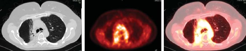
FIGURE 6. Thoracic lymph node sta-
tions. Subcategories include superior
mediastinal nodes, aortic nodes, inferior
mediastinal nodes, and N1 nodes (64).
a. 5 artery; v. 5 vein; Inf. pulm. ligt. 5
inferior pulmonary ligament; Ao 5 aorta;
PA 5 pulmonary artery; A-P 5 aortopul-
monary; L. pulmonary a. 5 left pulmonary
artery; Phrenic n. 5 phrenic nerve. (Re-
printed with permission of (64).)

indication that there is no direct extension into the medi-        patient’s clinical stage to stage IIIB or higher. At stage III,
astinum. Extension into the chest wall, diaphragm, medi-           evaluation of the mediastinum for either direct extension to
astinal pleura or pericardium, or main bronchus is defined         vital structures or contralateral mediastinal lymph node
as a T3 lesion. The presence of T3 lesions does not                disease determines resectability.
necessarily preclude curative resection. Invasion of the              The CT evaluation of mediastinal lymph nodes has ex-
mediastinum, vertebrae, and vital structures, such as the          tremely variable sensitivity and specificity, with false-negative
great vessels, trachea, esophagus, or heart, is classified as a    results of 7%239% and false-positive results of 20%245%
T4 lesion and does preclude curative resection.                    (9). Size criteria alone are not very reliable in the staging of
   Lymph node status (N status) is integral to determining         mediastinal lymph nodes (53,54). Lymph nodes of greater
the resectability of a tumor; it describes the presence or         than 1 cm in the short axis are considered abnormal by CT
absence and extent of regional lymph node metastasis.              criteria (55). Fifteen percent of patients with clinical stage I
Metastasis to lymph nodes in the ipsilateral peribronchial or      disease may have micrometastases in normal-size lymph
hilar regions is classified as N1 disease, a classification that   nodes (56). Other morphologic features of lymph nodes are
alters the stage and prognosis of disease. The presence of         unlikely to be helpful in differentiating benign disease from
N1 lymph nodes, however, does not preclude curative                malignant disease (57). Fat within a lymph node hilum is
resection and does not accurately predict mediastinal lymph        believed to be a sign of benignity. Adenopathy detected by CT
node involvement. Metastatic involvement of ipsilateral            is useful in directing invasive sampling techniques. Medias-
mediastinal lymph nodes is defined as N2 disease and               tinoscopy traditionally has been used for tissue diagnosis of
represents at least stage IIIA disease. The presence of con-       mediastinal lymph node metastasis; however, additional
tralateral mediastinal, hilar, scalene, or supraclavicular lymph   techniques, such as transbronchial, percutaneous, or video-
node involvement is defined as N3 disease and increases the        scopic biopsy, may be used when appropriate.

456      THE JOURNAL    OF   NUCLEAR MEDICINE • Vol. 47 • No. 3 • March 2006
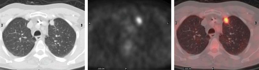
TABLE 2
                                                  Lymph Node Map Definitions

     Nodal station anatomic landmarks                                            Description

  N2 nodes—all N2 nodes lie within
    mediastinal pleural envelope
      Highest mediastinal nodes          Nodes lying above horizontal line at upper rim of bracheocephalic (left innominate)
                                           vein, where it ascends to left, crossing in front of trachea at its midline
       Upper paratracheal nodes          Nodes lying above horizontal line drawn tangential to upper margin of aortic arch
                                           and below inferior boundary of highest mediastinal nodes
       Prevascular and retrotracheal     Prevascular and retrotracheal nodes may be designated 3A and 3P; midline nodes
         nodes                             are considered to be ipsilateral
       Lower paratracheal nodes          Lower paratracheal nodes on right lie to right of midline of trachea between horizontal
                                           line drawn tangential to upper margin of aortic arch and line extending across right
                                           main bronchus at upper margin of upper-lobe bronchus and are contained within
                                           mediastinal pleural envelope; lower paratracheal nodes on left lie to left of midline
                                           of trachea between horizontal line drawn tangential to upper margin of aortic arch
                                           and line extending across left main bronchus at level of upper margin of left
                                           upper-lobe bronchus, medial to ligamentum arteriosum, and are contained within
                                           mediastinal pleural envelope
       Subaortic (aortopulmonary window) Subaortic nodes are lateral to ligamentum arteriosum, aorta, or left pulmonary artery
                                           and proximal to first branch of left pulmonary artery and lie within mediastinal
                                           pleural envelope
       Paraaortic nodes (ascending       Nodes lying anterior and lateral to ascending aorta and aortic arch or innominate artery,
         aorta or phrenic)                 beneath line tangential to upper margin of aortic arch
       Subcarinal nodes                  Nodes lying caudad to carina of trachea but not associated with lower-lobe bronchi or
                                           arteries within lung
       Paraesophageal nodes              Nodes lying adjacent to wall of esophagus and to right or left of midline, excluding
         (below carina)                    subcarinal nodes
       Pulmonary ligament nodes          Nodes lying within pulmonary ligament, including those in posterior wall and lower
                                           part of inferior pulmonary vein

  N1 nodes—all N1 nodes lie distal to
    mediastinal pleural reflection and
    within visceral pleura
      Hilar nodes                          Proximal lobar nodes, distal to mediastinal pleural reflection, and nodes adjacent to
                                             bronchus intermedius on right; radiographically, hilar shadow may be created by
                                             enlargement of both hilar and interlobar nodes
       Interlobar nodes                    Nodes lying between lobar bronchi
       Lobar nodes                         Nodes adjacent to distal lobar bronchi
       Segmental nodes                     Nodes adjacent to segmental bronchi
       Subsegmental nodes                  Nodes around subsegmental bronchi

  Reprinted with permission of (64).

   Evaluation of distant metastasis (M status) also is a            nervous system, bone, liver, and adrenal glands are com-
critical step in determining the resectability of a tumor.          mon sites for distant metastases, and such extension is
M status defines the presence or absence of tumor spread            considered to represent M1 disease (58). Metastases to the
to distant lymph node or organ sites. The brain, central            contralateral lung also are considered distant metastases.
                                                       TABLE 3
                              Procedures Used to Sample Lymph Nodes, by Lymph Node Level

  Lymph node level            Mediastinoscopy          Thoracotomy            Chamberlain/VATS             Esophageal sonography

  2L, 2R                               U                     U
  4L, 4R                               U                     U                                                         U
  5, 6                                                       U                         U                               U
  7                                    U                     U                                                         U
  8, 9                                                       U                                                         U
  10L, 10R                                                   U                         U
  11–14                                                      U

  Reprinted with permission of Society of Thoracic Surgeons (49).

                                                 PET EVALUATION      OF   LUNG CANCER • Bunyaviroch and Coleman                    457
TABLE 4
TNM Classification of Lung Cancer
Classification Description
Primary tumor (T)
TX Primary tumor cannot be assessed; or tumor proven by presence of malignant cells in sputum or
bronchial washes but not visualized by imaging or bronchoscopy
T0 No evidence of primary tumor
Tis Carcinoma in situ
T1 Tumor 3 cm or less in greatest dimension, surrounded by lung or visceral pleura, without bronchoscopic
evidence of invasion more proximal than lobar bronchus (i.e., not in main bronchus)
T2 Tumor with any of the following features of size or extent: more than 3 cm in greatest dimension; involves
main bronchus, 2 cm or more distal to carina; invades visceral pleura; or associated with atelectasis or
obstructive pneumonitis that extends to hilar region but does not involve entire lung
T3 Tumor of any size that directly invades any of following: chest wall (including superior sulcus tumors),
diaphragm, mediastinal pleura, or parietal pericardium; tumor in main bronchus less than 2 cm distal to
carina but without involvement of carina; or associated atelectasis or obstructive pneumonitis of entire lung
T4 Tumor of any size that invades any of following: mediastinum, heart, great vessels, trachea, esophagus,
vertebral body, or carina; or tumor with malignant pleural or pericardial effusion or with satellite tumor
nodule(s) within lobe of lung ipsilateral to lobe with primary tumor
Regional lymph
nodes (N)
NX Regional lymph nodes cannot be assessed
N0 No regional lymph node metastasis
N1 Metastasis to ipsilateral peribronchial or ipsilateral hilar lymph nodes or both and involvement of
intrapulmonary nodes by direct extension of primary tumor
N2 Metastasis to ipsilateral mediastinal or subcarinal lymph nodes
N3 Metastasis to contralateral mediastinal, contralateral hilar, ipsilateral or contralateral scalene,
or supraclavicular lymph nodes
Distant metastasis (M)
MX Presence of distant metastasis cannot be assessed
M0 No distant metastasis
M1 Distant metastasis present
Reprinted with permission of (51).
The radiologic workup for metastatic disease often begins evaluation of distant metastasis. Unenhanced CT followed
with clinical history, physical examination, and laboratory by MRI is reported as the most cost-effective morphologic
studies. The frequency of occult metastasis at the time of evaluation of suggestive adrenal lesions (63). Adrenal
presentation may be as high as 30% in patients with lesions that measure less than 10 HU on unenhanced CT
adenocarcinoma or large cell carcinoma of the lung (59). are considered benign. Adrenal lesions that do not have CT
Squamous cell carcinoma of the lung appears to have a signs of benignity are followed up with MRI with opposed-
lower frequency of occult metastasis (,15%) at presenta- phase imaging. 18F-FDG PET is sensitive for the detection
tion. Routine radiologic evaluation for occult metastases of adrenal metastases, but some benign adrenal adenomas
without clinical or laboratory findings is not clearly indi- may be abnormal on PET.
cated (60). The adrenal glands and liver are the most The International System for Staging Lung Cancer was
common sites for occult extrathoracic metastases. The developed in response to the need for a classification scheme to
adrenal glands occasionally may be the only sites for unify the variations in staging definitions and provide consis-
metastasis; however, incidental benign adenomas occur tent meaning and interpretation for different stages. The value
with a similar frequency in patients with bronchogenic of this system in predicting prognosis relies on the identifica-
carcinomas. Three to 5% of the overall population has tion of consistent and reproducible patient groups with similar
incidental nonfunctioning cortical adenomas, whereas ap- outcomes. The International System for Staging Lung Cancer
proximately 10% of patients with bronchogenic cancer applies to all 4 major cell types of lung cancer: squamous cell,
have an adrenal mass on CT (61,62). In the absence of adenocarcinoma (including bronchioalveolar cell), large cell,
other known extrathoracic metastases, adrenal masses usu- and small cell. Multiple factors are directly related to the
ally are benign. The liver usually is never the only site for extent of disease at diagnosis; these include the proportion of
metastasis, unless the primary malignancy is an adenocar- patients achieving a complete response, the duration of the
cinoma. CT and MRI traditionally have been used for the response, and recurrence after a complete response.
458 THE JOURNAL OF NUCLEAR MEDICINE • Vol. 47 • No. 3 • March 2006The TNM system is used to define 7 stages of disease
(Table 5) (51). Stage IA includes small tumors of less than
or equal to 3 cm, without invasion proximal to a lobar bron-
chus, and without metastasis. Stage IB includes larger
tumors, tumors with invasion of the visceral pleura or main
bronchus (.2 cm distal to the carina), or both, and tumors
without metastasis. There is a significant difference in sur-
vival between IA disease and IB disease, with 5-y survival
rates of 61% and 38%, respectively (64). Stage IIA includes
T1 tumors with metastases to ipsilateral peribronchial
lymph nodes, hilar lymph nodes, or both. These metastases
are difficult to document radiographically. Stage IIB includes
T2 lesions with metastases to ipsilateral peribronchial lymph
nodes, hilar lymph nodes, or both and T3 tumors without
metastasis. The 5-y survival rates for stage IIA and stage
IIB are 37% and 24%, respectively (64). Stage IIIA
includes T3 tumors with metastases to intrapulmonary
lymph nodes, hilar lymph nodes, or both (N1). T1 through
T3 tumors with ipsilateral mediastinal lymph node metas-
tases (N2) also are included in IIIA disease. This stage
includes limited invasion of the mediastinum or chest wall
(T3). Such lesions have an improved outcome and are
potentially resectable if vital structures in the mediastinum
are not involved. Stage IIIB involves extensive extrapul-
monary involvement, with invasion of the mediastinal
structures, esophagus, trachea, carina, heart, major vessels, FIGURE 7. Patient survival in relation to stage of disease. c 5
or vertebral bodies. An associated pleural effusion also is clinical stage. (Reprinted with permission of (51).)
considered to represent stage IIIB disease. No distant meta-
static disease is present. This stage of disease is virtually COMBINING CONVENTIONAL STAGING WITH
always nonresectable (9). The 5-y survival rates for stage METABOLIC IMAGING
IIIA and stage IIIB are 9%213% and 5%, respectively As a measure of metabolic activity, 18F-FDG PET adds a
(Fig. 7) (64). Stage IV includes any T status and N status functional evaluation to the staging of lung cancer. The
with distant metastases. Non–lymph-node metastases in ipsi- PET in Lung Cancer Staging trial attempted to determine
lateral lobes not involved by the primary tumor also are the value of 18F-FDG PET in lung cancer staging (65). The
considered stage IV disease. Stage IV disease is considered a goal was to determine whether unnecessary surgery could
contraindication to surgical resection (9). As expected, survival be reduced. The researchers enrolled 188 patients in a
with stage IV disease is poor, with less than 1% survival at randomized controlled trial comparing a conventional ra-
5 y (64). diologic staging workup (CWU) to CWU and PET. The
conclusions of the study were that the addition of PET to
CWU prevented unnecessary surgery in 1 of 5 patients with
suspected NSCLC. In addition, the staging of disease was
TABLE 5 increased for 27% of patients. The researchers believed that
International Staging System for NSCLC, Including the negative predictive value of PET for mediastinal lymph
Stage Groups and TNM Subsets node involvement was sufficiently high to avoid mediasti-
Stage TNM subset noscopy for noncentral tumors. Another prospective study
of 102 patients went further to conclude that invasive proce-
0 Carcinoma in situ dures probably are not necessary in a patient with negative
IA T1 N0 M0
IB T2 N0 M0
findings on PET for the mediastinum (66). In that study, the
IIA T1 N1 M0 sensitivity and specificity of PET in detecting mediastinal
IIB T2 N1 M0, T3 N0 M0 lymph node metastasis were 91% and 86%, respectively.
IIIA T3 N1 M0, T1 N2 M0, T2 N2 M0, T3 N2 M0 The high negative predictive value of PET led some
IIIB T4 N0 M0, T4 N1 M0, T4 N2 M0, T1 N3 M0, institutions to accept negative PET results without pathologic
T2 N3 M0, T3 N3 M0, T4 N3 M0
IV Any T any N M1
confirmation and to proceed to curative surgical resection.
This management scheme has led to much controversy with
regard to the role of PET in mediastinal staging. Although
Reprinted with permission of (51).
the PET in Lung Cancer Staging study demonstrated a clear
PET EVALUATION OF LUNG CANCER • Bunyaviroch and Coleman 459benefit of PET in predicting disease, the results may not be demonstrated that PET changed the staging of lung cancer generalizable to other populations (67). The accuracy of in 44% of cases (83). The use of PET in stage IV disease is clinical evaluation for distant metastasis in NSCLC has less of an issue for mediastinal staging, as the patient’s N been investigated for each stage of the disease. These status is no longer relevant. The use of PET in stage IV studies reported a 5% false-negative rate for the clinical disease will be discussed further with regard to identifying evaluation of stage I and II diseases (68–70). The false- and monitoring distant metastasis. negative rate for stage III disease was reported to be Because 18F-FDG describes metabolic activity, it cannot 15%220%. Without clinical evidence of distant metastatic distinguish malignancy from inflammation or infection. disease, mediastinal involvement becomes a crucial issue in 18F-FDG uptake is demonstrated in sites of active acute determining the stage of the disease. CT evaluation of the inflammation because of increased glucose uptake by acti- mediastinum has a false-negative rate of 15% overall; the vated macrophages and inflammatory cells (85). Multiple false-negative rate increases to 20%225% for central lung studies have demonstrated a positive predictive value for tumors (71,72). A meta-analysis of the diagnostic perfor- PET of 74%293% for evaluation of the mediastinum (66,86). mance of PET versus CT for mediastinal staging was per- A study comparing PET and mediastinoscopy evaluations formed by Dwamena at al. (73). For 14 PET and 29 CT case of 202 patients showed a positive predictive value for PET series, they determined that PET was statistically superior of 44.6% (81). The high rate of false-positive results dem- to CT for mediastinal staging. With respect to CT, PET has onstrates the necessity for mediastinoscopy in the staging been shown to have a higher negative predictive value, and of PET-positive mediastinal lymph nodes (80,87). The combined PET/CT has an even higher negative predictive added benefits of PET in this setting include the ability to value (49,74–76). direct mediastinal lymph node biopsy and to aid in selecting The use of PET to exclude mediastinal metastasis additional invasive methods for lymph nodes inaccessible remains controversial. From the data available, classifica- to mediastinoscopy (Table 3). tion of disease as stage I on the basis of a clinical exami- nation and negative results from CT and PET examinations appears sufficient to exclude mediastinal disease. Classifi- STAGING OF SMALL CELL LUNG CANCER (SCLC) cation of stage II and III diseases is more controversial; the SCLC represents approximately 18%225% of all cases negative predictive value of PET decreases in relation to the of lung cancer (88,89). SCLC is a neuroendocrine tumor size of the metastasis, the presence of centrally located that has an aggressive growth pattern, that commonly dis- primary disease or N1 nodes, and the avidity of the primary plays early widespread metastases, and that has a rapid tu- tumor for 18F-FDG (77,78). Micrometastatic disease cannot mor doubling time (90). Consequently, patients often present be imaged effectively on PET because of the spatial with bulky hilar and mediastinal lymph node metastases resolution of the imaging system (79,80). Takamochi et al. (91). The tumors usually are located centrally (89,92), often found that the diameters of false-negative lesions ranged with encasement of mediastinal structures and tracheobron- from 1 to 7.5 mm (80). In addition, the presence of hyper- chial compression (91,93). The primary tumor may be small metabolic central tumors or hilar lymph nodes can decrease or undetectable by radiographic methods, whereas early the detectability of mediastinal lymph nodes and thus the extrathoracic metastases are common and can present negative predictive value of mediastinal PET (78). Finally, before clinical symptoms (94,95). Unlike the situation for the metabolic activity of low-grade malignancies cannot be NSCLC, there is a 2-stage classification scheme proposed expected to be any greater than that of the primary tumor by the Veterans Administration Lung Cancer Study Group. (77). Mediastinal activity is a source of potential error at- Patients with SCLC are classified as having either limited tributable to random inhomogeneity and misregistration or extensive disease (96). Limited disease refers to tumor from respiratory, cardiac, and body motions. For stage II that is confined to the thorax. Extensive disease includes and III diseases, the incidence of false-negative results is distant metastases, including those to the contralateral lung. still greater with PET than with mediastinoscopy. In a Whether 18F-FDG PET has a role in the staging of SCLC is comparative study, the false-negative rates of mediastinos- controversial. Detterbeck et al. stated that the clinical pre- copy and PET were 3% and 11.7%, respectively (81). sentation and radiographic appearance are sufficiently char- Mediastinoscopy likely will remain part of the standard acteristic of the disease to eliminate the need for further protocol for mediastinal staging for stage II and III dis- confirmation (97). A few studies have been performed to eases. The clinical importance of differentiating stage IIIA compare the staging of SCLC by conventional radiography and IIIB diseases, with regard to denying curative resection, with that by 18F-FDG PET. PET changed patient manage- is a significant factor in the continued use of mediastinos- ment in 8.3%229% of these cases (98–101). Patients with copy. Several studies have demonstrated the potential of limited disease were given chemoradiation, whereas pa- PET to alter patient management (82–84). Hicks et al. tients with extensive disease were given chemotherapy alone. found that PET caused a major management change in The available studies show a possible role for 18F-FDG 40 of 63 patients (63%) who had previously undergone PET in the staging of SCLC; however, further study is potential curative surgery for NSCLC (82). Seltzer et al. necessary to evaluate the clinical necessity. 460 THE JOURNAL OF NUCLEAR MEDICINE • Vol. 47 • No. 3 • March 2006
COST-EFFECTIVENESS OF STAGING BY PET with greater specificity than can conventional imaging,
The cost-effectiveness of PET for the staging of NSCLC including CT (109). The mean frequency of extrathoracic
has been extensively studied in multiple health care sys- metastases in these studies was 13%. In 18% of the cases,
tems. Cost-effectiveness is analyzed with respect to the cost the PET results altered management. As expected, the
of patient care and life expectancy. The incremental cost- frequency of distant metastases was shown to increase with
effectiveness ratio quantifies the difference in cost for dif- higher stages: 7.5% in stage I, 18% in stage II, and 24% in
ferent therapeutic strategies versus the difference in life stage III (110).
expectancy (102). A study comparing 5 different clinical As discussed earlier, the adrenal glands and liver are the
strategies was performed with Medicare reimbursements in most common sites of extrathoracic metastases in lung
the United States as the basis for the cost analysis. Conven- cancer. At the time of presentation, up to 10% of patients
tional CT staging followed by biopsy and surgical versus will have an adrenal mass. Approximately two thirds of
nonsurgical therapy was compared with 4 strategies inte- these masses will be benign (111,112). In a study of 27
grating PET. Three strategies used confirmatory biopsy patients with 33 adrenal masses, the ability of PET to dif-
before diverting patients from curative resection. The final ferentiate benign from malignant adrenal masses was inves-
strategy eliminated confirmatory biopsy and proceeded to tigated (113). The sensitivity of PET for detecting adrenal
surgical or nonsurgical therapy. That study demonstrated metastasis was 100%, and the specificity was 80%. A
that the most cost-effective strategy involved the use of subsequent study of adrenal lesions demonstrated a sensi-
PET for CT evaluations with negative results followed by tivity of 100%, a specificity of 94%, and an accuracy of
confirmatory biopsy. The strategy involving the elimination 96% for detecting metastasis (114). The evaluation of liver
of confirmatory biopsy after CT and PET evaluations with metastasis by PET is less well studied. Liver metastases are
positive results had the lowest cost but also the lowest life rarely the only demonstrable site of metastatic disease (9).
expectancy (103). A direct comparison of the cost-effectiveness In a study of 110 patients with NSCLC, 18F-FDG PET
of PET for demonstrating additional or unanticipated re- was compared with methylene diphosphonate bone scan-
sults using PET with confirmatory mediastinoscopy and ning for the evaluation of bone metastases (115). 18F-FDG
PET with selective mediastinoscopy demonstrated a sav- PET had a higher specificity for detecting bone metastases
ings in both instances. Selective mediastinoscopy showed (98% vs. 61%). Some additional studies demonstrated a
approximately double the cost savings per patient ($2,267 higher specificity (116,117), and some demonstrated a higher
vs. $1,154) but missed 1.7% of patients who might be cured accuracy (115,118,119). Marom et al. (120) found that,
(104). A comparison of cost-effectiveness in other health compared with bone scintigraphy, 18F-FDG PET had a higher
care systems is more difficult because of the use of different sensitivity but an equivalent specificity for 90 patients who
therapeutic strategies. A study of the French health care underwent both studies. Fogelman et al. (121) reviewed the
system involved a significant difference in staging strate- literature on this topic and concluded that, with regard to lung
gies (105). The therapeutic strategies in that study did not cancer, 18F-FDG PET had a sensitivity similar to that of bone
mandate confirmatory biopsy before surgical or nonsurgical scintigraphy but a specificity higher than that of bone scin-
therapy. That study determined that the most cost-effective tigraphy. The practical advantage of 18F-FDG PET over bone
strategy involved the use of PET after a CT examination scintigraphy remains controversial. Mechanistically, there are
with negative or positive results. The PET results then were different patterns of uptake related to the morphology of the
used to make decisions regarding biopsy, surgery, or che- lesion: lytic, sclerotic, or mixed (121). As demonstrated in a
motherapy. Similar findings were demonstrated in studies study of breast cancer patients with bone metastases, 18F-FDG
of the Italian (29), Canadian (106), and German (107) health PET appears to have the advantage of detecting osteolytic
care systems. Irrespective of the use of mediastinoscopy, PET lesions, whereas bone scintigraphy has the advantage of
for the evaluation of mediastinal disease in NSCLC has been detecting osteoblastic lesions (122).
shown to be cost-effective in several health care models. The detection of brain metastasis by PET also has been
evaluated. In a study of 1,026 patients with multiple dif-
ferent malignancies, unsuspected cerebral or skull metas-
DETECTION OF DISTANT METASTASIS tases were detected in only 0.4% of the patients (123). PET
The presence of distant metastasis is classified as stage is less effective than CT or MRI for the detection of cere-
IV disease, which precludes a patient from the possibility bral metastasis.
of curative surgical resection. The patient therefore is pre-
scribed palliative therapy. An inherent advantage of PET is
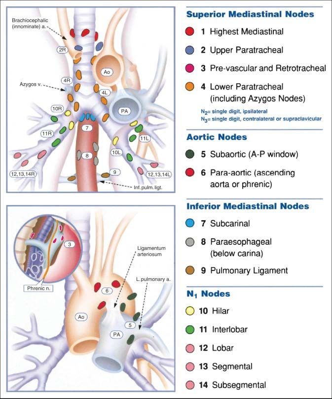
the use of whole-body scanning, which facilitates the sur- RESTAGING
vey of a much larger area than is possible with commonly The benefit of determining a metabolic response to ther-
used radiographic methods (Fig. 8). Distant metastases apy over a morphologic response has led to the investiga-
commonly involve the adrenal glands, bones, liver, and tion of PET for the restaging of NSCLC. The criteria for
brain (108). Multiple studies have demonstrated the ability conventional restaging were determined by the World
of 18F-FDG PET to detect distant metastasis of lung cancer Health Organization and later modified by the National
PET EVALUATION OF LUNG CANCER • Bunyaviroch and Coleman 461FIGURE 8. Lung cancer with osseous metastases. Hypermetabolic cavitary lung mass is seen in left upper lobe (A– C). Maximum-intensity-projection image (A) demonstrates additional lesions in contralateral thorax and hip. Additional focus of hypermetabolism is seen in right femoral neck (A–C) and corresponds to subtle lytic lesion on CT. Axial images (C) show hypermetabolism in right posterior 8th rib without osseous changes on CT. Cancer Institute and the European Association for Research cycle of chemotherapy (130). It was found that a reduction in and Treatment of Cancer. Tumor response is defined as a metabolic activity correlated closely with the final outcome therapy-induced reduction of the largest dimension of the of the therapy. An early metabolic response predicted better tumor by 30% (124). Complete and partial responses are survival, and a poor response predicted disease progression determined by the amount of tumor size reduction. Mea- within the first 3 cycles of chemotherapy. The impact of this suring and evaluating the morphologic response to therapy evaluation on the morbidity and cost of nonresponding is less than ideal. A morphologic response to therapy usu- tumors suggests much merit in this strategy. The third ally occurs over several weeks to months. During the in- scenario is the most commonly performed scenario for terim, patients with nonresponding tumors are treated without restaging. Multiple studies have demonstrated a high spec- benefit. In addition, morphologic evaluation can be inac- ificity for the characterization of viable tumor and scar tissue curate because of peritumoral scar tissue formation and after therapy (109). Furthermore, Patz et al. have shown that edema, which can mask tumor regression (125). 18F-FDG PET has prognostic value and correlates strongly PET has been investigated in 3 different scenarios: with rates of survival of patients with treated lung cancer; restaging after neoadjuvant therapy, early assessment of patients with positive 18F-FDG PET results have a signifi- response to therapy, and restaging after completion of therapy. cantly worse prognosis than patients with negative results In the first scenario, PET could be used after induction (131). Hicks et al. demonstrated a significant impact of PET chemotherapy or chemoradiation to evaluate for tumor re- on further management, with major changes being made in sectability. Few studies have been performed to investigate 63% of studied cases (82). the reliability of PET in assessing mediastinal ‘‘down- staging.’’ From the studies that are available, it appears that there is much variability in the results (97). Studies evaluat- RADIATION THERAPY PLANNING ing for a complete pathologic response appear to have high Radiation therapy currently involves CT-based planning false-positive and false-negative rates (126–129). The sec- to provide radiation selectively to a tumor. In lung cancer, ond scenario was investigated in a study of 57 patients who the chest is a critical area for treatment planning because of were evaluated by PET 1 wk before and 3 wk after the first the vital structures in close proximity to treatment ports. 462 THE JOURNAL OF NUCLEAR MEDICINE • Vol. 47 • No. 3 • March 2006
Limiting radiation strictly to tumor tissue may be nearly         involvement, as well as mediastinal involvement (142).
impossible, and nontarget tissues are inevitably affected.        Several studies have demonstrated a sufficient elevation in
PET has been investigated for refining treatment volumes          18F-FDG accumulation within malignant pleural mesothe-

for the purpose of limiting them to allow an increase in          liomas to distinguish benign from malignant pleural disease
dose to target tissues and a reduction in toxicity to nontarget   (143–146). In a study of 15 patients with MPM, the impact
tissues. In a retrospective study of 34 patients, Nestle et al.   of PET on staging was evaluated (147). For 13% of
determined that the use of PET would have led to a                patients, the staging of disease was increased, whereas for
substantial reduction in the size of radiation portals (132).     27% of patients, the staging of disease was decreased,
Multiple studies have demonstrated significant changes in         leading to the conclusion that 18F-FDG PET played a
target volumes after planning with PET (133–137).                 worthwhile role in staging. Erasmus et al. evaluated the
                                                                  role of PET/CT in patients who had MPM and who were
PLEURAL DISEASE                                                   being evaluated for surgery (extrapleural pneumonectomy)
  18F-FDG    PET has been used to evaluate pleural fluid and      (148). That study of 29 patients determined that PET/CT
pleural masses for evidence of malignancy. Erasmus et al.         can improve the accuracy of M staging, resulting in a more
evaluated 25 patients with suspected malignant pleural            appropriate selection of surgical candidates. Erasmus et al.
effusions (138). They reported the sensitivity, specificity,      found limitations of PET in T staging and N staging. Fur-
and positive predictive value to be 95%, 67%, and 95%,            ther investigation is necessary to determine the specific uses
respectively. With a high positive predictive value, 18F-         of PET in the staging of MPM. In addition to staging, 18F-
FDG PET is likely to improve staging in patients with             FDG PET may be useful in the prognosis of patients with
NSCLC. A later study of 92 patients compared the utility          MPM. Flores evaluated the risk of mortality from MPM in
of 18F-FDG PET with that of CT in the differentiation of          65 patients and determined that patients with tumors with
benign from malignant pleural effusions (139). A total of         an SUV of greater than 4 had a 3.3-fold greater risk of death
71% of pleural effusions seen on CT were indeterminate for        than did patients with tumors with a lower SUV (149).
malignancy. With 18F-FDG PET, the sensitivity, specificity,
and positive predictive value were 100%, 71%, and 63%,            EVALUATING PET SCANS
respectively. The difference in positive predictive values           In the examination of thoracic PET studies, it is helpful
may be attributable to the larger number of benign pleural        to review regions of physiologic 18F-FDG uptake, normal
effusions included in the more recent study. Despite some         variants, and nonmalignant causes of 18F-FDG uptake. Areas
differences in results, 18F-FDG PET was found to be useful        directly relevant to thoracic PET include the neck, thorax, and
for the evaluation of suspected malignant pleural effusions       upper abdomen. Table 6 describes potential false-positive
(Fig. 9). 18F-FDG PET likely will provide information             findings on 18F-FDG PET. Commonly demonstrated physio-
complementary to that obtained with other methods, be-            logic 18F-FDG uptake is seen in the salivary glands, vocal
cause the results of fluid cytologic analysis have been           cords, heart, and solid organs of the abdomen. 18F-FDG
reported to be positive for only 66% of malignant pleural         uptake has been demonstrated within the walls of the aorta
effusions from NSCLC (140).                                       and great vessels. This finding correlates with the patient’s age
   CT is commonly used to diagnose, stage, and monitor            and hypercholesterolemia and may represent areas of athero-
treatment response for malignant pleural mesothelioma             sclerosis (150–152). The esophagus may show physiologic
(MPM). The CT findings associated with mesothelioma in-           18F-FDG uptake or uptake within areas of esophagitis,

clude a unilateral pleural effusion, nodular pleural thicken-     Barrett’s esophagus, and gastroesophageal reflux (153). The
ing, interlobar fissure thickening, and tumor invasion of         thymus typically shows physiologic 18F-FDG uptake in chil-
the chest wall, mediastinum, and diaphragm (141). In a study      dren less than 13 y old. Uptake within the thymus also can be
of 20 patients, CT was shown to have limitations in the           seen after chemotherapy (154). Thymic hyperplasia is thought
evaluation of chest wall, transdiaphragmatic, and peritoneal      to be due to chemotherapeutic drugs causing increased uptake

FIGURE 9. Malignant pleural effusion in right hemithorax. Hypermetabolism is associated with this effusion, consistent with
malignant pleural effusion.

                                                PET EVALUATION     OF   LUNG CANCER • Bunyaviroch and Coleman                 463
You can also read