Mesenchymal stromal exosome-functionalized scaffolds induce innate and adaptive immunomodulatory responses toward tissue repair
←
→
Page content transcription
If your browser does not render page correctly, please read the page content below
SCIENCE ADVANCES | RESEARCH ARTICLE
MATERIALS SCIENCE Copyright © 2021
The Authors, some
Mesenchymal stromal exosome–functionalized rights reserved;
exclusive licensee
scaffolds induce innate and adaptive American Association
for the Advancement
immunomodulatory responses toward tissue repair of Science. No claim to
original U.S. Government
Works. Distributed
Ni Su1,2, Yaoyao Hao1, Fang Wang1, Wenda Hou1, Haifeng Chen1, Ying Luo1*† under a Creative
Commons Attribution
Designing scaffolds capable of inducing and guiding appropriate immune responses holds promise for tissue NonCommercial
repair/regeneration. Biofunctional scaffolds were here prepared by immobilizing mesenchymal stromal exo- License 4.0 (CC BY-NC).
somes onto fibrous polyester materials and allowed cell-mediated delivery of membrane-bound vesicles. Quanti-
tative cell-level analyses revealed that immune cells dominated the uptake of exosomes from scaffolds in vivo,
with materials and exosomes acting as the recruiter and trainer for immune cells, respectively, to synergistically
promote beneficial macrophage and regulatory T cell responses in skin wounds in mice. Adaptive T helper cell
responses were found active in remote immune organs, and exosome-laden scaffolds facilitated tissue repair in
large skin injury models. This study demonstrated important mechanisms involved in local and systemic immune
responses to biological implants, and understanding tissue-reparative immunomodulation may guide the design
Downloaded from http://advances.sciencemag.org/ on July 17, 2021
of new biofunctional scaffolds.
INTRODUCTION however, is still to be explored. Toward this end, a new type of cell-free
The immune system not only is our body’s first line of defense biological scaffolds was constructed here by immobilizing exo-
against the invasion of pathogens but also plays pivotal roles in somes derived from mesenchymal stromal cells (MSCs) onto the
mediating tissue development, homeostasis, and reparative pro- fibrous polymer meshes; the innate and adaptive immune responses
cesses. Through a variety of orchestrated events, immune cells and in the local tissue as well as in the remote lymphatic organs in the
mediators affect tissue repair and regeneration processes by being skin wound models were investigated.
involved in inflammation, angiogenesis, and stem/progenitor cell In the form of membrane-surrounded extracellular vesicles,
activities such as proliferation and differentiation (1–3). Among the exosomes shed from cells shuttle the messaging cargos of proteins
innate immune responses, the macrophage (M) polarization from and nucleic acids to mediate intercellular communications. In par-
the proinflammatory M1-like toward the reparative M2-like pheno- ticular, MSCs harvested from various tissues are known to have
types was found abundant to promote tissue repair (4, 5). The adapt immunomodulatory functions and offer promising therapeutic op-
ive immune cells are also active in their reciprocal communications tions in transplant surgeries and treatment of graft-versus-host
with the innate cells: CD4+ T helper 2 (TH2) cells and regulatory diseases (13, 14). Similar to the parent MSCs, the immunomodula-
T cells (Tregs) were capable of secreting cytokines such as interleukin-4 tion function associated with MSC exosomes involves promoting
(IL-4) and IL-10 and transforming growth factor– (TGF-) to the M2-like M phenotype, Treg population, and TH2 immune re-
favor the M transition toward the immunomodulatory M2-like sponses (15–18). As an alternative to live cells, MSC exosomes are
phenotype (1–3, 6). Tregs also produce growth factors that can directly promising for use as biological entities to develop therapies with
induce the proliferation and differentiation of the tissue-resident equivalent or superior efficacies (19–21). Here, it is of great interest
progenitors/stem cells (7, 8). On the other hand, CD8+ T cells and to understand whether scaffolds and exosomes could be developed
CD4+ TH1 cells secrete tumor necrosis factor– (TNF-) and interferon- as a combinatorial cell-free system to initiate synergistic immune
(IFN-), which would prolong the existence of proinflammatory responses on local and systemic levels. This study could potentially
M and induce stem cell apoptosis (9). facilitate the development of new biofunctional scaffolds on the
Designing biomaterials to modulate the immune system toward basis of the action mechanisms of these systems within the complex
pro-regenerative responses can shed light on the future direction of biological environment in the host.
regenerative medicine. Investigations into the innate and adaptive
immune responses within the context of biomaterials began to
emerge in recent years, showing that biofunctional scaffolds could RESULTS
promote tissue regeneration by modulating local Ms and their Preparation of exosome-functionalized scaffolds
cross-talk with T cells (10–12). Understanding and engineering the and cellular uptake of exosomes from scaffolds in vitro
respective roles of the scaffold materials and biologics for initiating and in vivo
the beneficial immune responses particularly on the systemic level, To prepare exosome-functionalized scaffolds, polycaprolactone (PCL)
fibers were fabricated through an electrospinning process followed
1
Department of Biomedical Engineering, College of Engineering, Peking University, by a multistep chemical treatment to conjugate polyethylenimine
Haidian District, Beijing 100871, China. 2Department of Orthopedic Surgery, Stanford (PEI) molecules onto the porous structure (fig. S1A). After chemi-
University School of Medicine, Stanford, CA 94305, USA. cal modification, the nitrogen element and amine groups corre-
*Corresponding author. Email: ying.luo@pku.edu.cn
†Present address: Department of Biomedical Engineering, Tufts University, Medford, sponding to the conjugated PEI on the fiber surface were detected
MA 02155, USA. (fig. S1, B and C); the diameters of PCL fibers remained unchanged,
Su et al., Sci. Adv. 2021; 7 : eabf7207 12 May 2021 1 of 12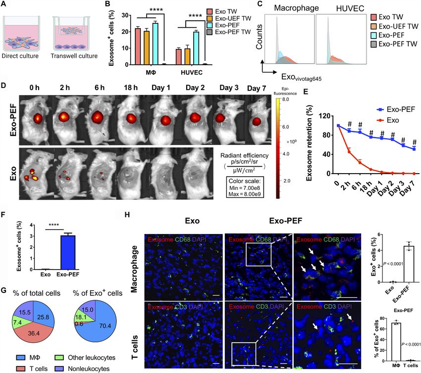
SCIENCE ADVANCES | RESEARCH ARTICLE
yet the hydrophilicity of the material surfaces was increased (fig. S1, noted that the gene expression of the M2 marker CD206 and the
D and E). MSC exosomes were analyzed for purity to show exosome anti-inflammatory cytokines including IL-4 and IL-10 were en-
makers and the absence of cellular or serum contaminants (tables hanced by exosome-functionalized scaffolds, possibly because of
S1 and S2). Vesicles had the classic “cup-shaped” morphology the added influence of the scaffold’s microenvironment (Fig. 1,
(Fig. 1B), with an average diameter of 76.7 ± 25.4 nm and zeta F to H) (22, 23).
potential of −30.8 ± 9.8 mV. The PEI-modified electrospun fibers To understand the delivery of exosomes from scaffolds to cells,
(PEFs) became positively charged and enabled tethering of MSC M cell line RAW 264.7 or human umbilical vein endothelial cells
exosomes to PCL fiber surfaces as a result of the negative potential (HUVECs) were seeded on Exo-PEF scaffolds and compared to the
of exosome membranes (Fig. 1, A to D). The amounts of exosomes counterpart system where cells were cultured in the bottom cham-
immobilized onto PEF were controllable and plateaued to reach a bers in separation with the scaffold materials placed in the upper
maximum level that the PCL scaffold could carry (Fig. 1E). The transwell (Fig. 2A). When the labeled exosomes were examined,
binding between MSC exosomes and PCL scaffolds was found to be only those cells that were cultured directly on the surface of Exo-PEF
stable in the buffer solutions, only showing a minimal initial de- scaffolds showed exosome+ signals (Fig. 2, B and C). The results indi-
tachment of the exosome residuals (fig. S1F). PEF bearing MSC cated that exosomes immobilized onto scaffolds would not be released
exosomes (Exo-PEF) was then compared to suspended exosome unless they were in direct contact with cells and then sequestered by
vesicles. Notably, being tethered to the polyester surface did not them, suggesting a possible interactive process involved in exosomes
compromise the function of MSC exosomes to modulate M to- reaching to cells. The two control groups, with exosomes applied to
ward the immunomodulatory M2-like phenotype in vitro. It was the upper chamber with or without plain unmodified electrospun
Downloaded from http://advances.sciencemag.org/ on July 17, 2021
Fig. 1. MSC exosome–functionalized fibrous scaffolds showed immunomodulatory effects on Ms cultured in vitro. (A) Schematic of immobilizing exosomes to
polycaprolactone (PCL) scaffolds through electrostatic interaction. (B) TEM imaging of MSC exosomes. (C) Scanning electron microscopy (SEM) imaging of Exo-PEF fibers.
(D) Transverse (left) and vertical (right) imaging of PCL fibers modified with DI-labeled exosomes visualized via red fluorescence. (E) Quantities of exosomes immobilized
onto PEF in variation with the initial exosome quantities added for reaction and comparison to the unmodified substrates (UEF). (F) Lipopolysaccharides (LPS)–stimulated bone
marrow–derived Ms (BMDMs) cultured on Exo-PEF. (G) Expression of M1 M-related genes (CD86, CD38, IL-1, and TNF-) and (H) M2 M-related genes (CD206, Egr2,
IL-10, and IL-4) in the LPS-stimulated Ms treated with suspended exosomes (Exo), PEF, or Exo-PEF; BMDMs with or without LPS stimulation cultured on tissue plates
serving as +LPS and –LPS controls, respectively. Exosome dosage: 10 g of protein mass (n = 5). Scale bars, 50 nm (B), 2 m (C), and 100 m (D, left)/20 m (D, right).
*P < 0.05, **P < 0.01, ***P < 0.001, and #P < 0.0001.
Su et al., Sci. Adv. 2021; 7 : eabf7207 12 May 2021 2 of 12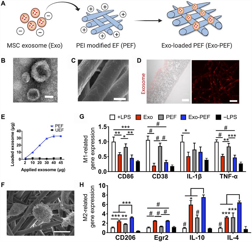
SCIENCE ADVANCES | RESEARCH ARTICLE
Downloaded from http://advances.sciencemag.org/ on July 17, 2021
Fig. 2. The uptake of exosomes from scaffolds was mediated through cell contact, and Exo-PEF prolonged exosome retention, showing dominant exosome
uptake by Ms in wounds. (A) Schematic of studying the exosome uptake where cells were either directly seeded on or separated from Exo-PEF through the transwell
culture. (B and C) Using liquid exosomes directly supplied to the upper chamber of the transwells with or without UEF as control groups (Exo TW and Exo-UEF TW), the
exosome uptake by Ms and HUVECs cultured on Exo-PEF or without the contact of Exo-PEF (Exo-PEF TW) was compared: (B) percent exosome-positive cells and (C)
representative flow cytometry histograms. (n = 5) (D) In vivo imaging of fluorophore-labeled exosomes in tissues receiving subcutaneous exosome injection (Exo) or
Exo-PEF implantation and (E) comparison of exosome retention over 7 days based on quantitative analysis. Exosome dosage: 15 g of protein mass. (F) Percentages of
exosome+ cells in tissues harvested on day 7 after injury. (G) Average percent distributions of different cell types in total cells or in pregated exosome+ cells as analyzed
by flow cytometry (n = 5). (H) Fluorescent imaging and statistic calculation of the exosome (red) uptake by M (CD68 in green) or T cells (CD3 in green) in the wounds on
day 7; arrows show the exosome colocalization with CD68 receptors on M cell surfaces but no overlap with T cells. Scale bar, 20 m. # or ****P < 0.0001. Blue, DAPI
showing cell nuclei.
fibers (UEFs), indicated that exosomes were able to permeate the subcutaneously injected exosomes were undetectable after 24 hours
transwell membrane and reach cells at the bottom chamber if exo- in vivo, whereas the exosome signals persisted in the Exo-PEF
somes were not immobilized to scaffolds. group, with 50.8 ± 5.8% fluorescence remaining on day 7 versus day 0
To investigate the cellular uptake of exosomes in vivo, the (Fig. 2, D and E). Specific cell types in exosome+ cells in the Exo-PEF
wounded skin tissues in mice were treated with Exo-PEF and har- group were divided and identified in four groups: M (CD45+F4/80+),
vested on day 7 after injury. The exosome retention was monitored T cells (CD45+CD3+), other leukocytes (CD45+CD3–F4/80–), and non-
through the labeled fluorescent exosomes. It was shown that the leukocytes (CD45–). In general, about 3% of the cells in the harvested
Su et al., Sci. Adv. 2021; 7 : eabf7207 12 May 2021 3 of 12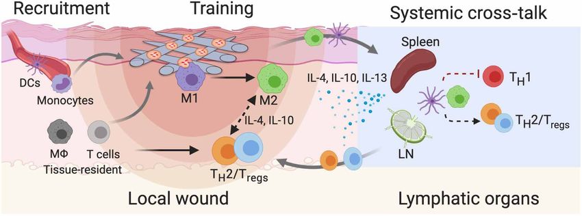
SCIENCE ADVANCES | RESEARCH ARTICLE
tissue carried the exosome fluorescent signals (Fig. 2F). Notably, an early and most significant dominance of immunomodulatory
Ms accounted for 70.4 ± 4.8% of the exosome+ cells, with the M M2-like Ms on day 7 (Fig. 3E). To validate the effects of Exo-PEF,
population at the level of 25.8 ± 2.5% within the wounds on day 7. scaffolds were also studied in a subcutaneous implantation model in
With a total cell population comparable to M, T cells carrying the the nonpathological skin tissue in mice. Analyses showed similar
exosome signals, however, were at a much lower level, making only proinflammatory- and immunomodulatory-biased M responses
3% of the exosome+ cell population (Fig. 2G). Immunostaining and in vivo resulting from PEF and Exo-PEF implantation, respectively
microscopic imaging validated the flow cytometry identification re- (fig. S2, A to D). In that model, Exo-PEF also mitigated foreign body
sults: Exosomes were found to colocalize with CD68+ Ms rather reaction to the implanted polyester scaffolds (fig. S2E).
than CD3+ T cells (Fig. 2H). Other types of leukocytes and non- T cells constitute an active group of adaptive immune cells during
leukocytes had similar levels on taking up exosomes from Exo-PEF the skin wound-healing process. Tregs were particularly reported to
scaffolds according to quantitative analyses, and each category con- be essential for the hair follicle regeneration (24, 25). By looking at
tributed to lower than 20% of the exosome+ cells (Fig. 2G). the pan-T cell subsets in different groups, it was observed that CD4+
TH2 and CD4+CD25+FoxP3+ Tregs were two pronounced groups in
Immunomodulatory responses induced in local tissue the Exo-PEF–treated wounds. Notably, Exo-PEF promoted the
To understand the immunomodulatory effects of Exo-PEF scaffolds presence of Tregs in higher than twofold quantity compared to
in vivo, Exo-PEF materials were studied in skin wounds in mice with untreated wounds on day 7 (Fig. 3, G and H). Moreover, at this time point,
the sham, injected exosomes (Exo) and blank PEF controls for over Exo-PEF also led to a higher portion of the T cells (18.6 ± 6.4%) secreting
2 weeks (Fig. 3A). As shown in Fig. 3 (B and E), the implantation of the anti-inflammatory cytokine IL-10 (fig. S3A), with most CD4+ cells
the scaffold materials, PEF or Exo-PEF, increased the number of Ms and all Tregs identified to be IL-10+ (fig. S3, B and C). Without exosomes,
Downloaded from http://advances.sciencemag.org/ on July 17, 2021
(Fig. 3B), which was most pronounced on day 7. The M response the blank PEF slightly increased the total number of T cells (Fig. 3F)
was differentiated through the analysis of M subtypes: PEF treat- that did not contain the beneficial CD4+ T cells or Tregs (Fig. 3, G and H),
ment significantly led to the accumulation of the proinflammatory, suggesting the possible presence of other T cell subtypes, such as CD8
CD86+ M1-like Ms at all three time points (Fig. 3C), while Exo- or T cells in the wounds treated with PEF alone.
PEF resulted in a onefold greater number of immunomodulatory,
CD206+ M2-like Ms compared to wound controls on days 3 and Remote adaptive immune responses induced
7 (Fig. 3D). In comparison to M2/M1-like M ratios, M polariza- in lymphatic organs
tion from the M1-like (day 3) to the M2-like (day 7 and beyond) The TH1 and TH2 lymphocytes expressing the CD4 co-receptors are
subtype was evident in all groups, with the Exo-PEF group inducing important adaptive cells by releasing T cell cytokines to mediate
Fig. 3. Exo-PEF promoted the M2 M subtype and Treg populations in the skin wound model. (A) Full excisional skin wounds were treated with injected exosomes,
PEF, or Exo-PEF. Exosome dosage: 30 g of protein mass. Analyses of immune cell numbers per milligram of tissue (with untreated wounds as the negative control):
(B) total Ms, (C) CD86+ M1 Ms, (D) CD206+ M2 Ms, (E) ratio of CD206+ M2 versus CD86+ M1 M, (F) total T cells, (G) CD4+ T cells, and (H) CD4+CD25+FoxP3+ Tregs on
days 3, 7, and 14 after injury. n = 6. *P < 0.05, **P < 0.01, ***P < 0.001, and ****P < 0.0001.
Su et al., Sci. Adv. 2021; 7 : eabf7207 12 May 2021 4 of 12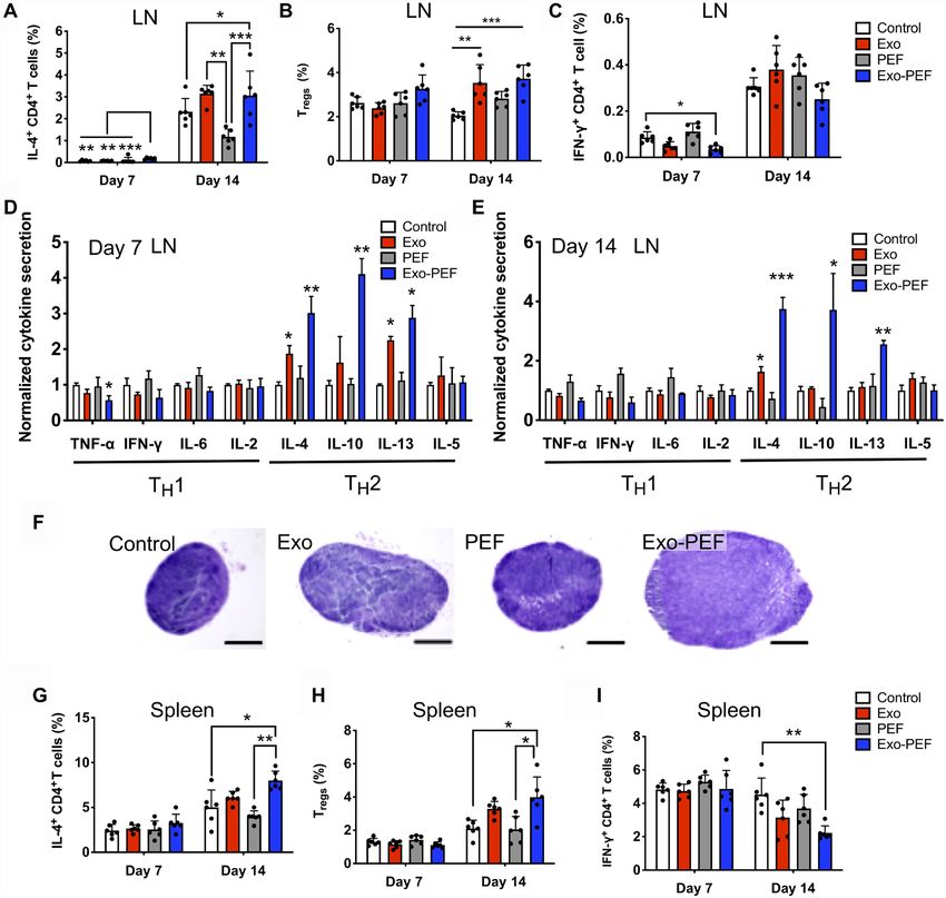
SCIENCE ADVANCES | RESEARCH ARTICLE
inflammation and tissue remodeling. In analyzing inguinal lymph (2.9-fold) and decreased that of TNF- (1.8-fold) and IFN- (1.6-fold);
nodes (LNs) near skin wounds, IL-4–secreting CD4+ TH2 cells the trend continued on day 14, showing the elevated secretion of
seemed to emerge in the Exo-PEF group on day 7, albeit at a low IL-4 (3.8-fold), IL-10 (3.7-fold), and IL-13 (2.6-fold) (Fig. 4E). The
level (Fig. 4A); the injected or immobilized exosomes both induced TH2 cytokine responses were less potent in the Exo group on day 7
higher populations of CD4+IL4+ TH2 and CD4+CD25+FoxP3+ Treg and almost unnoticeable on day 14 except for the secretion of the
responses on day 14 (Fig. 4, A and B). The Exo-PEF treatment also IL-4 cytokine (Fig. 4D). The hypertrophy of the LNs in the Exo and
demonstrated a trend to mitigate the IFN-+ TH1 cell number in the Exo-PEF groups was observed on day 14, indicating immune acti-
wounds, with the effect most noticeable on day 7, showing a more vation in the LNs (Fig. 4F).
than 50% reduction of this cell population compared to the wound In the spleen, only the Exo-PEF treatments induced the respons-
control (Fig. 4C). es on day 14 similar to those in the LNs, showing IL-4–secreting
The cytokine array experiments further suggested the activation CD4+ T cells and Tregs in higher numbers and IFN-+CD4+ T cells
of the TH2 immune responses induced by the immobilized or liquid being decreased (Fig. 4, G to I). To understand the origin of TH2
MSC exosomes, with the stronger effects observed in the former. immunoactivation and the possible immunogenic effects of any
Specifically, in comparison to untreated wounds on day 7, Exo-PEF exosome components diffusing into the tissue or organs, T cells
promoted the secretion of IL-4 (3.0-fold), IL-10 (2.9-fold), and IL-13 were isolated from the LNs harvested from the Exo and Exo-PEF
Downloaded from http://advances.sciencemag.org/ on July 17, 2021
Fig. 4. Exo-PEF activated the TH2 immune response in lymphatic organs. T cell subtypes were analyzed in lymph nodes (LN) and spleen following skin excisional
wounds and treatments. (A to C and G to I) Percent analyses of IL-4+CD4+ T cells, CD4+CD25+FoxP3+ Tregs, and IFN-+CD4+ T cells in the inguinal LNs (A, B, and C, respec-
tively) and spleen (G, H, and I, respectively) (n = 6; *P < 0.05, **P < 0.01, and ***P < 0.001). (D and E) Cytokine array studies on supernatants produced by ex vivo LN cells
isolated from the LNs harvested on day 7 (D) and day 14 (E) after injury. (F) Representative hematoxylin and eosin (H&E)–stained images of the LNs harvested from mice
(scale bar, 500 m).
Su et al., Sci. Adv. 2021; 7 : eabf7207 12 May 2021 5 of 12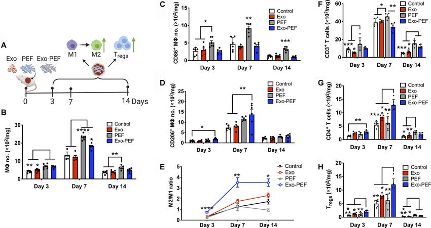
SCIENCE ADVANCES | RESEARCH ARTICLE
hosts at week 2. Dosing MSC exosomes directly in vitro failed to membranous vesicles (Fig. 2). One direct benefit is the prolonged
stimulate the proliferation peak from these T cells ex vivo, suggest- retention of exosomes in effecting their function in the hostile injury
ing that the adaptive immunomodulation in vivo induced by environment. Tethering exosomes through electrostatic interac-
Exo-PEF was not due to any inherent immunogenicity of MSC tions seemed to be reversible, but in an interactive way involving
exosomes (fig. S4). the active uptake by cells (Fig. 2, A to C). The delivery may therefore
be considered a dynamic “on-demand” process, and this is attrac-
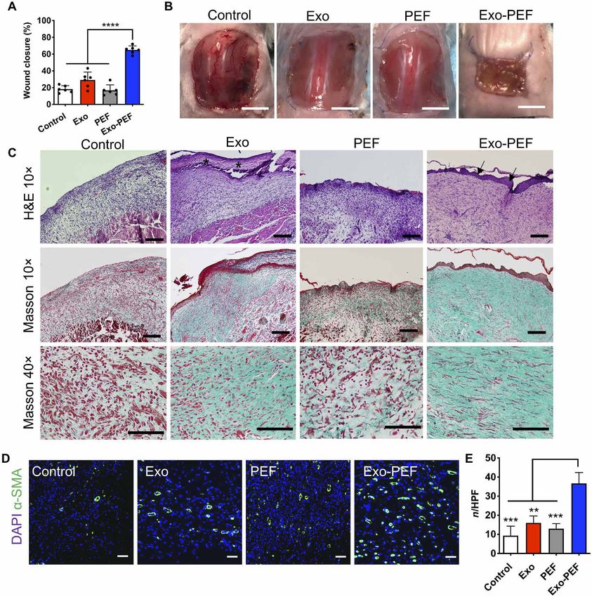
Effects of exosome-laden scaffolds on healing large tive and in contrast with the passive transport through diffusion, as
skin wounds shown in the previous exosome-laden or many drug delivery systems
To understand the pro-regenerative therapeutic effects of Exo-PEF (28–30). Another interesting property of Exo-PEF scaffolds was the
scaffolds, large square skin excisional wounds with an area of synergy between materials and exosomes, as observed in promoting
225 mm2 were created on the dorsal skin of mice. By measuring the the beneficial immunomodulation. In the experiments, the stand-
wound areas from day 0 to week 2, we found that Exo-PEF scaffolds alone PEF scaffolds increased the expression of anti-inflammatory
significantly accelerated wound closure (65.1 ± 5.3%) compared to cytokines and the M2 marker in vitro (Fig. 1H), presumably be-
untreated wounds (18.2 ± 4.7%), treatments of injected exosomes cause of the topological cues of fibrous materials (31, 32). The effects,
(29.2 ± 10.5%), and blank PEF scaffolds (16.8 ± 7.2%) (Fig. 5, however, were not translated at the wound sites, as PEF scaffolds
A and B). Histological analysis was performed to evaluate wound recruited M and T cells locally, but leaning toward the M1-biased
reepithelialization and collagen deposition through hematoxylin M and T cells likely in favor of inflammation. Increasing the pop-
and eosin (H&E) and Masson’s trichrome (MT) staining, respec- ulation of immune cells nevertheless is pivotal in the circumstance
tively. Histology images were acquired near the wound margin as of exosomes with the presence of PEF, as immune cells recruited by
Downloaded from http://advances.sciencemag.org/ on July 17, 2021
shown in Fig. 5C. Reconstitution of epidermal covering was ob- materials could be turned around showing the beneficial responses
served in the Exo-PEF group, while the Exo group exhibited a fi- (Fig. 3). Exo-PEF scaffolds therefore synergized the respective ef-
brin-like granulocyte-rich tissue surface. In comparison, other fects of scaffolds and exosomes as the “recruiter” and “trainer” for
groups showed no epidermal covering. Granulation tissues (GTs) immune cells in the process (Fig. 6).
were formed in the wound bed in all groups, but morphology varied. In the context of tissue repair and regeneration, the immune re-
GT in Exo-PEF was much thicker than that in other groups and sponses across different organisms and tissues are divergent in
exhibited pinkish color in H&E staining, indicating a healthier terms of suppression and activation. This would affect the inflam-
GT formation. In addition, Exo-PEF allowed collagenous extracel- matory and angiogenic processes as well as the progenitor activities
lular matrix deposition, with elongated fibroblasts resembling tis- and the regeneration outcome of a tissue (33–35). In our study, it
sue organization in the normal skin. In comparison, in control and was shown that the tissue reaction to Exo-PEF scaffolds started with
PEF groups, there was minimal collagen deposition (Fig. 5C). The the modulation of the M response manifested in the M2-biased
wound beds in the Exo-PEF group also contained a significantly polarization (Fig. 2, B to E). The increased population of the M2-like
higher number of blood vessels that were stained positive for M subtype has been recognized as a crucial event (35), and a de-
-smooth muscle actin (-SMA) than that in the other groups layed transition from the proinflammatory M1-like to immuno-
(Fig. 5, D and E). modulatory M2-like phenotypes would cause fibrosis and scarring
reactions to inhibit the regeneration of bone, muscle, heart, and
the skin tissue (33–35). Exo-PEF scaffolds also notably increased
DISCUSSION the adaptive immune responses both locally and remotely (Figs. 3,
Investigation of immune responses following implantation of bio- F to H, and 4). On the time scale, the adaptive responses were gen-
materials in the body is a long-standing topic in developing bio- erally active on day 14 in a phase later than the peak of the macro-
compatible medical implants. Incorporation of biological components, phage response occurring on day 7. Treg differentiation and other
such as proteins, exosomes, and cells, into material scaffolds for remote adaptive responses could be induced by the cytokines se-
application in tissue engineering and regenerative medicine has creted from the innate immune cells, e.g., IL-10 and TGF- that
added another layer of complexity with respect to understanding communicate to T cells (36, 37). It was worth mentioning that in-
the host response and how it would affect the integration of the creases in dendritic cells (DCs), Ms, and IL-10–secreting cells
implants. In essence, the primary function of the immune system is were detectable within the LNs (fig. S5, A to D) and were accompa-
to defend the host and eliminate foreign invaders. Developing bio- nied by decreases in T cells including CD4+ T cells (fig. S5, E and F)
active constructs appropriately interacting with the immune system in mice treated by Exo-PEF scaffolds. Innate and adaptive cells may
therefore faces formidable challenges (26). In our experiments, it thereby exist trafficking in and out of the lymphatic organs (fig. S5).
was found that most of the cells absorbing exosomes from scaffolds Using technologies such as molecular imaging to trace immune cell
were immune cells (Fig. 2, F to H). This may also be relevant to trafficking may be carried out to further investigate the cell migra-
other types of biological scaffolds, implying the importance of im- tion pattern.
mune responses in tissue reactions and the necessity to understand Scaffolds carrying therapeutic exosomes may potentially offer a
and use this the mechanism to guide the biological communica- promising category of cell-free regenerative system as demonstrated
tion network. by the effects of Exo-PEF on healing large skin wounds in mice (Fig. 5).
Given the diversity of their components from proteins to micro Skin wounds as large as 225 mm2 in size are considered a critical
RNAs, exosomes carry a multitude of signals (27). Through proteomic skin injury that cannot self-heal properly despite the regeneration
profiling, both immunomodulatory factors and proangiogenic fac- capability of skin tissue. The prohealing effects observed for Exo-
tors could be detected in MSC exosomes (table S1). Binding exo- PEF are therefore substantial. Accelerated M transition from pro
somes to fibrous scaffolds were shown to store, localize, and deliver inflammatory to immunomodulatory phenotypes, increased local
Su et al., Sci. Adv. 2021; 7 : eabf7207 12 May 2021 6 of 12SCIENCE ADVANCES | RESEARCH ARTICLE
Downloaded from http://advances.sciencemag.org/ on July 17, 2021
Fig. 5. Exo-PEF healed large skin wounds in 2 weeks, showing wound closure, reepithelialization, collagen deposition, and blood vessel formation. (A) Percent
wound closures and (B) representative images of the wounds after 2-week treatments. (C) H&E and Masson’s trichrome (MT) histological staining of wound tissues after
2-week treatments. Asterisks, fibrin-like granulocyte-rich layer in the Exo group; arrows, neo-formed epidermis in the Exo-PEF group. Scale bars, 200 m (10×) and 100 m
(40×). (D) Fluorescent staining of -smooth muscle actin (-SMA) (green) and DAPI (blue) in the wound bed. Scale bar, 50 m. (E) Statistical calculation of -SMA–positive
blood vessel numbers per high-power field (HPF). Exosome dosage: 50 g of protein mass. n = 6. **P < 0.01, ***P < 0.001, and ****P < 0.0001.
T reg population, and activation of TH2 response all favor anti- to identify the complex and versatile populations of immune cells,
inflammatory events to benefit the healing. In addition, the signals as well as other niche cells such as endothelial cells, fibroblasts, and
conveyed through the anti-inflammatory cytokines including IL-4, progenitor cells. The techniques may include mass cytometry with
IL-10, and IL-13 have recently been expanded in the regenerative the heavy metal–labeled antibodies or single-cell sequencing to
biology by maintaining the trophic factors such as insulin-link reveal the dynamics of the cell transcriptome. Especially, high-
growth factor, vascular endothelial growth factor, TGF-, and throughput techniques can be applied to characterize the plasticity
amphiregulin, which are essential to promoting progenitor activity and dynamics of the M population in different stages of tissue
(38–40). healing, as it has been recognized that the classically activated M1
Despite the potential of exosome-functionalized scaffolds pro- phenotype and the alternatively activated M2 phenotype cannot
viding an effective and potent immunomodulatory system, the accurately reflect the polarization and complexity of M subtypes
cellular communication network between the immunomodulation during tissue responses (41). Moving forward to clinical applica-
and regeneration processes is still to be elucidated in detail (Fig. 6). In tions, key components in MSC exosomes contributing to the im-
this regard, high-throughput cell-level analyses need to be adopted munomodulation effects need to be further identified. Studies should
Su et al., Sci. Adv. 2021; 7 : eabf7207 12 May 2021 7 of 12SCIENCE ADVANCES | RESEARCH ARTICLE
Fig. 6. Possible immunomodulation mechanisms involved in Exo-PEF materials promoting tissue repair may include (i) the recruitment and training of immune
cells, (ii) the action of anti-inflammatory mediators, and (iii) the effects from remote immune organs. M1, M1-like M; M2, M2-like M; Tregs, regulatory T cells;
TH1/2, T helper 1/2 cells; DCs, dendritic cells; LN, lymph nodes.
also be conducted to further rule out the possibility of exosome Cell isolation and culture
immunogenicity, especially among individuals or even across species. All cells were cultured at 37°C under humidified atmosphere with
Downloaded from http://advances.sciencemag.org/ on July 17, 2021
To conclude, immobilizing exosomes to polymeric fibrous scaf- 5% CO2. The MSCs were cultured in basal medium supplemented
folds generated multifunctional scaffolds capable of storing biologics with 10% fetal bovine serum (FBS) and 0.4% penicillin-streptomycin
and delivering exosome signals. The synergy between scaffold ma- (P/S). MSCs between passages 5 and 9 were used for the following
terials and exosomes was found effective to proactively recruit im- experiments. HUVECs were maintained in ECM supplemented
mune cells and modulate orchestrated M2/TH2/Treg responses with 5% FBS, 1% P/S solution, and 1% ECGS. RAW 264.7 Ms
locally and on the systemic level to benefit tissue repair. The study were cultured in DMEM supplemented with 10% FBS and 1%
lays the foundation for establishing the principles of designing bio- P/S. To isolate the primary macrophages, the bone marrow cells
logical scaffolds with immunomodulatory functions for regenera- from the femur and tibia of Balb/c mice were harvested and cultured
tive medicine. in the RPMI medium supplemented with 0.25 mM -mercaptoethanol,
1× nonessential amino acid, 10% heat-inactivated FBS (incubated at
56°C in water bath for 30 min), 1% P/S, and M-CSF (20 ng/ml). The
MATERIALS AND METHODS cell culture media were changed every other day. The bone marrow–
Materials and reagents derived macrophages (BMDMs) were stained by the CD11b and F4/80
The ultrapure water (≥18 megohms) was prepared from the deionized antibody and analyzed by flow cytometry at day 7, showing >90%
(DI) water through the Milli-Q system (Millipore, Billerica, MA). purity (fig. S6).
PCL (CAS: 24980-41-4) and lipopolysaccharide (LPS) were obtained
from Sigma-Aldrich (Milwaukee, WI). 1-Ethyl-3-(3-dimethylami- Isolation and characterization of MSC exosomes
nopropyl)carbodiimide (EDC) and N-hydroxysuccinimide (NHS) Exosomes were isolated from BM-MSCs from Balb/c mice. Cells at
were purchased from Pierce Biotechnology (Rockford, IL, USA). 80% confluency were washed with PBS and changed to serum-free
The branched PEI with the average Mw of 70 kDa was from Macklin medium. Supernatants were collected after 24 hours of incubation,
(Shanghai, China). The TRIzol kit, Dulbecco’s phosphate-buffered followed by centrifugation at 1200g for 15 min and 10,000g for
saline (DPBS), RPMI 1640 medium, Dulbecco’s modified Eagle’s me- another 30 min to remove cell debris. Exosomes were precipitated
dium (DMEM), and other cell culture reagents were purchased from by ultracentrifugation at 100,000g for 1 hour and then washed twice
Invitrogen (CA, USA) unless otherwise specified. 4′,6-Diamidino-2- with PBS. The precipitated exosomes were resuspended in 200 l of
phenylindole (DAPI) was obtained from Thermo Fisher Scientific PBS and stored at −80°C until further use. The protein contents of
(MA, USA). HUVECs, endothelial cell medium (ECM), and endothelial the collected exosomes were measured using the Micro BCA Protein
cell growth supplements (ECGS) were purchased from ScienCell Assay Kit (Thermo Fisher Scientific). For morphology observation,
(Carlsbad, CA). The mouse bone marrow MSCs (BM-MSCs) and the exosomes were fixed with 4% paraformaldehyde (PFA), stained
basal culture medium were purchased from Cyagen Biosciences Inc. with 2% (w/v) uranyl acetate (Sigma-Aldrich) for 5 min, washed
(CA, USA). The quality control report provided by the vendor for three times with DI water, and air-dried overnight. The exosome
this MSC product included the absence of endotoxin, >98% cell morphologies were observed with a transmission electron micro-
purity, and the adipogenic, osteogenic, and chondrogenic differen- scope (TEM) (Tecnai G2 T20, Thermo Fisher Scientific). The purity
tiation potential. The murine macrophage cell line RAW 264.7 was of the collected exosomes was analyzed on the basis of the standards
obtained from the Cell Culture Center of the Institute of Basic Med- suggested by the International Society for Extracellular Vesicles
ical Sciences (Beijing, China). Growth factors including macrophage (42) and showed the presence of positive exosomal markers without
colony-stimulating factors (M-CSFs) and cytokine IL-4 were pur- contamination because of other cellular organelles or culture medium.
chased from PeproTech (Canada) unless otherwise specified. The (tables S1 and S2).
flow cytometry antibodies and reagents were purchased from For mass spectrometry (MS), exosomes were treated by lysing
BioLegend (USA) unless otherwise specified. The suppliers of other buffer solutions [6% SDS, 200 mM dithiothreitol, and 200 mM tris-
chemicals, biological reagents, and equipment were specified below. HCl (pH 7.6)] at 95°C for 5 min followed by SDS–polyacrylamide
Su et al., Sci. Adv. 2021; 7 : eabf7207 12 May 2021 8 of 12SCIENCE ADVANCES | RESEARCH ARTICLE
gel electrophoresis. For protein identification, Coomassie-stained relative humidity using the sessile drop method on a contact angle
total aggregated proteins from each sample were cut out of the gel analyzer (USA Kino Industry Co. Ltd.).
and destained with a solution of 100 mM ammonium bicarbonate For sterilization, the EF membranes were rinsed with 75% ethanol
in 50% acetonitrile. After dithiothreitol reduction and iodoacetamide solutions for 30 min, washed with sterilized DI water three times, and
alkylation, the proteins were digested with porcine trypsin (Promega, air-dried in a biosafety cabinet (Thermo Fisher Scientific, Germany).
Madison, WI) overnight at 37°C. The resulting tryptic peptides EF scaffolds were further sterilized by exposure to ultraviolet radia-
were extracted from the gel pieces with 80% acetonitrile and 0.1% tion for 30 min on each side in a biosafety cabinet and were kept at
formic acid (FA). The samples were dried in a vacuum centrifuge 4°C until use. To immobilize MSC exosomes, the prescribed amount
concentrator and resuspended in 0.1% FA. The resulting peptides of exosomes was suspended in 1 ml of PBS, added to PEF scaffolds
were loaded to Thermo Orbitrap Fusion Lumos, and full MS spectra pretailored into appropriate sizes and shapes, and incubated for
were acquired in an Orbitrap mass analyzer. The MS data were 30 min at 4°C on a rotator. The mass of exosomes immobilized onto
aligned with the Escherichia coli Reviewed Swiss-Prot database by scaffolds was determined by measuring the difference of protein con-
the Proteome Discoverer 2.2 software. tents in PBS solutions before and after the binding reaction. The amount
of exosome added in each experiment was lower than the maximal
Fabrication and characterization of exosome-laden loading capacity of PEF to ensure the full binding of exosomes in the
PEF scaffolds solution to the scaffold to reduce the waste of exosomes. To visualize
The electrospun fibrous scaffolds were prepared from 21% (w/v) exosome loaded to PEF scaffolds, exosomes were incubated with 0.1 mM
PCL solutions in a mixture of CHCl3 and dimethylformamide at a 1,1′-dioctadecyl-3,3,3′,3′-tetramethylindocarbocyanine perchlorate (DiI)
volume ratio of 9:1. After stirring for 3 hours until the PCL was solutions (Invitrogen) and washed with PBS three times to remove
Downloaded from http://advances.sciencemag.org/ on July 17, 2021
completely dissolved, the solution was loaded into a syringe with a excessive dye molecules. The DiI-labeled exosomes were loaded to
blunt-ended stainless 21-gauge steel needle. The feeding rate and PEF scaffold and imaged via confocal microscopy (Zeiss) after em-
the applied voltage were controlled at 2 ml/hour and 10 kV, respec- bedding in the Tissue-Tek O.C.T. compound (VWR) and cryosection.
tively. The electrospun fibers were collected on a flat steel plate cov-
ered with a foil paper at a distance of 14 cm from the needle tip. The Exosome uptake and retention
EF membranes with a thickness of 80 to 90 m were collected and Transwell culture
manually trimmed into round shapes with a diameter of 12 mm and RAW 264.7 cells or HUVECs were seeded on the bottom chamber
mass at 2.7 ± 0.3 mg. Scaffolds of this size were used in all experi- of the transwell microplates, which contained a 0.4-m porous
ments except those for experiments in the large skin injury model. polyester membrane between the two chambers. Ten micrograms
To immobilize exosomes onto PCL EF scaffolds, the tailored scaf- of fluorophore-labeled exosomes were added to the upper chamber
fold membrane was immersed in a mixture of 1 M aqueous NaOH with or without the unmodified EF scaffolds as two control groups.
solution and 75% ethanol solution at a ratio of 1:1 (v/v) for 30 min One purpose of including the UEF scaffold in the upper chamber
with gentle agitation at room temperature. The treatment was to was to determine whether the presence of EF scaffolds would affect
enhance the PCL EF hydrophilicity and to generate carboxyl groups the permeation of free exosomes through the transwell membrane.
on the fiber surface via hydrolysis. Scaffolds were washed thoroughly The same amount of exosome was loaded to PEF scaffolds, which
afterward with DI water. were then placed in the upper chamber. RAW 264.7 cells or HUVECs
To covalently conjugate PEI, 60 pieces of NaOH-treated circular were also directly seeded to the Exo-PEF. After culture for 24 hours,
scaffolds were incubated in 25 ml of 0.1 M MES buffer (pH 5.1) cells were collected and flow cytometry analysis was performed to
containing 9.3 mM EDC and 5.8 mM NHS for 2 hours on a rotary determine cellular uptake of exosome.
shaker at room temperature. The 70-kDa branched PEI was dis- Exosome retention and uptake in vivo
solved in 25 ml of 0.2 M MES buffer (pH 6.5) to prepare a 3.5 mM The full excisional wounds with a diameter of 8 mm were created
PEI solution, which was added to the EFs and EDC/NHS reaction with a biopsy puncher on the dorsal skin of Balb/c mice under
and shaken for another 2 hours. The EDC:NHS:PEI molar ratio was isoflurane anesthesia (more details were described in the “Animal
at 8:5:3. PEF was washed four times using excessive 1 M NaCl solu- wound models and wound treatments” section). Exosomes were
tions to remove the physically adsorbed PEI and then multiple covalently labeled with Vivo Tag 645 (PerkinElmer), and 15 g of
times with water for 2 days. fluorescence-labeled exosomes was either subcutaneously injected
To confirm the success of PEI conjugation, the 2,4,6-trinitrobenzene at three points around the wound or immobilized onto round PEF
sulfonic acid assay (Sigma-Aldrich) was performed to detect amine (d = 12 mm) and applied on top of the wound. The 3M Tegaderm
groups on PEF fiber surfaces by forming of a highly chromogenic transparent wound dressing was used to cover the wound. Mice
(orange) product. Conjugation to EF scaffolds was also characterized under isoflurane anesthesia were imaged at multiple time points up
by x-ray photoelectron spectroscopy (AXIS Supra, Kratos Analytical to day 7 using the IVIS 200 Optical Imaging System (Xenogen, Caliper
Ltd., UK). The UEF and PEF samples (d = 12 mm) were directly Life Sciences). The wound area was selected as the region of interest
mounted on the test stage with a total acquisition time of 15 min for (ROI), and the radiant efficiency was calculated within the ROIs.
each sample. The overlapping peaks were resolved using the peak Exosome retention (%) at a time (t) was calculated by comparing the
synthesis method by applying the Gaussian peak components after radiant efficiency at t to that at 0 h (t0) according to the following
Shirley-type background subtraction. The morphology of the EF formula: [Radiant efficiency (t)]/[Radiant efficiency (t0)] × 100%.
materials in each modification steps was characterized with a scan- At day 7, wound tissues were harvested, digested into single-cell sus-
ning electron microscope (SEM) (Quanta FEG450, FEI, USA). Water pensions, stained with F4/80, CD45, and CD3 antibodies, and ana-
contact angle measurements of PCL EF scaffolds before and after lyzed by flow cytometry to determine exosome uptake by macrophages
PEI modification were conducted at room temperature under 60% (CD45+F4/80+) and T cells (CD45+CD3+). (Details for single-cell
Su et al., Sci. Adv. 2021; 7 : eabf7207 12 May 2021 9 of 12SCIENCE ADVANCES | RESEARCH ARTICLE
preparation and flow cytometry staining were described in the “Flow wound treatments were prepared similarly as described above ex-
cytometry analyses” section.) The skin wound tissues were embedded cept that a higher dose of 50 g of exosomes loaded to 230-mm2
in O.C.T., cryosectioned, and stained with primary antibody rabbit square EF scaffolds was applied to each wound. The digital photo-
anti-mouse CD68 or rabbit anti-mouse CD3 followed by fluorescein graphs of wounds were taken at day 14. The area of the wounds was
isothiocyanate (FITC) goat anti-rabbit secondary antibody staining. measured and calculated by ImageJ software. The percent of wound
Nuclei were stained with DAPI. The colocalization of exosome and cell closures was calculated on the basis of the average wound area (A)
markers was examined to identify cells that sequestered exosomes. measured on days 0 and 14 and the following equation: (Aday0 −
Aday14)/Aday0 × 100%. Wound tissue was harvested for histological
Macrophage modulation assays in vitro analyses after mice were euthanized at day 14.
BMDMs were activated with LPS (100 ng/ml; Sigma-Aldrich, L2880),
seeded on 48-well tissue culture plates at a density of 2 × 10⁵ cells Flow cytometry analyses
per well, and then supplemented with or without 10 g of MSC exo- All flow cytometry data were collected using BD FACSAria II and
somes. An equal number of BMDM were also seeded on the pretailored analyzed using the FlowJo software. Gating strategies were listed in
PEF or Exo-PEF scaffolds (d = 12 mm) loaded with 10 g of MSC figs. S7 and S8.
exosomes. After 12 hours, cells were treated with a TRIzol kit and Explanted scaffolds or skin wound tissues were harvested, and
the total RNA was extracted and reverse-transcribed using TransScript the attached fat tissues were first removed before weighing the wound.
First-Strand cDNA Synthesis SuperMix (Transgene, Beijing, China). The skin samples were diced and digested with the Opti-MEM
Gene expression levels of cytokines (IL-10, IL-4, IL-1, and TNF-) (Invitrogen) solutions containing Liberase TM (100 ug/ml) (Roche),
and phenotypic markers (CD206, Egr2, CD86, and CD38) were an- collagenase D (1 mg/ml) (Roche), and hyaluronidase (1 mg/ml)
Downloaded from http://advances.sciencemag.org/ on July 17, 2021
alyzed by SYBR Green real-time polymerase chain reaction (PCR) (Sigma-Aldrich) for 30 min at 37°C on a shaker. The digested cell
using the CFX Connect Real-Time PCR Detection System (Bio-Rad, suspensions were poured through a 70-m cell strainer. The re-
Hercules, CA). The primers for qPCR were ordered from Sangon maining tissue chunks were minced with a syringe plunger and
(Shanghai, China), and the primer sequence was listed in table S3. washed with PBS containing 2% FBS, followed by lysis treatment of
The gene expression values in one sample were all normalized to the the red blood cells (BioLegend).
-actin level. Relative expressions were calculated using the com- The inguinal LNs were harvested, minced with a syringe plunger,
parative Ct method. The mean minimal cycle threshold values were and filtered through a 70-m cell strainer. The spleen was diced and
calculated from triplicate reactions. digested in collagenase D (2 mg/ml) (Roche) for 30 min at 37°C on
a rotator. Cell suspensions were filtered through a 70-m cell strainer
Animal wound models and wound treatments followed by red blood cell lysis (BioLegend). After washing, cells
All procedures were performed according to the regulations ap- were directly processed for flow cytometry staining.
proved by the Institutional Animal Care and Use Committee of Cell surface staining for flow cytometry experiments was per-
Peking University. Balb/c female mice (Vital River, Beijing, PR China) formed on ice in the dark. Cell viability staining was conducted
at 6 weeks old were anesthetized by isoflurane during procedure. using the Zombie Violet Fixable Viability Kit (BioLegend), followed
Subcutaneous implantation in nonpathological tissue by nonspecific binding blockage by anti-CD16/32 incubation. Cells
The scaffold membranes (d = 12 mm) of UEF, PEF, or Exo-PEF were then stained with surface antibody cocktail for 45 min on ice.
(with 30 g of exosomes loaded) were implanted into the subcuta- The surface markers of the macrophage population included CD45,
neous space beneath the abdomen skin. Free exosomes (30 g) were F4/80, and CD86, and those of T cells included CD45, CD3, CD4,
also injected subcutaneously. The injection sites were marked with and CD25. Cell suspension were then fixed and permeabilized with
an indelible marker pen for the tissue harvest at later time points. the Cytofix/Cytoperm Fixation/Permeabilization Kit (BD Biosciences)
Scissions and pockets were created in all mice regardless of the scaf- according to the manufacturer’s instructions. Intracellular markers,
fold implantation. After euthanizing the mice at day 14, implanted including CD206 for M2 macrophage or FoxP3 for Tregs, were stained
scaffolds, inguinal LNs, and spleen were harvested for immune cell for 45 min on ice. To analyze immune cell populations in the wounds,
analysis using flow cytometry. The mice with implants were also the cell numbers were normalized to wound weight, reported as cell
kept for up to 5 weeks and analyzed by H&E histology staining. number per milligram.
Excisional wound model To perform intracellular cytokine staining, cells from LNs were
For mechanistic immunomodulation studies, round full-thickness stimulated and prevented from cytokine secretion with the Cell
excisional skin wounds with a diameter of 8 mm were created using Activation Cocktail containing brefeldin A (BioLegend) for 5 hours
a biopsy puncher after hair removal from the dorsal skin. The scaf- at 37°C in RPMI medium supplemented with 10% FBS. Cell surfac-
fold membranes (d = 12 mm) of UEF, PEF, or Exo-PEF (with 30 g es were stained for CD3 and CD4, followed by intracellular staining
of exosomes loaded) were applied on top of the wounds. Free exo- of IL-4 and IFN-. For cytokine array analysis, cells isolated from
somes (30 g) were injected subcutaneously around the wounds for LNs were cultured in RPMI medium without FBS for 24 hours.
the Exo group. Untreated skin wounds were included as a control. Supernatants were collected and analyzed to determine the level of
3M Tegaderm transparent wound dressing was used to cover the TH1 (TNF-, IFN-, IL-6, and IL-2) and TH2 (IL-4, IL-10, IL-13,
wounds. Wound tissues, inguinal LNs, and spleens were harvested and IL-5) cytokines by LEGENDplex bead-based immunoassays
on days 3, 7, and 14 after euthanizing the mice. Analyses of immune according to the manufacturer’s instructions (BioLegend).
cell populations were performed using flow cytometry.
For wound-healing studies, the 225-mm2 square wounds were Histological analyses
created, with four corners sutured to the dorsal muscles to prevent Skin wound tissues were harvested and cut into two parts from the
skin contraction. The Exo-PEFs and exosome controls for the large middle line, fixed with 4% PFA, embedded in paraffin, and sectioned
Su et al., Sci. Adv. 2021; 7 : eabf7207 12 May 2021 10 of 12SCIENCE ADVANCES | RESEARCH ARTICLE
into 5-m-thick slices. Tissue slices were stained with H&E or MT host disease with a novel mesenchymal stromal cell product (MSC-FFM). Bone Marrow
staining, and images were captured with a light optical microscope Transplant. 53, 852–862 (2018).
15. C. Lo Sicco, D. Reverberi, C. Balbi, V. Ulivi, E. Principi, L. Pascucci, P. Becherini, M. C. Bosco,
(Olympus, Japan).
L. Varesio, C. Franzin, M. Pozzobon, R. Cancedda, R. Tasso, Mesenchymal stem cell–
For immunohistochemical staining, the harvested skin wound derived extracellular vesicles as mediators of anti-inflammatory effects: Endorsement
tissues were embedded in O.C.T., cryosectioned at a thickness of of macrophage polarization. Stem Cells Transl. Med. 6, 1018–1028 (2017).
8 m, and stained with primary antibody and rabbit anti-mouse 16. W. Chen, Y. Huang, J. Han, L. Yu, Y. Li, Z. Lu, H. Li, Z. Liu, C. Shi, F. Duan, Y. Xiao,
-SMA, followed by the FITC goat anti-rabbit secondary antibody. Immunomodulatory effects of mesenchymal stromal cells-derived exosome. Immunol.
Res. 64, 831–840 (2016).
Nuclei were stained with DAPI. Fluorescent images were taken with
17. S. K. Crain, S. R. Robinson, K. E. Thane, A. M. Davis, D. M. Meola, B. A. Barton, V. K. Yang,
a confocal microscope, and positive signals per high-power field A. M. Hoffman, Extracellular vesicles from Wharton's jelly mesenchymal stem cells
were counted. For quantitative image analysis, data from three ani- suppress CD4-expressing T cells through transforming growth factor beta and adenosine
mals, eight sections per animal, and four high-magnification images signaling in a canine model. Stem Cells Dev. 28, 212–226 (2019).
per section for each group were analyzed and compared. 18. B. Zhang, R. W. Y. Yeo, R. C. Lai, E. W. K. Sim, K. C. Chin, S. K. Lim, Mesenchymal stromal cell
exosome-enhanced regulatory T-cell production through an antigen-presenting
cell-mediated pathway. Cytotherapy 20, 687–696 (2018).
Statistical analyses 19. S. Zhang, S. J. Chuah, R. C. Lai, J. H. P. Hui, S. K. Lim, W. S. Toh, MSC exosomes mediate
Quantitative results were presented as means ± SD. Statistical com- cartilage repair by enhancing proliferation, attenuating apoptosis and modulating
parisons were performed by one-way analysis of variance (ANOVA) immune reactivity. Biomaterials 156, 16–27 (2018).
followed by Holm-Sidak tests to compare the selected data pairs. 20. T. Li, Y. Yan, B. Wang, H. Qian, X. Zhang, L. Shen, M. Wang, Y. Zhou, W. Zhu, W. Li, W. Xu,
Exosomes derived from human umbilical cord mesenchymal stem cells alleviate liver
The values of P < 0.05 were considered statistically significant. fibrosis. Stem Cells Dev. 22, 845–854 (2013).
Downloaded from http://advances.sciencemag.org/ on July 17, 2021
21. L. Hu, J. Wang, X. Zhou, Z. Xiong, J. Zhao, R. Yu, F. Huang, H. Zhang, L. Chen, Exosomes
SUPPLEMENTARY MATERIALS derived from human adipose mensenchymal stem cells accelerates cutaneous wound
Supplementary material for this article is available at http://advances.sciencemag.org/cgi/ healing via optimizing the characteristics of fibroblasts. Sci. Rep. 6, 32993 (2016).
content/full/7/20/eabf7207/DC1 22. N. Goonoo, Modulating immunological responses of electrospun fibers for tissue
engineering. Adv. Biosyst. 1, (2017).
View/request a protocol for this paper from Bio-protocol.
23. A. D. Schoenenberger, H. Tempfer, C. Lehner, J. Egloff, M. Mauracher, A. Bird, J. Widmer,
K. Maniura-Weber, S. F. Fucentese, A. Traweger, U. Silvan, J. G. Snedeker, Macromechanics
and polycaprolactone fiber organization drive macrophage polarization and regulate
REFERENCES AND NOTES
inflammatory activation of tendon in vitro and in vivo. Biomaterials 249, 120034 (2020).
1. L. Chung, D. R. Maestas Jr., F. Housseau, J. H. Elisseeff, Key players in the immune
24. A. N. Mathur, B. Zirak, I. C. Boothby, M. Tan, J. N. Cohen, T. M. Mauro, P. Mehta,
response to biomaterial scaffolds for regenerative medicine. Adv. Drug Deliv. Rev. 114,
M. M. Lowe, A. K. Abbas, N. Ali, M. D. Rosenblum, Treg-cell control of a CXCL5-IL-17
184–192 (2017).
inflammatory axis promotes hair-follicle-stem-cell differentiation during skin-barrier
2. A. L. Mescher, A. W. Neff, M. W. King, Inflammation and immunity in organ regeneration.
repair. Immunity 50, 655–667.e4 (2019).
Dev. Comp. Immunol. 66, 98–110 (2017).
25. A. Nosbaum, N. Prevel, H. A. Truong, P. Mehta, M. Ettinger, T. C. Scharschmidt, N. H. Ali,
3. M. Karin, H. Clevers, Reparative inflammation takes charge of tissue regeneration. Nature
M. L. Pauli, A. K. Abbas, M. D. Rosenblum, Cutting edge: Regulatory T cells facilitate
529, 307–315 (2016).
cutaneous wound healing. J. Immunol. 196, 2010–2014 (2016).
4. B. Chazaud, Macrophages: Supportive cells for tissue repair and regeneration.
Immunobiology 219, 172–178 (2014). 26. M. O. Dellacherie, B. R. Seo, D. J. Mooney, Macroscale biomaterials strategies for local
5. K. L. Spiller, T. J. Koh, Macrophage-based therapeutic strategies in regenerative medicine. immunomodulation. Nat. Rev. Mater. 4, 379–397 (2019).
Adv. Drug Deliv. Rev. 122, 74–83 (2017). 27. F. Gao, S. M. Chiu, D. A. L. Motan, Z. Zhang, L. Chen, H. L. Ji, H. F. Tse, Q. L. Fu, Q. Lian,
6. Z. Julier, A. J. Park, P. S. Briquez, M. M. Martino, Promoting tissue regeneration by Mesenchymal stem cells and immunomodulation: Current status and future prospects.
modulating the immune system. Acta Biomater. 53, 13–28 (2017). Cell Death Dis. 7, e2062 (2016).
7. D. Burzyn, W. Kuswanto, D. Kolodin, J. L. Shadrach, M. Cerletti, Y. Jang, E. Sefik, T. G. Tan, 28. K. Zhang, X. Zhao, X. Chen, Y. Wei, W. du, Y. Wang, L. Liu, W. Zhao, Z. Han, D. Kong,
A. J. Wagers, C. Benoist, D. Mathis, A special population of regulatory T cells potentiates Q. Zhao, Z. Guo, Z. Han, N. Liu, F. Ma, Z. Li, Enhanced therapeutic effects of mesenchymal
muscle repair. Cell 155, 1282–1295 (2013). stem cell-derived exosomes with an injectable hydrogel for hindlimb ischemia treatment.
8. Y. Dombrowski, T. O'Hagan, M. Dittmer, R. Penalva, S. R. Mayoral, P. Bankhead, S. Fleville, ACS Appl. Mater. Interfaces 10, 30081–30091 (2018).
G. Eleftheriadis, C. Zhao, M. Naughton, R. Hassan, J. Moffat, J. Falconer, A. Boyd, 29. K. Lv, Q. Li, L. Zhang, Y. Wang, Z. Zhong, J. Zhao, X. Lin, J. Wang, K. Zhu, C. Xiao, C. Ke,
P. Hamilton, I. V. Allen, A. Kissenpfennig, P. N. Moynagh, E. Evergren, B. Perbal, S. Zhong, X. Wu, J. Chen, H. Yu, W. Zhu, X. Li, B. Wang, R. Tang, J.'. Wang, J. Huang, X. Hu,
A. C. Williams, R. J. Ingram, J. R. Chan, R. J. M. Franklin, D. C. Fitzgerald, Regulatory T cells Incorporation of small extracellular vesicles in sodium alginate hydrogel as a novel
promote myelin regeneration in the central nervous system. Nat. Neurosci. 20, 674–680 therapeutic strategy for myocardial infarction. Theranostics 9, 7403–7416 (2019).
(2017). 30. M. Á. Brennan, P. Layrolle, D. J. Mooney, Biomaterials functionalized with msc secreted
9. Y. Liu, L. Wang, T. Kikuiri, K. Akiyama, C. Chen, X. Xu, R. Yang, W. Chen, S. Wang, S. Shi, extracellular vesicles and soluble factors for tissue regeneration. Adv. Funct. Mater. 30,
Mesenchymal stem cell-based tissue regeneration is governed by recipient T 1909125 (2020).
lymphocytes via IFN- and TNF-. Nat. Med. 17, 1594–1601 (2011). 31. J. E. Won, Y. S. Lee, J. H. Park, J. H. Lee, Y. S. Shin, C. H. Kim, J. C. Knowles, H. W. Kim,
10. K. Sadtler, K. Estrellas, B. W. Allen, M. T. Wolf, H. Fan, A. J. Tam, C. H. Patel, B. S. Luber, Hierarchical microchanneled scaffolds modulate multiple tissue-regenerative processes
H. Wang, K. R. Wagner, J. D. Powell, F. Housseau, D. M. Pardoll, J. H. Elisseeff, Developing of immune-responses, angiogenesis, and stem cell homing. Biomaterials 227, 119548
a pro-regenerative biomaterial scaffold microenvironment requires T helper 2 cells. (2020).
Science 352, 366–370 (2016). 32. N. Jain, V. Vogel, Spatial confinement downsizes the inflammatory response
11. Z. Liu, X. Chen, Z. Zhang, X. Zhang, L. Saunders, Y. Zhou, P. X. Ma, Nanofibrous spongy of macrophages. Nat. Mater. 17, 1134–1144 (2018).
microspheres to distinctly release miRNA and growth factors to enrich regulatory T cells 33. R. Mirza, L. A. DiPietro, T. J. Koh, Selective and specific macrophage ablation is
and rescue periodontal bone loss. ACS Nano 12, 9785–9799 (2018). detrimental to wound healing in mice. Am. J. Pathol. 175, 2454–2462 (2009).
12. T. M. Raimondo, D. J. Mooney, Functional muscle recovery with nanoparticle-directed M2 34. B. Mahdavian Delavary, W. M. van der Veer, M. van Egmond, F. B. Niessen, R. H. Beelen,
macrophage polarization in mice. Proc. Natl. Acad. Sci. U.S.A. 115, 10648–10653 (2018). Macrophages in skin injury and repair. Immunobiology 216, 753–762 (2011).
13. M. J. Fontaine, H. Shih, R. Schäfer, M. F. Pittenger, Unraveling the mesenchymal stromal 35. B. Shook, E. Xiao, Y. Kumamoto, A. Iwasaki, V. Horsley, CD301b+ macrophages are
cells' paracrine immunomodulatory effects. Transfus. Med. Rev. 30, 37–43 (2016). essential for effective skin wound healing. J. Invest. Dermatol. 136, 1885–1891 (2016).
14. P. Bader, Z. Kuçi, S. Bakhtiar, O. Basu, G. Bug, M. Dennis, J. Greil, A. Barta, K. M. Kállay, 36. Q. Jin, L. Gui, F. Niu, B. Yu, N. Lauda, J. Liu, X. Mao, Y. Chen, Macrophages in keloid are
P. Lang, G. Lucchini, R. Pol, A. Schulz, K. W. Sykora, I. von Luettichau, G. Herter-Sprie, potent at promoting the differentiation and function of regulatory T cells. Exp. Cell Res.
M. A. Uddin, P. Jenkin, A. Alsultan, J. Buechner, J. Stein, A. Kelemen, A. Jarisch, 362, 472–476 (2018).
J. Soerensen, E. Salzmann-Manrique, M. Hutter, R. Schäfer, E. Seifried, T. Klingebiel, 37. D. C. Nascimento, P. H. Melo, A. R. Piñeros, R. G. Ferreira, D. F. Colón, P. B. Donate, F. V. Castanheira,
H. Bonig, S. Kuçi, Effective treatment of steroid and therapy-refractory acute graft-versus- A. Gozzi, P. G. Czaikoski, W. Niedbala, M. C. Borges, D. S. Zamboni, F. Y. Liew, F. Q. Cunha,
Su et al., Sci. Adv. 2021; 7 : eabf7207 12 May 2021 11 of 12You can also read