Membrane and synaptic defects leading to neurodegeneration in Adar mutant Drosophila are rescued by increased autophagy - BMC Biology
←
→
Page content transcription
If your browser does not render page correctly, please read the page content below
Khan et al. BMC Biology      (2020) 18:15
https://doi.org/10.1186/s12915-020-0747-0

 RESEARCH ARTICLE                                                                                                                               Open Access

Membrane and synaptic defects leading to
neurodegeneration in Adar mutant
Drosophila are rescued by increased
autophagy
Anzer Khan1,2†, Simona Paro3†, Leeanne McGurk3†, Nagraj Sambrani1, Marion C. Hogg3, James Brindle3,
Giuseppa Pennetta4, Liam P. Keegan1,3* and Mary A. O’Connell1,3*

  Abstract
  Background: In fly brains, the Drosophila Adar (adenosine deaminase acting on RNA) enzyme edits hundreds of
  transcripts to generate edited isoforms of encoded proteins. Nearly all editing events are absent or less efficient in
  larvae but increase at metamorphosis; the larger number and higher levels of editing suggest editing is most
  required when the brain is most complex. This idea is consistent with the fact that Adar mutations affect the adult
  brain most dramatically. However, it is unknown whether Drosophila Adar RNA editing events mediate some
  coherent physiological effect. To address this question, we performed a genetic screen for suppressors of Adar
  mutant defects. Adar5G1 null mutant flies are partially viable, severely locomotion defective, aberrantly accumulate
  axonal neurotransmitter pre-synaptic vesicles and associated proteins, and develop an age-dependent vacuolar
  brain neurodegeneration.
  Results: A genetic screen revealed suppression of all Adar5G1 mutant phenotypes tested by reduced dosage of the
  Tor gene, which encodes a pro-growth kinase that increases translation and reduces autophagy in well-fed
  conditions. Suppression of Adar5G1 phenotypes by reduced Tor is due to increased autophagy; overexpression of
  Atg5, which increases canonical autophagy initiation, reduces aberrant accumulation of synaptic vesicle proteins
  and suppresses all Adar mutant phenotypes tested. Endosomal microautophagy (eMI) is another Tor-inhibited
  autophagy pathway involved in synaptic homeostasis in Drosophila. Increased expression of the key eMI protein
  Hsc70-4 also reduces aberrant accumulation of synaptic vesicle proteins and suppresses all Adar5G1 mutant
  phenotypes tested.
  Conclusions: These findings link Drosophila Adar mutant synaptic and neurotransmission defects to more general
  cellular defects in autophagy; presumably, edited isoforms of CNS proteins are required for optimum synaptic
  response capabilities in the brain during the behaviorally complex adult life stage.
  Keywords: RNA editing, ADAR, Drosophila, Neurodegeneration, TOR, Autophagy

* Correspondence: Liam.Keegan@ceitec.muni.cz;
mary.oconnell@ceitec.muni.cz
†
 Anzer Khan, Simona Paro, and Leeanne McGurk are joint first authors.
1
 CEITEC Masaryk University, Kamenice 735/5, A35, CZ 62 500 Brno, Czech
Republic
Full list of author information is available at the end of the article

                                       © The Author(s). 2020 Open Access This article is distributed under the terms of the Creative Commons Attribution 4.0
                                       International License (http://creativecommons.org/licenses/by/4.0/), which permits unrestricted use, distribution, and
                                       reproduction in any medium, provided you give appropriate credit to the original author(s) and the source, provide a link to
                                       the Creative Commons license, and indicate if changes were made. The Creative Commons Public Domain Dedication waiver
                                       (http://creativecommons.org/publicdomain/zero/1.0/) applies to the data made available in this article, unless otherwise stated.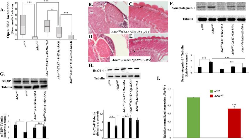
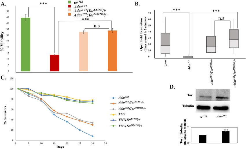
Khan et al. BMC Biology   (2020) 18:15                                                                           Page 2 of 16

Background                                                    and conserved editing include paralytic (para) [16],
Drosophila melanogaster has a single Adar (adenosine          shaker, shaker b, and cacophony (cac) [17] transcripts
deaminase acting on RNA) gene encoding an orthologue          which encode the pore-forming subunits of axonal
of the vertebrate ADAR2 RNA editing enzyme [1]. In            voltage-gated sodium, potassium, or calcium channels,
both vertebrates and Drosophila, ADAR RNA editing in          respectively. At the axon terminus, presynaptic active
CNS transcripts is targeted to pre-mRNA exons that            zones are formed above cacophony channels clustered in
form RNA duplexes with flanking intron sequences.             the presynaptic membrane; in the active zones, neuro-
Editing events are frequently located in coding regions,      transmitter synaptic vesicles are tethered for rapid
leading to the generation of alternative edited and un-       neurotransmitter release followed by rapid endocytosis
edited isoforms of CNS proteins (for review [2]). ADAR2       to recycle and refill the vesicles [18]. The cacophony
in mammals is required for editing a glutamine codon to       channel triggers calcium entry into presynaptic boutons
arginine at the Gria2 Q/R site in the transcript encoding     when it is activated in response to an action potential
a key glutamate receptor subunit [3]. This editing event      [19]. Other transcripts that are edited, especially in the
regulates the calcium permeability of AMPA class glu-         adult brain, such as Synapsin [20], Synaptotagmin 1,
tamate receptors, and loss of this editing event leads to     Endophilin A, and Munc [21], encode key proteins in-
seizures and neuronal cell death. Thus, mice lacking          volved in the formation and function of neurotransmit-
Adar2 die within 3 weeks of birth; however, Adar2;            ter synaptic vesicles.
Gria2R transgenic mice with the chromosomal Gria2                The Drosophila Adar5G1 null mutant fly shows reduced
gene mutated to encode arginine are normal indicating         viability, lack of locomotion, ataxia, and age-related neuro-
that Gria2 Q/R is the key editing site in vertebrates [4].    degeneration [6]. In larval motor neurons, targeted Adar
The number of edited transcripts and edited sites is very     RNAi knockdown leads to increased motor neuron excit-
much greater in Drosophila than in vertebrates. Editing       ability; reciprocally, Adar overexpression in motor neurons
site recognition is conserved; human ADAR2 expressed          leads to reduced neuronal excitability [22]. Adar5G1 mutant
in Drosophila rescues Adar5G1 null mutant phenotypes          larval neuromuscular junctions have defects in calcium-
[5] and correctly edits hundreds of Drosophila tran-          regulated synaptic transmission and increased numbers of
scripts encoding ion channels subunits and other CNS          boutons [23] with increased numbers of synaptic vesicles
proteins [6–10].                                              and increased levels of the pre-synaptic proteins Synapsin
   Our hypothesis is that during the evolutionary increase    [20], Endophilin A, Synaptotagmin 1, and others [24]. A
in site-specific RNA editing events in advanced insects,      much weaker hypomorphic Adarhyp mutant that has a
there has been selection for editing events that allow        nearly normal capacity for locomotion when stimulated ex-
production of alternative edited and unedited isoforms        hibits an aberrantly increased sleep pressure associated with
of CNS proteins [11]; edited isoforms are also more           the inability to achieve a normal sleep-mediated reduction
abundant in adult brains than in larval brains in Dros-       of pre-synaptic vesicles and associated proteins and synap-
ophila. RNA editing has also been evolutionarily ex-          tic signaling [25]. This defective locomotion due to persist-
panded in cephalopod molluscs [12], consistent with the       ent halting in the hypomorphic Drosophila Adarhyp mutant
idea that more RNA editing may be able to enhance             is similar to what we observe in the more severely affected
some brain function(s). Recent results reveal the com-        Adar5G1 null mutant. In the Adarhyp adult brain, the sleep
plexity of RNA editing in Drosophila neurons, showing         defect is due to neuronal excesses of neurotransmitter syn-
that different neuronal populations have distinct editing     aptic vesicles held in a reserve pool that is not readily re-
signatures [13]. The extreme opposite hypothesis to           leasable and difficult to deplete, and the level of presynaptic
ours, that editing events are evolutionary accidents, ap-     proteins is elevated, consistent with defects in axonal active
pears less likely since many editing events are well con-     zones in brain neurons similar to those observed at larval
served within insects or cephalopods, respectively, and       neuromuscular junctions [25].
are under positive selection during evolution [14]. How-         To elucidate whether Adar null mutant phenotypes
ever, it is still possible that the many different editing    have a coherent underlying basis, we performed a pilot
events serve many different and unconnected purposes.         genetic screen on chromosome II for suppressors of the
We set out to define the key effects of Drosophila Adar       Adar5G1 null mutant reduced viability. We find that re-
RNA editing by identifying genetic suppressors of Adar        duced dosage of Tor (target of rapamycin) is a potent
null mutant phenotypes and determining the mecha-             suppressor of Adar mutant phenotypes. Tor is a member
nisms of action of these suppressors.                         of the phosphatidylinositol 3-kinase-related kinase family
   Adar expression increases strongly at pupation, and        and is essential for several cellular processes including
the number of edited sites and editing efficiencies at        increased translation and reduced autophagy under well-
most sites is higher after metamorphosis in the brain of      fed conditions (for review [26, 27]). Electron microscopic
the adult fly [6, 15]. In Drosophila, transcripts with high   analysis reveals that neurodegeneration in Adar5G1
Khan et al. BMC Biology   (2020) 18:15                                                                            Page 3 of 16

mutant fly retina is associated with abnormal, large,               We tested mutations in individual genes within the rescu-
intracellular membrane-bounded vacuoles. These vacu-             ing Df(2 L)ED778 deficiency and the partially overlapping
oles appear to contain cellular components and are likely        Df(2 L)ED784 deficiency, and within some other partially
to result from aberrant activity of the endosome/autoph-         rescuing deficiencies, for rescue of Adar mutant viability.
agy/lysosome system. Tor protein levels are increased in         DrosDel deletions are excellent for rapid genome coverage
the Adar5G1 mutant, and reducing Tor gene dosage sup-            in genetic screens, but for unknown reasons, inability to
presses these defects by increasing autophagy and clear-         map effects of deletions down to reduced copy numbers of
ing excess pre-synaptic proteins. There is no extensive          single genes within the deletions is very common. In this
cell death in the Adar-mutant CNS. The findings are              case, single gene mutations in the Tor gene, but not muta-
consistent with the hypothesis that Drosophila Adar              tions in other genes within the deleted regions, were found
function has an evolutionarily selected biological role re-      to increase viability (Fig. 1a) and open field locomotion
lated to synaptic plasticity and CNS protection.                 (Fig. 1b) [29, 30] in Adar5G1;Tork17004 / + and Adar5G1;
                                                                 TorMB07988 / + flies; lifespan also appears to be increased
                                                                 (Fig. 1c) (we are unable to perform the appropriate
Results                                                          Kolmogorov-Smirnov test for statistical significance with
Reduced Tor gene dosage suppresses Adar mutant                   our small sample size in 3 replicates). These Tor mutants
reduced viability, open field locomotion defects, and            are homozygous lethal P-element insertions at different po-
reduced longevity                                                sitions in Tor that are presumed null mutants.
To elucidate which mechanisms mediate Adar mutant phe-              Open field locomotion was measured by recording
notypes, we performed a pilot screen for heterozygous dele-      crossing of individual flies over lines in a gridded Petri
tions that increase the number of adult male Adar5G1 flies       dish (three 2-min measurements on each of 10 or more
eclosing from pupae in crosses (Adar is on Chr. X and            flies for each line) as previously described [17]. In this
males have one gene copy). When virgin female y,Adar5G1,         assay, even wild-type flies may stop moving for part of
w /FM7, Bar flies are crossed with male w1118 and male           the 2-min measurement period. However, the Adar mu-
progeny that eclose from pupae are counted, the ratio of         tant flies tend to stop within a few tens of seconds and
male y,Adar5G1,w to male FM7 Bar progeny obtained is             to not move again thereafter. The Adar5G1 mutant flies
only about 20% (see w1118 control cross at the bottom of         also show leg tremors and difficulty in walking straight
Additional file 1: Figure S1). This reduced viability at eclo-   without stumbling (Additional file 7: Video S1 show
sion from the pupa reflects the death of Adar5G1 mutants         Adar5G1 mutant walking defects, and Additional file 8:
during embryonic, larval, and pupal stages. Therefore,           Video S2 shows rescue in Adar5G1; TorMB07988 / +).
when virgin female y,Adar5G1,w /FM7, Bar flies are crossed          Reduced Tor gene dosage may directly correct an ab-
with male w, Df(2)/SM5 Cy, suppression of this Adar5G1 re-       errantly increased activity of Tor in Adar5G1. Immuno-
duced viability, measured by the proportion of live Adar5G1;     blot analysis of Adar5G1 mutant total head protein
Df(2)/+ mutant flies eclosing from pupae can be used for a       extracts show that Tor protein is present at a signifi-
genome-wide screen of deficiencies.                              cantly increased level in Adar5G1 (Fig. 1d). Increased Tor
   We performed a trial screen of 35 DrosDel deficiencies        protein is likely to lead to increased levels of activated
[28] covering 70% of the left arm of chromosome II for           Tor but, unfortunately, there is no available antibody to
deficiencies that when heterozygous act as suppressors           detect specifically the active, phosphorylated form of
of the reduced viability of male Adar5G1mutant flies             Drosophila Tor.
(Additional file 1: Figure S1). DrosDel deficiencies are a
series of genetically engineered deficiencies covering           Reduced Tor gene dosage also suppresses Adar mutant
most of the Drosophila euchromatin that each deletes             age-dependent neurodegeneration
about 30 genes on average [28]. The most robustly res-           The Adar5G1 null mutant neurodegeneration has been
cuing deficiency identified by the screen, Df(2 L)ED778,         described previously [5, 6, 8, 31]. The Drosophila ADAR
substantially increases (to 80%), and the partially over-        protein is normally present in nuclei of all brain neurons
lapping Df(2 L)ED784 deficiency somewhat increases,              in wild type and is entirely absent in the Adar5G1 null
Adar5G1 mutant viability. The viability of Adar5G1 is in-        mutant that deletes the entire Adar transcribed region
creased by 8 deficiencies and decreased by others. The           [6]. Neurodegeneration develops more quickly in certain
level of suppression differs greatly between deficiencies,       brain regions. In brains of 23-day and 30-day Adar5G1
with many giving slight suppression that makes the re-           mutant flies, the calyces of the mushroom bodies (MB)
sults noisy and not ideal for a larger genome-wide               and the retina (Fig. 2c, d, Additional file 2: Figure S2)
screen. As we obtained a robust result from two defi-            show filled vacuoles not observed in 23-day w1118 flies
ciencies in this pilot screen, we decided to study these         (Fig. 2a, b). Within the retina, neurodegeneration is evi-
further.                                                         dent at 23 days as a narrowing of photoreceptors with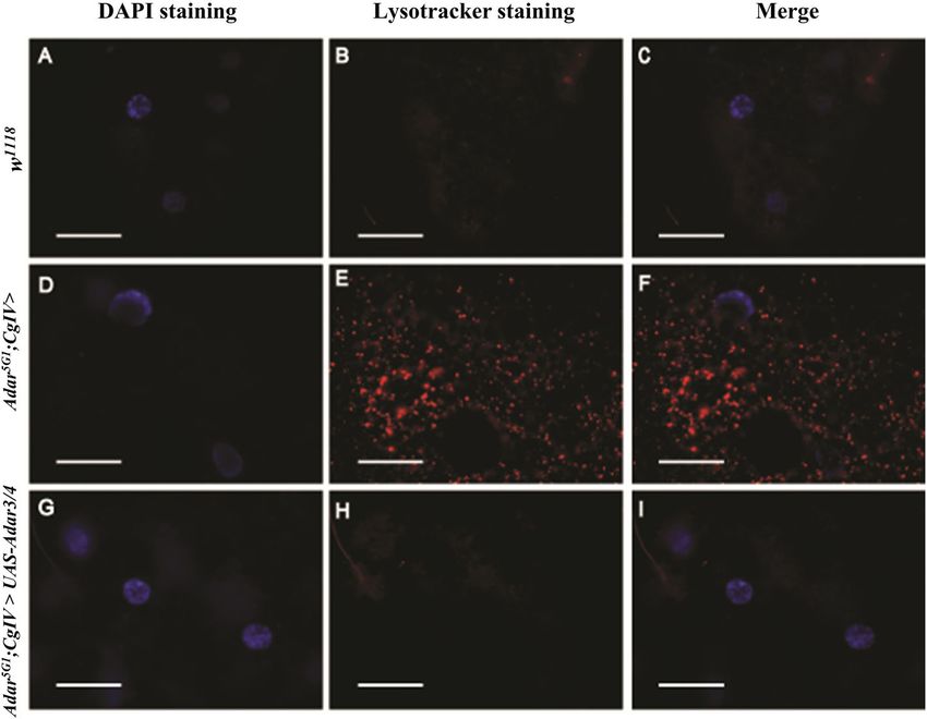
Khan et al. BMC Biology       (2020) 18:15                                                                                                Page 4 of 16

 Fig. 1 Reduced Tor gene dosage rescues Adar5G1 mutant phenotypes. Tor mutations increase a viability at eclosion from the pupae, n = 3; b open
 field locomotion, n > 8; and c lifespan in Adar5G1mutant flies. FM7 is a first chromosome balancer strain. n = 3. d Immunoblot with antibody to
 Drosophila Tor protein of Adar5G1mutant and wild-type (w1118) fly head protein extracts. n = 3. Quantitation of immunoblot data shows increased
 level of Tor in Adar5G1. p values in a and b were calculated by a one-way ANOVA followed by Tukey’s test. The significance of differences
 between variables was described based on p values: *p value < 0.05, **p value < 0.005, ***p value < 0.001, and n.s (not significant). Error bars: SEM
 (standard error of mean for biological replicates). p values in d were calculated by Student’s t test. Source data values are included
 in Additional file 6

separations appearing between ommatidia (Fig. 2d, Add-                       dendrites. Both olfactory projection neurons and Kenyon
itional file 2: Figure S2). Heterozygous Tor mutations                       cells have now been shown to be cholinergic [33], con-
suppress the Adar mutant neurodegeneration in retina                         sistent with our earlier observations that Adar5G1; ChA-
and mushroom body neuropil in Adar5G1;Tork17004 / +                          T>Adar 3/4 flies expressing active ADAR under choline
(Fig. 2e, f) and Adar5G1;Tor MB07988 / + (Fig. 2g, h). Neu-                  acetyltransferase ChAT-GAL4 driver control in choliner-
rodegeneration in the Adar5G1 null mutant is 100%                            gic neurons [34] show rescue of vacuolization in MB ca-
penetrant and is never observed in the brain of wild-type                    lyces and retinas of 30-day Adar5G1 brains [1, 17, 35].
flies. We do not quantitate the number of the vacuoles
as their size variation is too large; instead, we state                      The Adar mutant neurodegeneration involves aberrant
whether it occurs or not.                                                    membrane processes and formation of large brain
   Prominent vacuoles in the brain appear particularly in                    vacuoles
the mushroom body (MB) calyces. The mushroom body                            What is the defect underlying the Adar5G1 mutant neu-
calyces are neuropil regions composed of olfactory pro-                      rodegeneration that is strongly suppressed by reduced
jection neuron axons and dendrites of mushroom body                          Tor dosage? To examine the Adar5G1 mutant neurode-
Kenyon cells; the cell bodies of the Kenyon cells are lo-                    generation at higher resolution, we performed an elec-
cated above the calyces and their nuclei stain darkly with                   tron microscopic analysis of retinas and optic laminae of
hematoxylin. Vacuoles may develop within the large                           aged Adar5G1 mutant flies. Transmission electron micro-
boutons at the pre-synaptic boutons of olfactory projec-                     scope (TEM) sections parallel to the surface of the eye
tion neurons which extend axons from the olfactory                           are particularly suitable for study because these sections
lobes beneath the brain reach to the mushroom body ca-                       show a highly regular pattern of photoreceptors and
lyces [32]. Large round boutons at the ends of projection                    support cells within the repeating ommatidia (Fig. 3a, b).
neuron axons are surrounded by many fine Kenyon cell                         TEM images of sections through the retina of 25-day-
Khan et al. BMC Biology      (2020) 18:15                                                                                                Page 5 of 16

 Fig. 2 Rescue of Adar5G1 mutant neurodegeneration by reduced Tor gene dosage. Images show representative 6-μm-thick hematoxylin and eosin
 stained sections through mushroom body calyces (left panels (× 63)) and retinas (right panels (× 40)) of a, b 23-day w1118, c, d 23-day Adar5G1 e, f
 25-day Adar5G1; TorK170048 / +, and g, h 23-day Adar5G1; TorMB07988 / +. Scale bars, 20 μm

old Adar5G1 show large membrane-bounded vacuoles                             autophagy. It is likely that the aberrant vacuoles between
between or within support cells that surround the pho-                       ommatidia develop within the retinal pigment cells that
toreceptors (R1-R7/8) (Fig. 3c, arrows). Other defects in                    import red and brown pigment precursors from the
Adar5G1 resemble those seen with autophagy mutants,                          hemolymph and process and store them in membrane-
such as autophagic-like vesicles (Fig. 3d–f), multilamellar                  bounded pigment granules that are a type of lysosome-
vesicles (Fig. 3g, h), and membrane-bounded vesicles                         related organelle. We did not obtain TEM sections
budding from the rhabdomeres of photoreceptors in                            through mushroom body calyces, but sections through the
more advanced stages of degeneration (Fig. 3i–l).                            optic lamina where the cellular arrangements are more
  This data suggests that the Adar mutant neurodegener-                      difficult to interpret in EM also show aberrant multilamel-
ation does not involve death of neurons in the first in-                     lar vesicles and membrane overgrowth.
stance, but it does reflect development and enlargement                        Aberrant intracellular membrane processes typify the
of aberrant intracellular vacuoles like those observed in                    Adar mutant neurodegeneration, which does not appear to
lysosomal storages diseases that cause defects in                            involve extensive neuronal death. TUNEL assays did not
Khan et al. BMC Biology (2020) 18:15 Page 6 of 16
Fig. 3 EM analysis of retinal degeneration in the Adar5G1 mutant. a The ommatidia of w1118 at 25 days. Each ommatidium comprises seven
photoreceptor cells surrounded by and separated from neighboring ommatidia by thin pigment cells containing red pigment granules. b An
ommatidium of 25-day-old w1118 at higher resolution. The photoreceptor cells with light-detecting rhabdomeres (Rb) appear normal. The R7/R8
photoreceptor is indicated. Organelles such as mitochondria are identifiable (arrow). c Retina of the Adar5G1 mutant at 25 days showing pigment
cells with large vacuoles between ommatidia (arrows). d Higher resolution image of a single ommatidium in 25-day-old Adar5G1 with vacuole (V)
between photoreceptor cells of two ommatidia. e Magnification of area within the circle in d. Interrupted membrane (arrow) was observed inside
the vacuole. f Magnification of area within the square in d. Membrane-bounded vesicles (arrows) in the photoreceptors contain cellular
components in an autophagosome-like structure surrounded by two or more membrane layers. g, h Multilamellar membrane structures (arrows)
in a photoreceptor cell and within a glial cell close to the basement membrane between the retina and the lamina in Adar5G1. i Single
membrane-bounded vesicles pinching off from the photoreceptor (arrows) in early stages of photoreceptor degeneration in Adar5G1. j Larger
multilamellar membrane structures budding off from the extracellular membrane of photoreceptor cells into the ommatidial cavity (arrows) at
more advanced stages of degeneration in Adar5G1. k Extensive loss of pigment cells separating ommatidia in advanced stages of
neurodegeneration in Adar5G1. Photoreceptor cell cytoplasm and extracellular membrane are abnormal, and vesicles bud from the rhabdomeres
(arrows). l Abnormal exocytosis from the rhabdomere in late stages. The extracellular membrane of the photoreceptor is not well defined
detect neuronal death in the Adar5G1 mutant brain (Add- gene dosage. Tor is a key gene controlling growth and
itional file 3: Figure S3A-D), and few Lysotracker-positive autophagy [27]; suppression of Adar mutant phenotypes
nuclei are seen in the brain (Additional file 3: Figure S3B), by reduced Tor gene dosage may be due to decreased
although cell death does occur outside the brain in head fat translation or to increased autophagy in the Adar5G1;
cells (Additional file 3: Figure S3A-D). Adar5G1;ChAT>p35 Tor / + flies.
flies expressing the viral anti-apoptotic protein p35, which Tor is a protein kinase that, when active, increases
inhibits most Drosophila caspases [36, 37], still show vacuo- translation by phosphorylation of the ribosomal protein
lization in the MB calyces and retina at 30 days (Add- S6 kinase (S6K) protein that increases its activity and by
itional file 2: Figure S2E, F), indicating that vacuolization is phosphorylation of the eIF 4E BP translation inhibitor
not prevented by blocking apoptosis. that reduces its inhibitory activity [38, 39]. Reduced Tor
gene dosage should reduce translation in the Adar5G1;
Suppression of Adar mutant phenotypes by reduced Tor Tor/+ double mutants. However, mimicking translation-
or by increased expression of Atg5 decreasing effects of reduced Tor gene dosage by de-
We next focused on understanding the mechanism of creasing S6 kinase activity in cholinergic neurons in
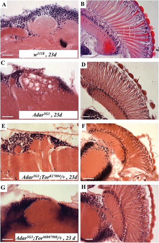
suppression of Adar mutant phenotypes by reduced Tor Adar5G1; ChAT>S6KKQ flies expressing a dominantKhan et al. BMC Biology (2020) 18:15 Page 7 of 16 negative S6K [40], or Adar5G1; ChAT>Thor flies with in- viability and rescue of Adar5G1 mutant locomotion de- creased expression of translation-inhibiting eIF 4E-BP fects (Fig. 4a) and neurodegeneration (Fig. 4b, c). There- (Thor), did not show suppression of Adar5G1 mutant fore, suppression of Adar5G1 mutant phenotypes appears open field locomotion (Fig. 4a). This indicates that re- to be due to increased autophagy caused by the reduced duced translation is not the primary mechanism by Tor gene dosage. which reduced Tor suppresses the Adar mutant Tor is activated by growth-promoting extracellular sig- phenotypes. nals such as insulin as well as by intracellular signals; Since suppression of the Adar mutant phenotypes by Tor locates to the surface of the lysosome and is acti- reduced Tor does not appear to be due to reduced trans- vated there by amino acids being returned from the lyso- lation, the suppression may instead be due to increases some to the cytoplasm. The insulin receptor acts in some type of autophagy. Increased autophagy could through PI3 kinase (PI3K) and the serine-threonine pro- be consistent with the clearing of the large vacuoles in tein kinase AKT to phosphorylate the Tuberous Scler- aged Adar mutant brains and retinas by reduced Tor osis Complex (TSC), releasing it from the Rheb (Ras dosage. Activated Tor suppresses autophagy by phos- homolog enriched in brain) protein in the lysosomal Tor phorylating Atg1, the key protein for activation of ca- protein complex and activating Tor [42]. If reduced Tor nonical autophagy. Increased expression of key gene dosage suppresses Adar mutant phenotypes be- autophagy proteins is able to increase canonical autoph- cause it reduces effects of growth-promoting signals agy [27]; Adar5G1; ChAT>Atg5 flies [41] show increased such as insulin, then the effect of reduced Tor gene Fig. 4 Decreased Tor, or increased Atg5 to increase autophagy, suppresses Adar5G1 mutant phenotypes. a Rescue of Adar5G1 mutant open field locomotion defects in Adar5G1; TorK170048 / +, Adar5G1; TorMB07988 / +, Adar5G1; ChAT>Atg5, and Adar5G1; ChAT>Atg1 flies but not in Adar5G1; ChAT>Thor or Adar5G1; ChAT>S6KKD and very partially in Adar5G1; ChAT>TSC1,TSC2 flies. n > 8. b Representative images of MB calyx (× 63) and c retina (× 40) in 30-day Adar5G1; ChAT>Atg5. Scale bars, 20 μm. d Immunoblot with antibody to Synaptotagmin 1 of head protein extracts of Adar5G1, w1118, Adar5G1; TorK17004 / +, and Adar5G1; ChAT > Atg5 flies. Quantitation of immunoblot data shows increased Synaptotagmin 1 in Adar5G1 is reduced by decreased Tor or by increased Atg5. n ≤ 3. e Immunoblot with antibody to ref(2)p, the Drosophila p62 canonical autophagy protein, of head protein extracts of w1118, Adar5G1mutant, Adar5G1; TorK17004 / +, and Adar5G1; ChAT > Atg5 flies. Quantitation of immunoblot data shows that increased ref(2)p, Drosophila p62 protein, in Adar5G1 is not reduced but increased by decreasing Tor or by increasing Atg5. n ≥ 3. p values were calculated by a one-way ANOVA followed by Tukey’s test. The significance of differences between variables was described based on p values: *p value < 0.05, **p value < 0.005, ***p value < 0.001, and n.s (not significant). Error bars: SEM (standard error of mean for biological replicates). Source data values are included in Additional file 6
Khan et al. BMC Biology   (2020) 18:15                                                                      Page 8 of 16

dosage should be mimicked by increasing TSC protein          Lysotracker dye. Staining larval fat cells from well-fed
dosage. Surprisingly, Adar5G1; ChAT>TSC1, TSC2               larvae of the Adar5G1 mutant with Lysotracker dye
(Fig. 4a) with reduced signaling to Tor through the insu-    shows the presence of increased numbers of lysosomes
lin pathway do not show strong rescue of Adar5G1mu-          in the Adar5G1 mutant, even in the absence of starvation
tant locomotion defects. This suggests that any aberrant     (Fig. 5e, f) relative to equivalent wild-type w1118 cells
axonal growth signal in the Adar5G1mutant is not due to      (Fig. 5b, c). Starvation increases the number of lyso-
alteration in an upstream signal through the insulin re-     somes further in the Adar5G1 mutant cells (data not
ceptor, nor through the anaplastic lymphoma kinase that      shown). Expression of Adar 3/4 (Fig. 5h, i) in Adar5G1
may substitute for insulin receptor in the brain that also   mutant fat cells under the control of the CollagenIV-
signals through PI3K [43] to the Tor complex 1               GAL4 (CgIV-GAL4) driver is sufficient to eliminate the
(TORC1). If suppression of the Adar mutant phenotype         elevated basal autophagy in the Adar5G1 mutant, as indi-
by reduced Tor is not due to changed responsiveness to       cated by the loss of Lysotracker vesicle staining.
external signals such as insulin, then it may be due to an
intracellular effect. Since Tor is activated on lysosomes,   Rescue of Adar mutant phenotypes by increased
there may be an aberrant intracellular feedback from au-     expression of the endosomal microautophagy (eMI)
tophagy that leads to increased Tor.                         protein Hsc70-4
   To determine whether increased autophagy may be           Recent studies have shown that a different type of
rescuing Adar mutant defects by clearing aberrant accu-      starvation-inducible, Tor-inhibited autophagy called
mulations of synaptic vesicles, we measured levels of the    endosomal microautophagy (eMI) occurs in Drosophila
presynaptic protein Synaptotagmin1 that is associated        neurons and is especially important in presynaptic active
with the synaptic vesicles in heads of Adar5G1 mutant        zones [46–49]. To test whether increased eMI rescues
and rescued flies by immunoblotting. Immunoblotting of       Adar5G1 mutant phenotypes, we used the ChAT-GAL4
head protein extracts with anti-Synaptotagmin 1 anti-        and Act 5C-GAL4 drivers to increase expression of the
bodies demonstrates that there is an aberrant accumula-      Hsc70-4 protein by directing expression of UAS-Hsc70-
tion of Synaptotagmin 1 in Adar5G1mutant heads [25]          4. Increasing Hsc70-4 in cholinergic neurons increases
(Fig. 4d) that is lowered by reduced Tor or by increased     locomotion (Fig. 6a); on the other hand, knocking down
Atg5 expression.                                             of Hsc70-4 in cholinergic neurons does not improve the
                                                             Adar5G1 mutant phenotype (Fig. 6a). When acting as a
Increased autophagic vesicles but incomplete clearance       chaperone for neurotransmitter synaptic vesicles, Hsc70-
of ref(2)p in the Adar5G1 mutant                             4 acts together with an interacting partner protein called
To assess canonical autophagy in the Adar5G1 mutant          small glutamine-rich tetratricopeptide repeat protein
and rescues, we examined levels of ref(2)p protein.          (Sgt), as an ATP-driven molecular chaperone protein. In
ref(2)p is the Drosophila orthologue of the mammalian        eMI, Hsp70-4 acts without Sgt to recruit KFERQ-motif
p62 canonical autophagy adapter protein (also called         proteins to endosomes [46]. The Sgt protein favors the
Sequestosome1) that brings ubiquitinated cargo to ca-        more general chaperone role of Hsc70-4 in synaptic
nonical autophagosomes; p62 is degraded in the process       vesicle cycling and suppresses its function in eMI.
and p62 accumulates when canonical autophagy is de-          Therefore, we also increased eMI with an UAS-Sgt RNAi
fective [44]. If canonical autophagy is operating normally   construct to decrease expression of Sgt specifically in
in Adar5G1 and increased in heads of Adar5G1; Tor k17004     cholinergic neurons and this also dramatically sup-
/+ double mutant or Adar5G1; ChAT>Atg5 flies, then           pressed the Adar5G1 mutant locomotion defect (Fig. 5a);
levels of p62 protein should be normal in Adar5G1 and        knockdown of Sgt with the ubiquitous Act 5C-GAL4
reduced in the double mutants [45]. However, p62 pro-        driver is lethal. Increased eMI in the Adar5G1 mutant
tein levels are twofold higher than normal in Adar5G1        background also suppresses neurodegeneration. Overex-
head protein extracts and increase further in the double     pression of Hsc70-4 (Fig. 6b, c) or knocking down Sgt
mutants (Fig. 4e), in particular with increased Atg5. This   (Fig. 6d, e) in Adar5G1 with ChAT-GAL4 suppresses the
suggests that canonical autophagy might not be func-         Adar5G1 mutant neurodegeneration in retina and mush-
tioning perfectly in the Adar5G1 mutant background,          room body.
even though it partially clears excess synaptic vesicle        Immunoblotting of head protein extracts with anti-
proteins (see below).                                        Synaptotagmin 1 antibodies demonstrates that the aber-
  Larval fat cells are used to study autophagy in Dros-      rant accumulation of Synaptotagmin 1 in Adar5G1mu-
ophila, as these cells are much larger than brain neurons    tant heads (Fig. 6f) is dramatically reduced by increased
and form a single sheet of cells in which autophagy is       Hsc70-4 expression. We conclude that increased eMI
readily induced by starvation of the larvae and detected     suppresses the Adar5G1 mutant phenotypes. The reduc-
by staining the lysosomes in live cells with acidic          tion of Synaptotagmin 1 to below wild-type levels is
Khan et al. BMC Biology (2020) 18:15 Page 9 of 16 Fig. 5 ADAR protein expression rescues the autophagy-related phenotype in Adar5G1 larval fat cells. The fat bodies of a–c wild-type strain w1118, d–f Adar5G1;CgIV>, and g–i Adar5G1;CgIV>UAS-Adar3/4 have been dissected and live-stained with DAPI (a, d, g) and Lysotracker (b, e, h) dyes (merges in c, f, i). Wild-type fat body does not show any Lysotracker staining (b, c). Adar5G1 mutant fat cells have an increased activation of autophagy as indicated by increased Lysotracker staining in lysosomes (e, f). Expression of the UAS-Adar3/4 transgene in the Adar5G1 mutant fat cells is sufficient to rescue the elevated basal autophagy (h, i). Scale bars, 50 μm surprising, but synaptic vesicle-associated proteins are (Fig. 6i). By both methods, we observe a small but sig- normally present at levels that probably reflect retention nificant decrease in Hsc70-4 level in Adar5G1. of a reserve of older protein molecules in association with the no-longer readily releasable reserve pool of syn- Discussion aptic vesicles [50–52]. We also see a less dramatic de- RNA editing by Adar is required to maintain the integ- crease in the level of Synaptotagmin 1 when reducing rity of the CNS in adult Drosophila [6]. To find suppres- the level of Tor or overexpressing Atg5 in the Adar5G1 sors of the Adar5G1 null mutant phenotype, we mutant background (Fig. 4d). Increased Atg5 is likely to performed an initial screen for genetic suppressors that be lowering Synaptotagmin 1 through increased canon- increase the viability of Adar5G1 and discovered a key ical autophagy and is unlikely to be acting within the role for Tor-regulated autophagy in all Adar mutant eMI pathway as Atg5 has been reported to not be re- phenotypes (Fig. 1a–c, Fig. 2e–h). Tor protein is abnor- quired for eMI [47]. mally increased in Adar5G1 mutant heads (Fig. 1d); We also examined the level of ref(2)p when overex- therefore, suppression of Adar mutant defects by re- pressing Hsc70-4 or knocking down Sgt in Adar5G1 duced Tor gene dosage is, at least in part, a true rescue, (Fig. 6g). We did not observe any significant difference i.e., reducing Tor directly corrects a defect in the Adar in ref(2)p levels between head extracts of Adar5G1 mu- mutant rather than simply activating some entirely unre- tant, Adar5G1; ChAT > Hsc70-4 or Adar5G1; ChAT > Sgt lated bypass pathway. RNAi flies. This suggests that, as expected, increased Consistent with an autophagy defect, the Adar5G1 mu- Hsc70-4 does not increase canonical autophagy or sig- tant neurodegeneration shows resemblances to neurode- nificantly change levels of ref(2)p. generations in Drosophila models of lysosomal storage Since increasing eMI suppresses the Adar5G1 mutant diseases, a class of neurodegenerations in which lyso- phenotypes, it is possible that eMI might be insufficient somes accumulate different intracellular components in Adar5G1. To investigate this, we determined the level [53]. The most distinctive abnormal intracellular compo- of Hsc70-4 protein by immunoblotting head protein ex- nents in the Adar5G1 mutant eyes and brains (Fig. 2a–f), tracts (Fig. 6h) and by measuring its expression by qPCR apart from double membrane autophagosomes (Fig. 3f),
Khan et al. BMC Biology       (2020) 18:15                                                                                                 Page 10 of 16

 Fig. 6 Increased Hsc70-4 suppresses Adar5G1 mutant phenotypes. a Rescue of Adar5G1 mutant open field locomotion defects in Adar5G1; ChAT >
 Hsc70-4 and Adar5G1; ChAT > Sgt RNAi flies with increased endosomal microautophagy. n ≥ 10. b Representative images of MB calyx (× 40) and c
 retina in 30-day Adar5G1; ChAT>Hsc70-4 (× 40). d Representative images of MB calyx (× 40) and e retina in 30-day Adar5G1; ChAT>SgtRNAi (× 40). f
 Immunoblot detection of the presynaptic protein Synaptotagmin1 in w1118, Adar5G1 mutant, Adar5G1; ChAT>Hsc70-4, Adar5G1; ChAT>Sgt RNAi, and
 Adar5G1; Act5c>Hsc70-4 head protein extracts. Quantitation of the immunoblot data is shown below; levels of Synaptotagmin 1 compared to
 tubulin in each of the different head protein extracts. n ≤ 3. g Immunoblot to detect ref(2)p, the Drosophila p62 autophagy protein, in total head
 proteins of Adar5G1 mutant, w1118 wild type, Adar5G1; ChAT>Hsc70-4, and Adar5G1; ChAT>Sgt RNAi flies. n ≤ 3. h Immunoblot to detect Hsc70-4
 protein in total head protein extracts of w1118 wild type, Adar5G1 mutant, Adar5G1; ChAT>Hsc70-4, and Adar5G1; ChAT>Sgt RNAi flies and Adar5G1;
 Act5c>Hsc70-4. n = 3. i qPCR of Hsc70-4 from w1118 wild type and Adar5G1 heads showing that Hsc70-4 is significantly decreased in Adar5G1 heads.
 n = 6. p values in a, e, g, and h were calculated by a one-way ANOVA followed by Tukey’s test. The significance of differences between variables
 was described based on p values: *p value < 0.05, **p value < 0.005, ***p value < 0.001, and n.s (not significant). Error bars: SEM (standard error of
 mean for biological replicates). p values in h were calculated by Student’s t test. Source data values are included in Additional file 6

are the multilamellar membrane whorls (Fig. 3h). These                        homolog of the vertebrate p62 adapter for canonical au-
have been identified in cell bodies in other Drosophila                       tophagy of ubiquitinated proteins, is increased in
mutants such as eggroll [54], swiss cheese [55–57], and                       Adar5G1 and increased much more with reduced Tor or
benchwarmer/spinster [58] and are characteristic of the                       increased Atg5 (Fig. 4e). Adar5G1 larval fat cells also
human neurodegenerative Tay-Sachs disease [53, 59].                           show increased Lysotracker-positive acidic autophagoso-
The formation of large vacuoles in Adar mutant mush-                          mal and lysosomal vesicles (Fig. 5e, f). This impeded CA
room body calyces might be directly related to accumu-                        in Adar5G1 might arise because some proteins that have
lation of large numbers of neurotransmitter-containing                        edited isoforms play important roles in CA [60]. Tran-
presynaptic vesicles and associated presynaptic proteins                      scripts of cacophony (cac) and straightjacket (stj) encode
such as Synaptotagmin 1 in the brain [25], which is pre-                      subunits of the pre-synaptic voltage-gated calcium chan-
vented by reduced Tor gene dosage or by increased Atg5                        nel that is also required for fusion of lysosomes with
(Fig. 4d) or increased Hsc70-4 (Fig. 6e) expression to in-                    autophagosomes and endosomes. Loss of function muta-
crease autophagy.                                                             tions of cac or stj impairs neurotransmission and lyso-
  Which type of Tor-regulated autophagy is involved in                        some function in neurons, leading to some accumulation
the suppression of Adar mutant phenotypes? Canonical                          of p62 protein [61], although it is not known whether
autophagy (CA) is still sufficiently functional to mediate                    loss of only the edited isoforms of these proteins is suffi-
rescue of Adar5G1 mutant phenotypes (Fig. 4a–d), even                         cient to cause any similar defect. Other edited tran-
though it may also be somewhat impaired in Adar5G1.                           scripts encoding CA-associated proteins include Atg14,
Immunoblots show that ref(2)p protein, the Drosophila                         Atg17, AMPKalpha, and Foxo (Additional file 4: Table
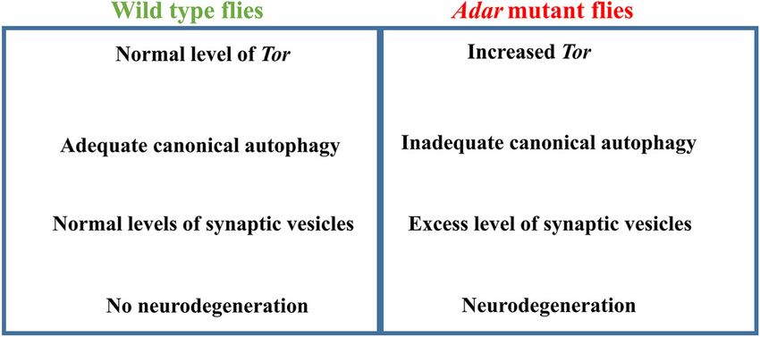
Khan et al. BMC Biology   (2020) 18:15                                                                        Page 11 of 16

S1); all of these, in addition to probable involvement of     protein is at a lower level in Adar mutant heads (Fig. 6h,
edited synaptic vesicle-associated proteins in membrane       i); this suggests that eMI may be insufficient or sup-
fusion events in CA [61], suggest that both CA and syn-       pressed by increased Tor in the Adar mutant. Similar to
aptic vesicle are among processes affected by proteins        the p62 adapter during CA, the Hsc70-4 cargo selector
encoded by edited transcripts in CNS. An additional           is believed to also be turned over as KFERQ target pro-
possible explanation for why ref(2)p clearance is im-         teins are recruited and destroyed during eMI. It is not
peded in Adar5G1 is that CA is affected by Dicer-2-           known how activated Tor suppresses eMI; it has been
mediated aberrant innate antiviral immune induction           proposed that Atg1 is also involved [47]; possibly, the re-
that occurs in Adar5G1-mutant heads (Deng et al., 2020,       duced Hsc70-4 observed in Adar5G1 is part of the mech-
Nat. Comms, in press), which is likely to result from ac-     anism of eMI suppression by increased Tor.
cumulated unedited intracellular dsRNA in Adar5G1,               Since rescue of Adar mutant locomotion defects by
paralleling the mouse Adar1 mutant interferon induc-          expression of Adar requires expression of the catalytic-
tion through antiviral dsRNA sensors [62–64]. In mam-         ally active Adar protein, we expected that RNA editing
malian cells, innate immune induction impedes CA by           of some target transcript might be essential to rescue
diverting p62 from its role as the receptor for ubiquiti-     locomotion [1]. For instance, editing of the transcript
nated proteins in CA to instead form a cytoplasmic in-        encoding Synaptotagmin 1 might be required because
nate immune signaling platform; p62 and other CA              this leads to production of an edited isoform with a dif-
substrates then accumulate because they are less effi-        ferent residue close to those that determine the calcium
ciently turned over by CA [60]. This cross-regulation of      responsiveness of synaptic vesicle exocytosis, potentially
p62 by innate immune signaling helps to redirect CA to        affecting the calcium dependence of the synaptic vesicle
innate immune defense, and it is likely that a similar ef-    cycle [24]. Or editing of the transcript encoding Synap-
fect also acts on ref(2)p in Drosophila; this could in part   sin might be required because this changes an important
account for the Adar5G1 mutant ref(2)p protein                residue that is phosphorylated by cAMP-dependent pro-
accumulation.                                                 tein kinase A (PKA); edited synapsin may limit aberrant
  The increased ref(2)p in the Adar mutant may also           synaptic vesicle accumulation and clustering [20, 25].
lead to the increased Tor activation. In vertebrates, the     Therefore, rescue of locomotion defects by reduced Tor
p62 protein associates with TORC1 on the cytosolic sur-       or increased autophagy without restoring editing of any
face of the lysosome; increased p62 contributes to in-        target transcript is surprising.
creased Tor activation by intracellular amino acids
returning from the lysosome [65]. Aberrant Tor activa-        Conclusion
tion through this cell-autonomous pathway in Drosoph-         Altering flows of membranes and proteins through Tor-
ila [66] might explain why we could not mimic the Tor/        regulated autophagy processes is surprisingly sufficient
+ rescue of Adar mutant phenotypes by genetic manipu-         to overcome Drosophila Adar mutant synaptic synaptic
lations that interfere with extracellular hormone and         defects, locomotion defects, and age-dependent neurode-
growth-related signaling to TORC1, e.g., by increased         generation, presumably by rejuvenating synaptic vesicle
expression of the TSC1 and TSC2 proteins that repress         pools (these Adar mutant defects are summarized in
Tor in the growth signaling pathways (Fig. 4a).               Fig. 7). This suggests that controlling such flows is also a
  Endosomal microautophagy (eMI) has recently been            major biological role of Adar RNA editing in Drosophila.
described as an important new autophagy pathway in-           Can we therefore propose an overall coherent role of
volved in proteostasis at presynaptic active zones in         ADAR2-type RNA editing in CNS of vertebrates and in-
Drosophila [46, 47]. Drosophila eMI targets proteins          vertebrates? The independent evolutionary expansions of
containing KFERQ motifs to endosomes using the                ADAR2-type RNA editing events in transcripts encoding
KFERQ-recognition protein (Hsc70-4 in Drosophila,             CNS proteins in advanced insect groups and in cephalo-
HSPA8 in humans) that is also used in lysosomal               pods suggests involvement in brain function and more
chaperone-mediated autophagy (CMA) in vertebrates.            complex cognition, behavior, and life cycles. In verte-
Drosophila is believed to lack CMA as it does not have a      brates, the homologous ADAR2 is a cycling protein that
homolog of the alternatively spliced isoform of lysosomal     mediates circadian effects [67]; ADAR2 editing also me-
LAMP2A protein required to recruit HSPA8 to lyso-             diates a type of homeostatic postsynaptic plasticity
somes [46, 47]. Increased expression of the key Hsc70-4       through regulated editing of transcripts encoding glu-
protein or decreased Sgt increases eMI and rescues Adar       tamate receptor subunits [68, 69], and the seizures that
mutant locomotion defects (Fig. 6a), neurodegeneration        develop in Adar2 mutant mouse pups also involve wide-
(Fig. 6b–d), and elevated Synaptotagmin 1 levels in Adar      spread effects of aberrant synaptic plasticity [70]. It is
mutant heads (Fig. 6f), without affecting ref(2)p levels      likely that Drosophila Adar is also involved in circadian
(Fig. 6g). Immunoblots for Hsc70-4 indicate that this         rhythms [71], and Drosophila Adar is also involved in
Khan et al. BMC Biology     (2020) 18:15                                                                                        Page 12 of 16

 Fig. 7 Summary of Adar mutant phenotypes. In the Adar mutant, aberrantly increased Tor leads to inadequate autophagy, reduced synaptic
 vesicle clearance, and neurodegeneration

synaptic plasticity during sleep [25]. Aberrantly in-                      BDSC #28709 - y1 v1; P{TRiP.JF03136}attP2 ( Hsc70-4
creased sleep drive arises because the increased reserve                   RNAi)
pools of presynaptic neurotransmitter synaptic vesicles                    BDSC #61267 - y1 v1; P{TRiP.HMJ23046}attP40 (sgt
cannot be reduced normally during sleep. The role of                       RNAi)
Adar we outline here acts to protect the brain through
effects on synaptic plasticity. Adar RNA editing may be                   The GAL4 binary system was used to express trans-
involved in circadian changes in synaptic plasticity and                genes in the Adar mutant background. The Adar5G1
may even mediate beneficial effects of sleep on the                     mutant strain was combined with ChAT>-GAL4, and
brain.                                                                  virgin females of these strains were crossed to males of
                                                                        the transgenic lines bearing the Drosophila UAS-cDNA
Methods                                                                 constructs. Female genotype is y, Adar5G1, w / w, FM7,
Fly maintenance and fly strains                                         Bar; (ChAT-GAL4.7.4)19B,(UASGFP.S65T)T2 / (ChAT-
All fly stocks were raised on standard corn meal-agar                   GAL4.7.4)19B,(UASGFP.S65T)T2.
medium. Fly stocks were maintained at 18 °C, and
crosses were performed at 25 °C. Flies used in aging ex-                DrosDel screen for suppressors of reduced viability in the
periments were maintained in tubes not supplemented                     Adar5G1 mutant
with additional yeast, to prevent flies from becoming                   To screen for suppressors of Adar5G1 mutant reduced
stuck to the yeast. A single fly was maintained in a vial,              viability, we crossed virgin female y, Adar5G1, w /FM7,
and each vial was tipped-on daily. The wild-type control                Bar in groups of five with males from the DrosDel /
strains were either w1118. The GAL4 driver lines and bal-               SM5 Cy lines. Taking male non-Curly progeny, we
ancer lines were obtained from the Bloomington Stock                    counted the Adar5G1; DrosDel / + and FM7 Bar; DrosDel
Centre. Detailed genotypes of individual strains used are               / + flies that eclosed from pupae and determined the ra-
as follows;                                                             tio of male y, Adar5G1, w; Df / + to sibling male FM7; Df
                                                                        / +progeny for each deficiency. DrosDel deficiencies are
  Tork17004: y[1] w[67c23]; P{w[+mC]=lacW}Tor[k17004]/                  marked with mini-w+. Tests of Tor mutants were per-
  CyO,                                                                  formed in the same way.
  TorMB07988: w[1118]; Mi{ET1}Tor[MB07988]
  S6KKQ (dominant negative): w[1118]; P{w[+mC]=UAS-                     Open field locomotion assays
  S6k.KQ}2                                                              Open field locomotion was measured by recording
  Thor: w[*]; P{w[+mC]=UAS-Thor.wt}2                                    crossing of individual flies over lines in a gridded Petri
  Atg6: y; UAS-Atg6-2D; Sb/Tm6b (from U. Pandey)                        dish (three 2-min measurements on each of 10 or more
  Atg5: y[1] w[1118]; wg[Sp-1]/CyO; P{w[+mC]=UAS-                       flies for each line) as previously described [17]. The data
  eGFP-drAtg5}16                                                        are presented as the average number of lines crossed by
  Atg1[6A]: y,w,hsflp;; UAS-Atg1[6A], (from T. Neufeld)                 a fly in the 2-min period. The flies are collected on the
  Atg1[GS10797](EP line): y,w,hsflp; Atg1[GS10797], (from               day of eclosion from the pupae. Next morning, when ef-
  T. Neufeld)                                                           fects of CO2 anesthesia have worn off, they are individu-
  TSC1, TSC2: y,w,hsFlp; UAS-TSC1, UAS-TSC2, (from                      ally introduced to the measuring dish and the measuring
  T. Neufeld)                                                           period begins after tapping the dish once on the bench.
  UAS-Hsc70-4: w[126]; P{w[+mC]=UAS-Hsc70-4.K71S}G                      The test measures the flies maximized movement
Khan et al. BMC Biology (2020) 18:15 Page 13 of 16
response to an initial stimulation and to a new advanced histogram section in either IP Lab Spectrum
environment. or Adobe Photoshop by setting the minimum and max-
imum pixel intensities on the histogram. If necessary,
Histology techniques the gamma was altered on the histogram.
For standard hematoxylin-eosin stained sections, Dros-
ophila heads were fixed at room temperature in Carnoy’s Immunoblotting
fixative for 4 h. For detecting cell death, the terminal Male flies (minimum 15 flies) of the desired genotype
deoxynucleotidyl transferase Biotin-dUTP nick end- were collected and aged for 2 days and then homoge-
labelling (TUNEL) kit from Roche was used. Drosophila nized in NB Buffer (150 mM NaCl, 50 mM Tris-HCl pH
heads were fixed for 4 h at room temperature in 4% 7.5, 2 mM EDTA, 0.1% NP-40). Protein concentration
paraformaldehyde. The heads were embedded into paraf- was determined with Pierce BCA Protein Assay Kit. An
fin wax with standard histology procedures. Sections equal amount of protein was loaded in each lane of a
were cut at 6 μm and either stained with hematoxylin Tris-Glycine Gel and transferred to a nitrocellulose
and eosin for pathological analysis or stained for cell membrane. The membrane was blocked with 5% BSA,
death according to the TUNEL kit instructions. Images incubated with primary antibody overnight. The next
were captured using a compound microscope, which day, the membrane was incubated with secondary anti-
comprised a Coolsnap HQ CCD camera (Photometrics body and developed with Pierce ECL Western Blotting
Ltd., Tucson, AZ) with Plan-neofluar objectives (Carl Substrate. Anti-Ref2P (antibody registry ID: AB_2570151
Zeiss, Welwyn Garden City, UK). Images were captured (1:1000) was a gift from Tor Erik Rusten (University of
with neofluar objectives at × 40 (with a numerical aper- Oslo), anti-synaptotagmin (1:500) (Developmental Stud-
ture of 1.3) for eyes and at × 63 and × 40 (with a numer- ies Hybridoma Bank, DSHB Hybridoma Product 3H2
ical aperture of 1.25) for mushroom bodies. Color 2D7, Antibody Registry ID: AB_528483), anti-Hsc70-4
additive filters (Andover Corporation, Salem, NH) in- (1;1000) was a gift from Konrad Zinsmaier (Bronk et.al,
stalled in a motorized filter wheel (Ludl Electronic Prod- Neuron 2001), anti-Tor antibody (antibody registry ID:
ucts, Hawthorne, NY) were used sequentially to collect AB_2568971) (1:1000) was a gift from Gábor Juhász,
red, green, and blue images, which were then superim- anti-Tublin (Developmental Studies Hybridoma Bank,
posed to form a color image. Image capture and analysis DSHB Hybridoma Product 12G10, antibody registry ID:
were performed with in-house scripts written for IPLab AB_1157911) (1:5000). Imaging was performed with
Spectrum (Scanalytics Corp, Fairfax, VA). The bright- ChemiDoc™ XRS+ System, signal intensity was quanti-
ness and contrast were altered with the advanced histo- fied with Image J software, and statistical analyses were
gram section in either IP Lab Spectrum or Adobe done with the t test.
Photoshop. This was done by manually setting the mini-
mum and maximum pixel intensities on the histogram. qPCR
If necessary, the gamma was altered on the histogram. RNA from approximately 20 fly heads was isolated with
The images shown are representative examples from Tripure, and cDNA generated with RevertAid First
samples of 10–20 heads sectioned for each age and Strand cDNA Synthesis Kit (Thermo Scientific). qPCR
genotype. reactions were performed with The LightCycler® 480
SYBR Green I Master mix, and the primers listed in
Electron microscopy Additional file 5: Table S2 were used to measure expres-
The Adar5G1 mutants and w1118 controls were aged to sion levels. Expression levels were normalized to those
25 days or longer from parallel collections. The probos- of RP49, and t tests were used for statistical analysis.
cis was removed in Schneider’s insect media, and the
heads were fixed for at least 1 h in 2.5% glutaraldehyde Lysotracker staining of larval fat cells
and subsequently fixed in 1% osmium tetroxide in Sor- Drosophila larvae were collected, and brains and fat
enson’s buffer. The heads were dehydrated and embed- body dissected in cold PBS. The tissue of interest was in-
ded into resin. Survey sections of 0.5 μm were cut cubated with LysoTracker® Red DND-99, Molecular
through the frontal brain, and ultra-thin sections were Probes, Invitrogen (l μl of dye in 10 ml of cold PBS), for
cut at the regions of interest. The sections were stained 5 min in ice. After five 2-min washes in PBS, the tissue
with 2% aqueous uranyl acetate for 15 min and lead cit- was mounted in Vectashield DAPI and viewed with a
rate (supplied by Leica) for 5 min. The tissue sections fluorescent microscope.
were viewed with a Philips CM 100 Compustage (FEI)
transmission electron microscope, and digital images are Statistical analyses
collected with an AMT CCD camera (Deben). The Two sample data were analyzed by Student’s t test. A p
brightness and contrast were altered manually with the value of < 0.05 was considered statistically significant. InKhan et al. BMC Biology (2020) 18:15 Page 14 of 16
more than three groups, p values were calculated by a Funding
one-way ANOVA followed by Tukey’s test. The signifi- This work was funded from the Medical Research Council UK to SP and
MCH, by an MRC Capacity Building Area Research Studentship to L. McG, by
cance of differences between variables was described the Medical Research Council UK, (U.1275.01.005.00001.01) to MAO, and by
based on p values: *p value < 0.05, **p value < 0.005, ***p the European Union’s Seventh Framework Programme for research,
value < 0.001, and n.s (not significant). Error bars: SEM technological development and demonstration under grant agreement no.
621368 to MAO, Czech Science Foundation, project no. 19-16963S to LK.
(standard error of mean for biological replicates).
Availability of data and materials
All data generated or analyzed during this study are included in this
Supplementary information published article and its supplementary information files.
Supplementary information accompanies this paper at https://doi.org/10.
1186/s12915-020-0747-0.
Ethics approval and consent to participate
Not applicable.
Additional file 1: Figure S1. Screen of DrosDel deletions on
Chromosome 2 L for rescue of Adar5G1 viability. Ratio of Adar5G1 to FM7
Bar genotypes among male progeny in the presence of DrosDel Competing interests
deficiencies, or in their absence (w1118 cross at the bottom) (expressed as The authors declare that they have no competing interests.
a percentage). Progeny are obtained by crossing Adar5G1 / FM7, Bar virgin
females to males to w1118 males or males of DrosDel/SM5, Cy deficiency Author details
1
stocks. CEITEC Masaryk University, Kamenice 735/5, A35, CZ 62 500 Brno, Czech
Additional file 2: Figure S2. Adar5G1 neurodegeneration at 30 days. Republic. 2National Centre for Biomolecular Research, Faculty of Science,
Images of 6 micron thick haematoxylin and eosin stained sections Masaryk University, Kamenice 5, 625 00 Brno, Czech Republic. 3MRC Human
through mushroom body calyces (left panels, (63X)) and retinas (right Genetics Unit, Institute of Genetics and Molecular Medicine at the University
panels, 40X) of 30-day Adar5G1. of Edinburgh, Crewe Road, Edinburgh EH4 2XU, UK. 4Centre for Integrative
Physiology, Euan MacDonald Centre for Motor Neurone Disease Research,
Additional file 3: Figure S3. Neuronal cell death is not prominent in Hugh Robson Building, University of Edinburgh, George Square, Edinburgh
heads of 25-day-old Adar5G1 mutant flies. (A) TUNEL staining to detect EH8 9XD, UK.
apoptotic cells in head sections from 25-day-old Adar5G1 mutant flies
stained with DAPI to detect nuclei. TUNEL-positive nuclei are not de- Received: 5 September 2019 Accepted: 5 February 2020
tected in neurons However TUNEL-positive nuclei are conspicuous in
head fat bodies of 25-day-old Adar5G1 mutant flies (boxed area in A). (B)
Magnification of area boxed in A (C) Haematoxylin and eosin stained sec-
tion serial to A, white box indicates fat body tissue. (D) Magnification of References
area boxed in C. (E, F) Images show representative 6 micron thick haema- 1. Keegan LP, McGurk L, Palavicini JP, Brindle J, Paro S, Li X, Rosenthal JJ,
toxylin and eosin stained sections through mushroom body calyces (left O'Connell MA. Functional conservation in human and Drosophila of
panels, (63X)) and retinas (right panels, 40X) of 30-day Adar5G1; ChA- Metazoan ADAR2 involved in RNA editing: loss of ADAR1 in insects. Nucleic
T>UAS-p35. Scale bars: 20 μm. Acids Res. 2011;39(16):7249–62.
Additional file 4: Table S1. List of Adar edited transcripts encoding 2. Keegan L, Khan A, Vukic D, O'Connell M. ADAR RNA editing below the
proteins required for autophagy. backbone. Rna. 2017;23(9):1317–28.
3. Sommer B, Köhler M, Sprengel R, Seeburg PH. RNA editing in brain controls
Additional file 5: Table S2. Primers used for qPCR. a determinant of ion flow in glutamate-gated channels. Cell. 1991;67:11–9.
Additional file 6. Excel sheet containing source data file for Figure 1A 4. Higuchi M, Maas S, Single F, Hartner J, Rozov A, Burnashev N, Feldmeyer D,
,1B ,1C.1D ,4A,4D,4E,6A,6F,6G,6H and 6I. Sprengel R, Seeburg P. Point mutation in an AMPA receptor gene rescues
Additional file 7. Video of Adar5G1 null mutant showing locomotion lethality in mice deficient in the RNA-editing enzyme ADAR2. Nature. 2000;
defect. 406:78–81.
5. Hogg M, Paro S, Keegan LP, O'Connell MA. RNA editing by mammalian
Additional file 8. Video of Adar5G1; TorMB07988 Double mutant, which ADARs. Adv Genet. 2011;73:87–120.
shows locomotion defect is recued when Tor dosage is reduced in the 6. Palladino MJ, Keegan LP, O'Connell MA, Reenan RA. A-to-I pre-mRNA
Adar null mutant background. editing in Drosophila is primarily involved in adult nervous system function
and integrity. Cell. 2000;102(4):437–49.
7. Chen L, Rio DC, Haddad GG, Ma E. Regulatory role of dADAR in ROS
metabolism in Drosophila CNS. Brain Res Mol Brain Res. 2004;131(1–2):93–
Acknowledgements 100.
The electron micrographs were captured at the electron microscopy 8. Ma E, Gu XQ, Wu X, Xu T, Haddad GG. Mutation in pre-mRNA adenosine
research services at Newcastle University. We wish to thank Vivian Thomson deaminase markedly attenuates neuronal tolerance to O2 deprivation in
for technical assistance with EM, and Ian Meinertzhagen for advice on EM. Drosophila melanogaster. J Clin Invest. 2001;107(6):685–93.
Drosophila stocks were from the Bloomington Drosophila Stock Centre and 9. Stapleton M, Carlson JW, Celniker SE. RNA editing in Drosophila
from Thomas Neufeld. We are grateful to the different research groups that melanogaster: new targets and functional consequences. Rna. 2006;12(11):
have given us antibodies. Tragically, during the preparation of this 1922–32.
manuscript, James Brindle died from natural causes; he is sorely missed. 10. Graveley BR, Brooks AN, Carlson JW, Duff MO, Landolin JM, Yang L, Artieri
CG, van Baren MJ, Boley N, Booth BW, et al. The developmental
transcriptome of Drosophila melanogaster. Nature. 2011;471(7339):473–9.
Authors’ contributions 11. St Laurent G, Tackett M, Nechkin S, Shtokalo D, Antonets D, Savva Y,
All authors read and approved the final manuscript. AK performed the Maloney R, Kapranov P, Lawrence C, Reenan R. Genome-wide analysis of A-
immunoblots, qPCR, and other assays. SP performed the Drosophila screen. to-I RNA editing by single-molecule sequencing in Drosophila. Nat Struct
LM performed the TEM and microscopy. NS generated some of the Mol Biol. 2013;20:1333–9.
Drosophila strains. MCH performed the microscopy. JB assisted with the 12. Liscovitch-Brauer N, Alon S, Porath HT, Elstein B, Unger R, Ziv T, Admon A,
experiments. GP designed the experiments. LPKn designed the experiments Levanon EY, Rosenthal JJ, Eisenberg E. Trade-off between transcriptome
and wrote the manuscript. MAO the designed experiments and wrote the plasticity and genome evolution in cephalopods. Cell. 2017;169(2):191–202
manuscript. e111.You can also read