Control of oviductal fluid flow by the G-protein coupled receptor Adgrd1 is essential for murine embryo transit - Nature
←
→
Page content transcription
If your browser does not render page correctly, please read the page content below
ARTICLE
                  https://doi.org/10.1038/s41467-021-21512-w                 OPEN

                  Control of oviductal fluid flow by the G-protein
                  coupled receptor Adgrd1 is essential for murine
                  embryo transit
                  Enrica Bianchi 1, Yi Sun 2,3, Alexandra Almansa-Ordonez1, Michael Woods4, David Goulding5,
                  Nadia Martinez-Martin2 & Gavin J. Wright 1,6 ✉
1234567890():,;

                  Dysfunction of embryo transport causes ectopic pregnancy which affects approximately 2%
                  of conceptions in the US and Europe, and is the most common cause of pregnancy-related
                  death in the first trimester. Embryo transit involves a valve-like tubal-locking phenomenon
                  that temporarily arrests oocytes at the ampullary-isthmic junction (AIJ) where fertilisation
                  occurs, but the mechanisms involved are unknown. Here we show that female mice lacking
                  the orphan adhesion G-protein coupled receptor Adgrd1 are sterile because they do not
                  relieve the AIJ restraining mechanism, inappropriately retaining embryos within the oviduct.
                  Adgrd1 is expressed on the oviductal epithelium and the post-ovulatory attenuation of tubal
                  fluid flow is dysregulated in Adgrd1-deficient mice. Using a large-scale extracellular protein
                  interaction screen, we identified Plxdc2 as an activating ligand for Adgrd1 displayed on
                  cumulus cells. Our findings demonstrate that regulating oviductal fluid flow by Adgrd1
                  controls embryo transit and we present a model where embryo arrest at the AIJ is due to the
                  balance of abovarial ciliary action and the force of adovarial tubal fluid flow, and in wild-type
                  oviducts, fluid flow is gradually attenuated through Adgrd1 activation to enable embryo
                  release. Our findings provide important insights into the molecular mechanisms involved in
                  embryo transport in mice.

                  1 Cell Surface Signalling Laboratory, Wellcome Sanger Institute, Cambridge, UK. 2 Receptor Discovery Group, Microchemistry, Proteomics and Lipidomics

                  Department, San Francisco, CA, USA. 3 Institute of Cardiovascular Sciences, University of Birmingham, Birmingham, UK. 4 Mouse Production Team,
                  Wellcome Sanger Institute, Cambridge, UK. 5 Electron and Advanced Light Microscopy Suite, Wellcome Sanger Institute, Cambridge, UK. 6 Department of
                  Biology, Hull York Medical School, York Biomedical Research Institute, University of York, Wentworth Way, York, UK. ✉email: gw2@sanger.ac.uk

                  NATURE COMMUNICATIONS | (2021)12:1251 | https://doi.org/10.1038/s41467-021-21512-w | www.nature.com/naturecommunications                           1
ARTICLE                                                                    NATURE COMMUNICATIONS | https://doi.org/10.1038/s41467-021-21512-w

E
      ctopic pregnancies occur when a fertilised egg implants and              International Mouse Phenotyping Consortium database and
      develops outside of the uterus, and in almost all cases, this            identified a gene encoding an orphan member of the family of
      occurs in the Fallopian tube resulting in a tubal pregnancy.             adhesion G-protein coupled receptors, Adgrd121. We confirmed
The control of embryo movement through the oviduct is there-                   that female mice containing a targeted Adgrd1 gene-trap allele
fore thought to have an important role in this condition, but                  (Supplementary Fig. 1) were sterile, irrespective of the male
while generic risk factors that include previous IVF treatment and             genotype (Fig. 1a). Female Adgrd1-mutant reproductive tissues
surgery have been identified, the underlying genetic causes and                 were morphologically normal, and ovulated the same number of
mechanisms are poorly characterised1,2.                                        eggs as wild-type littermates (Fig. 1b). Fertilisation of Adgrd1-
   The oviduct promotes successful fertilisation by storing and                deficient oocytes was unaffected both in vivo (Fig. 1c) and in vitro
supporting the capacitation of sperm, and providing a conduit for              (Fig. 1d). We did not observe any embryo implantation in
eggs and early embryos to reach the uterus. The transit of eggs                Adgrd1-deficient mothers (Fig. 1e), although Adgrd1−/− uteri
and embryos through the oviduct is thought to be due to the                    could support embryo development (Fig. 1f, g) since transferred
combined action of several different factors which include the                 wild-type embryos developed normally until at least day 12.5
beating of cilia in an abovarial direction, periodic contractions of           (Fig. 1g—inset). We consistently observed embryos denuded of
the surrounding muscle, and regulated secretions from the ovi-                 cumulus cells that were ectopically located in the ampulla of
ductal epithelium. One general feature of mammalian oviductal                  mutant mothers (Fig. 1h, i), which had reached the expected
transport is that ovulated cumulus-oocyte-complexes initially                  morula stage by 2.5 dpc (Fig. 1j). The remnants of unfertilised
move rapidly through the infundibulum to the ampulla, and are                  eggs such as empty zonae pellucidae and dead oocytes were also
then halted for many hours at the ampullary-isthmic junction                   frequently observed in the ampullae of mutant mothers (Fig. 1h).
(AIJ) before continuing their journey to the uterus3–7. In humans,             Together, these data demonstrate that Adgrd1−/− females are
the AIJ is the site of fertilisation and the arrest of oocytes is likely       sterile because embryos are inappropriately retained in the
to promote successful reproduction by providing a suitable                     ampulla, perhaps because they are unable to reverse the tubal lock
environment for the gametes to meet and suppress polyspermy8.                  that retains eggs at the AIJ.
The pausing of tubal passage is a conserved feature of mammalian
oviducts and has been described in several mammals including
                                                                               Adgrd1 is expressed in the oviductal epithelium. To understand
mouse3,6, rabbits7, horses9, sheep, pig, guinea pig and cat10. None
                                                                               how loss of Adgrd1 regulates embryo passage through the AIJ, we
of the factors known to be involved in tubal transport provide a
                                                                               first asked in which tissues Adgrd1 was expressed. Using the beta-
satisfactory explanation for this valve-like behaviour of the ovi-
                                                                               galactosidase enzyme that is incorporated into the gene trap allele,
duct, and especially how it is unlocked to allow the developing
                                                                               we used whole mount X-gal staining to show that the Adgrd1
embryos continued passage to the uterus.
                                                                               promoter was highly active in the isthmus in fertile Adgrd1 het-
   Adhesion G-protein coupled receptors are a subfamily of 33
                                                                               erozygous mice (Fig. 2a, Supplementary Fig. 2a). The expression in
cell surface receptor proteins in humans that contain the arche-
                                                                               the isthmus was clearer when the highly coiled oviduct was dis-
typal seven transmembrane-spanning region that couples extra-
                                                                               sected away from the ovary and uterus and partially extended
cellular stimuli to intracellular signalling and appropriate cellular
                                                                               (Fig. 2b). To determine the location of the protein, we raised an
responses through G-protein activation. They are distinguished
                                                                               antibody to the entire extracellular regions of mouse Adgrd1 and
by the presence of an N-terminal extracellular region that varies
                                                                               observed that the protein was expressed throughout the oviductal
considerably in length within the subfamily and contains protein
                                                                               epithelial cells in the ampulla in both secretory and ciliated cells
domains whose known functions are to mediate cell and extra-
                                                                               (Fig. 2c); the antibody did not stain oviductal epithelium from
cellular matrix interactions11. These receptors are expressed on a
                                                                               Adgrd1-deficient mice, demonstrating the specificity of the anti-
wide range of different cell types and gene-deficient mice have
                                                                               body (Fig. 2c). At higher magnification, the staining was con-
shown that they have a variety of functions ranging from
                                                                               centrated at the cell surface in both ciliated and non-ciliated
immunoregulation12,13, development of the nervous system14,
                                                                               secretory cells (Fig. 2d). Staining sections of the isthmic region of
angiogenesis15,16 and male fertility17,18 although there have been
                                                                               the oviduct showed Adgrd1 was expressed in the epithelial cells
no previous reports of a role in female reproduction. One char-
                                                                               that line the lumen of the oviduct and not the underlying smooth
acteristic feature is the presence of the GAIN (GPCR-Autopro-
                                                                               muscle (Fig. 2e). We did not observe any major changes in the
teolysis INducing) domain which proteolytically cleaves the
                                                                               level of transcription of Adgrd1 in either the ampullary or isthmic
extracellular region at the GPS (GPCR Proteolytic Site) and re-
                                                                               epithelium between the ovulatory period (0.5 dpc) and after the
associates non-covalently to form a heterodimeric receptor.
                                                                               embryos had transited through the AIJ (1.5 dpc) (Supplementary
Activation of these receptors is triggered by the binding of specific
                                                                               Fig. 2b). These data show that Adgrd1 is expressed on the plasma
extracellular ligands which relieves an autoinhibitory region
                                                                               membrane of both secretory and ciliated cells in the epithelium of
thereby releasing an activatory peptide19,20; however, many
                                                                               the oviduct and is not regulated during the oestrous cycle.
receptors are orphans, having no known ligand.
   Here, we report on the action of a gene called Adhesion G-
protein coupled receptor D1 (Adgrd1) which is expressed in the                 The structure and function of oviductal cilia and muscle are
oviductal epithelium and female mice lacking this gene are sterile             normal. Embryo transport through the oviduct is thought to
because embryos are retained within the ampulla. Our results                   involve the concerted effects of oviductal fluid secretions, muscle
demonstrate an essential role for Adgrd1 in embryo transport and               contractions, and the action of cilia which beat in an abovarial
because oviductal fluid flows are dysregulated in these animals,                 direction towards the uterus, although their relative contributions
provides a plausible explanation for oviductal tubal locking and               have not been determined. To provide a mechanistic explanation
how it is regulated.                                                           for the lack of embryo transport, we first stained oviductal sections
                                                                               with a ciliary marker which revealed no overt differences in the
                                                                               ultrastructure of the epithelium nor the number or distribution of
Results                                                                        ciliated cells in the Adgrd1-deficient oviduct compared to het-
Adgrd1-deficient female mice are sterile due to defective                       erozygous controls (Fig. 3a). Using electron microscopy, we
embryo transport. To identify genes required for female                        similarly observed no differences in the ultrastructure of the ovi-
fertility with unknown mechanisms of action, we interrogated the               ductal cilia (Fig. 3b), nor the typical 9 + 2 arrangement of the

2                  NATURE COMMUNICATIONS | (2021)12:1251 | https://doi.org/10.1038/s41467-021-21512-w | www.nature.com/naturecommunications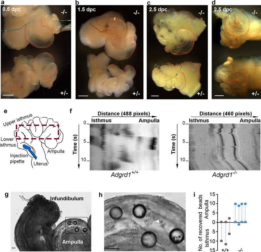
NATURE COMMUNICATIONS | https://doi.org/10.1038/s41467-021-21512-w                                                                      ARTICLE

Fig. 1 Adgrd1−/− female mice are infertile due to defective embryo transport. a Adgrd1−/− females are sterile irrespective of the male genotype.
b Adgrd1−/− female mice ovulate comparable numbers of oocytes compared to wild-type controls. Adgrd1−/− oocytes are fertilised at normal
frequencies both in vivo (c) and in vitro (d). e No implantation sites were observed in Adgrd1−/− females. f Adgrd1 mutant uteri support implantation of
transferred wild-type embryos. g Images showing implantation of transferred wild-type embryos in Adgrd1−/− and control uteri at 12.5 dpc; embryos
developed normally (insets). h 2.5 dpc embryos are ectopically located within the ampulla of an Adgrd1−/− oviduct. Arrow identifies morula-stage
embryo, arrowhead identifies a dead oocyte. Scale bar represents 100 µm. i Embryos are ectopically located in the ampulla of Adgrd1−/− mice at 2.5
dpc and 3.5 dpc. j Mutant Adgrd1−/− embryos recovered from the ampulla at 2.5 dpc have reached the morula stage. Scale bar represents 80 µm. Bars
in a, b, c, d, and i represent mean ± SEM. Each data point in a represents a biologically independent mating pair. In b, c, d, e, f and i each data point
represents a single mouse; source data are provided as a Source Data file. h and j are representative examples of at least five independent
experiments.

microtubules (Fig. 3c). To investigate ciliary function more                     mutant (Fig. 3f, f’). Consistent with this, there were no differences
directly, we placed beads on explants of oviductal ampullary                     in the thickness of the myosalpinx in either the ampullary or
epithelium and quantified their velocity. Beads placed on both                    isthmic regions of the oviduct (Fig. 3g). By in situ observation of
Adgrd1-mutant explants and controls moved in an abovarial                        the oviduct, we did not detect any overt differences in the rhythm
direction (Supplementary Movie 1), and at similar speeds                         or spatial locations of muscle contractions in Adgrd1-deficient
(Fig. 3d). These findings were consistent with the transport of the               oviducts. To examine this in more detail, we placed beads into the
cumulus complexes from the infundibulum to the ampulla which                     lumen of an Adgrd1-mutant oviductal explant to determine the
was normal in Adgrd1−/− mice, and thought to be largely driven                   effects of muscle contractions on their movement and observed
by ciliary action. We next examined the smooth muscle that lines                 that they were not rigidly held in position, but rather were moved
the oviduct (the myosalpinx) and observed no overt defects in                    back and forth in pendulum-like movements due to the contrac-
muscle organisation or structure in the mutant oviducts (Fig. 3e).               tions of the surrounding muscle (Supplementary Movie 2). These
We stained sections of the oviduct with a marker of smooth                       observations were consistent with the view that muscle contrac-
muscle and again observed no differences between wild type and                   tions mix rather than vectorially transport the contents of the

NATURE COMMUNICATIONS | (2021)12:1251 | https://doi.org/10.1038/s41467-021-21512-w | www.nature.com/naturecommunications                               3
ARTICLE                                                                     NATURE COMMUNICATIONS | https://doi.org/10.1038/s41467-021-21512-w

Fig. 2 Adgrd1 is expressed on the plasma membrane of both secretory and ciliated cells in the epithelium of the oviduct. a Adgrd1 promoter is active in
the isthmus of the oviduct as detected by whole mount X-gal staining using the lacZ reporter enzyme. Staining is detected in the homozygous oviducts
(left) but not in the wild-type sibling control (right). b Whole mount X-gal staining of homozygous Adgrd1 oviducts that have been removed from the ovary
and uterus and partially extended to show Adgrd1 promoter activity in the isthmus. c An Adgrd1 polyclonal antibody raised against the entire ectodomain
specifically stains the ampullary epithelium (purple) from wild type (upper panels) but not Adgrd1-deficient mice lower panels); anti-acetylated tubulin
staining was used to mark ciliated cells (green). d Adgrd1 is localised on the apical plasma membrane of ciliated and non-ciliated cells in the oviductal
epithelium. Scale bar represents 10 μm. e, Expression of Adgrd1 in the epithelial cells of the isthmus is (green, upper panel) and nuclei counterstained with
DAPI (blue, lower panel). c and e scale bars represent 50 μm. Representative examples of three independent experiments are shown.

tube10. Finally, and similar to experiments in other mammals7,22,               Consistent with the reduction in fluid production after ovulation,
tissue sections of wild type and Adgrd1−/− oviducts throughout                  ligating heterozygous oviducts in the post-ovulatory period (1.5
the oestrus cycle failed to reveal a constriction or occlusion at the           and 2.5 dpc) resulted in a much reduced accumulation of fluid
AIJ that could explain embryo retention, and this was consistent                (Fig. 4b and c). Adgrd1−/− mutant oviducts ligated at 0.5 dpc
with the ease of flushing trapped embryos from the ampulla of                    exhibited the same ampullary distention (Fig. 4a); however, by
Adgrd1−/− oviducts through the isthmus.                                         contrast to heterozygous littermates, did not show the same
                                                                                reduction at 1.5 and 2.5 dpc demonstrating that the post-ovulatory
                                                                                attenuation of fluid production in mutant oviducts is dysregulated
Attenuation of post-ovulatory fluid flow is dysregulated in                       (Fig. 4b and c). This was confirmed by performing the ligations
Adgrd1-deficient mouse oviducts. Oviductal fluid influences                        in vivo at 2.5 dpc, which resulted in a more conspicuous distension
embryo transport and varies throughout the oestrous cycle,                      of the ampulla due to the accumulated fluid in the mutant com-
peaking at ovulation and reducing in volume prior to the next                   pared to heterozygous controls (Fig. 4d and Supplementary
cycle23–27. Due to the small size and flexuous morphology of the                 Fig. 3a). Dysregulated fluid production was specifically observed in
mouse oviduct, ligation is a practical method of evaluating fluid                the isthmus by ligating oviducts within the isthmus itself (Sup-
production27, and so heterozygous Adgrd1+/− oviductal explants                  plementary Fig. 3b). While these ligation experiments demon-
were ligated at the infundibulum in vitro at 0.5 dpc and 1.5 dpc.               strated defects in oviductal fluid production, they did not directly
Four hours after ligation of 0.5 dpc oviducts, we observed a striking           show how oviductal fluid flow was altered in Adgrd1-mutant
distention of the ampulla due to the accumulated fluid (Fig. 4a).                oviducts. To address this, we developed a surgery setup similar to
This demonstrated that fluid exits the oviduct at the ovarial end                that used by Hino and Yanagimachi27 using a heating jacket that
creating a flow that opposes the movement of embryos - agreeing                  permitted experimental access to the female reproductive organs to
with observations in mice27, and larger mammals22,24,28,29.                     inject a tracer dye into the oviduct lumen and directly observe fluid

4                   NATURE COMMUNICATIONS | (2021)12:1251 | https://doi.org/10.1038/s41467-021-21512-w | www.nature.com/naturecommunications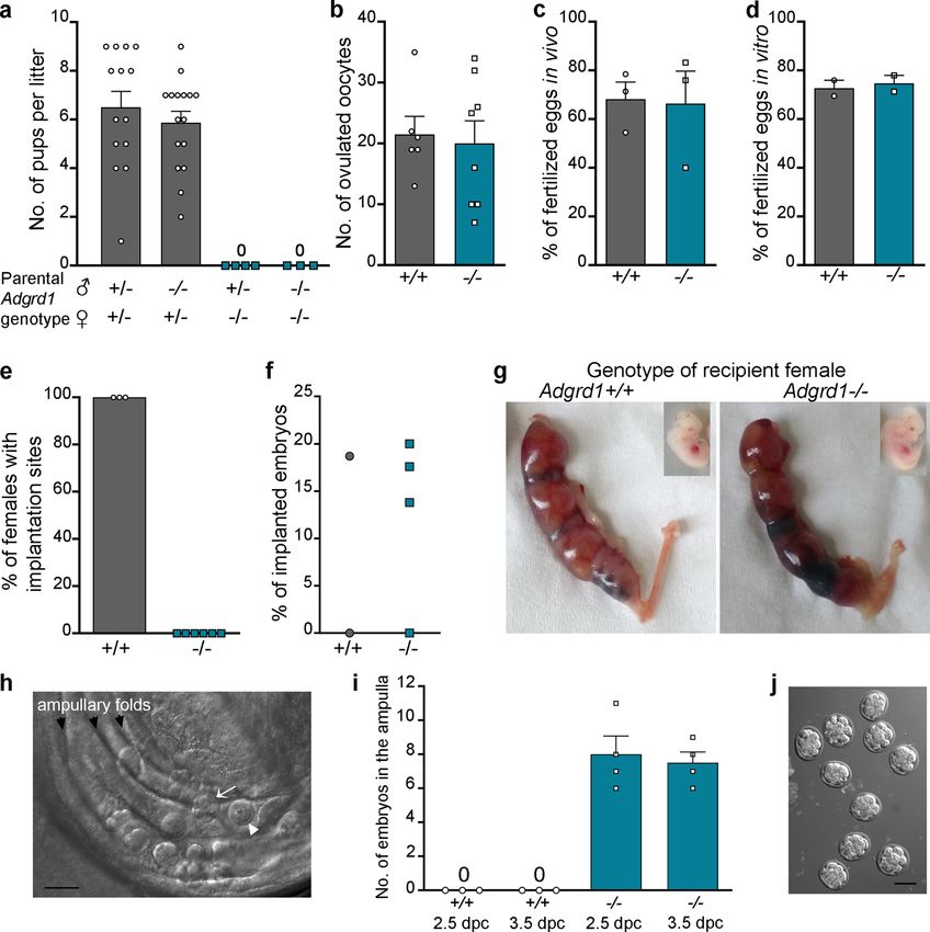
NATURE COMMUNICATIONS | https://doi.org/10.1038/s41467-021-21512-w                                                                      ARTICLE

Fig. 3 Adgrd1-deficient oviducts have morphologically normal cilia and muscle. a The distribution of ciliated cells is similar in the epithelium of control
Adgrd+/− and Adgrd1−/− oviducts. Oviductal sections from adult females in diestrous were stained with an antibody against acetylated tubulin to mark cilia
(green), and nuclei counterstained with DAPI (blue, right panel). b, b’ Ciliated cells analysed by scanning electron microscopy did not differ in mutant
Adgrd1−/− oviducts compared to wild-type controls. c Transmission electron microscopy images of Adgrd1−/− cilia showed the usual 9 + 2 organisation
of microtubules. d Polystyrene beads placed on oviductal epithelium explants moved at equivalent speeds in an abovarial direction in both control and
Adgrd1−/− epithelial tissues. Individual data points are bead velocity quantified with Image J manual tracking for a minimum of 15 s. Bars represent the
mean ± SEM, measurements were performed on 3 Adgrd+/+ and 3 Adgrd1−/− ampullae; an unpaired t-test analysis showed no significant (ns) difference
between the groups. e Image shows 3D projection of oviducts from 17-day-old mice stained with a phalloidin-Texas Red conjugate, demonstrating no overt
difference in muscle structure and organisation between wild type and mutant. f and f’ Representative examples of sections of the isthmus stained with
anti-smooth muscle alpha actin (magenta) and counterstained with DAPI (Cyan). g The thickness of the myosalpinx is similar in controls (hues of grey)
and Adgrd1−/− (hues of red) in the different oviductal regions. Bars represent the mean ± SEM, measurements were performed on 3 Adgrd+/− and 3
Adgrd1−/− oviducts; a minimum of two sections per mouse were analysed. A two-way ANOVA analysis found that the genotype has no effect (ns) while
the difference between the regions of the oviduct is extremely significant (p < 0.0001). a, b, b’, c, e, f, and f’ are representative examples of three
independent experiments.

NATURE COMMUNICATIONS | (2021)12:1251 | https://doi.org/10.1038/s41467-021-21512-w | www.nature.com/naturecommunications                                5
ARTICLE                                                                     NATURE COMMUNICATIONS | https://doi.org/10.1038/s41467-021-21512-w

Fig. 4 Adgrd1 regulates post-ovulatory attenuation of oviductal fluid production. a Accumulation of fluid is comparable in ligated control (+/−)
and mutant (−/−) oviductal explants in vitro at 0.5 dpc; ampullary distension is marked with red lines. b No reduction in fluid accumulation in mutant
Adgrd1−/− oviducts at 1.5 dpc by contrast to heterozygous control. c Oviductal ligation at 2.5 dpc in vitro: more fluid has accumulated in the ampulla of the
mutant compared to control. d The ampulla of mutant (−/−) oviducts exhibited a more pronounced distension compared to heterozygous control (+/−)
after in vivo ligated at 2.5 dpc and collected four hours later; the ampullary region is highlighted by dotted red lines. Images in a, b, c and d are
representative of at least three independent experiments and the scale bar represents 500 μm. e Schematic showing the region of the oviduct used for
producing the kymographs. f Kymographs showing the behaviour of a tracer dye injected into the oviduct of Adgrd1-mutants compared to wild type
littermate control. Adgrd1-mutant oviducts are rapidly filled with the dye which does not change over the time of observation whereas control oviducts
segregate the dye into boluses which are gradually moved towards the ampulla. g Glass beads (~100 μm diameter) were inappropriately retained in the
ampulla of Adgrd1−/− oviducts. Image taken 24 h after bead transplantation. h Magnification of the boxed area in g, showing glass beads and retained
embryo (arrowhead). Scale bars represent 80 μm, images are representative of four independent experiments. i Distribution of 10 to 12 transplanted glass
beads in Adgrd1−/− (n = 4) and control (n = 3) oviducts at 1.5 dpc.

movement in situ while maintaining a physiological temperature,                 the Adgrd1-deficient oviducts, the tracer dye instantaneously dis-
blood circulation and tissue innervation (Supplementary Fig. 4).                persed along the entire length of the oviduct so that once filled,
We focussed on comparing the fluid flow in Adgrd1-deficient and                    there was relatively little change in the distribution of the dye along
control oviducts at 1.5 dpc where the earlier ligation experiments              the length of the oviduct over the course of the observation
had demonstrated differences. Consistent with findings from other                (Supplementary Fig. 5 and Supplementary Movie 3). To visualise
groups and our ligation experiments, we observed adovarial flow of               these different behaviours, we drew kymographs which emphasised
oviductal fluid in both Adgrd1-deficient and control oviducts                     the pulsatile character observed in the fertile controls compared to
(Supplementary Fig. 5 and Supplementary Movie 3). In the control                the continuous rapid flow in the Adgrd1-deficient oviducts (Fig. 4e
animals, the dye behaved similarly to that observed by Hino and                 and f). Given these differences in fluid flow, we next asked if there
Yanagimachi: the dye segregated into several boluses within the                 were differences in secretory cell development in Adgrd1-mutant
oviduct close to the uterus which then gradually moved towards                  oviducts. Using a marker of secretory cells, we observed no dif-
the ovary in a saltatory manner that preserved the quantised nature             ference in either the relative number or organisation of these cells
of the dye distribution (Supplementary Movie 3). By contrast, in                within the mutant epithelium (Supplementary Fig. 6). Finally, we

6                   NATURE COMMUNICATIONS | (2021)12:1251 | https://doi.org/10.1038/s41467-021-21512-w | www.nature.com/naturecommunications
NATURE COMMUNICATIONS | https://doi.org/10.1038/s41467-021-21512-w ARTICLE
reasoned that if embryo retention was due to dysregulated ovi- which has prevented progress on developing treatments and
ductal fluid production, the transport of particles other than diagnostics. The currently known risk factors are generic (previous
embryos would be affected. Consistent with this, we found that IVF treatment, smoking, damage to the Fallopian tube due to
appropriately-sized glass beads were similarly retained within the surgery or infection) and therefore lack any insight into the
ampulla of Adgrd1−/− but not control oviducts (Fig. 4g, h and i). underlying mechanisms involved. The few genetic studies in
Together, these results show that the postovulatory cessation of humans have reported dysregulated expression of genes at the site
oviductal fluid flow is misregulated in the oviduct of Adgrd1- of ectopic implantation including HOXA1038, leukaemia inhibi-
deficient mice which could prevent embryo passage of the AIJ and tory factor39, and MUC140, but these observational studies do not
cause infertility. distinguish between cause and consequence. Female mice lacking
functional cannabinoid receptor1 (Cnr1) have reported defects in
embryo transport, although the phenotype differs from Adgrd1
The ADGRD1 ligand PLXDC2 is expressed on cumulus cells.
mutant mice because they are only subfertile, with 65% of mothers
Adgrd1 encodes a cell surface receptor belonging to the adhesion
still showing embryo implantation within the uterus. Moreover,
G-protein coupled receptor family, a subset of over 30 proteins
the defect results in a delay of embryo transport rather than
that typically contain a large N-terminal ectodomain, most of
complete block, because embryos are recovered from the isthmus
which have no identified ligand30. Adgrd1 contains a pentraxin
and are not retained within the ampulla41. Our finding that female
domain in its extracellular region and is known to initiate
mice lacking functional Adgrd1 are sterile due to embryo reten-
intracellular signalling by stimulatory G proteins leading to
tion in the ampulla provides strong evidence that embryo trans-
increases in cAMP levels by activating adenylate cyclase19. One
port is genetically controlled.
mechanism to trigger G-protein signalling used by this family of
Beyond unequivocally demonstrating a genetic basis for
receptors is through the ligand-dependent relief of an auto-
embryo transport, our results also provide a mechanism to
inhibitory ectodomain30–32. To identify a ligand for ADGRD1, we
explain the valve-like behaviour of the mammalian oviduct. We
first expressed the entire ectodomain as a multimeric binding
present a model (Fig. 6) where ovulated COCs are propelled
probe to increase binding avidity and thereby circumvent the
towards the uterus by ciliary action but are halted at the AIJ due
often weak binding affinities of extracellular receptor-ligand
to the opposing force of oviductal fluid flowing towards the ovary
interactions33. The highly avid ADGRD1 binding probe was then
caused by the narrowing of the oviduct at the isthmus. Adgrd1 is
systematically tested in an unbiased manner for binding to a
locally activated in the oviductal epithelium by Plxdc2 displayed
panel of 1132 unique human receptor ectodomains34, and the
on the surface of cumulus cells that are progressively released as
Plexin Domain-Containing Protein 2 (PLXDC2) was identified as
the jelly-like hyaluronic acid matrix surrounding the cumulus
a candidate ligand (Supplementary Fig. 7a). To investigate this
mass gradually disintegrates. The triggering of Adgrd1 decreases
further, we used an assay designed to detect direct extracellular
oviductal fluid production and consequently the flow that
receptor-ligand interactions called AVEXIS35 and demonstrated
opposes the constitutive abovarial ciliary action to permit con-
that ADGRD1 and PLXDC2 interacted in both bait-prey orien-
tinued passage of embryos through the isthmus. This suggests
tations (Fig. 5a). To further validate the interaction, we expressed
that cumulus cells not only support oocyte development, but also
the entire ectodomains of the mouse orthologues of both Adgrd1
communicate with the oviduct to regulate tubal transit. While the
and Plxdc2, and showed that they could directly interact (Fig. 5a).
flow of the oviductal fluid towards the ovary which opposes the
Cells transfected with a plasmid encoding Plxdc2 gained the
movement of embryos appears counterintuitive, this behaviour
ability to bind the highly avid Adgrd1 binding probe, and this
has been reported in several mammals and likely assists the ascent
binding was specifically blocked by preincubating the transfected
of sperm from the storage regions in the isthmus towards the site
cells with an anti-Plxdc2 antibody (Fig. 5b). We next mapped the
of fertilisation. It was first reported in larger animals such as
interaction interface to the pentraxin domain of Adgrd1 and the
cattle22, sheep24 and rabbits28 where the surgical implantation of
PSI domain of Plxdc2 by creating a series of truncated ectodo-
cannulas can be achieved more easily, but more recently observed
mains that encompassed known domains of both proteins and
directly using video microscopy in mice27. In mice, the fluid
using the AVEXIS assay to quantify binding (Fig. 5c, d and
empties out into the peritoneal cavity through a hole in the
Supplementary Fig. 7b). To understand how the Adgrd1 receptor
ovarian bursa and the rate of flow is surprisingly high, estimated
could be activated in the oviduct, we characterised the tissue
at 2.2 microlitres per hour in the periovulatory period but
expression patterns of Plxdc2. Within the female reproductive
decreasing by 1.5 dpc27.
system, Plxdc2 was most highly transcribed in the ovary and
One area for future investigation is to understand how Plxdc2
cumulus-oocyte complexes (COCs) compared to the uterus and
mediated activation of Adgrd1 and the consequent increase in
oviduct and other tissues such as brain, liver and spleen (Sup-
cAMP within the oviductal epithelial cells leads to changes in
plementary Fig. 7c). We confirmed the expression of Plxdc2
fluid flow to regulate embryo passage. Consistent with our find-
protein in the ovary and COCs by Western blotting (Fig. 5e), and
ings, prior research using perfused and cannulated oviducts has
showed using immunocytochemistry that Plxdc2 was highly
shown that increasing cAMP concentrations by adding a cell
expressed on the surface of cumulus cells (Fig. 5f). We demon-
permeable analogue of cAMP (dibutyryl cAMP) or agents that
strated that Plxdc2 is an activating ligand for Adgrd1 using an
increase cAMP levels such as forskolin, theophylline and cholera
established GPCR activation assay which measures the increase in
toxin all resulted in a decrease or abolition of oviductal secretion
cellular cAMP levels36. Adgrd1-transfected HEK293 cells showed
in humans42 and rabbits23,43. These results are in keeping with a
an expected increase in cAMP levels37 which was augmented by
regulatory role for fluid secretion and the fact that we observed no
Plxdc2 (Fig. 5g). Together, these results identify Plxdc2, which is
changes in the relative number and organization of the secretory
expressed on cumulus cells as an activating ligand for Adgrd1.
cells in the oviducts of Adgrd1-deficient mice. The addition of
inhibitors of ion channels that may be regulated by cAMP levels
Discussion to oviducts such as bumetanide and 5-nitro-2- (3-phenylpropyl-
Ectopic pregnancy is a relatively common complication which amino) benzoic acid did not provide any further insight, but these
affects up to 2% of all pregnancies in the United States and Europe experiments were compromised because they were performed on
and is a major risk factor for maternal health. Despite being so explanted oviducts which are separated from the circulation and
prevalent, we know remarkably little about the underlying causes innervation. One possibility is that fluid flow could be regulated
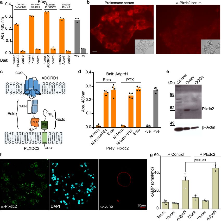
NATURE COMMUNICATIONS | (2021)12:1251 | https://doi.org/10.1038/s41467-021-21512-w | www.nature.com/naturecommunications 7ARTICLE NATURE COMMUNICATIONS | https://doi.org/10.1038/s41467-021-21512-w Fig. 5 Plxdc2 is an activating ligand for Adgrd1 and expressed on cumulus cells. a Direct interactions between human and mouse ADGRD1 and PLXDC2 ectodomains in both bait-prey orientations using the AVEXIS assay (n = 3 independent replicates). b Adgrd1 ectodomain probe bound Plxdc2-transfected cells (left panel) and was blocked by preincubation with Plxdc2 antiserum (right). Scale bar represents 10 μm. c Schematic of ADGRD1 and PLXDC2 domain organisation. d Domain truncations demonstrate that the pentraxin (PTX) domain of Adgrd1 and PSI domain of Plxdc2 are sufficient for binding (n = 4 independent replicates). e Western blotting and immunofluorescence f demonstrates that Plxdc2 is expressed on the plasma membrane of cumulus cells. HEK293T cells were used as negative control in e, anti-Juno shows the localisation of the oocyte in f. g The overexpression of Adgrd1 induces a significant increase of intracellular cAMP compared to non-transfected cells (mock) and to cells transfected with a plasmid encoding GFP (vector). The Plxdc2 ectodomain induces a significant increase of cAMP levels in Adgrd1-expressing cells only. (n = 2 independent experiments). Bars in a, d, and g represent mean ± SEM. by changes in the tone of the myosalpinx to control the rigidity of gradual, and will therefore require advanced longer term in vivo the oviduct. In our characterisation of the Adgrd1-deficient ovi- imaging to investigate. ducts, however, we observed no morphological or developmental The roles of PLXDC2 and ADGRD1 in humans are poorly defects within the oviductal smooth muscle, and when observed characterised and our findings warrant further investigations, in situ, the contractions appeared completely normal in terms of particularly for any role they may have in reproduction and their frequency, strength, and localisation. It has been convin- ectopic pregnancy. Both genes are additionally expressed outside cingly shown by others that ablating oviductal muscle contrac- of the female reproductive system suggesting other functions, and tions using nicardipine had no effect on the rate of oviductal fluid systematic mouse knockout phenotyping have reported bone flow27 suggesting no direct role of muscle in regulating oviductal mineralisation phenotypes as well as female sterility for Adgrd1 fluid flow. Similarly, previous attempts to explain the tubal- (see the significant phenotypes reported by the International locking phenomenon led to careful histological examination of Mouse Consortium https://www.mousephenotype.org/data/ the muscles at the AIJ, and did not reveal a sphincter-like genes/MGI:3041203). Mice containing a transgene targeting the organization7,22, and complete resection of the AIJ did not affect majority of the signal peptide of Plxdc2 resulted in a ten-fold the fertility of rabbits44. If the myosalpinx does play a regulatory decrease in Plxdc2 transcript levels in the cerebella45 but no role in fluid flow, then these changes are likely to be subtle and abnormal phenotypes were reported. One suggested possibility is 8 NATURE COMMUNICATIONS | (2021)12:1251 | https://doi.org/10.1038/s41467-021-21512-w | www.nature.com/naturecommunications
NATURE COMMUNICATIONS | https://doi.org/10.1038/s41467-021-21512-w ARTICLE
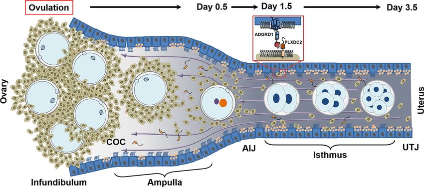
Fig. 6 Model describing Adgrd1-mediated control of fluid flow underlies the valve-like behaviour of the mammalian oviduct. Cumulus-oocyte-
complexes are ovulated into the infundibulum and rapidly transported to the AIJ by the action of the cilia lining the oviductal epithelium beating towards
the uterus. At the AIJ, the COCs are halted due to the balance of abovarial ciliary action and the force of adovarial oviductal fluid flow (purple arrows). In
wild-type oviducts, the gradual release of cumulus cells as the hyaluronic acid matrix which surrounds them slowly disintegrates releases the Plxdc2 ligand
and triggers a reduction of oviductal fluid production by activating Adgrd1 on the oviductal epithelium to licence embryo release. COC = cumulus-oocyte-
complex; AIJ = ampullary-isthmic junction; UTJ = uterotubal junction.
that a gene encoding a related protein, Plxdc1, might compensate gonadotropin (PMSG) followed by 5 IU of human chorionic gonadotropin (hCG)
for the role of Plxdc2. Genome-wide association studies have 48 h later. In vivo fertilisation was assessed by scoring the number of zygotes and
the number of non-fertilised oocytes present in the ampulla at 0.5 dpc. Eggs and
implicated polymorphisms linked to ADGRD1 with variations in embryos were fixed in 4% formalin (28906 Thermo Scientific Pierce) and stained
adult height46,47 and heart beating frequency in electro- with DAPI (62248 Thermo Fisher). In vitro fertilisation was performed essentially
cardiograms48. Subsequent studies focussed on ADGRD1, repor- as described52. Briefly, sperm were collected from the cauda epididymis of adult
ted no clinical phenotypes in adults that are heterozygous for male mice, capacitated for 1 h in HTF medium at 37 °C and added to cumulus-
predicted loss of function alleles consistent with our findings in enclosed oocytes that had been collected 13 h after hCG treatment. Groups of 20 to
30 eggs were inseminated in 100 μL drops of HTF medium containing 1 × 10⁵
mice49. Importantly, tubal ectopic pregnancies are restricted to sperm; four hours later, eggs were washed and cultured in KSOM (MR-121D
primates and are not observed in other animals1. In mice, EmbryoMax, Millipore) for the following four days. Formation of pronuclei was
embryos recovered from the ampulla of Adgrd1-deficient mice scored and embryo development recorded daily. Mutant and age-matched control
had not developed beyond the blastocyst stage in agreement with females were mated with proven males and the day of vaginal plug formation was
counted as 0.5 dpc. The female reproductive tracts were visually inspected for the
the reported ability of the ampulla to sustain the development presence of implantation sites at 5.5 dpc, 6.5 dpc and 8.5 dpc. The embryo position
in vitro to that stage50, and suggesting that the ampulla cannot in the oviducts was determined using the Zeiss Axioplan 2 microscope and
support embryo implantation and further development. embryos were counted after flushing the oviducts with M2 medium (Sigma).
Our demonstration that Adgrd1 is essential for embryo Non-surgical embryo transfer (NSET) was performed as described53. Wild-type
2-cell embryos were cultured in KSOM for 48 h and blastocysts were transferred to
transport provides strong evidence that embryo transport is pseudopregnant Adgrd1+/+ and Adgrd1−/− recipient females at 2.5 dpc using the
genetically controlled, and provides a plausible mechanism to NSET device (Paratechs, catalogue #60010). 14 and 16 blastocysts were transferred
explain how the tubal locking phenomenon described many years to two wild-type females, and 14, 17, 29 and 30 blastocysts were transferred to four
ago in the mammalian oviduct is regulated. Ectopic pregnancy is Adgrd1-deficient females. The number of implanted embryos was counted ten days
after the transfer.
a common and potentially deadly condition for which we have
very little mechanistic understanding, and so our findings here
not only provide potential starting points to develop diagnostics Time-lapse video microscopy. Oviducts were carefully dissected from 8-week-old
and treatments but also an animal model to study this condition females at the required stage of the oestrus cycle. Entire oviducts were separated
from the ovary by opening the ovarian bursa, and scission of the ovarian ligament;
further. isolation from the rest of the reproductive tract was achieved by trimming the
broad ligament whilst maintaining the utero-tubal junction. When cutting the
mesosalpinx, care was taken to avoid pulling and damaging the tubes. Spontaneous
Methods rhythmic contractions of oviducts were documented by video recording immedi-
Generation, breeding and fertility phenotyping of Adgrd1-deficient mice. All ately after dissection using a Zeiss Axioplan 2 microscope equipped with a
animal experiments were performed under UK Home Office governmental reg- Hamamatsu 1394 ORCA-ERA digital camera and the Velocity imaging software.
ulations and European directive 2010/63/EU. Research was approved by the Sanger Fifty seconds of real time were acquired at a frame frequency of 1 Hz. To record
Institute Animal Welfare and Ethical Review Board. Adgrd1−/− mice were ciliary beating, the ampullary region of the oviducts were opened longitudinally,
obtained from the Knockout Mouse Project resource (IKMC Project: 22527) and placed in PBS on a microscope glass and microparticles (Sigma 74964) were gently
contain a lacZ-tagged allele targeted to the Adgrd1 genomic locus located on added to the tissue. Movies were acquired at 8.5 frames per second.
chromosome 5, Adgrd1tm1b(EUCOMM/Wtsi). Mice were generated by injecting blas- Micro beads (Micro particles based on polystyrene, 15μm of diameter; no.
tocysts with the targeted mouse embryonic stem cells which were transferred to 74964 Merck Life Science) were applied to longitudinally opened ampullae in
pseudopregnant females to generate chimeras. Germline transmission of the tar- DPBS with Ca2+ and Mg2+, and live videos were recorded with a stereomicroscope
geted allele was confirmed by PCR after mating of chimeric males with C57BL/ (Leica M205FA) equipped with the Leica DFC7000T camera. The velocity of bead
6NTac females. To obtain the reporter-tagged deletion allele, females heterozygous transport was quantified with Image J manual tracking for a minimum of 15 s
for the ‘knockout-first’ allele Adgrd1tm1a(EUCOMM/Wtsi) were crossed to hemizygous per bead.
males ubiquitously expressing the Cre enzyme51. Mice with the recombined allele
were identified using PCR and diagnostic primers using genomic DNA extracted
from ear biopsies (4403319 DNA Extract All Reagents Kit, Life Technologies) as Visualisation and analysis of the oviductal fluid flow. The design of the plastic
the template for short range PCR using Platinum® Taq DNA Polymerase jacket was drawn with AutoCAD Inventor and printed with Ultimaker 2+ Connect
(10966034 Invitrogen). The mouse colony was maintained by crossing hetero- 3Dprinter using a PolySmooth printing filament. After printing the surface was
zygous males and females. Male and female fertility was quantified by pairing smoothed with Isopropanol and the lid was sealed, finally the surface was treated
homozygous and heterozygous Adgrd1 transgenic adult mice with homozygous with the Polymaker Polysher from 3DJake UK to prevent leakage. In preparation
and heterozygous animals of proven fertility and the number of resulting pups was for surgery the jacket was connected to a motor water pump immersed in warm
monitored continuously for three months. The number of ovulated oocytes was water. For the visualisation of oviductal fluid flow female mice were mated, selected
counted after induction of ovulation with 5 IU of pregnant mare serum at the required time, and prepared for surgery as described above. The ovary and
NATURE COMMUNICATIONS | (2021)12:1251 | https://doi.org/10.1038/s41467-021-21512-w | www.nature.com/naturecommunications 9ARTICLE NATURE COMMUNICATIONS | https://doi.org/10.1038/s41467-021-21512-w
the oviduct were exposed on the surface of the surgical incision and manipulated removed, a surgeon’s knot was tied around the oviduct with polypropylene suture
with care to avoid damaging the tissues. A topical tissue adhesive (Gluture, Zoetis) (Ethicon Prolene 10-0, W2794). The ligated oviducts were maintained in culture at
was used to hold the fat pad and the ovary onto the skin on one side of the incision. 37 °C in 500 µL of pre-warmed KSOM media, fixed in NBF overnight, and imaged
The jacket containing circulating warm water was sealed to the mouse skin and with a Leica M205FA stereomicroscope.
pre-warmed DPBS with Ca2+ and Mg2+ (Hyclone SH30264) was added to Glass beads with a diameter of ~100 μm (Sigma G4649) for transfer into
maintain the tissues at ~37 °C. A 40 μm bevelled glass pipette (BioMedical oviducts were sterilised by autoclaving and washed in M2 medium. Female
Instruments VESbv-40-0-0-55) was used to inject a tracer dye (India Ink) in the recipient mice whose mating had been timed were prepared for surgery using
lower isthmus. The dye was dialysed overnight in DPBS and diluted twofold in the gaseous anaesthesia under aseptic conditions, shaved, and given analgesia. Using a
same buffer before injections. Videos were recorded with the Leica MC170HD stereomicroscope (Leica MZ7.5), a small longitudinal skin incision was made at the
camera at 24fps and retrospectively analysed. Kymographs were generated with the midline, level with the last rib. The ovary was located through the muscle wall and
dedicated Image J plug-in by selecting a region encompassing the oviducts for a micro scissors were used to make a small incision, followed by blunt dissection
total of 12.5 seconds. and exteriorisation of the ovary and oviduct by holding the associated fat pad,
anchored using a serrefine clamp. After removing the bursa covering the ovary and
oviduct, glass beads were taken up using a mouth pipette into a 230 mm Pasteur
Wholemount LacZ staining, immunofluorescence and immunohistochemistry. glass pipette with the aid of a stereomicroscope (Leica MZ9.5) and transferred into
To determine the expression of the lacZ reporter gene in the targeted allele at the the oviducts of recipient mice at 0.5 dpc. The location of the beads in the ampulla
Adgrd1 locus, dissected tissues were fixed in 10% Neutral Buffered Formalin (NBF, and isthmus were counted at 1.5 dpc. In vivo ligation of oviducts was performed
Cellpath) overnight, washed with PBS, stained with a LacZ staining solution (2 mM using the same surgical procedure with polypropylene suture which was slid under
MgCl2, 0.02% IGEPAL CA-630, 5 mM potassium ferrocyanide, 5 mM potassium the oviduct ~1 mm from the infundibulum and fine forceps used to tie a surgeon’s
ferricyanide, 0.01% deoxycholic acid, 0.1% X-Gal (5-bromo-4-chloro-3-indolyl knot. When assessing fluid accumulation in the isthmus, additional ligatures were
β-D-galactopyranoside) in dimethylformamide) at 4 °C, post-fixed in 4% formalin tied at the ampullary-isthmic junction and the isthmus. The ovary and oviduct
and stored in 70% glycerol. were carefully replaced within the body cavity using blunt forceps, and the skin
Polyclonal antiserum was raised by immunising rabbits with the entire incision closed using a single wound clip.
ectodomains of mouse ADGRD1 or mouse PLXDC2 expressed as soluble
recombinant 6-his-tagged proteins in HEK293-6E cells and purified using Ni2+-
NTA chromatography, essentially as described54. Immunisations were carried out Preparation of the protein library for large-scale receptor screening. A library
by Cambridge Research Biochemicals in accordance with UK Home Office of single-transmembrane-spanning (STM) human proteins was compiled using
regulations and the Sanger Institute animal welfare ethical review board. computational prediction algorithms and experimental evidence, as described25.
To prepare wholemount tissues for immunofluorescence, they were fixed in 4% The ectodomain of each protein was synthesised and cloned into the pRK5 vector
formalin for 30 min at room temperature, incubated with the primary antibody for (Genentech) in frame with a C-terminal Fc (hIgG1) tag. The resulting library
1 h, and washed extensively with PBS and finally stained with the secondary consists of 1364 human proteins, including 1132 unique receptors and a number of
antibody conjugated with the appropriate fluorochrome (Alexa Fluor 488 goat anti- replicate constructs as assay controls. Expression of the human STM protein library
mouse supplied by Thermo Fisher, and Alexa Fluor 647 goat anti-rabbit supplied was performed as described34. In brief, conditioned media enriched in individual
by Jackson Immunoresearch). Where tissues were sectioned, tissue was fixed in 4% receptors was prepared in Expi293F cells (Thermo Fisher), transiently transfected
formalin for 60 min, rinsed extensively in PBS and soaked in a sucrose solution with receptors expressed as ectodomains fused to a human Fc (IgG1). Expi293F
overnight before embedding in OCT and 8 micron sections cut with a Leica cells were cultured in Expi293 Expression Media (Cell Technologies) in flasks at
CM1950 cryostat. Sections were treated with 1% SDS in PBS for 5 min, blocked 37 °C and 150 r.p.m. agitation in a humidified incubator. Human cells were chosen
before adding primary antibody overnight at 4 °C. Sections were washed and for expression of the collection of STM receptors to maximise protein quality and
incubated with a goat anti-rabbit – Alexa488 secondary antibody for 1 h at room incorporation of relevant post-translational modifications. One mL cell transfec-
temperature. Sections were then washed and mounted in slowFade Gold mounting tions were performed using an automated system consisting of a TECAN liquid
solution with DAPI (Thermo). handling system and a MultiDrop reagent dispenser. Transfections were processed
Binding of recombinant proteins to cells was quantified by incubating in batches of 96 clones using a Biomek FX liquid handling robot and conditioned
transfected cells with highly avid ectodomains expressed as FLAG-tagged media was harvested 7 days post-transfection. Subsequently, the receptor Fc-tagged
pentameric preys in cell culture medium at 37 °C before fixation in a phosphate- ectodomains present in the conditioned media were captured on protein A-coated
buffered 4% formalin solution. Staining of muscle was performed by first 384-plates (Thermo Scientific), and stored at 4 °C until use. For the binding screen,
permeabilizing fixed oviducts with 0.2% Triton X-100 and incubating with Texas- the ectodomain of ADGRD1, truncated at S600, was expressed in HEK293-6E cells
Red-phalloidin conjugate for 30 minutes. Optical sections of whole oviductal tissue as a pentameric probe fused to a beta-lactamase enzyme55. A Thr to Gly mutation
were acquired with a Leica SP5 laser confocal microscope (z-step size = 2.52μm). at position 574 in the GPCR proteolytic site (GPS) of human ADGRD1 was
The 3D projection of the whole z-stack was generated with the LAS AF software. introduced to prevent the autoproteolysis56.
The antibodies and probes and the dilutions used in this study were: polyclonal
rabbit-anti-mouse Adgrd1 (this study) 1:100; polyclonal rabbit-anti-mouse Plxdc2
(this study) 1:100; rat monoclonal anti-mouse Juno (clone TH6, Biolegend) 1:200; Identification of binding extracellular ligands for ADGRD1. The Cell Surface
mouse monoclonal anti-acetylated Tubulin (clone 6-11B-1, Sigma Aldrich); anti- Receptor Interaction screening was based on the AVEXIS method35, which was
Pax8 rabbit polyclonal antibody (10336-1-AP, Proteintech) 1:400; mouse further implemented for automated high throughput screening in 384 well plate
monoclonal anti-alpha smooth muscle Actin (clone 1A4, A2547, Sigma) 1:400; format, as recently described34. In brief, screens were performed using an inte-
mouse monoclonal anti-Flag-Cy3 conjugate (clone M2, Sigma Aldrich) 1:500; grated robotic system consisting of automated liquid handling devices for high
Texas-red conjugated phalloidin (Thermo Fisher, T7471); Alexa Fluor 633 goat throughput analysis of receptor-ligand interactions. On the day of the assays, the
anti-mouse IgG (Thermo Fisher A-21052)1:500; Alexa Fluor 488 goat anti-mouse protein A plates coated with the receptor library were washed with PBS containing
IgG (405319 Biolegend) 1:500; Alexa Fluor 488 goat anti-rabbit IgG (111-545-003 Ca2+ and Mg2+, followed by incubation with the beta-lactamase-tagged penta-
Jackson Immunoresearch) 1:500; pentameric FLAG-tagged PLXDC2 (this meric ADGRD1 probe for 1 h at room temperature. Plates were then washed to
study) 1:10. remove free protein prior to addition of nitrocefin (Calbiochem) for 1 h at room
temperature. Hydrolysis of the beta-lactamase substrate nitrocefin was quantified
by measuring absorbance at 485 nm using an integrated TECAN plate reader. The
Transmission and scanning electron microscopy. Mouse oviducts were fixed at raw enzymatic absorbance values were analysed to calculate Z-scores across all
room temperature in a 2% formalin/2.5% glutaraldehyde mixture buffered in 0.1 M proteins in the library. Comparisons to previous screens of this type identified
sodium cacodylate at pH 7.4 for one hour, rinsed and fixed in 1% osmium tetroxide immobilised ligands which repeatedly gave positive binding signals irrespective of
for another hour followed by 1% tannic acid and 1% sodium sulphite for 30 the binding probes used, and included known lectins (e.g., SIGLEC family mem-
minutes respectively. Samples were then dehydrated in an ethanol series, staining bers and MRC1) which are likely to directly interact with common glycans pre-
en bloc with uranyl acetate at the 30% stage before embedding (Epoxy embedding sented on the binding probe.
medium kit – Sigma). 50 nm ultrathin sections were cut on a Leica UC6 ultra-
microtome and imaged on an FEI 120 kV Spirit Biotwin TEM with a F4.15 Tietz
camera. Recombinant protein production and protein interaction screening. The
For scanning electron microscopy, mouse oviducts were fixed as for TEM extracellular domains of ADGRD1 and PLXDC2 were expressed as soluble secreted
(replacing tannic acid with osmium-thiocarbohydrazide), dehydrated in an ethanol proteins by transient transfection in HEK293-6E cells55. NCBI reference sequences
series, critical point dried in a Leica CPD300, mounted and sputter-coated with for the expressed proteins were: mouse Adgrd1: NP_001074811; human ADGRD1:
2 nm of platinum using a Leica ACE600 evaporator. Samples were imaged on a NP_001317426; mouse Plxdc2: NP_080438; human PLXDC2: NP_116201. The
Hitachi SU8030 SEM. region encoding the entire predicted ectodomain was chemically synthesised by
gene synthesis (GeneArt, Life Technologies) flanked by unique NotI, AscI
restriction sites to facilitate cloning into mammalian expression plasmids to pro-
Oviduct ligation, surgical procedures and transfer of beads. Oviducts from duce enzymatically biotinylatable monomeric ‘baits’ or pentameric FLAG-tagged
mice at the required stage in the oestrus cycle were dissected under a Nikon ‘preys’; both bait and preys contained a rat Cd4d3 + 4 epitope tag as described.
SMZ800 stereo microscope. Once the ovary was separated from the oviduct and Biotinylated proteins were produced by co-transfecting a plasmid encoding a
10 NATURE COMMUNICATIONS | (2021)12:1251 | https://doi.org/10.1038/s41467-021-21512-w | www.nature.com/naturecommunicationsNATURE COMMUNICATIONS | https://doi.org/10.1038/s41467-021-21512-w ARTICLE
secreted version of the BirA enzyme55. To produce recombinant proteins corre- 5. Pauerstein, C. J. & Eddy, C. A. The role of the oviduct in reproduction; our
sponding to specific regions of both ADGRD1 and PLXDC2 ectodomains, primers knowledge and our ignorance. J. Reprod. Fertil. 55, 223–229 (1979).
were designed that would amplify the appropriate region from the plasmid 6. Humphrey, K. W. Observations on transport of ova in the oviduct of the
encoding the entire ectodomain by PCR and the products cloned into both bait and mouse. J. Endocrinol. 40, 267–273 (1968).
prey expression plasmids using unique NotI and AscI sites54. Bait proteins were 7. Greenwald, G. S. A study of the transport of ova through the rabbit oviduct.
normalised by ELISA using a monoclonal antibody (Ox68) recognising the rat Fertil. Steril. 12, 80–95 (1961).
Cd4d3 + 4 tag. Prey proteins were normalised to their β-lactamase activity to levels 8. Coy, P., García-Vázquez, F. A., Visconti, P. E. & Avilés, M. Roles of the
suitable for the AVEXIS assay as described57. Biotinylated baits that had been oviduct in mammalian fertilization. Reproduction 144, 649–660 (2012).
either purified or dialysed against PBS to remove excess free D-biotin were 9. Weber, J. A., Woods, G. L. & Aguilar, J. J. Location of equine oviductal
immobilised in streptavidin-coated 96-well microtitre plates (NUNC). Preys were embryos on Day 5 post ovulation and oviductal transport time of Day 5
incubated for one hour, washed three times in PBS/0.1% Tween-20 and once in embryos autotransferred to the contralateral oviduct. Theriogenology 46,
PBS. Finally, 60 µl of 125 µg/ml nitrocefin was added and the absorbance measured 1477–1483 (1996).
at 485 nm on a Pherastar plus (BMG laboratories). A biotinylated protein con- 10. Koester, H. Ovum Transport. in Mammalian Reproduction 189–228 (Springer
sisting of just the rat Cd4d3 + 4 tag alone was used as a negative control bait and a Berlin Heidelberg, 1970).
biotinylated Ox68 monoclonal antibody (anti-prey) diluted 1:2,000 was used as an 11. Bassilana, F., Nash, M. & Ludwig, M.-G. Adhesion G protein-coupled
internal control. The rat Cd200-Cd200R interaction was used as a positive control.
receptors: opportunities for drug discovery. Nat. Rev. Drug Discov. 18,
869–884 (2019).
Western blot. To perform Western blotting, proteins from mouse ovary, ovulated 12. Leemans, J. C. et al. The epidermal growth factor-seven transmembrane (EGF-
COCs, and HEK293-T were extracted with RIPA buffer and quantified with TM7) receptor CD97 is required for neutrophil migration and host defense. J.
Bradford assay (Thermo) following manufacturer’s instructions. Briefly, normalised Immunol. 172, 1125–1131 (2004).
protein amounts were resolved under reducing conditions by SDS–PAGE and 13. Yona, S. et al. Ligation of the adhesion-GPCR EMR2 regulates human
blotted to Hybond-P PVDF membrane (GE Healthcare) for 1 h at 30 V. After neutrophil function. FASEB J. 22, 741–751 (2008).
blocking for 1 h with 2% BSA, the membrane was incubated for 1 h with Plxdc2 14. O’Sullivan, M. L. et al. FLRT proteins are endogenous latrophilin ligands and
antiserum diluted 1:100 or with anti beta-Actin (Abcam, ab8227) diluted 1:1000 in regulate excitatory synapse development. Neuron 73, 903–910 (2012).
PBST(PBS/0.1% Tween-20) added with 2% BSA, washed three times and incubated 15. Cullen, M. et al. GPR124, an orphan G protein-coupled receptor, is required
with a horseradish peroxidase (HRP)-conjugated anti-rabbit antibody (Thermo- for CNS-specific vascularization and establishment of the blood-brain barrier.
Fisher Scientific Cat. No. G21234) diluted 1:5,000. Proteins were detected using Proc. Natl Acad. Sci. USA 108, 5759–5764 (2011).
SuperSignalWest Pico Chemiluminescent substrate (Thermo Scientific) and devel- 16. Lu, S. et al. Developmental vascular remodeling defects and postnatal kidney
oped on photographic film. Uncropped blots can be found in the source data file. failure in mice lacking Gpr116 (Adgrf5) and Eltd1 (Adgrl4). PLoS ONE 12,
e0183166 (2017).
RT-PCR. Total RNA was extracted from mouse tissues using Trizol reagent 17. Davies, B. et al. Targeted deletion of the epididymal receptor HE6 results in
(Invitrogen) as per manufacturer’s instructions, resuspended in water, and quan- fluid dysregulation and male infertility. Mol. Cell. Biol. 24, 8642–8648 (2004).
tified with a Nanodrop 1000 spectrophotometer (Thermo Scientific). SuperScript 18. Chen, G., Yang, L., Begum, S. & Xu, L. GPR56 is essential for testis
III reverse transcriptase (Invitrogen) was used to produce cDNA from 1 μg of RNA development and male fertility in mice. Dev. Dyn. 239, 3358–3367 (2010).
and subsequent amplification was obtained with the KOD hot start DNA poly- 19. Liebscher, I. et al. A tethered agonist within the ectodomain activates the
merase (Novagen). A list of all primers is provided in Supplementary Table 1. PCR adhesion G protein-coupled receptors GPR126 and GPR133. Cell Rep. 10,
products were resolved on a 1.5% agarose gel and imaged with an Azure c-600 gel 1021 (2015).
documentation system. 20. Stoveken, H. M., Hajduczok, A. G., Xu, L. & Tall, G. G. Adhesion G protein-
coupled receptors are activated by exposure of a cryptic tethered agonist. Proc.
cAMP ELISA. Adherent HEK293-T cells were transiently transfected with either a Natl Acad. Sci. USA 112, 6194–6199 (2015).
plasmid encoding mouse Adgrd1, or a negative control encoding membrane-tethered 21. White, J. K. et al. Genome-wide generation and systematic phenotyping of
EGFP (vector), or were treated with the transfection reagent Lipofectamine 2000 alone knockout mice reveals new roles for many genes. Cell 154, 452–464 (2013).
(mock). Twenty-four hours after transfection, cells were seeded on a streptavidin- 22. Black, D. L. & Davis, J. A blocking mechanism in the cow oviduct. J. Reprod.
coated microtitre plate that had been pre-incubated with the mouse PLXDC2 or Fertil. 4, 21–26 (1962).
control rat Cd200 biotinylated ectodomains. After three hours at 37 °C, the levels of 23. Gott, A. L., Gray, S. M., James, A. F. & Leese, H. J. The mechanism and control
cyclic AMP were determined in duplicate using a cAMP ELISA kit (ADI-900-066, of rabbit oviduct fluid formation. Biol. Reprod. 39, 758–763 (1988).
Enzo Life Science) according to the manufacturer’s protocol, and normalised to the 24. Belve, A. R. & McDonald, M. F. Directional flow of fallopian tube secretion in
total protein concentration determined using the Bradford assay (ThermoFisher the Romney ewe. J. Reprod. Fertil. 15, 357–364 (1968).
Scientific). One-way ANOVA showed a significant variation among conditions 25. Kanayama, K. & Osada, H. Relationship between changes in volume of the
(F5,12 = 43, p < 0.0001) and a post-hoc Bonferroni test indicated that intracellular oviductal fluid in the ampulla and the descent of ovulated eggs from the
cAMP differed significantly in Adgrd1-transfected cells treated with Plxdc2 compared ampulla to the isthmus in mice. J. Int. Med. Res. 28, 20–23 (2000).
to Adgrd1-transfected cells treated with the control protein CD200 (p = 0.0391). 26. Black, D. L., Duby, R. T. & Riesen, J. Apparatus for the continuous collection
of sheep oviduct fluid. J. Reprod. Fertil. 6, 257–260 (1963).
Reporting summary. Further information on research design is available in the Nature 27. Hino, T. & Yanagimachi, R. Active peristaltic movements and fluid
Research Reporting Summary linked to this article. production of the mouse oviduct: their roles in fluid and sperm transport and
fertilization. Biol. Reprod. 101, 40–49 (2019).
Data availability 28. Black, D. L. & Asdell, S. A. Transport through the rabbit oviduct. Am. J.
Data supporting the findings of this manuscript are available in the Source Data file and Physiol. 192, 63–68 (1958).
from the corresponding author upon reasonable request. Source data are provided with 29. Edgar, D. G. & Asdell, S. A. The valve-like action of the uterotubal function of
this paper. the ewe. J. Endocrinol. 21, 315–320 (1960).
30. Hamann, J. et al. International Union of Basic and Clinical Pharmacology.
XCIV. Adhesion G protein–coupled receptors. Pharmacol. Rev. 67, 338–367
Received: 6 December 2019; Accepted: 29 January 2021; (2015).
31. Araç, D. et al. A novel evolutionarily conserved domain of cell-adhesion
GPCRs mediates autoproteolysis: Cell-adhesion GPCRs mediates
autoproteolysis. EMBO J. 31, 1364–1378 (2012).
32. Coleman, J. L. J., Ngo, T. & Smith, N. J. The G protein-coupled receptor N-
terminus and receptor signalling: N-tering a new era. Cell. Signal. 33, 1–9
References (2017).
1. Corpa, J. M. Ectopic pregnancy in animals and humans. Reproduction 131, 33. Wright, G. J. Signal initiation in biological systems: the properties and
631–640 (2006). detection of transient extracellular protein interactions. Mol. Biosyst. 5,
2. Shaw, J. L. V., Dey, S. K., Critchley, H. O. D. & Horne, A. W. Current 1405–1412 (2009).
knowledge of the aetiology of human tubal ectopic pregnancy. Hum. Reprod. 34. Martinez-Martin, N. et al. An unbiased screen for human cytomegalovirus
Update 16, 432–444 (2010). identifies neuropilin-2 as a central viral receptor. Cell 174, 1158–1171 (2018).
3. Croxatto, H. B. & Ortiz, M. E. Egg transport in the fallopian tube. Gynecol. e19.
Invest. 6, 215–225 (1975). 35. Bushell, K. M., Söllner, C., Schuster-Boeckler, B., Bateman, A. & Wright, G. J.
4. Knobil, E. et al. The physiology of reproduction, second edition. Large-scale screening for novel low-affinity extracellular protein interactions.
Endocrinologist 5, 77–78 (1995). Genome Res. 18, 622–630 (2008).
NATURE COMMUNICATIONS | (2021)12:1251 | https://doi.org/10.1038/s41467-021-21512-w | www.nature.com/naturecommunications 11You can also read