The potential role of necroptosis in clinical diseases (Review)
←
→
Page content transcription
If your browser does not render page correctly, please read the page content below
INTERNATIONAL JOURNAL OF MOlecular medicine 47: 89, 2021
The potential role of necroptosis in clinical diseases (Review)
WENLI DAI1*, JIN CHENG1*, XI LENG2, XIAOQING HU1 and YINGFANG AO1
1
Institute of Sports Medicine, Beijing Key Laboratory of Sports Injuries, Peking University Third Hospital,
Beijing 100191; 2Medical Imaging Center, The First Affiliated Hospital of Guangzhou University
of Chinese Medicine, Guangzhou, Guangdong 510405, P.R. China
Received November 19, 2020; Accepted March 8, 2021
DOI: 10.3892/ijmm.2021.4922
Abstract. As an important type of programmed cell death in 1. Introduction
addition to apoptosis, necroptosis occurs in a variety of patho‑
physiological processes, including infections, liver diseases, Necroptosis, an emerging field closely related to apoptosis, is a
kidney injury, neurodegenerative diseases, cardiovascular non‑caspase‑dependent cell death that has been implicated in
diseases, and human tumors. It can be triggered by a variety the pathological processes of various diseases. It is regulated
of factors, such as tumor necrosis factor receptor and Toll‑like by various genes that cause regular and ordered cell death.
receptor families, intracellular DNA and RNA sensors, and Through activating specific death signaling pathways, it shares
interferon, and is mainly mediated by receptor‑interacting typical characteristics of necrosis, including loss of metabolic
protein kinase 1 (RIP1), RIP3, and mixed lineage kinase function and subcellular changes (1,2). Receptor‑interacting
domain‑like protein. A better understanding of the mechanism protein kinase 1 (RIP1) was the first signaling molecule
of necroptosis may be useful in the development of novel drugs identified in the necrosome (3). RIP1 and RIP3 interact
for necroptosis‑related diseases. In this review, the focus is on with the receptor protein, transducing death signals, and
the molecular mechanisms of necroptosis, exploring the role of further recruiting and phosphorylating mixed lineage kinase
necroptosis in different pathologies, discussing their potential domain‑like protein (MLKL) (4‑7). Necroptosis can be
as a novel therapeutic target for disease therapy, and providing involved in the regulation of several signaling pathways,
suggestions for further study in this area. including the caspase‑8‑dependent apoptotic pathway, the
mitogen‑activated protein (MAP) kinase cascade, and activa‑
tion of the nuclear factor‑κ B (NF‑κ B) pathway.
Contents To explore the potential role of necroptosis in human
diseases, researchers have developed various methods, such as
1. Introduction gene knockdown and knockout, and pharmacological inhibitors.
2. Overview of the molecular mechanism of necroptosis By using these methods, it has been found that necroptosis plays
3. Difference of the key characteristics between apoptosis an important role in pathophysiological processes of several
and necroptosis clinical diseases, including infections, liver diseases, kidney
4. Identification of necroptosis injury, neurodegenerative diseases, cardiovascular diseases, and
5. Potential role of necroptosis in clinical diseases human tumors (8). In the current review, we aimed to explore
6. Drugs and agents that regulate necroptosis the potential role of necroptosis in various clinical diseases.
7. Conclusions and perspectives
2. Overview of the molecular mechanism of necroptosis
Necroptosis can be triggered by a variety of factors, such as
tumor necrosis factor receptor (TNFR) and toll‑like receptor
(TLR) families, intracellular DNA and RNA sensors, and
Correspondence to: Professor Yingfang Ao or Dr Xiaoqing Hu, interferon (IFN) (9‑11). TNF‑dependent TNFR1 stimulation
Institute of Sports Medicine, Beijing Key Laboratory of Sports has three consequences that depend on the assembly of regula‑
Injuries, Peking University Third Hospital, 49 North Garden Road,
tory proteins. These different pathways ultimately stimulated
Haidian, Beijing 100191, P.R. China
NF‑κ B‑dependent inflammation, caspase‑8‑dependent apop‑
E‑mail: aoyingfang@126.com
E‑mail: xiaoqinghubj@163.com tosis, or selective activation of necroptosis under caspase‑8
inhibition (Fig. 1) (12). TNF‑dependent necroptosis is regu‑
*
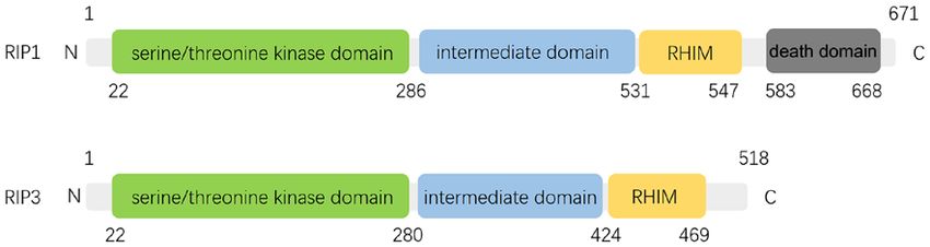
Contributed equally lated by RIP1 and RIP3, which interact through unique RIP
homotypic‑interacting motifs (RHIMs) (Fig. 2) (13,14).
Key words: necroptosis, cell death, mechanism, physiological The interaction of RIP1 and RIP3 results in autophospho
function, clinical diseases rylation, transphosphorylation, and assembly of ‘necrosome’
complex (5). RIP3 and MLKL are essential for necroptosis,
whereas RIP1 is only sometimes involved in this process.
2                                          DAI et al: NECROPTOSIS IN CLINICAL DISEASES

RIP3 and MLKL knockout mice do not show deficiency in                have been investigated. Conditional deletion of RIP1 in kera‑
embryogenesis, homeostasis and development, indicating the           tinocytes or intestinal epithelial cells suggested RIP1 plays an
role of necroptosis may be not essential in non‑challenged           essential role in maintaining epithelial homeostasis (29,30). It is
conditions (15,16).                                                  worth noting that the role of RIP1 in maintaining the intestinal
                                                                     barrier is similar to caspase‑8 (31). In addition, mice with Birc2,
3. Difference of the key characteristics between apoptosis           Birc3, and Xiap codeletion in the myeloid lineage have high
and necroptosis                                                      levels of circulating inflammatory cytokines, sterile inflamma‑
                                                                     tion, and granulocytes, which can be partially corrected by the
Although necroptosis is characterized by caspase indepen‑            lack of RIP1 or RIP3 (32). Tamoxifen‑induced systemic RIP1
dence, the molecular pathway involved is similar to and shares       gene knockout in adult mice is fatal due to a surge in cell death
features of apoptosis. However, the immunological and morpho‑        and intestinal bone marrow failure, which accumulates and
logical consequences of necroptosis are vastly different (Fig. 3).   causes fatal systemic inflammation (33,34). Fetal hepatocytes
Necroptosis shares the major morphological features of necrosis,     that received tamoxifen‑induced RIP1 deletion or RIP1‑/‑
such as the swelling of organelles, gradually translucent cyto‑      progenitor cells are unable to repopulate irradiated receptors.
plasm, and rupture of the cellular membrane (12). By contrast,       This defect can be partially corrected by the concomitant
apoptosis is characterized by membrane blebbing, cell shrinkage,     lack of RIP3, indicating that RIP1 plays a key role in the
nuclear fragmentation, and chromatin concentration (17).             survival of hematopoietic stem and progenitor cells (33,34).
The rupture of the cellular membrane results in the release of       Furthermore, systemic inflammation caused by RIP1‑/‑ can be
cellular contents, leading to the exposure of damage‑associated      restricted in RIP1‑/‑ RIP3‑/‑ Casp8 ‑/‑ hosts (34,35). These hosts
molecular patterns (DAMPs), triggering a strong inflammatory         show age‑related lymphoproliferative disorders similar to
response in necroptosis, suggesting necroptotic cells are more       those developed by RIP3‑/‑ Casp8‑/‑ mice (11,34,36). In addition,
immunogenic than apoptotic cells, which is relatively intact,        compared to control animals, RIP1+/‑ mice, mice treated with
with DAMP restricted to the plasma membrane, or encapsulated         intravenous siRNA targeting RIP1, and Nec‑1‑treated mice
in the apoptotic bodies (17). It has also been shown that necrop‑    showed a higher rate of physiological intestinal epithelial cell
tosis was associated with maintenance of T‑cell homeostasis, as      regeneration in the small intestine (37). In addition, the negative
it has been found to be able to clear excess and abnormal T cells    effects of Nec‑1 on the regeneration of intestinal epithelial cells
in the absence of caspase‑8 (18), which can prevent abnormal         are also present in RIP3‑/‑ mice (37). Findings of those studies
proliferation of lymphocytes (19).                                   suggest that there may be a delicate balance between different
                                                                     cell deaths in maintaining homeostasis in adults.
4. Identification of necroptosis
                                                                     Infectious diseases
As there is currently no specific marker for necroptosis, multiple   Viral infections. Findings have shown the crucial role of necrop‑
methods are usually required to identify necroptosis (Fig. 4). In    tosis in inflammation during viral infection (Table I). The
cultured cells, transmission electron microscopy can be used         viruses use the host's signaling pathways, such as anti‑apoptotic
to identify necroptotic cells (20). Detection of key molecular,      proteins, to enhance infection, thereby increasing its ability
including RIP1, RIP3 and MLKL activation, necrosome                  to replicate in the host cell. It has been reported that viral
formation, MLKL oligomerization, and membrane transloca‑             encoding protein involving the RHIM domain interacts with
tion can also be used to identify necroptosis (21). Activation of    RIP1 and RIP3 to inhibit virus‑induced cell death (38). Viral
RIP3 and MLKL can be monitored by western blot analysis              inhibitor of RIP activation (vRIA) disrupts the combination of
to assess phosphorylation status (22,23). Phosphorylation of         DAI and RIP3, thereby suppressing cytomegalovirus‑mediated
MLKL at Ser358 and Thr357 and RIP3 at S227 indicates the             necroptosis (9). By contrast, human cytomegalovirus differs
activation of necroptosis (24). In particular, MLKL phos‑            in protein, which does not disrupt RIP3 binding with DAI;
phorylation has been used as a biomarker for certain disease         it works via blocking signaling downstream of MLKL (39).
diagnosis and prognosis (25). In addition, several pharmaco‑         Experimental studies in mice lacking RIP3 have shown
logical inhibitors such as the necrostatin (Nec)‑1, GSK872,          impaired virus‑induced necroptosis and increased suscepti‑
and necrosulfonamide (NSA) have also been used to detect             bility to viral infections such as vaccinia virus, influenza A
necroptosis (7,26). In vivo, the activation of necroptosis can       virus, and HSV‑1 (5,25,38,40) (Fig. 5).
be identified by the elevated levels of RIP1, RIP3, or MLKL
mRNA or protein. Additionally, previous findings suggested           Bacterial infections. Necroptosis also plays an important
that RIP3 and MLKL are more specific molecular biomarkers            role in the inflammation caused by bacterial infections.
than RIP1 for the detection of necroptosis (27).                     Enteropathogenic E. coli (EPEC) has been shown to
                                                                     synthesize and secrete large amounts of the immunogenic
5. Potential role of necroptosis in clinical diseases                effector protein NleB1 and modify the arginine residues of
                                                                     the Fas‑associated death domain (FADD) and RIP1 death
Physiological functions. Over the last decade, researchers have      domains to prevent apoptosis and necroptosis (41,42). EPEC
put a lot of effort into the development of effective RIP1, RIP3,    lacking NleB1 fails to colonize intestinal epithelial cells, indi‑
and MLKL inhibitors, and created mouse models that lack one          cating that bacterial necroptosis is a protective mechanism of
or more components of the necroptotic pathway at systemic            the organism (41,42). Similarly, the absence of RIP3 sensitizes
level or in specific tissues (28). Due to the existence of these     host cells to Yelsonella. Moreover, the simultaneous knockout
models, the physiological function of proteins of necroptosis        of FADD or caspase‑8 could make cells more sensitive (43,44).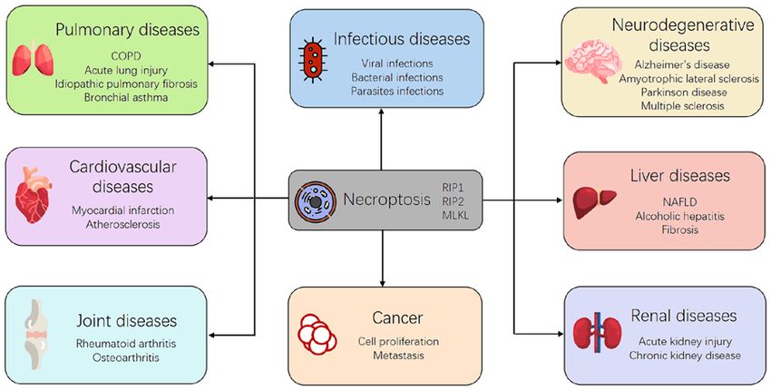
INTERNATIONAL JOURNAL OF MOlecular medicine 47: 89, 2021                                                          3

Figure 1. TNFR1‑mediated survival and cell death pathways. After TNF binding, TNFR1 recruits TRADD and RIP1 to complex I via their respective death
domains. TRADD recruits TRAF2 and cIAP1/2, after which cIAP1/2 ubiquitinate components of complex I. The ubiquitination of RIP1 promotes the formation
and activation of the TAK1/TAB complex and the IKKα/IKKβ/NEMO complex, which induced the NF‑κ B pathway and cell survival. Deubiquitination of RIP1
by cylindromatosis (CYLD) induces the dissociation of TRADD and RIP1 from TNFR1, which leads to the formation of either complex IIa or complex IIb.
FADD and pro‑caspase‑8 are recruited to TRADD and RIP1 to form complex IIa, resulting in the activation of caspase‑8 by oligomerization and cleavage. In
the absence of cIAP1/2, TAK1 or IKK complex, complex IIb, which contains RIP1, FADD and pro‑caspase‑8 except TRADD, is formed and then activates
caspase‑8, after which caspase‑8 induces apoptosis. When caspase‑8 activity is blocked, for example by zVAD‑fmk, complex IIc/necrosome is formed, and
RIP3‑dependent necroptosis is induced. In the necrosome, RIP3 phosphorylates MLKL, and translocation of phosphorylated MLKL to the cell membrane leads
to direct pore formation with the release of DAMPs. In spite of pore formation, MLKL also mediates its effect after interacting with ion channels.

Figure 2. Domain structure of RIP1 and RIP3. The intermediate domain of RIP1 contains the RIP homotypic interaction motif (RHIM) that enables the protein
to combine with the RHIM in RIP3 to activate necroptosis. Length is indicated in a number of amino acids.

In vitro, Salmonella typhimurium is able to escape TNFα,                      Parasite infections. Parasitic diseases such as malaria and
causing RIP1‑ and RIP3‑dependent necroptosis in infected                      leishmaniasis usually cause hemolysis, anemia, and bleeding.
macrophages. In a model of Salmonella typhimurium venous                      These are due to the release of hemoglobin (Hb) into the
infection, RIP3 knockout significantly reduces splenic                        circulation by the rupture of red blood cells. When Hb is
macrophage death, thereby reducing bacterial numbers and                      oxidized, heme is generated, the Fenton reaction starts, and
prolonging mouse survival (45,46). Findings focusing on oral                  peaks with the generation of reactive oxygen species (ROS).
Salmonella typhimurium infection have also shown that outer                   Heme is also involved in the activation of TLR4, causing
protein B was downregulated during infection, which resulted                  autocrine secretion of ROS and TNF, and synergistically
in promoting bacterial translocation, increasing macrophage                   activating RIP1/3‑dependent necroptosis (48). In addition,
necroptosis, and exacerbating bacterial infection (Table I) (47).             it has been shown that 10 ng/ml TNFα can induce infected
4                                                  DAI et al: NECROPTOSIS IN CLINICAL DISEASES

Figure 3. Difference of the key characteristics between apoptosis and necroptosis. Necroptosis is characterized by the swelling of organelles, gradually translu‑
cent cytoplasm, and rupture of the cellular membrane. The rupture of the cellular membrane results in the release of cellular contents, leading to the exposure
of DAMPs, triggering a strong inflammatory response in necroptosis. Apoptosis is characterized by membrane blebbing, cell shrinkage, nuclear fragmentation,
and chromatin concentration, with DAMPs restricted to the plasma membrane, or encapsulated in the apoptotic bodies.

human foreskin fibroblasts egressing Toxoplasma gondii                            proliferation, and induce DNA damage accumulation in colon
(Table I) (49).                                                                   cancer (68). These findings indicate that inhibiting activities
                                                                                  of necroptosis components may be a strategy in the treatment
Cancers                                                                           of cancers.
Type of cancers. Studies on necroptosis highlight its role
in cancer because of its necroptosis‑inducing function                            Metastasis. Metastasis is the most common cause of
(Table II) (50). Chen et al suggested that necroptosis is an                      cancer‑related death. Researchers have found that metastasis
important cell death mechanism for blocked apoptosis, and                         involves a complex interaction between cancer cells and the
has been proposed as an alternative cell death procedure                          microenvironment. By promoting inflammation, necroptosis
to prevent cancer (20). Previous studies have shown that a                        may be able to promote metastasis (69). It has been shown that
decreased expression of RIP3 or MLKL is associated with                           TNFα plays a critical role in cancer progression. However, the
worse prognosis and poor survival in breast cancer (51,52),                       exact mechanism of this process has not been fully understood.
colorectal cancer (53‑55), acute myeloid leukemia (56,57),                        Increased expression of TNFα in cancer is a key characteristic
melanoma (58,59), head and neck squamous cell carci‑                              in numerous malignancies and is usually associated with
noma (60), gastric cancer (61), ovarian cancer (62), and cervical                 a poor prognosis and decreased survival (69). Consistent
squamous cell carcinoma (63). However, increased RIP3 or                          with the pro‑inflammatory properties of necroptosis and the
RIP1 expression was also correlated with cancer development,                      cancer‑promoting effect of inflammation, Nec‑1 was able to
including glioblastoma (64), lung cancer (65), and pancreatic                     reduce inflammation and colitis‑related tumor formation (70),
cancer (66,67). SN38, the topoisomerase inhibitor, was found                      indicating that targeting necroptosis may be a strategy for
to be able to promote necroptosis progression, inhibit cell                       preventing cancer metastasis.
INTERNATIONAL JOURNAL OF MOlecular medicine 47: 89, 2021 5
Table I. The role of necroptosis in infectious diseases.
Type of infections Observations (Refs.)
Viral infections HSV‑1 HSV‑1‑Induced necroptosis is partially dependent on RIP1, and fully (38)
dependent on RIP3 and MLKL
Influenza A virus Mice deficient in RIPK3 is more susceptible to influenza A (40)
virus than wild‑type counterparts
MCMV RIP3‑/‑ murine embryonic fibroblasts were resistant to (9,156)
MCMV‑induced necrosis
HIV‑1 Necrostatin‑1 restrains HIV‑1‑induced cytopathic effect and inhibits (157)
the formation of HIV‑induced syncytia in CD4+ T‑cell lines
Reovirus Cell death following reovirus infection was sensitive to (158)
inhibition of RIP1
Vaccinia virus RIP1‑/‑ mice cells infected with Vaccinia virus was resistant to (159)
TNF‑α induced death
RIP3‑/‑ mice exhibited severely impaired virus‑induced tissue (5)
necrosis and inflammation
Bacterial infections Clostridium prefringens RIP1 or RIP3 inhibitors reduced both bacteria‑induced (160)
β‑toxin apoptosis and necrosis
Salmonella Inhibition of the RIP1 or RIP3 prevented the bacteria‑induced (47)
typhimurium death of wild‑type macrophages
Deletion of MLKL rescued severity of bacteria‑induced tissue (45)
inflammatory
M. tuberculosis RIP1 and RIP3 morpholino knockdown reduced susceptibility of (161)
zebrafish to Mycobacterium marinum
Yersinia pestis Deficiency of both RIP3 and caspase‑8 completely abrogated (44)
Yersinia‑induced cell death
Staphylococcus aureus RIP3‑/‑ mice exhibited significantly improved staphylococcal clearance (162)
Parasite infections Toxoplasma gondii Blocking necroptosis by necrostatin‑1 has little impact on (49)
TNF‑α‑induced egress of T. gondii
Leishmaniasis and Inhibition of the RIP1 or RIP3 protected macrophages from (48)
Malaria heme‑induced cell death
HSV‑1, herpes simplex virus type 1; RIP, receptor‑interacting protein kinase; MLKL, mixed lineage kinase domain‑like protein; MCMV,
murine cytomegalovirus; TNFα, tumor necrosis factor‑α; HIV‑1, human immunodeficiency virus type‑1.
Neurodegenerative diseases levels of necroptosis components, such as RIP1 and MLKL,
Parkinson's disease. Researchers have shown that necroptosis were significantly higher than the normal brain (Table III) (75).
was activated in Parkinson's disease (PD), and may be asso‑ Treatment of AD in brain of mice with the necroptosis inhibitor
ciated with mitochondrial defects which led to necroptosis can significantly suppress necroptosis and prevent neuronal
(Table III) (71). Compared with healthy brain, the level of loss (75). This indicates that targeting necroptosis may be a
necroptosis components, including RIP1, RIP3 and MLKL, new therapeutic strategy for AD treatment.
was significantly increased in the substantia nigra of PD
brain (72). Moreover, researchers have found Nec‑1 could Amyotrophic lateral sclerosis. Amyotrophic lateral sclerosis
protect PC12 cells from death in PD models (73). This suggests (ALS) is a neurodegenerative disease that is characterized by
that the activation of RIP1 may be a risk factor for dopami‑ loss of motor neurons. In a previous study, the ALS spinal cord
nergic neurons lost in PD patients. In addition, leucine‑rich was shown to have a significant increase in necroptosis compo‑
repeat kinase 2, which was identified in a systematic RNAi nents including RIP1, RIP3, and MLKL in the ALS mouse
screen, is encoded by a gene that is frequently mutated in PD model compared to healthy mouse spinal cord (77). In addition,
and is able to promote activation of RIP1 (74). loss of optineurin, an ALS‑related gene, resulted in suscep‑
tibility to necroptosis. Nec‑1 inhibition of RIP1 or knockout
Alzheimer's disease. Alzheimer's disease (AD) is a degenera‑ of RIP3 could prevent demyelination and reduce axonal
tive brain disease featured by loss of neurons. Previous studies pathological hallmarks in ALS mouse models (Table III) (77).
have found that there were activated necroptosis in both Those findings suggested that targeting necroptosis may have
human (75) and mouse (76) AD brain. In the AD brain, the potential therapeutic value in ALS patients.6 DAI et al: NECROPTOSIS IN CLINICAL DISEASES Figure 4. Methods to identify necroptosis. In cultured cells, transmission electron microscopy can be used to identify necroptotic cells. Detection of key molecular, including RIP1, RIP3 and MLKL activation, necrosome formation, MLKL oligomerization, and membrane translocation can also be used to identify necroptosis. Activation of RIP3 and MLKL can be monitored by western blot (WB) analysis to assess phosphorylation status. Several pharmacological inhibitors such as the Nec‑1, GSK872, and NSA have also been used to detect necroptosis. In vivo, the activation of necroptosis can be identified by the elevated levels of RIP1, RIP3, or MLKL mRNA or protein. Figure 5. The potential role of necroptosis in clinical diseases. Necroptosis has been implicated in pathophysiological processes of several clinical diseases, including infectious diseases, neurodegenerative diseases, liver diseases, pulmonary diseases, renal diseases, cardiovascular diseases, joint diseases, and human tumors. Multiple sclerosis. Multiple sclerosis (MS) is degenerative model (79). Notably, MLKL was shown to be involved in the disease characterized by oligodendrocyte loss and demye‑ MS process (79). lination. Previous findings have shown a significant increase in necroptosis components including RIP1, RIP3, and Liver diseases. Non‑alcoholic fatty liver disease (NAFLD), MLKL in MS patients. In addition, MLKL oligomers were Non‑alcoholic fatty liver disease (NAFLD) is a chronic significantly increased in MS pathology samples compared disease characterized by excess triglyceride accumulation with controls (Table III) (78). This suggests necroptosis is in the liver. Several studies have used different models to activated in the pathogenesis of MS. In MS mouse model, assess the effects of necroptosis on NAFLD (Table IV). oral administration of RIP1 inhibitor can suppress oligo‑ Studies have found necroptosis components, including RIP1, dendrocyte degeneration and reduce disease severity (78). RIP3 and MLKL, were increased in NAFLD models, as Moreover, researchers have shown that inhibition of RIP1 well as in RIP3KO mice (80‑85). In addition, an increased reduced demyelination and disease progression in an MS expression of RIP3 and MLKL in the human NAFLD was
INTERNATIONAL JOURNAL OF MOlecular medicine 47: 89, 2021                                              7

Table II. The role of necroptosis in cancer.

Cancer type                                                           Observations                                             (Refs.)

Breast cancer	Decrease of RIP3 expression was associated with worse prognosis                                                  (51,52)
Colorectal cancer	Decrease RIP3 and MLKL expression were associated with decreased overall survival                            (53‑55)
Gastric cancer          Low MLKL expression was significantly associated with decreased overall survival                         (61)
Ovarian cancer	Decrease of MLKL expression was associated with worse prognosis                                                   (62)
Pancreatic cancer       Increase of RIP1, RIP3, FADD and MLKL expression were associated with worse                            (66,67)
                        prognosis
Lung cancer             Increased RIP1 expression was associated with worse prognosis                                            (65)
Acute myeloid leukemia	Decrease of RIP3 expression was associated with worse prognosis                                         (56,57)
Melanoma	Decrease of RIP3 expression was associated with worse prognosis                                                       (58,59)
Head and neck squamous	Decrease of RIP1 expression was associated with worse prognosis                                           (60)
cell carcinoma
Cervical squamous cell 	Decrease of MLKL expression was associated with worse prognosis                                         (63)
carcinoma
Glioblastoma            Increased RIP1 expression was associated with worse prognosis                                           (64)

RIP, receptor‑interacting protein kinase; MLKL, mixed lineage kinase domain‑like protein; FADD, Fas‑associated death domain.

Table III. The role of necroptosis in neurodegenerative diseases.

Neurodegenerative diseases                                              Observations                                           (Refs.)

Parkinson's disease RIP1 inhibition improved survival of optic atrophy 1‑mutant human induced pluripotent                       (72)
                    stem cell‑derived neurons in vitro. RIP1 inhibition attenuated MPTP‑induced
                    dopaminergic neuronal loss
Alzheimer's disease RIP1 inhibition reduced Aβ burden, levels of inflammatory cytokines, and memory                             (76)
                    deficits in a mouse model of Alzheimer's disease
Amyotrophic lateral RIP1 inhibition or RIP3 deficiency blocked oligodendrocyte death, microglial                                (77)
sclerosis           inflammation, and axonal degeneration
Multiple sclerosis	Cortical lesions in human multiple sclerosis brain samples showed increased activation of                    (78)
                    RIP1, RIP3 and MLKL
                    Inhibition of RIP1 inhibited the progression of demyelination and disease development                       (79)
                    in a cuprizone‑induced model for multiple sclerosis

RIP, receptor‑interacting protein kinase; MPTP, 1‑methyl‑4‑phenyl‑1,2,3,6‑tetrahydropyridine; MLKL, mixed lineage kinase domain‑like
protein.

identified (83,85). Increased level of RIP3 and p‑MLKL                PSMC1 KO mice could increase RIP3 expression, indi‑
was also found in visceral adipose tissue of obese patients.          cating RIP3 expression was post‑translationally regulated in
Furthermore, RIP3 expression correlated with p‑MLKL and               ethanol‑mediated liver injury (87). Additionally, when RIP3
metabolic serum markers including blood insulin levels and            was deleted, the steatosis and inflammatory effects of ethanol
Hemoglobin A1c (84).                                                  in hepatocyte could be reduced (86). However, the inflamma‑
                                                                      tory and steatosis effects of high fat diet for hepatocyte were
Alcoholic hepatitis. Similar to the pathologies of NAFLD,             increased when RIP3 was deleted (80).
alcoholic hepatitis is an inflammatory syndrome in liver, which
can result in high morbidity and mortality. Several studies have      Fibrosis. Hepatic fibrosis is one of the most common liver
found that RIP3 was increased following ethanol feeding, and          diseases, which is closely related to liver failure and hepato‑
RIP3 deletion could protect the liver from ethanol‑mediated           cellular cancer. RIP3 deletion reportedly aggravated hepatic
injury (Table IV). In addition, p‑JNK was regulated by RIP3           fibrosis by increasing insulin resistance (80). Furthermore,
in a model of alcoholic hepatitis, and RIP3 deletion reduced          inhibition of RIP3 did not result in protective effect in
ethanol‑induced p‑JNK expression (86). Another study found            carbon tetrachloride (CCL4)‑induced fibrosis (83) (Table IV).
pharmacological inhibition of proteasome and liver‑specific           Additionally, curcumol suppressed serum inflammatory
8 DAI et al: NECROPTOSIS IN CLINICAL DISEASES
Table IV. The role of necroptosis in liver diseases.
Liver diseases Observations (Refs.)
NAFLD MLKL deficiency and necrostatin‑1 administration improves insulin sensitivity without affecting (81)
inflammation
RIP3KO mice had increased hepatic steatosis but reduced inflammation (82)
MCD diet‑fed RIP3KO mice were protected, but CCL4‑induced fibrosis model mice were not (83)
protected
RIP3 maintains WAT homeostasis and has a role in WAT insulin signaling (84)
RIP3 deficiency protects from steatosis, inflammation, and fibrosis (85)
Alcoholic hepatitis RIP3 expression increased following ethanol feeding (163)
RIP3 but not RIP1 inhibition protects from ethanol‑induced hepatic injury and steatosis (86)
RIP3 ablation and necrostatin‑1 decreased hepatic inflammation (87)
Curcumin reduced ethanol‑induced necroptosis in Nrf/p53‑dependent mechanism (164)
Fibrosis RIP3KO mice were not protected against steatosis, inflammation and fibrosis (80)
RIP3 deficiency protects from steatosis, inflammation, and fibrosis (85)
RIP3KO MCD diet‑fed mice were protected from inflammation and fibrosis, while CCL4‑induced (83)
fibrosis was not reduced in RIP3KO mice
RIP3 deficiency reduced inflammation, oxidative stress, and fibrosis in 3‑day CBD ligation model (165)
Melatonin protects from CCL4‑induced fibrosis (166)
Curcumol‑mediated decreased fibrosis is associated with increased necroptosis in hepatic (88)
stellate cells
NAFLD, non‑alcoholic fatty liver disease; RIP, receptor‑interacting protein kinase; KO, knockout; MCD, methionine and choline‑deficient;
CCL4, carbon tetrachloride; WAT, white adipose tissue; CBD, common bile duct.
markers, transaminases, and fibrosis in a dose‑dependent and lipopolysaccharide‑induced necroptosis (95). Researchers
manner by inducing necroptosis of hepatic stellate cells in a have also found that mice lacking RIP3 were protected from
liver fibrosis model (88). Curcumol‑induced increased necrop‑ ventilator‑induced lung injury (96). Additionally, inhibition of
tosis was mediated by increased expression of p‑RIP3 and RIP1 can reduce systemic and pulmonary inflammation and
p‑JNK (88). Those results indicated that pharmacotherapy increase survival rate of septic neonatal mice (Table V) (97).
which induced increased necroptosis may be a notable strategy
for the treatment of hepatic fibrosis in the future. Idiopathic pulmonary fibrosis. Idiopathic pulmonary
fibrosis (IPF) is a chronic, progressive, fibrotic lung disease
Pulmonary diseases characterized by the usual interstitial pneumonia pattern at
Chronic obstructive pulmonary disease. Chronic obstructive histopathologic examination (98). In a previous study using
pulmonary disease (COPD) is characterized by persistent and alveolar epithelial cells, RIP3‑mediated necroptosis was
progressive airway inflammation and narrowing, and is a major associated with IPF development by releasing DAMP (99).
source of the high healthcare expenditure in the elderly (89). Additionally, RIP3 and p‑MLKL levels in the lungs of
An increasing number of studies have shown that necroptosis IPF patients are significantly higher than those in healthy
is associated with the etiology of COPD (Table V). In addition, lungs (99). Mice with RIP3 knockout showed a reduced cell
necroptosis of epithelial cell is associated with COPD (90). death, with a decrease of p‑MLKL level in alveolar epithe‑
Cigarette smoking (CS)‑related necroptosis and DAMP release lial cells (99). RIP3 knockout could effectively suppress the
could cause neutrophil inflammation in mice, and Nec‑1 could DAMP releasing, cell death, and pulmonary fibrosis without
reduce the inflammation (91). In addition, researchers have found reducing the expression of cleaved caspase‑3 (Table V) (99).
that in airway epithelial cells, endoplasmic reticulum chaperone These indicate that inhibiting activities of necroptosis compo‑
protein GRP78 could promote CS‑induced inflammation. This nents may be a strategy in the treatment of IPF.
may be due to the upregulation of necroptosis and subsequent
activation of the NF‑κB pathway (92). Bronchial asthma. Bronchial asthma is the most common
chronic respiratory disease characterized by bronchial
Acute lung injury. Acute lung injury (ALI) is one of the most hyper‑responsiveness and airway obstruction (100).
common complications in critically ill patients (93). Recent find‑ Viral‑induced bronchial asthma exacerbation mimicked by
ings have shown the involvement of RIP3‑mediated necroptosis IFN‑β knockout mice treated with house dust mite is asso‑
in neonatal mice with hypoxia‑induced lung injury, which can be ciated with increased necroptosis components, including
attenuated by gene deletions in RIP3 (94). In additional, inhibi‑ p‑MLKL and LDH in the bronchoalveolar lavage fluid (101).
tion of RIP3 could significantly reduce inflammatory activation As a major inflammatory cytokine in bronchial asthma, IL‑33INTERNATIONAL JOURNAL OF MOlecular medicine 47: 89, 2021 9
Table V. The role of necroptosis in pulmonary diseases.
Pulmonary diseases Observations (Refs.)
COPD CS‑induced necroptosis and the release of DAMPs trigger neutrophilic inflammation in mice that (91)
was reduced with Nec‑1 treatment
Acute lung injury RIP3‑mediated necroptosis is observed in neonatal mice with HALI, which is attenuated by (94)
genetic deletion in RIP3
RIP3‑deficient mice are protected against ventilator‑induced lung injury (96)
Idiopathic RIP3 and p‑MLKL expression levels are significantly higher in the lungs of IPF patients than in (99)
pulmonary fibrosis healthy control lungs
Bleomycin‑treated AECs isolated from RIP3 knockout mice show attenuation of cell death with
decreased p‑MLKL expression
Bronchial asthma GW806742X can abrogates IL‑33 reaction in vitro and attenuates eosinophilia in a mouse (102)
model of asthma
COPD, chronic obstructive pulmonary disease; CS, cigarette smoking; DAMPs, damage‑associated molecular patterns; RIP, receptor‑inter‑
acting protein kinase; MLKL, mixed lineage kinase domain‑like protein; HALI, hypoxia‑induced lung injury; IPF, idiopathic pulmonary
fibrosis; AECs, alveolar epithelial cells; IL‑33, interleukin 33.
Table VI. The role of necroptosis in renal diseases.
Renal diseases Observations (Refs.)
Acute kidney injury RIP3‑/‑ and MLKL‑/‑ mice are less sensitive to oxalate crystal–induced and cisplatin‑driven (107,110)
AKI than are their wild‑type counterparts
The RIP3‑/‑ genotype confers considerable protection against mild IRI, and such a (104)
protection can be extended to severe IRI by the concomitant deletion of Ppif
Chronic kidney disease RIP1 and RIP3 participate in the loss of renal cells of subtotal nephrectomised rats (112)
Gene deletion of RIP3 or MLKL ameliorated renal tubular cell necroptosis, and then finally (113)
reduced interstitial fibrogenesis in the long term after IRI
RIP, receptor‑interacting protein kinase; MLKL, mixed lineage kinase domain‑like protein; AKI, acute kidney injury; IRI, ischemia‑reperfusion
injury.
is released in response to necroptosis and causes eosinophil the protection can be extended to severe IRIs with the deletion
and basophil activation (102). Moreover, in a mouse model of of Ppif (104). Similar results are also found when Nec‑1, SfA,
asthma induced by Aspergillus fumigatus extract, the necrop‑ and 16‑86 are employed alone or in combination (104,109). The
tosis inhibitor GW806742X can eliminate necroptosis and abovementioned results suggest that inhibition of necroptosis
IL‑33 response, and attenuates eosinophilia (102). Additionally, may be a therapeutic option for AKI treatment.
The TNFα‑induced necroptosis enhanced by mucin 1 can be
reduced by Nec‑1 in human bronchial epithelial cells (103) Chronic kidney disease. Similar to the AKI, necroptosis was
(Table V). also found in chronic kidney disease (CKD) after unilateral
nephrectomy (Table VI) (112). Researchers have shown that
Renal diseases necroptosis and the highest levels of RIP1 and RIP3 occurred
Acute kidney injury. Acute kidney injury (AKI) is a common 8 weeks after subtotal nephrectomy (112). Notably, the renal
and severe clinical disease that often requires renal replacement pathological changes and renal function could be significantly
therapy (Table VI). Signs of an ongoing necroptotic response improved after Nec‑1 treatment, and the overexpression of
have been found in AKI caused by ischemia‑reperfusion injury RIP1, RIP3, MLKL could be significantly reduced (112). These
(IRI) (104‑106), urolithiasis (107), cisplatin‑based chemo‑ results suggest that necroptosis contributes to the loss of renal
therapy or radiocontrast (104,108‑111). Previous findings have cells in subtotal nephrectomized rats. Furthermore, during the
shown that compared with wild‑type counterparts, RIP3‑/‑ and AKI to CKD process, upregulation of expression and inter‑
MLKL ‑/‑ mice are less sensitive to oxalate crystal‑induced action between RIP3 and MLKL can induce necroptosis in
AKI, and are associated with reduced plasma creatinine levels, proximal renal tubular cells and promote inflammasome acti‑
neutrophil infiltration, and limited tubular injury (107,110). vation under IRI conditions (113). RIP3 or MLKL knockout
Moreover, the RIP3‑/‑ mice confer protection from mild IRI, and could protect the renal tubular cells from necroptosis and10 DAI et al: NECROPTOSIS IN CLINICAL DISEASES
Table VII. The role of necroptosis in cardiovascular diseases.
Cardiovascular diseases Observations (Refs.)
Myocardial infarction RIP3 deficiency protects mouse hearts from IR‑induced necroptosis and significantly (22)
reduces infarct size
Necrostatin‑1 prevents both short and long‑term effects of myocardial ischemia
Atherosclerosis Ox‑LDL deposited in the endothelium can upregulate the expression of RIP3 and (118)
ox‑LDL‑related genes, resulting in the necroptosis of macrophages
RIP, receptor‑interacting protein kinase; IR, ischemia‑reperfusion; ox‑LDL, oxidized‑low‑density lipoprotein.
Table VIII. The role of necroptosis in joint diseases.
Joint diseases Observations (Refs.)
Rheumatoid arthritis RIP1, RIP3 and MLKL were potently increased in the synovium of a collagen‑induced RA (120)
mouse model
RIP1 inhibitor significantly decreased the expression of RIP1, RIP3 and MLKL and (121)
suppressed the expression of IL‑17, IL‑1β, IL‑6 and TNFα in a RA mouse model
Osteoarthritis Gene expression levels of RIP3 and MLKL were elevated in highly degenerated cartilage tissue (123)
Trauma induced cell death and subsequent release of pro‑inflammatory mediators could be
largely attenuated by necrostatin‑1 or N‑acetylcysteine
RA, rheumatoid arthritis; RIP, receptor‑interacting protein kinase; MLKL, mixed lineage kinase domain‑like protein; IL, interleukin; TNFα:
tumor necrosis factor‑α.
inflammasome activation, which prevent kidney from intersti‑ suggest that inhibition of necroptosis may be a therapeutic
tial fibrogenesis after IRI (113). option for atherosclerosis treatment.
Cardiovascular diseases Joint diseases
Myocardial infarction. Myocardial infarction, characterized Rheumatoid arthritis. Rheumatoid arthritis (RA), characterized
by regional myocardial ischemia and hypoxia, is one of the by synovial membrane inflammation, is a chronic systemic inflam‑
leading causes of death worldwide (114). Previous findings matory autoimmune disease that affects 0.5‑1% of the population
have shown that compared to wild‑type mice, the levels of worldwide (119). Previous findings have shown a significant
RIP1 and RIP3 were significantly higher in hearts of ischemic increase in necroptosis components including RIP1, RIP3, and
mice (22,115). In an acute IRI mouse model, RIP3 deficiency MLKL in the synovium of an arthritis mouse model (120).
was able to protect heart from IRI‑induced necroptosis and Additionally, researchers have also found in an arthritis mouse
reduce the infarct size (116). Notably, researchers have found model, Nec‑1 could significantly reduce these key components of
that Nec‑1 could protect heart against short‑term and long‑term necroptosis and IL‑17, IL‑1β, IL‑6, and TNFα (Table VIII) (121).
effects of myocardial ischemia, including reduced necrotic These results suggested that inhibiting activities of necroptosis
cell death and size of myocardial infarction, which helped to components may be a strategy in the treatment of RA.
maintain long‑term cardiac function (22) (Table VII).
Atherosclerosis, Atherosclerosis, a chronic inflammatory Osteoarthritis. Osteoarthritis (OA) is the leading cause of pain
disease, is frequently observed in middle‑aged individuals and and disability among chronic disease, which affects about 10%
the elderly, and is a major cause of cardiovascular death (117). of men and 18% of women older than 60 years (122). A signifi‑
It has been demonstrated that necroptosis may promote the cant increase in necroptosis components including RIP3, and
inflammatory response and atherosclerosis development. MLKL was found in highly degenerated cartilage tissue (123).
Oxidized low‑density lipoprotein (LDL) is able to upregulate Moreover, it has been shown that Nec‑1 could significantly
RIP3 and oxidized LDL‑related gene expression in macro‑ reduce cell death and subsequent release proinflammatory
phages, leading to macrophage necroptosis (118). It triggers mediators in the OA model (Table VIII) (123).
an inflammatory response, which leads to atherosclerosis.
During the progression of the disease, some cytokines are 6. Drugs and agents that regulate necroptosis
released and monocytes accumulate in the lesion, exacerbating
the accumulation of inflammation. Additionally, necroptosis As necroptosis not only participate in the maintenance
can lead to the death of foam cells, which in turn aggravates of organismal homeostasis, but also constitute etiological
the progression of the disease (Table VII) (118). These results determinants of diverse human pathologies (124), at least twoINTERNATIONAL JOURNAL OF MOlecular medicine 47: 89, 2021 11
Table IX. Drugs and agents that regulate necroptosis.
Drugs and agents Target Disease condition (Refs.)
Drugs and agents that induce necroptosis
Interferons RIP3 and MLKL Different diseases (127)
Valproic acid RIP1 Epilepsy and mood disorders (128)
Decitabine and 5‑azacytidine RIP3 Breast cancer (51)
Shikonin RIP1 and RIP3 Pancreatic and non‑small cell lung cancers, osteosarcoma (129)
Emodin RIP1, RIP3 and MLKL Renal cancer (130)
Bufalin RIP1 and RIP3 Pancreatic and breast cancers (131)
Resibufogenin RIP3 and MLKL Pancreatic and colorectal cancers (132)
Radiotherapy Caspase‑8 Different cancers (147)
5‑fluorouracil RIP1 and RIP3 Different cancers (148)
Cisplatin RIP1, RIP3 and MLKL Different cancers (149)
Anthracyclines and oxaliplatin RIP3 and MLKL Lung cancer (150)
Obatoclax RIP1, RIP3 and MLKL Different cancers (151)
Neoalbaconol RIP1 and RIP3 Nasopharyngeal carcinoma (152)
Tanshinone RIP1 and RIP3 Hepatocellular carcinoma (153)
Drugs and agents that inhibit necroptosis
Cyclosporine A RIP1 and RIP3 Immunosuppressive drug (133)
Rapamycin RIP1 Restenosis in coronary arteries, transplant rejection in (134)
lymphangioleiomyomatosis, and retinal detachment
Patchouli alcohol RIP3 and MLKL Colitis (139)
Pazopanib RIP1 Renal cell carcinoma and advanced soft tissue sarcoma (140)
Ponatinib RIP1 and RIP3 Leukemia (140)
GSK2982772 RIP1 Inflammatory diseases (colitis, rheumatoid arthritis, psoriasis) (167)
GSK3145095 RIP1 Pancreatic cancer (141)
Dabrafenib RIP3 Melanoma (88)
Carfilzomib RIP3 and MLKL Multiple myeloma (142)
Sorafenib RIP1 and RIP3 Thyroid and renal cell cancers, hepatocellular carcinoma (143)
Phenytoin RIP1 Epilepsy and breast cancer (144)
Aucubin RIP1 and MLKL Epilepsy (145)
Wogonin RIP1 Acute kidney injury (146)
therapeutic paradigms can be envisioned: i) the activation Rapamycin (134), inhibitors of the HSP90 [(G‑TPP) (135),
of necroptosis, as a means to bypass the accrued resistance Kongensin A (136), 17‑demethoxy‑reblastatin (137),
of most tumors to apoptosis (125); ii) the inhibition of DHQ3 (137), gamitrinib (18), and geldanamycin (138)],
necroptosis, as a strategy to limit the loss of post‑mitotic as well as traditional Chinese medicine such as patchouli
cells in pathologies such as inflammatory, ischemic, and alcohol (139).
toxic syndromes (126). Therefore, drugs affecting either the Promising specific inhibitors are also being developed for
expression or the activity of necroptosis mediators may have the central molecules of necroptosis. Currently, several drugs
therapeutic potential (Table IX). with anti‑necroptotic activity have been used for the treatment
Several drugs have been found to upregulate the expression of different types of cancer [Pazopanib (140), Ponatinib (140),
of the key molecules of necroptosis, including interferons (127), GSK3145095 (141), Dabrafenib (26), Carfilzomib (142), and
histone deacetylase inhibitor valproic acid (128), and hypo‑ Sorafenib (143)]. Moreover, a clinically used anti‑convulsant,
methylating agents such as decitabine and 5‑azacytidine (51). Phenytoin (144) as well as components found in different plants
Additionally, several traditional Chinese medicine drugs [aucubin (145) and wogonin (146)] could inhibit RIPK1 activity.
such as shikonin (129), emodin (130), bufalin (131), and By contrast, radiotherapy (147), chemotherapeutic agents such
resibufogenin (132) were also found to upregulate RIP1 and as 5‑fluorouracil (148), cisplatin (149), oxaliplatin (150), and
RIP3, which finally induced necroptosis. By contrast, various anthracyclines (150), pan‑BCL‑2 inhibitor Obatoclax (151),
drugs have been documented to downregulate necroptosis, or traditional Chinese medicines such as neoalbaconol (152)
including immunosuppressive drug Cyclosporine A (133) and and tanshinone (153) have been documented to upregulate12 DAI et al: NECROPTOSIS IN CLINICAL DISEASES
necroptosis. Although these drugs did not affect the expression Availability of data and materials
of necroptotic component, these medicines may increase the
effect of the drugs affecting the expression of the necroptotic Not applicable.
molecules in combination therapy in cancer cells.
Authors' contributions
7. Conclusions and perspectives
YFA and WLD conceived the study; JC and XQH were
During the last decade, necroptosis has been recognized as an involved in data curation; XL, WLD and JC were involved in
alternative to apoptosis when cells are exposed to various stimuli collection of references as well as writing and editing; YFA,
under specific conditions (154). The necrosome components, XQH and JC supervised the study. All authors have read and
RIP1, RIP3, and MLKL, are critical regulators of necroptotic agreed to the published version of the manuscript.
cell death. RIP1 functions as a traffic cop for mechanisms of
cell death. MLKL acts as the executioner of necroptosis, based Ethics approval and consent to participate
on the phosphorylation, oligomerization and membrane trans‑
location (155). Current understandings demonstrated a pathway Not applicable.
in which RIP3 activation, possibly mediated by RIP1, induces
MLKL activation, and finally results in permeabilization of the Patient consent for publication
plasma membrane and cell death.
Recent studies have revealed a complex role for necroptosis Not applicable.
in diverse clinical diseases, such as ischemia‑reperfusion injury,
neurological and inflammatory diseases, retinal disorders, Competing interests
acute kidney injury or bacterial infections. On the one hand,
it functioned as a cell‑death mechanism activated by various The authors declare that they have no competing interests.
signal transduction cascades in the same cell or the same tissue;
on the other hand, it acted as an inflammation inducer through References
the release of DAMPs. Cross‑regulation between necroptosis
and other modes of cell death increase the complexity of these 1. Galluzzi L, Vitale I, Abrams JM, Alnemri ES, Baehrecke EH,
pathways. The major necroptosis‑regulating proteins exert Blagosklonny MV, Dawson TM, Dawson VL, El‑Deiry WS,
Fulda S, et al: Molecular definitions of cell death subroutines:
pleiotropic signaling functions that culminate in necroptotic Recommendations of the Nomenclature Committee on Cell
cell death and have cell death‑independent functions, such as Death 2012. Cell Death Differ 19: 107‑120, 2012.
regulation of inflammasome activation, mitochondrial func‑ 2. Moriwaki K, Balaji S, McQuade T, Malhotra N, Kang J
and Chan FK: The necroptosis adaptor RIPK3 promotes
tion and integrity, and cellular metabolic activities (96). injury‑induced cytokine expression and tissue repair.
As necroptosis constitutes etiological determinants of Immunity 41: 567‑578, 2014.
multiple human pathologies, targeting the necroptotic pathway 3. Holler N, Zaru R, Micheau O, Thome M, Attinger A, Valitutti S,
Bodmer JL, Schneider P, Seed B and Tschopp J: Fas triggers an
is a potential therapeutic approach for multiple diseases, and alternative, caspase‑8‑independent cell death pathway using the
several activators or inhibitors of the necroptosis pathway have kinase RIP as effector molecule. Nat Immunol 1: 489‑495, 2000.
been developed, such as dabrafenib, pazopanib, and ponatinib. 4. He S, Wang L, Miao L, Du F, Zhao L and Wang X: Receptor inter‑
acting protein kinase‑3 determines cellular necrotic response to
These small‑molecule activators or inhibitors of necroptosis TNF‑alpha. Cell 137: 1100‑1111, 2009.
may be useful as therapeutics in a specific clinical disease. 5. Cho YS, Challa S, Moquin D, Genga R, Ray TD, Guildford M and
However, most studies investigating the therapeutics targeting Chan FK: Phosphorylation‑driven assembly of the RIP1‑RIP3
complex regulates programmed necrosis and virus‑induced
necroptosis are based on in vitro experiments or animal models, inflammation. Cell 137: 1112‑1123, 2009.
thus the feasibility of the clinical use of these compounds 6. Zhang DW, Shao J, Lin J, Zhang N, Lu BJ, Lin SC, Dong MQ
and agents remains to be assessed in vivo and clinical trials. and Han J: RIP3, an energy metabolism regulator that switches
TNF‑induced cell death from apoptosis to necrosis. Science 325:
Additionally, the off‑target effects of the necroptosis‑targeting 332‑336, 2009.
therapeutics should be scrutinized, and novel approaches that 7. Sun L, Wang H, Wang Z, He S, Chen S, Liao D, Wang L, Yan J,
conjugate necroptosis inducers and disease‑guiding agents Liu W, Lei X and Wang X: Mixed lineage kinase domain‑like
protein mediates necrosis signaling downstream of RIP3 kinase.
should be developed to enhance selectivity and safety. Cell 148: 213‑227, 2012.
8. Jouan‑Lanhouet S, Riquet F, Duprez L, Vanden Berghe T,
Acknowledgements Takahashi N and Vandenabeele P: Necroptosis, in vivo detection in
experimental disease models. Semin Cell Dev Biol 35: 2‑13, 2014.
9. Upton JW, Kaiser WJ and Mocarski ES: DAI/ZBP1/DLM‑1
The authors are grateful to Dr He Xiao for his valuable advice complexes with RIP3 to mediate virus‑induced programmed
and English editing. necrosis that is targeted by murine cytomegalovirus vIRA. Cell
Host Microbe 11: 290‑297, 2012.
10. Oberst A and Green DR: It cuts both ways: Reconciling the dual
Funding roles of caspase 8 in cell death and survival. Nat Rev Mol Cell
Biol 12: 757‑763, 2011.
11. Kaiser WJ, Upton JW, Long AB, Livingston‑Rosanoff D,
This work was supported by grants from the National Natural Daley‑Bauer LP, Hakem R, Caspary T and Mocarski ES: RIP3
Science Foundation of China (nos. 81672212, 81802153 mediates the embryonic lethality of caspase‑8‑deficient mice.
and 81902205), the Beijing Natural Science Foundation Nature 471: 368‑372, 2011.
12. Weinlich R, Oberst A, Beere HM and Green DR: Necroptosis
(nos. 7171014, 7174361 and 7182175), and the Beijing Municipal in development, inflammation and disease. Nat Rev Mol Cell
Science and Technology Commission (no. Z171100001017085). Biol 18: 127‑136, 2017.INTERNATIONAL JOURNAL OF MOlecular medicine 47: 89, 2021 13
13. Sun X, Yin J, Starovasnik MA, Fairbrother WJ and Dixit VM: 35. Dillon CP, Weinlich R, Rodriguez DA, Cripps JG, Quarato G,
Identification of a novel homotypic interaction motif required Gurung P, Verbist KC, Brewer TL, Llambi F, Gong YN, et al:
for the phosphorylation of receptor‑interacting protein (RIP) by RIPK1 blocks early postnatal lethality mediated by caspase‑8
RIP3. J Biol Chem 277: 9505‑9511, 2002. and RIPK3. Cell 157: 1189‑1202, 2014.
14. Sun X, Lee J, Navas T, Baldwin DT, Stewart TA and Dixit VM: 36. Oberst A, Dillon CP, Weinlich R, McCormick LL, Fitzgerald P,
RIP3, a novel apoptosis‑inducing kinase. J Biol Chem 274: Pop C, Hakem R, Salvesen GS and Green DR: Catalytic activity
16871‑16875, 1999. of the caspase‑8‑FLIP(L) complex inhibits RIPK3‑dependent
15. Shan B, Pan H, Najafov A and Yuan J: Necroptosis in develop‑ necrosis. Nature 471: 363‑367, 2011.
ment and diseases. Genes Dev 32: 327‑340, 2018. 37. Matsuoka Y and Tsujimoto Y: Role of RIP1 in physiological
16. Dillon CP, Tummers B, Baran K and Green DR: Developmental enterocyte turnover in mouse small intestine via nonapoptotic
checkpoints guarded by regulated necrosis. Cell Mol Life Sci 73: death. Genes Cells 20: 11‑28, 2015.
2125‑2136, 2016. 38. Huang Z, Wu SQ, Liang Y, Zhou X, Chen W, Li L, Wu J,
17. Hacker G: The morphology of apoptosis. Cell Tissue Res 301: Zhuang Q, Chen C, Li J, et al: RIP1/RIP3 binding to HSV‑1 ICP6
5‑17, 2000. initiates necroptosis to restrict virus propagation in mice. Cell
18. Ch'en IL, Tsau JS, Molkentin JD, Komatsu M and Hedrick SM: Host Microbe 17: 229‑242, 2015.
Mechanisms of necroptosis in T cells. J Exp Med 208: 633‑641, 39. Omoto S, Guo H, Talekar GR, Roback L, Kaiser WJ and
2011. Mocarski ES: Suppression of RIP3‑dependent necroptosis
19. Lenardo M, Chan KM, Hornung F, McFarland H, Siegel R, by human cytomegalovirus. J Biol Chem 290: 11635‑11648,
Wang J and Zheng L: Mature T lymphocyte apoptosis‑immune 2015.
regulation in a dynamic and unpredictable antigenic environ‑ 40. Nogusa S, Thapa RJ, Dillon CP, Liedmann S, Oguin TH III,
ment. Annu Rev Immunol 17: 221‑253, 1999. Ingram JP, Rodriguez DA, Kosoff R, Sharma S, Sturm O, et al:
20. Chen D, Yu J and Zhang L: Necroptosis: An alternative cell death RIPK3 activates parallel pathways of MLKL‑Driven necroptosis
program defending against cancer. Biochim Biophys Acta 1865: and FADD‑Mediated apoptosis to protect against influenza a
228‑236, 2016. virus. Cell Host Microbe 20: 13‑24, 2016.
21. He S, Huang S and Shen Z: Biomarkers for the detection of 41. Pearson JS, Giogha C, Ong SY, Kennedy CL, Kelly M,
necroptosis. Cell Mol Life Sci 73: 2177‑2181, 2016. Robinson KS, Lung TW, Mansell A, Riedmaier P, Oates CV, et al:
22. Oerlemans MI, Liu J, Arslan F, den Ouden K, van Middelaar BJ, A type III effector antagonizes death receptor signalling during
Doevendans PA and Sluijter JP: Inhibition of RIP1‑dependent bacterial gut infection. Nature 501: 247‑251, 2013.
necrosis prevents adverse cardiac remodeling after myocardial 42. Li S, Zhang L, Yao Q, Li L, Dong N, Rong J, Gao W, Ding X,
ischemia‑reperfusion in vivo. Basic Res Cardiol 107: 270, 2012. Sun L, Chen X, et al: Pathogen blocks host death receptor signal‑
23. Dong K, Zhu H, Song Z, Gong Y, Wang F, Wang W, Zheng Z, ling by arginine GlcNAcylation of death domains. Nature 501:
Yu Z, Gu Q, Xu X and Sun X: Necrostatin‑1 protects photore‑ 242‑246, 2013.
ceptors from cell death and improves functional outcome after 43. Weng D, Marty‑Roix R, Ganesan S, Proulx MK, Vladimer GI,
experimental retinal detachment. Am J Pathol 181: 1634‑1641, Kaiser WJ, Mocarski ES, Pouliot K, Chan FK, Kelliher MA, et al:
2012. Caspase‑8 and RIP kinases regulate bacteria‑induced innate
24. McQuade T, Cho Y and Chan FK: Positive and negative phos‑ immune responses and cell death. Proc Natl Acad Sci USA 111:
phorylation regulates RIP1‑ and RIP3‑induced programmed 7391‑7396, 2014.
necrosis. Biochem J 456: 409‑415, 2013. 44. Ph i l ip N H, D i l lon C P, Snyd e r AG, Fit zger a ld P,
25. Wang H, Sun L, Su L, Rizo J, Liu L, Wang LF, Wang FS and Wynosky‑Dolfi MA, Zwack EE, Hu B, Fitzgerald L, Mauldin EA,
Wang X: Mixed lineage kinase domain‑like protein MLKL Copenhaver AM, et al: Caspase‑8 mediates caspase‑1 processing
causes necrotic membrane disruption upon phosphorylation by and innate immune defense in response to bacterial blockade
RIP3. Mol Cell 54: 133‑146, 2014. of NF‑κ B and MAPK signaling. Proc Natl Acad Sci USA 111:
26. Li JX, Feng JM, Wang Y, Li XH, Chen XX, Su Y, Shen YY, 7385‑7390, 2014.
Chen Y, Xiong B, Yang CH, et al: The B‑Raf(V600E) 45. Robinson N, McComb S, Mulligan R, Dudani R, Krishnan L
inhibitor dabrafenib selectively inhibits RIP3 and alleviates and Sad S: Type I interferon induces necroptosis in macrophages
acetaminophen‑induced liver injury. Cell Death Dis 5: e1278, during infection with Salmonella enterica serovar Typhimurium.
2014. Nat Immunol 13: 954‑962, 2012.
27. Kaiser WJ, Sridharan H, Huang C, Mandal P, Upton JW, 46. Bleriot C and Lecuit M: The interplay between regulated necrosis
Gough PJ, Sehon CA, Marquis RW, Bertin J and Mocarski ES: and bacterial infection. Cell Mol Life Sci 73: 2369‑2378, 2016.
Toll‑like receptor 3‑mediated necrosis via TRIF, RIP3, and 47. Hu GQ, Yang YJ, Qin XX, Qi S, Zhang J, Yu SX, Du CT and
MLKL. J Biol Chem 288: 31268‑31279, 2013. Chen W: Salmonella outer protein B suppresses colitis devel‑
28. Conrad M, Angeli JP, Vandenabeele P and Stockwell BR: opment via protecting cell from necroptosis. Front Cell Infect
Regulated necrosis: Disease relevance and therapeutic opportu‑ Microbiol 9: 87, 2019.
nities. Nat Rev Drug Discov 15: 348‑366, 2016. 48. Fortes GB, Alves LS, de Oliveira R, Dutra FF, Rodrigues D,
29. Dannappel M, Vlantis K, Kumari S, Polykratis A, Kim C, Fernandez PL, Souto‑Padron T, De Rosa MJ, Kelliher M,
Wachsmuth L, Eftychi C, Lin J, Corona T, Hermance N, et al: Golenbock D, et al: Heme induces programmed necrosis on
RIPK1 maintains epithelial homeostasis by inhibiting apoptosis macrophages through autocrine TNF and ROS production.
and necroptosis. Nature 513: 90‑94, 2014. Blood 119: 2368‑2375, 2012.
30. Takahashi N, Vereecke L, Bertrand MJ, Duprez L, Berger SB, 49. Yao Y, Liu M, Ren C, Shen J and Ji Y: Exogenous tumor necrosis
Divert T, Gonçalves A, Sze M, Gilbert B, Kourula S, et al: RIPK1 factor‑alpha could induce egress of Toxoplasma gondii from
ensures intestinal homeostasis by protecting the epithelium human foreskin fibroblast cells. Parasite 24: 45, 2017.
against apoptosis. Nature 513: 95‑99, 2014. 50. Liu Y, Liu T, Lei T, Zhang D, Du S, Girani L, Qi D, Lin C, Tong R
31. Gunther C, Martini E, Wittkopf N, Amann K, Weigmann B, and Wang Y: RIP1/RIP3‑regulated necroptosis as a target
Neumann H, Waldner MJ, Hedrick SM, Tenzer S, Neurath MF for multifaceted disease therapy (Review). Int J Mol Med 44:
and Becker C: Caspase‑8 regulates TNF‑ α‑induced epithelial 771‑786, 2019.
necroptosis and terminal ileitis. Nature 477: 335‑339, 2011. 51. Koo GB, Morgan MJ, Lee DG, Kim WJ, Yoon JH, Koo JS,
32. Wong WW, Vince JE, Lalaoui N, Lawlor KE, Chau D, Kim SI, Kim SJ, Son MK, Hong SS, et al: Methylation‑dependent
Bankovacki A, Anderton H, Metcalf D, O'Reilly L, Jost PJ, et al: loss of RIP3 expression in cancer represses programmed necrosis
cIAPs and XIAP regulate myelopoiesis through cytokine produc‑ in response to chemotherapeutics. Cell Res 25: 707‑725, 2015.
tion in an RIPK1‑ and RIPK3‑dependent manner. Blood 123: 52. Stoll G, Ma Y, Yang H, Kepp O, Zitvogel L and Kroemer G:
2562‑2572, 2014. Pro‑necrotic molecules impact local immunosurveillance in
33. Roderick JE, Hermance N, Zelic M, Simmons MJ, Polykratis A, human breast cancer. Oncoimmunology 6: e1299302, 2017.
Pasparakis M and Kelliher MA: Hematopoietic RIPK1 defi‑ 53. Feng X, Song Q, Yu A, Tang H, Peng Z and Wang X:
ciency results in bone marrow failure caused by apoptosis and Receptor‑interacting protein kinase 3 is a predictor of survival
RIPK3‑mediated necroptosis. Proc Natl Acad Sci USA 111: and plays a tumor suppressive role in colorectal cancer.
14436‑14441, 2014. Neoplasma 62: 592‑601, 2015.
34. Rickard JA, O'Donnell JA, Evans JM, Lalaoui N, Poh AR, 54. Moriwaki K, Bertin J, Gough PJ, Orlowski GM and Chan FK:
Rogers T, Vince JE, Lawlor KE, Ninnis RL, Anderton H, et al: Differential roles of RIPK1 and RIPK3 in TNF‑induced necrop‑
RIPK1 regulates RIPK3‑MLKL‑driven systemic inflammation tosis and chemotherapeutic agent‑induced cell death. Cell Death
and emergency hematopoiesis. Cell 157: 1175‑1188, 2014. Dis 6: e1636, 2015.You can also read