Impact of high salinity and the compatible solute glycine betaine on gene expression of Bacillus subtilis
←
→
Page content transcription
If your browser does not render page correctly, please read the page content below
Environmental Microbiology (2020) 22(8), 3266–3286                                                         doi:10.1111/1462-2920.15087

Impact of high salinity and the compatible solute
glycine betaine on gene expression of Bacillus subtilis

Hermann Rath,1 Praveen K. Sappa,1                                       additionally analysed the global effects of glycine
Tamara Hoffmann,2 Manuela Gesell Salazar,1                              betaine on the transcriptome and proteome of B. sub-
Alexander Reder,1 Leif Steil,1 Michael Hecker,1,3                       tilis and revealed that it influences gene expression
Erhard Bremer ,2* Ulrike Mäder 1* and                                   not only under high-salinity, but also under standard
Uwe Völker1,3*                                                          growth conditions.
1
  Interfaculty Institute for Genetics and Functional
Genomics, University Medicine Greifswald, Greifswald,                   Introduction
Germany.
2
  Laboratory for Microbiology, Department of Biology,                   Life depends on the availability of water (Stevenson
Philipps-University Marburg, Marburg, Germany.                          et al., 2015a), a parameter that can rapidly change, in
3
  Institute of Marine Biotechnology e.V. (IMaB),                        particular in terrestrial habitats where microorganisms
Greifswald, Germany.                                                    perform critical tasks for soil ecology (Stevenson and
                                                                        Hallsworth, 2014; Stevenson et al., 2015b). One of the
                                                                        main habitats of the Gram-positive bacterium Bacillus
Summary                                                                 subtilis is the upper layer of the soil (Mandic-Mulec
The Gram-positive bacterium Bacillus subtilis is fre-                   et al., 2015), where changes in the osmotic conditions
quently exposed to hyperosmotic conditions. In addi-                    occur frequently through flooding and desiccation. An
tion to the induction of genes involved in the                          increase in external osmolarity triggers water efflux from
accumulation of compatible solutes, high salinity                       the cell, thereby causes dehydration of the cytoplasm and
exerts widespread effects on B. subtilis physiology,                    a reduction in turgor pressure, which consequently impairs
including changes in cell wall metabolism, induction                    growth (Wood, 2011; Hoffmann and Bremer, 2017; Bremer
of an iron limitation response, reduced motility and                    and Krämer, 2019).
suppression of sporulation. We performed a com-                            To counteract these detrimental effects, cells initially
bined whole-transcriptome and proteome analysis of                      take up potassium ions (Whatmore and Reed, 1990),
B. subtilis 168 cells continuously cultivated at low or                 which are subsequently replaced by physiologically com-
high (1.2 M NaCl) salinity. Our study revealed signifi-                  pliant organic osmolytes, the compatible solutes (Kempf
cant changes in the expression of more than one-                        and Bremer, 1998; Hoffmann and Bremer, 2017; Gunde-
fourth of the protein-coding genes and of numerous                      Cimerman et al., 2018). The import of potassium ions by B.
non-coding RNAs. New aspects in understanding the                       subtilis is mediated by two Ktr-type transporters, KtrAB
impact of high salinity on B. subtilis include a                        and KtrCD (Holtmann et al., 2003; Mikusevic et al., 2019),
sustained low-level induction of the SigB-dependent                     and by KimA (Gundlach et al., 2017; Tascon et al., 2020).
general stress response and strong repression of                        These transporters are controlled by the second messen-
biofilm formation under high-salinity conditions. The                    ger c-di-AMP, a signalling molecule essential for potas-
accumulation of compatible solutes such as glycine                      sium homeostasis in B. subtilis (Corrigan et al., 2013;
betaine aids the cells to cope with water stress by                     Gundlach et al., 2019). The accumulation of compatible
maintaining physiologically adequate levels of turgor                   solutes allows the cell to reduce the cytoplasmic potassium
and also affects multiple cellular processes through                    concentration through the activities of potassium export
interactions with cellular components. Therefore, we                    systems (Whatmore et al., 1990; Fujisawa et al., 2007;
                                                                        Hoffmann and Bremer, 2017). Of the at least 15 naturally
Received 17 February, 2020; revised 30 April, 2020; accepted 13
                                                                        occurring compatible solutes employed by B. subtilis to
May, 2020. *For correspondence. E-mail bremer@staff.uni-marburg.        provide osmotic stress protection (Hoffmann and
de; Tel. +49-6421-2821529; Fax +49-6421-2828979. E-mail ulrike.         Bremer, 2017), proline is the only one that it can synthesize
maeder@uni-greifswald.de; Tel. +49-3834-4205813; Fax +49-3834-
4205809. E-mail voelker@uni-greifswald.de; Tel. +49-3834-               de novo (Whatmore et al., 1990; Brill et al., 2011). Bacillus
4205800; Fax +49-3834-4205809.                                          subtilis uses five osmotically regulated osmostress
© 2020 The Authors. Environmental Microbiology published by Society for Applied Microbiology and John Wiley & Sons Ltd.
This is an open access article under the terms of the Creative Commons Attribution-NonCommercial License, which permits use,
distribution and reproduction in any medium, provided the original work is properly cited and is not used for commercial purposes.
High salinity effects on B. subtilis gene expression 3267 protectant uptake systems, the Opu transporters, to import Nicolas et al., 2012), thus providing B. subtilis with a non- the various compatible solutes. Particularly effective specific and pre-emptive multiple stress resistance osmostress protection is conferred by glycine betaine, the (reviewed in Hecker et al., 2007; Price, 2011). In a system- probably most widely used compatible solute in nature atic analysis of stress survival rates of B. subtilis mutants (Yancey, 2005). Bacillus subtilis can either take up glycine defective in individual SigB-controlled genes, it was found betaine from the environment via OpuA, OpuC and OpuD that roughly 20% of the SigB-regulon members can be or synthesize it via the GbsAB enzymes from the precursor associated with cellular protection against severe salt choline imported via OpuB and OpuC (Boch et al., 1994; shock (Höper et al., 2005). Following a sudden osmotic Kappes et al., 1996). upshift, SigB contributes to the expression of opuD and The intracellular accumulation of compatible solutes opuE encoding transporters for glycine betaine and proline such as glycine betaine is not only important for mainte- respectively (Hoffmann and Bremer, 2017). The activity of nance of turgor pressure under hyperosmotic conditions SigB is regulated by a signal transduction cascade starting (Cayley and Record Jr, 2003; de Lima Alves et al., 2015), with two main signalling pathways that respond to environ- but it can also stabilize proteins and other cellular compo- mental stress and energy depletion respectively. Activation nents (Bourot et al., 2000; Ignatova and Gierasch, 2006; of SigB by environmental stresses, including ethanol, salt Street et al., 2006; Guinn et al., 2011; Stadmiller and heat, is predominantly described as a transient et al., 2017). The function-preserving attributes of compati- response (Boylan et al., 1993; Völker et al., 1995; Young ble solutes in general (Bolen and Baskakov, 2001; Street et al., 2013; Cabeen et al., 2017). et al., 2006), and that of glycine betaine in particular, led to In addition to the detrimental influences of high salinity the description of these organics osmolytes as chemical on hydration of the cytoplasm and magnitude of turgor, chaperones (Chattopadhyay et al., 2004). The amassing of salt stress exerts additional effects on the physiology of compatible solutes thus lets the cell to avoid water stress B. subtilis, in particular by alterations of the composition caused by high salinity or high osmolarity surroundings of the cytoplasmic membrane and of the properties of the (Kempf and Bremer, 1998; Gunde-Cimerman et al., 2018) cell wall (Lopez et al., 1998; 2000; 2006; Steil et al., 2003 and allows simultaneously the optimization of the solvent Hahne et al., 2010). The impairment of cell wall function properties of the bacterial cell to maintain the functionality might also cause the induction of the regulons controlled of key biochemical reactions (Wood, 2011; Bremer and by the extracytoplasmic function (ECF) sigma factors Krämer, 2019). It should be noted that water stress can SigM and SigW (Helmann, 2016) in response to salt also be induced by hydrophobic substances, compounds shock as revealed by transcriptional profiling (Petersohn that are frequently found as pollutants in soil ecosystems et al., 2001; Steil et al., 2003; Hahne et al., 2010) and (Bhaganna et al., 2010), and by various types of highly functional studies (Horsburgh and Moir, 1999). osmotically active chaotropes (e.g. NH4NO3, MgCl2, The response of B. subtilis to high salinity can be CaCl2; de Lima Alves et al., 2015). The accumulation of divided into two phases: an initial reaction to a sudden compatible solutes, kosmotropic compounds, thus pro- increase in salinity and the subsequent cellular adapta- vides a means to offset physicochemical constrains on tion to prolonged growth under high-salinity conditions. It cellular physiology and aids pro- and eukaryotic microor- was previously shown that only a small portion of the ganisms to cope with low water-activity (Kempf and genes that are immediately induced or repressed by salt Bremer, 1998; Cayley and Record Jr, 2003; Stevenson shock also display significant differences in their tran- and Hallsworth, 2014; Stevenson et al., 2015a; Gunde- scriptional profile in B. subtilis cells continuously culti- Cimerman et al., 2018). In addition to their role as vated at high versus low salinity (Steil et al., 2003). osmostress protectants and their beneficial effects on the These findings indicate that salt shock and prolonged functionality of proteins and various cellular components, growth at high salinity require partially different adapta- the amassing of compatible solutes can also influence gene tion reactions. expression, particularly affecting those genes that are Previous studies of salt-adapted B. subtilis cells had involved in the synthesis or uptake of these compounds revealed alterations in gene expression that affect multiple (Barron et al., 1986; Spiegelhalter and Bremer, 1998; aspects of cellular physiology, namely uptake and synthe- Romeo et al., 2003; Gunasekera et al., 2008; Brill sis of osmoprotectants (Kappes et al., 1996; von Blohn et al., 2011; Hoffmann et al., 2013). et al., 1997; Kappes et al., 1999; Brill et al., 2011; Hoff- Salt stress is one of the strongest inducers of the general mann et al., 2013), cell wall metabolism and cell division stress response, a cellular adaptation triggered by a variety (Dartois et al., 1998; Steil et al., 2003; Fischer and of environmental and cellular cues (Völker et al., 1995). Bremer, 2012), iron metabolism (Hoffmann et al., 2002; Activation of the general stress sigma factor, SigB, triggers Steil et al., 2003), degradative enzyme synthesis (Kunst coordinated expression of more than 200 genes (Helmann and Rapoport, 1995), endospore formation (Kunst and et al., 2001; Petersohn et al., 2001; Price et al., 2001; Rapoport, 1995; Ruzal et al., 1998; Widderich et al., 2016) © 2020 The Authors. Environmental Microbiology published by Society for Applied Microbiology and John Wiley & Sons Ltd., Environmental Microbiology, 22, 3266–3286
3268 H. Rath et al.
and motility (Steil et al., 2003). In one of these studies         Spizizen’s minimal medium (SMM) with glucose as the
global gene expression profiling of high-salinity adaptation        carbon source or in SMM that contained additional 1.2 M
of B. subtilis was performed using a first generation DNA           NaCl. This level of salt stress puts a considerable con-
array with PCR products spotted on a nylon membrane                strain on the growth of B. subtilis (Boch et al., 1994;
(Steil et al., 2003). This study was conducted with a SigB-        Fig. 1). Overall, our transcriptional analysis generated
deficient mutant of B. subtilis JH642, a widely used labo-          gene expression data for 4292 annotated coding
ratory strain derived from the legacy B. subtilis strain 168       sequences (CDSs) and 1445 RNA features (including
(Brehm et al., 1973).                                              potential sRNAs, asRNAs and new predicted CDSs) pre-
   In order to reveal additional facets of adaptation of B.        viously identified in a systematic study of the B. subtilis
subtilis to growth under sustained high-salinity conditions        transcriptome exposed to a wide range of experimental
(generated with 1.2 M NaCl; Boch et al., 1994), we per-            conditions (Nicolas et al., 2012).
formed a whole-transcriptome analysis of the B. subtilis              Triplicate cultures of the B. subtilis wild-type strain
168 wild-type using strand-specific tiling arrays. Addition-        BSB1 (168 Trp+) were grown in SMM and in SMM with
ally, the transcriptome data were complemented by ana-             1.2 M NaCl and total RNA was isolated from exponen-
lysing the proteome of cells cultured under the same               tially growing cells (OD600 of 1). After sample processing
conditions. Because the accumulation of compatible sol-            and quantification of the hybridization signals, an aggre-
utes (e.g. glycine betaine) affects multiple facets of the         gated expression value per gene was computed as the
cell’s response to sustained osmotic stress, we also               median intensity of probes lying within the respective
analysed the global effects of glycine betaine on the              genomic region (Nicolas et al., 2012). Genes were con-
transcriptome and proteome of B. subtilis cells cultured           sidered as significantly differentially expressed if their
either in the absence or presence of NaCl-induced                  mean transcript level ratio between the two conditions
stress.                                                            was ≤ 0.5 or ≥ 2 and the false discovery rate (FDR) was
                                                                   less than 0.05, a threshold met by more than 95% of all
                                                                   genes with the stringent fold-change cut-off of 2. A group
Results and discussion                                             of 832 genes (638 annotated CDSs and 194 RNA fea-
                                                                   tures) displayed at least twofold higher expression levels
Growth of B. subtilis under high-salinity conditions
                                                                   in cultures grown at high salinity, and 995 genes (694
is associated with profound changes in mRNA
                                                                   CDSs and 301 RNA features) displayed at least twofold
and protein levels
                                                                   lower expression under high-salinity growth conditions
In order to comprehensively characterize the transcrip-            (Table S1).
tional adaptation of B. subtilis 168 to growth under high-            In order to further characterize the RNA features
salinity conditions, we performed a whole-transcriptome            whose levels were altered in response to increased salin-
analysis using a strand-specific DNA tiling array (see              ity of the medium, we divided this group into RNA classes
Experimental procedures) of cells grown in either                  defined by the study by Nicolas and colleagues (2012)

                                                                                                   Fig 1. Growth of the B. subtilis
                                                                                                   strain BSB1 in SMM (□) sup-
                                                                                                   plemented either with 1 mM gly-
                                                                                                   cine betaine (■), 1.2 M NaCl ()
                                                                                                   or 1.2 M NaCl and 1 mM GB (●).

          © 2020 The Authors. Environmental Microbiology published by Society for Applied Microbiology and John Wiley & Sons Ltd.,
                                                                                       Environmental Microbiology, 22, 3266–3286
High salinity effects on B. subtilis gene expression 3269
Table 1. Summary of RNA features with altered transcript levels CDSs expressed at lower levels in high-salinity-grown
under high-salinity conditions (1.2 M NaCl).
compared to control cells, are members of the regulons
controlled by the sporulation-specific sigma factors SigF,
High-salinity-induced High-salinity-repressed
SigE, SigG and SigK (Arrieta-Ortiz et al., 2015; Fig. S2).
Featuresa n asRNA CDS n asRNA CDS
In order to complement the transcriptome data, we
0
5 70 14 3 121 35 7 analysed the proteomes of B. subtilis cells cultured under
30 UTR 21 11 – 17 4 4 the same conditions. Mass spectrometric analysis
30 NT 6 6 – 14 12 –
30 PT 14 13 – 15 14 – resulted in the quantification of 1493 proteins, of which
Indep 6 2 – 24 14 3 196 and 254 proteins were found in significantly higher or
Indep-NT 11 11 – 29 27 – lower amounts (absolute fold change of ≥ 2), respec-
Inter 33 11 1 59 24 1
Intra 28 8 1 20 1 – tively, during growth in high-salinity medium compared to
Total 189 76 5 299 131 15 SMM without additional salt (Table S3). Hence, the pres-
ence of 1.2 M NaCl had a pronounced effect on cellular
a. RNA-features were categorized according to Nicolas and col-
leagues (2012); indep/indep-NT RNA-features which were not protein levels. Similar to what has been reported before
assigned as asRNA or CDS were classified as potential sRNAs. (Kohlstedt et al., 2014), part of these changes was not
reflected in the transcriptome profiles (Fig. S3), as
observed, for example, for a number of proteins belong-
ing to the SigD regulon (see below). On the other hand,
(Table 1). Of these, potential regulatory RNAs, i.e. the effect of high salinity on sporulation was not reflected
sRNAs and asRNAs, and newly predicted small proteins at the proteome level, because the small fraction of spor-
are of particular interest for further study. Overall, we ulating cells in the cultures resulted in protein levels
found altered expression of 7 sRNAs, 207 asRNAs and below the limit of detection of the proteome analysis. Of
20 new potential CDSs (Table S2). In agreement with the the proteins with higher amounts under high-salt condi-
repressive effect of high salinity on sporulation-specific tions, more than 72% showed a similar change at the
gene expression (see below), we noted that 91 of the mRNA level (Fig. S3). This proportion was smaller (47%)
131 asRNAs with reduced expression levels are for the proteins found in lower amounts, whose differen-
predicted to be controlled by one of the sporulation sigma tial abundance partly reflects the lower growth rate of B.
factors (Nicolas et al., 2012; Table S2). In the group of 76 subtilis in the presence of 1.2 M NaCl, as observed, e.g.
high-salinity-induced asRNAs, we found 29 that are tran- for ribosomal proteins (Fig. S1) and proteins involved in
scribed from predicted SigB-type promoters, which will be nucleotide biosynthesis.
further discussed below.
Growth of B. subtilis in SMM is strongly impaired by
Cellular functions involved in the adaptation of B. subtilis
the addition of 1.2 M NaCl (Fig. 1). Surprisingly, growth-
to high salinity
rate related effects, e.g. changes in expression of ribo-
somal protein encoding genes, were only poorly reflected In the following, we focus on the specific adaptation reac-
in the transcriptome data (Fig. S1). This might be due to tions of B. subtilis to high-salinity growth conditions, as
differences in total cellular RNA amount associated with revealed by our genome-wide transcriptome study. In
the growth rate, which can cause a systematic bias not order to define the underlying transcriptional regulators,
eliminated by available data normalization approaches we assigned the significantly differentially expressed
(Borkowski et al., 2016). In order to prevent endospore genes to the known transcription factor regulons of B. sub-
formation during the course of the cultivation, a cultivation tilis. We used a Fisher’s exact test to specify the regulons
procedure was employed that minimized spore formation that were significantly enriched (Benjamini–Hochberg
in SMM-grown cells propagated in the absence of high adjusted p-value < 0.05) for up- and downregulated genes
salinity (see Experimental procedures). Nevertheless, our respectively. This analysis revealed, besides sporulation-
tiling array data revealed that the expression of sporula- specific regulons, six and nine regulons enriched for
tion genes was noticeable in the control cultures without genes expressed at higher and lower level, respectively, in
additional NaCl, reflecting the fact that a certain, albeit cultures grown at high versus low salinity (Fig. 2A). The
minor fraction of the culture initiates sporulation during majority of the affected regulons reflect cellular functions
growth in defined medium with glucose as the carbon previously implicated in the adaptation of B. subtilis to
source (Dawes and Thornley, 1970). It is well established high-salinity conditions, namely cell envelope and cell divi-
that B. subtilis cells propagated at high external salinity sion related functions (SigI, YvrHb, SigV, SigX and SigD),
are blocked at an early stage of the sporulation process motility and chemotaxis (SigD), high-salinity-induced iron
(Kunst and Rapoport, 1995; Ruzal et al., 1998; Widderich limitation (Fur) and cellular processes controlled by the
et al., 2016). Consistent with this finding, 420 of the 694 DegSU two-component regulatory system.
© 2020 The Authors. Environmental Microbiology published by Society for Applied Microbiology and John Wiley & Sons Ltd.,
Environmental Microbiology, 22, 3266–32863270 H. Rath et al.
Fig 2. Transcription factor regulons significantly enriched (Benjamini–Hochberg corrected p-value ≤ 0.05) for differentially expressed genes as
identified through Fisher’s exact test.
(A). For each regulon, the number of differentially expressed genes and the regulon size are shown in parentheses. The general function of the
transcription factor is based on the SubtiWiki database. (B). Fraction of high-salinity regulated genes whose induction or repression was reversed
by addition of 1 mM glycine betaine.
Moreover, the tiling array transcriptome profiles of our In the absence of exogenously supplied compatible sol-
study revealed a number of additional regulons enriched utes, B. subtilis only synthesizes proline as osmostress
for differentially expressed genes that have so far not protectant; hence, disruption of the osmoregulatory proline
been implicated in the adaptation of B. subtilis to growth biosynthetic proHJ operon causes a strong growth defect
under high-salinity conditions (Fig. 2A), namely the SigB under hyperosmotic conditions (Brill et al., 2011). Accord-
regulon, regulons involved in biofilm formation (SinR, ingly, the proHJ genes are strongly induced under high-
RemA) and the CymR regulon controlling cysteine salinity growth conditions (more than sixfold in the present
metabolism. These novel findings were further examined. study) in the absence of externally provided osmopro-
tectants. Of the five Opu transport systems, three dis-
played significantly higher expression of their encoding
Osmotic-stress specific response
genes during growth in high-salinity medium compared to
Uptake and synthesis of compatible solutes are central SMM without additional salt: the opuA operon (2.1-fold),
for the cellular adaptation of B. subtilis to high-salinity the opuB operon (twofold) and the opuE gene (14-fold).
conditions and are mediated by five osmoprotectant Transcript levels of opuD were slightly increased (1.5-fold),
uptake systems (OpuA to OpuE; Hoffmann and while those of the opuC operon were unaffected under
Bremer, 2017), the GbsB and GbsA enzymes for glycine steady-state high-salinity growth conditions. The OpuE
betaine synthesis from the precursor choline (Boch et al., transporter does not only function for the import of exoge-
1996) and the ProJ and ProH enzymes that, together nously provided proline as an osmoprotectant, but also to
with ProA, catalyse osmostress-adaptive proline biosyn- recapture proline that is synthesized by salt-stressed
thesis (Brill et al., 2011; Hoffmann et al., 2017). Expres- B. subtilis cells and partly released under high-osmotic
sion of the encoding genes is generally regulated at the steady state growth conditions (Hoffmann et al., 2012). As
transcriptional level by means of osmotically inducible already observed in previous studies (Hahne et al., 2010;
SigA-type promoters (Hoffmann and Bremer, 2017). Kohlstedt et al., 2014), proline release can lead to the
Notably, the opuD and opuE genes each possess a sec- induction of the catabolic putBCP operon needed for the
ond promoter that is recognized by the general stress utilization of proline as a nutrient, which encodes the trans-
sigma factor SigB (Spiegelhalter and Bremer, 1998). In porter PutP and the proline degrading enzymes PutB and
B. subtilis cells grown in the presence of 1.2 M NaCl we PutC (Moses et al., 2012). The putP gene was found to
observed significantly elevated expression of proHJ, the be induced more than threefold, the putB and putC genes
opuA and opuB operons, opuE, and gbsAB (Table S1). more than 15-fold in cells grown at high versus low
© 2020 The Authors. Environmental Microbiology published by Society for Applied Microbiology and John Wiley & Sons Ltd.,
Environmental Microbiology, 22, 3266–3286High salinity effects on B. subtilis gene expression 3271
salinity. However, it was previously demonstrated that the impairs growth of the bacterium under hyperosmotic condi-
transport activity of PutP is strongly inhibited in cells grown tions (Schuster et al., 2019).
at high salinity (Zaprasis et al., 2014), thus potentially limit-
ing a futile cycle of de novo proline synthesis and degra-
Cell envelope related functions and motility
dation. The gbsAB operon is known to be induced by the
addition of choline to the growth medium, a process medi- Among the regulons enriched for differentially expressed
ated by the release of the GbsR repressor from its opera- genes were five regulons associated with cell envelope
tor site (Nau-Wagner et al., 2012). Higher expression of and cell division related functions: SigI, YvrHb, SigV, SigX
the gbsAB operon during growth of B. subtilis in SMM and SigD (Fig. 2A). Alterations in the envelope composition
with 1.2 M NaCl revealed that high salinity leads to a nota- of B. subtilis cells grown under hyperosmotic conditions
ble degree of osmotic induction (by about 5.5-fold) of the were first reported based on altered sensitivity towards
gbsAB operon in the absence of the inducer choline. This phage infection or antibiotics targeting the cell wall (Lopez
finding is in agreement with previous transcriptome data et al., 1998). Along with the properties of the cell wall,
on the response of B. subtilis to a sudden osmotic upshift exposure of B. subtilis to high salinity also influences the
(Hahne et al., 2010) and derived from cells grown in a lipid and fatty acid composition of the cytoplasmic mem-
chemostat under sustained high salinity (Kohlstedt brane (Lopez et al., 1998; 2000; 2006). In the present
et al., 2014). study, all fatty acid biosynthesis genes regulated by FapR
After initially importing large quantities of potassium, were expressed at lower level under high-salinity condi-
osmotically stressed B. subtilis cells export part of this tions, though only half of them (5/10) passed the twofold
ion again, once the synthesis or import of compatible sol- criterion chosen for significant regulation. Downregulation
utes commences (Whatmore et al., 1990; Fujisawa of mRNAs and proteins involved in fatty acid and lipid bio-
et al., 2007). In this way a build-up of a long lasting high synthesis was also observed by Hahne and col-
ionic strength cytoplasm is prevented without impairing leagues (2010) in response to a sudden osmotic upshift
the osmotic potential of this cellular compartment. Of the elicited with NaCl. Their study also revealed expression
potassium exporter genes of B. subtilis (Hoffmann and changes apparently specific for the initial salt stress
Bremer, 2017), only the khtSTU operon was induced response, in particular induction of genes affecting mem-
(fourfold) in cells grown at high versus low salinity, brane fluidity, namely the desaturase gene des and the
whereas yugO mRNA levels were unchanged. In the SigW-regulated genes fabF and floT (Kingston et al., 2011;
study by Hahne and colleagues (2010), expression of Lee et al., 2012). One should note that changes in the lipid
both, khtSTU and yugO, was upregulated early (10 and profiles of cells can depend on the chaotropic (e.g. MgCl2)
30 min) after the osmotic upshift and khtSTU mRNA or non-chaotropic (e.g. NaCl) nature of the salts used to
levels remained high until the last time point (120 min) elicit water stress. This phenomenon is based upon the
analysed. Hence, these data collectively support a promi- ability of various salts to destabilize or disorder membranes
nent role of the KhtTU system in the export of potassium (Bhaganna et al., 2010).
ions during the adaptation phase and during continuous Among the osmotically regulated genes identified in the
growth of B. subtilis at high salinity. In addition, we transcriptome study by Steil and colleagues (2003) were
observed an upregulation of the mrp operon encoding genes involved in peptidoglycan remodelling (lytF, yocH,
the major sodium extrusion system of B. subtilis as was yabE, yqiI), the dlt operon involved in teichoic acid biosyn-
the case in response to a suddenly imposed salt stress thesis, wapA encoding a cell wall associated protein, and
(Hahne et al., 2010). The physiologically very important gpsB encoding a key player in the elongation-division cycle
role of extrusion systems for cytotoxic sodium ions in of B. subtilis, which recruits penicillin-binding protein 1
osmostress adaptation of B. subtilis is related to the activi- (PBP1) to the cell division site (Claessen et al., 2008). In
ties of the glycine betaine transporter OpuD [betaine-cho- addition, osmotic induction of the structural gene (pbpE) for
line-carnitine transporter (BCCT) family] and the proline the peptidoglycan endopeptidase PBP4* was observed
transporter OpuE [sodium:solute symporter (SSS) family]. (Palomino et al., 2009); notably, the individual disruption of
Both transporters import sodium ions together with the the gpsB and pbpE genes causes osmotic sensitivity
compatible solutes (Hoffmann and Bremer, 2017), which (Claessen et al., 2008; Palomino et al., 2009). We identified
therefore need to be timely removed from the cytoplasm in changes in the expression of 57 genes involved in cell enve-
order to prevent the strong growth-inhibiting effects of this lope functions and cell division (Table 2), including most of
ion (Gorecki et al., 2014). Finally, expression of the yrkA the genes previously shown to be affected during continu-
gene encoding a putative magnesium transporter was five- ous growth of B. subtilis at high salinity (Steil et al., 2003)
fold higher in cells grown at high versus low salinity. It can and the early adaptation phase (up to 120 min; Hahne
be noted in this context that inactivation of the magnesium et al., 2010) respectively. Of the 35 downregulated genes,
transporter MgtE of Staphylococcus aureus strongly 17 possess a promoter recognized by a sporulation-specific
© 2020 The Authors. Environmental Microbiology published by Society for Applied Microbiology and John Wiley & Sons Ltd.,
Environmental Microbiology, 22, 3266–32863272 H. Rath et al.
Table 2. High-salinity-regulated genes involved in cell envelope and cell division related functions identified in the present and previous studies.
Gene namea Locus Product/function Regulator Expression ratioa Reference(s)
(1.2 M NaCl/
no NaCl)
Cell wall synthesis (19 of 97 genes)b
tagC BSU35770 Biosynthesis of teichoic acid LexA 5.13
pbpE BSU34440 Penicillin-binding protein 4b SigW 1.01 Palomino et al., 2009
tagU BSU35650 Attachment of teichoic acid to TagU, SigX 0.49
peptidoglycan
amj BSU04230 Lipid II flippase SigM 0.46 Hahne et al., 2010
pbpC BSU04140 Penicillin-binding protein 3 0.43
yfhO BSU08610 Lipoteichoic acid glycosylation SigB, SigX 0.35
bcrC BSU36530 Undecaprenyl pyrophosphate SigI, SigM, SigW, SigX, 0.32 Hahne et al., 2010
phosphatase SigV
pbpH BSU13980 Penicillin-binding protein H AbrB, WalR 0.32
dltABCDE BSU38500 … Biosynthesis of teichoic acid SigD, SigM, SigX, Spo0A, 0.23 Steil et al., 2003
BSU38540 stringent resp., YvrHb,
SigV
spoVD BSU15170 Penicillin-binding protein SigE, SpoIIID 0.19
(spore cortex)
pbpI BSU27310 Penicillin-binding protein 4b SigE, SigF 0.18
dacB BSU23190 Penicillin-binding protein 5b SigE 0.15
spoVB BSU27670 Sporulation-specific MurJ SigE, SpoIIID 0.14
family lipid II flippase
pbpG BSU37510 Penicillin-binding protein 2d SigF, SigG 0.13
pbpX BSU16950 Penicillin-binding protein X SigM, SigX, SigV 0.08 Hahne et al., 2010
dacF BSU23480 Penicillin-binding protein I AbrB, SigF, SigG 0.04
Cell shape (2 of 10 genes)b
mreBH BSU14470 Cell shape-determining protein SigI, WalR 4.00
mbl BSU36410 MreB-like protein SigE, stringent resp. 0.47
Autolysis and peptidoglycan hydrolases (17 of 37 genes)b
blyA BSU21410 N-acetylmuramoyl-L-alanine 58.04
amidase
yqiI BSU24190 Peptidoglycan hydrolase 23.60 Steil et al., 2003;
Hahne et al., 2010;
Fischer and
Bremer, 2012
cwlT BSU04970 Cell wall hydrolase ImmR 21.08
xlyA BSU12810 N-acetylmuramoyl-L-alanine Xpf 6.52 Hahne et al., 2010
amidase
xepA BSU12780 PBSX prophage lytic Xpf 5.41 Hahne et al., 2010
exoenzyme
cwlP BSU21350 Cell wall hydrolase, wall 4.62
turnover
yocH BSU19210 Peptidoglycan hydrolase Spo0A, WalR, AbrB 3.78 Steil et al., 2003;
Hahne et al., 2010
lytE BSU09420 Major autolysin, cell elongation Spo0A, SigI, WalR, SigH 3.15
and separation
xlyB BSU12460 N-acetylmuramoyl-L-alanine 2.74
amidase
lytABC BSU35640 … Major autolysin, cell SinR, SigD, YvrHb, SlrR 2.04
BSU35620 separation, wall turnover
lytF BSU09370 Major autolysin, cell SinR, SigD, SlrR 0.78 Steil et al., 2003;
separation Hahne et al., 2010
lytD BSU35780 Major autolysin, cell SigD 0.29
separation
cwlH BSU25710 Cell wall hydrolase, N- 0.29
acetylmuramoyl-L-alanine
amidase GerE, SigK
spoIIP BSU25530 Cell wall hydrolase SigF, SpoVT, SigE 0.24
lytH BSU32340 Peptidoglycan hydrolase SigG 0.21
iseA BSU18380 Inhibitor of autolysins WalR 0.12 Steil et al., 2003
cwlD BSU01530 Cell wall hydrolase, N- LexA, SigE, SigG 0.11
acetylmuramoyl-L-alanine
amidase
cwlJ BSU02600 Cell wall hydrolase, spore coat SigE, SpoIIID 0.02
protein
(Continues)
© 2020 The Authors. Environmental Microbiology published by Society for Applied Microbiology and John Wiley & Sons Ltd.,
Environmental Microbiology, 22, 3266–3286High salinity effects on B. subtilis gene expression 3273
Table 2. Continued
Gene namea Locus Product/function Regulator Expression ratioa Reference(s)
(1.2 M NaCl/
no NaCl)
Capsule biosynthesis and degradation (7 of 10 genes)b
capBCAE BSU35900 … Capsule biosynthesis DegU 3.31 Hahne et al., 2010
BSU35870
ggt BSU18410 Gamma-glutamyltransferase 3.02
pghZ BSU20460 Phage-derived gamma 2.19
polyglutamic acid hydrolase
pghL BSU17820 Phage-derived gamma SigK 0.22 Hahne et al., 2010
polyglutamic acid hydrolase
Cell division (1 of 35 genes)b
gpsB BSU22180 Cell division adaptor protein, 1.9 Steil et al., 2003
member of the divisome
mciZ BSU23616 FtsZ inhibition peptide SigE 0.01
Other (11 of 25 genes)b
ybfG BSU02200 Putative pepdidoglycan 7.54 Steil et al., 2003
binding protein
slp BSU14620 Small peptidoglycan- 3.89 Hahne et al., 2010
associated lipoprotein
ykfC BSU12990 D-Glutamyl-L-amino acid CodY 2.38
peptidase
spoIIM BSU23530 Dissolution of the septal cell SigE 0.23
wall
spoIID BSU36750 Lytic transglycosylase SigE, SpoIIID 0.21
ydhD BSU05710 Spore coat glycosylase SigE 0.18
gamAP BSU02360, Glucosamine utilization GamR 0.17
BSU02350
wprA BSU10770 Secreted quality control YvrHb, CcpA 0.14 Hahne et al., 2010
protease
wapA BSU39230 Cell wall-associated protein YvrHb, WalR, DegU 0.02 Steil et al., 2003
precursor
a. Significantly differentially expressed genes of the present study are marked in bold (growth in SMM with 1.2 M NaCl compared to SMM with-
out additional salt, expression ratio ≤ 0.5 or ≥ 2 and Benjamini–Hochberg corrected p-value ≤ 0.05).
b. Number of significantly differentially expressed genes compared to the total number of genes belonging to a functional category (according to
the SubtiWiki database).
sigma factor. Previous studies reported transient induction cells apparently experience severe iron limitation, leading
of the regulons controlled by the ECF sigma factors SigM to the induction of the Fur regulon (Hoffmann et al., 2002;
and SigW following salt shock as well as downregulation of Steil et al., 2003), of which 30 genes were significantly
SigX-regulated genes (Steil et al., 2003; Hahne et al., 2010). upregulated in B. subtilis BSB1 grown at high versus low
In agreement with these data, we observed downregulation salinity (Fig. 2A, Table S1). Restrictions in iron availability
of significant portions of the SigV and SigX regulons, to osmotically challenged cells probably result from
whereas the SigM and SigW regulons were not significantly reduced solubility of iron in high salinity solutions and from
enriched for upregulated genes during prolonged growth the fact that B. subtilis strains derived from the domesti-
under high-salinity conditions. Among others, our study cated 168 lineage carry a mutation in sfp gene leading to
newly identified upregulation of mreBH encoding a cell insufficient production of the siderophore bacillibactin (May
shape-determining protein that is also required for the activ- et al., 2001; Hoffmann et al., 2002).
ity of the cell wall hydrolase LytE. The mreBH and lytE Expression of the SigD-dependent major autolysin
genes are regulated by the WalKR two-component regula- genes (lytC, lytD and lytF) was differently affected:
tory system and the alternative sigma factor SigI (Salzberg Whereas expression of the lytABC operon controlled by
et al., 2013). SigD and the SlrR-SinR complex (see below) was
SigI, which is mainly involved in cell wall metabolism, increased under high-salinity conditions, that of lytD was
also directly promotes transcription of genes subject to reg- decreased and lytF mRNA levels were nearly unaffected.
ulation by the Fur repressor (Baichoo et al., 2002), namely The alternative sigma factor SigD of B. subtilis controls
the dhbACEBF and ykuNOP operons (Ramaniuk genes involved in motility and chemotaxis. Downregulation
et al., 2018), which were more than 10-fold induced during of the SigD regulon under high-salinity conditions was
growth in high-salinity medium. Under these conditions, already observed by Steil and colleagues (2003) and could
© 2020 The Authors. Environmental Microbiology published by Society for Applied Microbiology and John Wiley & Sons Ltd.,
Environmental Microbiology, 22, 3266–32863274 H. Rath et al.
Fig 3. Influence of high salinity on motility of B. subtilis.
The B. subtilis strain BSB1 was inoculated onto soft agar plates without or with 1.2 M NaCl. (A) or soft agar plates containing 1 mM glycine beta-
ine without or with 1.2 M NaCl. (B) Bar chart displaying the proportion of SigD-regulated genes and proteins that exhibited significantly reduced
levels under high-salinity-conditions. In the proteome, 46 of 120 SigD regulon members were identified (set to 100%). (C) Swimming capability
was documented after growth at 37 C overnight. (D) Levels of hag mRNA and Hag protein under the different growth conditions. Values were
normalized to growth in SMM without NaCl and glycine betaine.
be linked to a role of the DegSU two-component regulatory higher as compared to the transcriptome (69% versus
system in the control of salt-mediated repression of the fla/ 27%; Fig. 3B; Table S3). The level of Hag, the structural
che operon (see below). It comprises 32 genes (including protein of the flagellum, was more than sixfold reduced in
the sigD gene), which encode flagella components and cells grown in the presence of 1.2 M NaCl, whereas the
chemotaxis constituents. SigD is involved in the control of level of the hag mRNA decreased only a 1.8-fold (Fig. 3D).
the fla/che operon and activates transcription of motility
genes outside the fla/che operon like hag (encoding flagel-
Global regulators involved in osmotic signalling and
lin) and the motAB operon (encoding flagellar stator pro-
adaptation
teins; Estacio et al., 1998; Serizawa et al., 2004). In the
present study, 34 genes of the SigD regulon including 12 Our data indicate that, in addition to Spo0A, the activity
genes of the fla/che operon (Cozy et al., 2012) were signifi- of three global regulators, DegU, AbrB and SigB, was
cantly lower expressed during growth of B. subtilis BSB1 influenced by high-salinity growth conditions (Fig. 2A).
at high salinity. For the B. subtilis JH642 sigB mutant Steil The DegSU two-component regulatory system is involved
and colleagues (2003) observed a strong downregulation in the complex network that mediates the regulation of
of 50 SigD controlled genes. Since the impact of high salin- cellular processes during the transition from exponential
ity on the expression of motility and chemotaxis genes is to stationary phase. DegU is crucial for competence
apparently less pronounced in the B. subtilis 168 Trp+ development, but is also involved in the regulation of
(BSB1) strain than in JH642, the swimming capability of B. degradative enzyme production, biofilm development and
subtilis BSB1 under low and high salt conditions was flagellar motility (reviewed in Murray et al., 2009). Salt-
examined on soft agar plates. Indeed, motility of the cells mediated differential expression of DegU-controlled
was severely impaired in the presence of 1.2 M NaCl genes including the fla/che operon and a role of the
(Fig. 3A). Strikingly, at the proteome level the fraction of the DegSU TCS in sensing salt stress were previously
SigD regulon affected by high salinity was significantly reported (Kunst and Rapoport, 1995; Dartois et al., 1998;
© 2020 The Authors. Environmental Microbiology published by Society for Applied Microbiology and John Wiley & Sons Ltd.,
Environmental Microbiology, 22, 3266–3286High salinity effects on B. subtilis gene expression 3275
Ruzal and Sanchez-Rivas, 1998; Steil et al., 2003). At SigB-regulated genes during continuous growth at high ver-
low phosphorylation levels, DegU activates transcription sus low salinity with those measured 10 min after an
of the fla/che operon in the presence of the regulator SwrA, osmotic upshift elicited by addition of 0.4 M NaCl using the
whereas high levels of phosphorylated DegU are assumed same tiling array design (Nicolas et al., 2012). The data
to inhibit flagellar-based motility in undomesticated strains revealed a clear but submaximal induction of the SigB
(Kearns and Losick, 2005; Verhamme et al., 2007; Mordini regulon even in cells subjected to prolonged growth under
et al., 2013). The laboratory strain B. subtilis 168 that lacks high-salinity conditions (Fig. 4A). The median induction was
a functional swrA gene (Kearns et al., 2004) shows strongly 18-fold in response to a sudden osmotic upshift but only
reduced expression of the fla/che operon in the presence threefold during continuous growth at high salinity. Using
of high levels of DegU-P (Mäder et al., 2002; Amati single cell measurements Young and colleagues (2013)
et al., 2004). Expression patterns of other DegU-controlled recently proposed a single-pulse transient activation of
genes revealed by our study (Table S1) support the notion SigB following exposure to environmental insults including
that B. subtilis cells exhibit high DegU-P levels under hyper- salt shock. While we confirm maximal transient activation
osmotic growth conditions. In particular, the aprE and cap- of SigB following the sudden imposition of salt stress, our
BCEA operons whose activation requires high DegU-P data clearly show that SigB activity remains higher than
levels were significantly higher expressed (Verhamme basal level following continuous growth at high salinity dur-
et al., 2007; Cairns et al., 2013). ing cultivation in shake flasks. This sustained SigB-
The transition state regulator AbrB contributes to the dependent response of B. subtilis is likely caused by the
regulation of numerous genes, including those for biofilm high NaCl concentration used (1.2 M) and in agreement
formation and the production of antimicrobial compounds with a previous study by Cabeen and colleagues (2017)
(Hamon et al., 2004; Strauch et al., 2007; Chu showing that high ethanol concentrations also provoke a
et al., 2008). Although the majority of AbrB-controlled sustained SigB-dependent stress response.
genes are subject to multiple regulation, 11 of 80 regulon Bacillus subtilis uses two distinct stress-sensing path-
members showing reduced expression at 1.2 M NaCl are ways for activation of SigB, which respond to energy
supposed to be exclusively regulated by AbrB, including stress (ATP depletion) and to environmental stressors
the ppsABCDE operon for biosynthesis of the lipopeptide respectively. SigB is kept inactive by its anti-sigma factor
antibiotic plipastatin. The abrB gene is under negative RsbW and is released from the SigB/RsbW complex by
control of Spo0A-P (Strauch et al., 1990) and the gene the unphosphorylated form of the anti-anti-sigma factor
encoding the inhibitor of AbrB activity, AbbA, is under RsbV (Alper et al., 1996; Yang et al., 1996). RsbV is
positive control of Spo0A-P (Banse et al., 2008). It can dephosphorylated by either of two phosphatases, RsbU
thus be assumed that low Spo0A-P levels found in B. and RsbP. Environmental stress signals are transduced
subtilis cells grown at high salinity (Widderich by a large multiprotein complex, the stressosome, that
et al., 2016) trigger the lower expression of AbrB- controls the activity of RsbT (reviewed in Marles-Wright
regulated genes. However, no differences in abrB and and Lewis, 2008), the positive regulator of the phospha-
abbA mRNA and protein levels were detected under the tase RsbU (Völker et al., 1995; Yang et al., 1996), while
conditions of our study, indicating that AbrB activity, energy stress signals are conveyed to SigB via RsbQ
which is presumably modulated by phosphorylation and the phosphatase RsbP (Vijay et al., 2000; Brody
(Kobir et al., 2014), rather than its expression level, might et al., 2001). In order to confirm that SigB activation dur-
be responsible for AbrB-dependent changes in gene ing continuous growth of B. subtilis at high salinity is
expression under high-salinity conditions. mediated by the RsbU-dependent ‘stress pathway’, we
made use of rsbP and rsbU deletion mutants (Brigulla
et al., 2003; Table 3). The strains were grown in SMM
Increased expression of the general stress regulon until OD600 of 0.3 and subjected to salt shock by the
under high-salinity growth conditions
addition of 0.4 M NaCl or continuously grown in SMM
More than half of the genes assigned to the SigB-controlled containing 1.2 M NaCl. RNA was isolated from cells
general stress regulon (Helmann et al., 2001; Petersohn harvested before and 10 min after salt shock and from
et al., 2001; Price et al., 2001; Nicolas et al., 2012) the high-salinity grown culture after reaching an OD600 of
exhibited significantly higher expression levels in cultures 1. Northern blot analysis of the transcript levels of the
grown at high salinity (Fig. 2A). The alternative sigma factor SigB-regulon members gspA and gsiB revealed that their
SigB is known to be activated by osmotic stress, but has basal SigB-dependent expression depends on the RsbP
been mainly implicated in the short-term response to a sud- phosphatase, whereas induction by salt shock and during
den osmotic upshift (Völker et al., 1995; Young continuous growth at high salinity depends on the RsbU
et al., 2013). Therefore, we compared induction ratios of phosphatase (Fig. 4B).
© 2020 The Authors. Environmental Microbiology published by Society for Applied Microbiology and John Wiley & Sons Ltd.,
Environmental Microbiology, 22, 3266–32863276 H. Rath et al.
Fig 4. (A) Induction of the general stress regulon in response to a sudden osmotic upshift (Nicolas et al., 2012) and during continuous growth of
B. subtilis BSB1 at high salinity. Genes classified as SigB regulon members by two previously reported independent studies (Nannapaneni
et al., 2012; Nicolas et al., 2012) are shown. (B) Northern blot analysis of the SigB-dependent gspA and gsiB transcript levels in B. subtilis 168
(wild type), BSM21 (168 ΔrsbU) and BSM30 (168 ΔrsbP), immediately before (co) and 10 min after salt shock (0.4 M NaCl) and during continu-
ous growth at high salinity (1.2 M NaCl). For each sample 3 μg of total RNA per lane were loaded.
Table 3. Bacillus subtilis strains used in this study. against reactive oxygen and electrophilic species
(reviewed in Chandrangsu et al., 2018). In line with this,
Strain Genotype Reference the CymR regulon has been implicated in oxidative stress
+
BSB1 trp Nicolas et al., 2012 adaptation of B. subtilis (Hullo et al., 2010) and S. aureus
168 trpC2 Anagnostopoulos and (Soutourina et al., 2010) and was shown to be induced by
Spizizen, 1961
BSM21 trpC2 rsbU::kan Brigulla et al., 2003 thiol-reactive electrophiles such as diamide and by superoxide
BSM30 trpC2 rsbP::spc Brigulla et al., 2003 stress (Leichert et al., 2003; Mostertz et al., 2004; Nguyen
et al., 2009).
Salt stress was suggested to trigger a secondary oxi-
dative stress response in B. subtilis (Höper et al., 2006;
Involvement of the CymR regulon and the oxidative Reder et al., 2012) and other bacteria, for example Bacil-
stress response in high-salinity adaptation lus licheniformis (Schroeter et al., 2013). Moreover,
growth of a B. subtilis mutant lacking bacillithiol is
Beside the regulons controlled by SigB, Fur and SigI,
strongly reduced under high-salinity conditions (Gaballa
the CymR regulon was particularly strongly enriched for
et al., 2010). In light of these findings, the observed
genes higher expressed during growth of B. subtilis at
induction of the CymR regulon during growth of B. subtilis
high salinity (Fig. 2A). The global regulator CymR
in the presence of 1.2 M NaCl supports a link between
represses the transcription of more than 40 genes
hyperosmotic conditions and oxidative stress in B. sub-
involved in cystine uptake and cysteine biosynthesis in
response to cysteine availability (Even et al., 2006; tilis. Our findings suggest an increased cellular require-
Tanous et al., 2008). Of these, 23 genes were signifi- ment for low molecular weight thiols that function to
cantly higher expressed during growth of B. subtilis 168 maintain the protein thiol redox state and to protect active
(Trp+) in SMM with 1.2 M NaCl compared to medium site cysteine residues by S-thiolation (Chi et al., 2011). In
without additional salt. Among them were the ssuBACD addition to the CymR regulon, a few other genes involved
operon for aliphatic sulfonate uptake and degradation, in the oxidative stress response of B. subtilis showed
cysK which encodes cysteine synthase, and mccAB induction by high-salinity, including the SigB-controlled
which is involved in methionine-to-cysteine conversion. catalase gene katE, mhqNO (involved in resistance to
Cysteine and other low molecular weight thiols such as quinones and diamide) and ohrA (peroxiredoxin involved
bacillithiol play important roles in protecting cells in protection against organic peroxides).
© 2020 The Authors. Environmental Microbiology published by Society for Applied Microbiology and John Wiley & Sons Ltd.,
Environmental Microbiology, 22, 3266–3286High salinity effects on B. subtilis gene expression 3277
Strong downregulation of the biofilm matrix operons observed expression pattern of the tapA and eps
operons is associated with low Spo0A-P levels, sinI
In addition to different types of cellular differentiation of
expression was 5.5-fold and 8-fold reduced on the mRNA
planctonic cells (e.g. sporulation, development of compe-
and protein level, respectively, and the slrR mRNA
tence), B. subtilis can form biofilms on surfaces. In these
amount was 16-fold reduced in cells grown at high versus
morphologically complex structures, cells of different
low salinity (Table S1). This result is in agreement with a
types coexist in a self-produced extracellular matrix
regulatory feedback loop that enhances expression of the
(Vlamakis et al., 2013; Kalamara et al., 2018). Among the
SinR target gene slrR. By forming a complex with SinR,
genes that displayed the strongest downregulation in the
SlrR switches the DNA binding specificity of SinR and
presence of 1.2 M NaCl were those of the tapA-sipW-
prevents binding of SinR to its target promoters. Interest-
tasA operon (48-, 178- and 157-fold lower expression;
ingly, in contrast to other conditions, during growth of B.
Table S1). The encoded proteins are required for poly-
subtilis in the presence of 1.2 M NaCl, strong repression
merization of the amyloid-like protein TasA, a major com-
of biofilm genes as well as reduced swimming motility are
ponent of the B. subtilis biofilm matrix (reviewed in
observed at the same time. As described above, lower
Cairns et al., 2014). The second biofilm matrix operon,
expression of SigD-dependent motility and autolysin
epsA-O encoding the enzymes for synthesis of the extra-
genes most probably results from high DegU-P levels
cellular polysaccharide, was also strongly downregulated
under high-salinity conditions.
(>20-fold). The tapA and eps operons are repressed by
To test if repression of biofilm genes results in a defect
SinR, the master regulator of biofilm formation, when
in biofilm formation, we tested the ability of B. subtilis to
Spo0A-P levels are low. Phosphorylation of Spo0A stimu-
form complex colonies as a function of growth medium
lates transcription of the gene encoding the antagonist
protein SinI, which inhibits the DNA-binding activity of osmolarity. It was previously shown that domesticated
SinR. The transcriptional regulator SlrR can also bind to B. subtilis 168 strains can develop architecturally com-
SinR, and the SlrR-SinR complex acts as a repressor of plex colonies on different media (Gallegos-Monterrosa
motility and autolysin genes (Chai et al., 2010). By con- et al., 2016). Bacillus subtilis BSB1 exhibited complex
comitant activation of biofilm matrix genes with repres- colony architecture when grown on MSgg medium with-
sion of motility genes, biofilm formation and motility are out additional NaCl (Fig. 5A). Strikingly, complex colony
mutually exclusive cell fates in B. subtilis (Vlamakis formation was already reduced at 0.25 M NaCl and
et al., 2013). Under high salinity growth conditions, completely abolished in the presence of 0.75 M NaCl.
Spo0A levels and activity are reduced (Widderich Bacillus subtilis was not able to grow on MSgg medium
et al., 2016). Supporting the assumption that the with higher NaCl concentration (Fig. 5A).
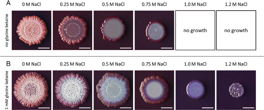
Fig 5. Effects of increasing salinity on biofilm formation in the absence or the presence of glycine betaine. Bacillus subtilis BSB1 cells were spot-
ted onto MSgg agar plates containing the indicated concentrations of NaCl in the absence (A) or in the presence (B) of 1 mM glycine betaine.
Colonies were imaged after 3 days of incubation at 30 C. Bacillus subtilis BSB1 cells were not able to grow on plates containing 1 M or 1.2 M
NaCl, respectively, when no glycine betaine was added. Scale bars: 0.5 cm.
© 2020 The Authors. Environmental Microbiology published by Society for Applied Microbiology and John Wiley & Sons Ltd.,
Environmental Microbiology, 22, 3266–32863278 H. Rath et al.
Effects of glycine betaine on gene expression not truly live stress-free (Hallsworth, 2018). However, to
what extent these levels of glycine betaine can affect gene
By accumulating compatible solutes, bacteria are able to
expression, has not been explored so far. Therefore, we
maintain turgor at physiologically adequate values, reduce
included in our study samples of B. subtilis BSB1 grown in
the level of molecular crowding, and optimize the solvent
SMM with 1 mM glycine betaine and in SMM with 1.2 M
properties of the cytoplasm (Wood, 2011; Stevenson
NaCl/1 mM glycine betaine in order to determine the
et al., 2015a; Bremer and Krämer, 2019). Collectively, this
effects of glycine betaine under both standard and high-
allows the cells to proliferate under unfavourable hyper-
salinity growth conditions.
osmotic conditions. One of the most widely used and par-
Under high-salinity conditions, the transcript levels of
ticularly effective osmoprotectants in microorganisms is
427 and 546 genes were up- and downregulated,
glycine betaine (Kempf and Bremer, 1998; Gunde-
respectively, when 1 mM glycine betaine was present in
Cimerman et al., 2018). Previous studies with different
the growth medium (Fig. 6; Table S1). Generally, two
microorganisms have shown that its import can influence
gene expression. In particular, genes involved in the syn- groups of high-salinity responsive genes can be distin-
thesis or uptake of compatible solutes exhibit reduced guished according to the effect of glycine betaine. The
induction under osmotic stress conditions (Spiegelhalter expression pattern of group 1 genes is not influenced
and Bremer, 1998; Brill et al., 2011; Hoffmann et al., 2013). by the presence of glycine betaine. For group 2, the
The intracellular glycine betaine pool generated by B. sub- high-salt-mediated regulatory pattern is at least partially
tilis through import is linearly dependent on the salt con- reversed by glycine betaine, e.g. Figure 2B: genes
centration of the growth medium and reaches 500 mM in belonging to the SigB, CymR, DegU, SigD, SinR and
the presence of 1 M NaCl (Hoffmann et al., 2013). Glycine AbrB regulons (Fig. S4). Interestingly, a transcriptome
betaine accumulates to substantial level (about 160 mM) study of Escherichia coli exposed to continuous
even in B. subtilis cells grown in the standard laboratory osmotic stress revealed only minor effects of glycine
medium SMM (about 350 mosmol kg−1) without additional betaine on the expression of osmotically induced genes
salt (Hoffmann et al., 2013), indicating that these cells do (Gunasekera et al., 2008).
Fig 6. Effects of glycine betaine
on high-salinity regulated genes.
Venn diagrams show the overlap
of genes differentially expressed
between SMM with 1.2 M NaCl
and SMM (left) and between
SMM with 1.2 M NaCl and 1 mM
glycine betaine and SMM with
1.2 M NaCl (right). Each group is
subdivided into coding
sequences (CDS; bold) and RNA
features according to Nicolas and
colleagues (2012) (italic).
© 2020 The Authors. Environmental Microbiology published by Society for Applied Microbiology and John Wiley & Sons Ltd.,
Environmental Microbiology, 22, 3266–3286You can also read