Chasing the Apomictic Factors in the Ranunculus auricomus Complex: Exploring Gene Expression Patterns in Microdissected Sexual and Apomictic ...
←
→
Page content transcription
If your browser does not render page correctly, please read the page content below
G C A T
T A C G
G C A T
           genes
Article
Chasing the Apomictic Factors in the
Ranunculus auricomus Complex: Exploring Gene
Expression Patterns in Microdissected Sexual and
Apomictic Ovules
Marco Pellino 1,2 , Diego Hojsgaard 3 , Elvira Hörandl 3, *        and Timothy F. Sharbel 1,2,†
 1   Leibniz Institute of Plant Genetics and Crop Plant Research (IPK), D-06466 Gatersleben, Germany;
     marco.pellino@gifs.ca (M.P.); tim.sharbel@usask.ca (T.F.S.)
 2   Seed Developmental Biology Program, Global Institute for Food Security (GIFS), University of Saskatchewan
     105 Administration Place, Saskatoon, SK S7N 5A2, Canada
 3   Department of Systematics, Biodiversity and Evolution of Plants, Albrecht-von-Haller Institute for Plant
     Sciences, Georg-August-University of Göttingen, Untere Karspüle 2, D-37073-1 Göttingen, Germany;
     dhojsga@gwdg.de
 *   Correspondence: Elvira.hoerandl@biologie.uni-goettingen.de; Tel.: +49-551-39-7843
 †   Present address: Department of Plant Sciences, University of Saskatchewan, 51 Campus Drive,
     Saskatoon, SK S7N 5A8, Canada.

 Received: 20 April 2020; Accepted: 27 June 2020; Published: 30 June 2020

 Abstract: Apomixis, the asexual reproduction via seeds, is associated to polyploidy and hybridization.
 To identify possible signatures of apomixis, and possible candidate genes underlying the shift from
 sex to apomixis, microarray-based gene expression patterns of live microdissected ovules at four
 different developmental stages were compared between apomictic and sexual individuals of the
 Ranunculus auricomus complex. Following predictions from previous work on mechanisms underlying
 apomixis penetrance and expressivity in the genus, gene expression patterns were classified into
 three categories based on their relative expression in apomicts compared to their sexual parental
 ancestors. We found evidence of misregulation and differential gene expression between apomicts and
 sexuals, with the highest number of differences detected during meiosis progression and emergence
 of aposporous initial (AI) cells, a key developmental stage in the ovule of apomicts where a decision
 between divergent reproductive pathways takes place. While most of the differentially expressed
 genes (DEGs) could not be annotated, gene expression was classified into transgressive, parent
 of origin and ploidy effects. Genes related to gametogenesis and meiosis demonstrated patterns
 reflective of transgressive and genome dosage effects, which support the hypothesis of a dominant
 factor controlling apomixis in Ranunculus and modulated by secondary modifiers. Three genes with
 probable functions in sporogenesis and gametogenesis development are identified and characterized
 for future studies.

 Keywords: apomixis; differentially expressed genes; hybridization; microarrays; polyploidy;
 Ranunculus; sexuality

1. Introduction
      The evolution of asexuality from sexual progenitors has occurred repeatedly and independently
in a wide range of plants and animals [1–3]. The evolutionary consequences of such a drastic functional
trait change have been the subject of many studies and have shed light onto mechanisms of species
dispersal and adaptation relevant to the evolutionary success of asexuality [4,5]. It has been suggested
that asexual reproduction is advantageous because it reduces to a half the costs associated with

Genes 2020, 11, 728; doi:10.3390/genes11070728                                     www.mdpi.com/journal/genes
Genes 2020, 11, 728 2 of 22
sex [6] and because of a greater adaptability to stable environments [7]. In addition, asexuality would
be beneficial for fixing advantageous mutations and genotypes highly adapted to the surrounding
environment [8,9]. Despite these expected advantages, limitations arise from the lack of meiotic
recombination and exploitation of recombinational DNA repair mechanisms [10]. The absence of
meiosis has also drastic consequences on purging deleterious mutations [11]. Asexuals are expected
to accumulate deleterious mutations in a non-reversible ratchet-like fashion [12], a process which
accelerates in small populations and can drive a lineage to extinction due to mutational meltdown [13].
In contrast, sexual recombination allows the purging of deleterious mutations [12,14] and provides
statistical chances for advantageous mutations from distinct individuals to recombine together in
a single offspring [8]. Moreover, sexuality is advantageous in changing environments whereby the
exploitation of genetic variability through meiotic and gametic recombination is central for adaptation
in natural populations [15,16].
Asexuality in plants can happen by different forms of vegetative propagation (like runners, bulbils,
tubers, etc.), or by apomixis (clonal seed formation [17]). Apomixis has been described in more than
400 species scattered throughout 293 angiosperms genera [18,19]. Apomixis is sporophytic when an
embryo develops directly from a somatic cell of the ovary, or gametophytic when the embryo develops
through the formation of an unreduced female gametophyte [17]. Two main types of gametophytic
apomixis have been characterized following the formation of an unreduced embryo sac from a nucellar
cell (apospory), or from the megaspore mother cell (MMC) via an altered meiosis (diplospory). In all
cases of gametophytic apomixis, three developmental steps must sequentially occur to produce a
functional clonal seed: (1) the development of a meiotically unreduced egg cell (apomeiosis), (2) the
development of the seed’s embryo without fertilization (parthenogenesis), and (3) the production of a
functional endosperm with (pseudogamy) or without (autonomous) fertilization [20].
The molecular control of apomixis is still uncertain. Different hypotheses have been proposed to
explain observations and experimental data from diverse apomictic plant species. Perhaps the two
most accepted hypothesis are the idea of apomixis being a consequence of changes to developmental
timing of reproduction, and the one considering apomixis as a mutation-based phenomenon [21–23].
In the first case, since polyploidy and hybridization are common traits of almost all apomictic
plants [20], both have been hypothesized to be triggers for the reproductive switch from sexuality
to apomixis. In this theoretical frame, it is proposed that global gene regulatory changes that
follow polyploidization and hybridization in general, and specifically de-regulation of the standard
sexual reproductive program lead to apomixis in plants [18], and can arise via chromosomal [24–26],
genomic [27,28], and transcriptomic [26,29] changes in gene regulatory networks. As support for this
hypothesis, studies demonstrated heterochronic gene expression (i.e., deregulation) between sexual
and apomictic reproductive tissues in Tripsacum spp. [30] and Boechera spp. [31].
In the second case, apomixis in plants has been often identified with one or two dominant factors
segregating according to Mendelian proportions, and which are altered via epistasis, genetic modifiers,
polyploidy, etc. [32]. In several studies using different apomictic species of the Ranunculus auricomus
complex throughout four generations of crossings and backcrossings, Nogler [33–35] established that
apomixis was controlled by a dominant factor A− with a recessive lethal behaviour. One of the main
consequences of such conclusion was that A− could not be transmitted though haploid gametes,
and thus nicely provided a causality for the intriguing rarity of apomixis in natural diploid plants.
In his studies, Nogler [35,36] further concluded that the frequency (i.e., expressivity) of apomixis in
individual plant genotypes was dosage dependent and relative to the wild type A+ . More recently,
part of Nogler’s results were contradicted by crossing experiments on other, more distantly related
species within the R. auricomus complex, in which experimental diploid hybrids displayed functional
apomixis in low frequencies and inheritance of apospory in diploids [37]. In this study, the expression
of apospory was dosage-dependent and higher in plants with two aposporous parents compared to
those with just one aposporous parent [37]. The apospory-specific factor appears to be not necessarily
lethal, but allelic dosage effects were confirmed.Genes 2020, 11, 728 3 of 22
The Ranunculus auricomus complex is becoming a model for studying genomic differences between
sexual and apomictic taxa. Hundreds of apomictic species are derived from a handful of sexual
progenitor species [38,39]. The closely-related diploid sexual species R. carpaticola, and diploid
and autotetraploid R. cassubicifolius extend from Switzerland to west Slovakia [39], while hexaploid
apomicts are located in central Slovakia. Analyses based upon AFLPs and SSRs, combined with DNA
sequence [39,40] and RNAseq SNPs [41], support a hybrid origin for hexaploid apomicts resulting from
a cross between a putative 4x R. cassubicifolius and a 2x R. carpaticola sometime during the last glacial
period (60,000–80,000 years ago). Apomictic Ranunculus species are aposporous (Hieracium-type),
pseudogamous and express apomixis facultatively [36,37,42–46]. Aposporous initial (AI) cells arise
from a somatic cell of the nucellus during the time in which the megaspore mother cell (MMC)
starts meiosis [35,43,44], and competes for the formation of the female gametophyte within the ovule.
Once the mature female gametophyte is formed, fertilization of the egg and central cells is required in
meiotic female gametophytes while in apomictic ones only the central cell needs to be fertilized to
form the endosperm [36,43,44].
Taking advantage of a previous study using SNPs and comparing sexual and asexual Ranunculus
genotypes using Illumina RNAseq data [41], we developed a custom expression microarray and
used it to transcriptionally profile microdissected ovules at four developmental stages from sexual
diploid R. carpaticola and tetraploid R. cassubicifolius genotypes, and hexaploid apomictic R. carpaticola
× cassubicifolius genotypes. Using SNPs and RNAseq data might be of help to distinguish the molecular
cause for apomixis in Ranunculus, i.e., whether is due to heterochronic gene expression or dominant
factors. Based on levels of gene expression changes and dysregulated genes, we would expect global
changes in gene expression in the first case or rather a few changes on master genes in the second case.
This, however, is at least challenging since one or a few dominant factors regulating apomixis would
likely trigger a wide range of downstream changes in gene regulatory networks, blurring possibilities
to segregate expression patterns into one of both hypotheses. Either way, our experimental approach
and in silico analyses were designed to overcome difficulties due to the occurrence of differing ploidies
in our target Ranunculus plants, exploiting multiple genotypes as biological replicates. The goals of
this study were to (1) identify transcripts showing differential expression between apomictic and
sexual ovules, (2) quantify heterochronic gene expression patterns during apomictic and sexual ovule
developments, (3) partition transcripts into groups whose expression signatures reflect hybridity,
parent of origin effects, or polyploidy, and (4) frame our findings according to previous and ongoing
studies to understand the inheritance of apomixis in Ranunculus and identify evidence of molecular
evolution between reproductively relevant sequences within the complex.
2. Materials and Methods
2.1. Custom Microarray Development
The RNAseq approach used to generate the data for microarray design was explained in detail in
Pellino et al. [41]. In short, flowers were collected from two sexual and two apomictic genotypes of the
Ranunculus auricomus complex within a two-week time window (Table 1). For each individual, total RNA
was isolated from pooled flowers of five different sizes (two to seven mm in length, from an early
stage when sepal primordia have enclosed the floral meristem, to the fully developed unopened flower
stage) using the Qiagen RNeasy Plant Mini Kit (Qiagen GmbH, Hilden, Germany). After isolation,
any DNA contamination was removed using Qiagen RNase-Free DNase, while contamination from
the DNase enzyme, polysaccharides, and proteins were removed with a second purification step using
the Qiagen RNeasy Mini Kit. The manufacturer’s instructions were followed in all purification steps.Genes 2020, 11, 728 4 of 22
Table 1. Diploid, tetraploid and hexaploid Ranunculus samples used in this study.
Taxon Apospory Collection Locality, Collector, Collection Number, and
Nind Ploidy 1 Reprod Mode
(Code) Expressivity 2 Vouchers
R. carpaticola Slovakia, Slovenské rudohorie, Revúca, hill Skalka (forest)
3 2x Sex 0.0
REV1 Hörandl 8483 (WU)
R. carpaticola × Slovakia, Strázovské vrchy (near Trençín), between Kubra
cassubicifolius 1 6x Apo 0.226 and Kubrica (margin of forest and meadow)
TRE Hörandl et al. C29 (SAV)
R. carpaticola ×
Slovakia, Turçianska kotlina, Vrútky-Piatrová (meadow)
cassubicifolius 2 6x Apo 0.339
Hörandl et al. C35 (SAV)
VRU 2
R. cassubicifolius Austria, Lower Austria, Wülfachgraben, SE Ybbsitz (forest)
1 4x Sex 0.0
YBB 1 Hörandl 8472 (WU)
1For ploidy identification see [47,48]; 2 Data from [44]. WU: herbarium of the University of Vienna, SAV: herbarium
of the Institute of Botany, Slovak Academy of science, Bratislava.
To avoid over-representation of highly transcribed genes during subsequent sequencing steps,
full length normalized cDNA libraries were produced, whereby each step of the normalization was
performed and optimized following the procedures described by Vogel et al. [49] and Vogel and
Wheat [50].
15 µL of normalized cDNA (200 ng/µL) was sent to Fasteris SA (Geneva, Switzerland) for RNAseq,
where a dual-sequencing approach (54-mer single read (SE) and 108-mer pair-end read (PE)) was chosen
in order to balance cost and efficiency of a de novo assembly (see Pellino et al. [41]). Both sequencing
strategies were conducted using the HiSeqTM 2000 sequencing system (Illumina Inc., San Diego, CA,
USA).
CLC Genomics Workbench (CLC bio version 4.9) was implemented for the sequencing assembly.
At first, sequences were trimmed for vector contamination, length and quality score using CLC
default values. As no reference genome for Ranunculus is available, de novo and iterated de novo
approaches were used to assemble the data. For the de novo assembly, all libraries were pooled
and assembled using CLC default parameters. In the iterated de novo assembly, each library was
assembled individually, and the resultant contigs from each individual assembly re-assembled together,
including all unassembled reads from each individual assembly. In both strategies the final assembly
was trimmed for contigs shorter than 300 bp (see details in Pellino et al. [41]). The two approaches
were evaluated based on the total number of matching reads (i.e reads that could be assembled into
longer contigs) and N50 values, and the de novo assembly was selected for array design. Both contigs
and singletons were forwarded to Roche NimbleGen Inc. (Madison, WI, USA) for design and
manufacture. The NimbleGen selection strategy bioinformatically designed three different probes for
each contig, and one to three probes for each singleton, followed by design of a 3 × 1.4 million-spot
array. The final array therefore contained multiple technical replicates for each gene expressed during
flower development in Ranunculus. All the microarrays data has been uploaded to Arrayexpress at
EMBL-EBI (“Experiment E-MTAB-3316”).
2.2. Transcriptomal Profiling of Sexual and Apomictic Ovules
2.2.1. Sample Selection, Ovule Microdissection, and RNA Extraction
Plants were grown from seedling to pre-flowering stages in outdoor plots at the Leibniz Institute
of Plant Genetics and Crop Plant Research (IPK), and were then moved into a phytotron for flowering
(day: 16 h, 21 ◦ C; night: 8 h, 16 ◦ C; humidity 70%; light intensity: 150 µmol/m2 ). Based on the
cytological observations of Hojsgaard et al. [44], ovules at four developmental stages were collected at
the pre-meiotic, meiotic (tetrad stage/aposporous initial), 2–4 nuclei embryo sac and mature embryo
sac stages (I, II, III, IV; Figure S1) at standardized times (between 7:00 and 9:00 AM) over multiple days
from both sexual and apomictic Ranunculus under a sterile laminar-flow hood with a stereoscopic
microscope (100 Stemi; Carl Zeiss AG, Oberkochen, Germany). Using sterile scalpel and forceps toGenes 2020, 11, 728 5 of 22
open the flower, carpels were collected and immediately immersed in a sterile 0.55 M mannitol solution
and placed on ice.
In the second step, microdissection of each single carpel was conducted in a in a sterile laminar
air-flow hood under an inverted microscope (Axiovert 200M; Carl Zeiss AG, Oberkochen, Germany)
using hand-crafted sterile glass needles (self-made with a Narishige PC-10 puller). Individual ovules
were collected using an Eppendorf Cell Tram Vario connected to a 150 µm inner diameter glass
capillary, immersed in 100 µL RNA-stabilizing buffer (RNA later; Sigma-Aldrich, St. Luis, MO, USA),
and immediately frozen in liquid nitrogen. 20 to 40 ovules for each genotype/stage were dissected
and stored at −80 ◦ C. RNA extraction was carried out using a PicoPure isolation kit (Thermo Fisher
Scientific, Carlsbad, CA, USA) and quantification and quality were assessed with RNA Pico chips on
an Agilent 2100 Bioanalyzer (Agilent Technologies, Santa Clara, CA, USA).
2.2.2. cDNA Synthesis and Amplification
For each of the samples from the seven Ranunculus individuals (Table 1), cDNA synthesis and
amplification were conducted using the Sigma TransPlex Complete WTA2 kit (Sigma-Aldrich) following
the producer’s instructions. Amplified cDNA was purified using the GenEluteTM PCR Cleanup kit
(Sigma-Aldrich) following the manufacturer’s protocol, and concentration and quality were measured
with a NanoDropTM 1000 Spectrophotometer (Thermo Fisher Scientific, Wilmington, DE, USA).
2.2.3. Microarray Hybridization and Data Processing
Twenty-eight 1.4 M probe custom microarrays were outsourced to Roche NimbleGen for design
and manufacture, each of which was used for an independent sample labeling and hybridization
reaction for each of the 28 classes of microdissected ovule samples (four stages for seven genotypes;
Table 1, Figure S1) following the NimbleGen microarray labeling protocol of their One-Color DNA
Labeling Kit. The labeled samples where then individually hybridized in random order, using the
NimbleGen Hybridization System 12 to the custom Ranunculus arrays according to the producer’s
instructions, and scanned using a NimbleGen MS 200 Microarray Scanner at 535 nm. Feature
intensities were extrapolated using the DEVA software (version 1.1, Roche NimbleGen), and the raw
expression data were normalized together using the DEVA implemented robust multiarray average
(RMA) algorithm.
The normalized data were analyzed using the Qlucore Omics Explorer software (version 2.3).
Principal Component Analysis (PCA) and Qlucore filtering by variance was implemented, whereby
contigs with variation (σ/σmax ) < 0.7 between apomictic and sexual groups were removed from
the dataset. This threshold was chosen, using the Qlucore software, such that the differentially
expressed apomictic and sexual probe groups could be visually separated on a PCA, while at the same
time retaining the maximum number of differentially expressed genes. Second, the resultant set of
differentially expressed genes was ranked according to p-value (2 and adjustment of the p-value for multiple tests using a false discovery rate (FDR) with
q-value < 0.05 were applied.
2.2.4. Analyses of Gene Expression throughout Development
In order to calculate significant changes in gene expression patterns throughout ovule development
between apomictic and sexual samples, the STEM software [51] was used to perform a similar analysis
to that made by Sharbel et al. [31]. First, a data set of genes showing significant expression differences
in at least one stage (but including the expression over all other stages) was constructed and used for
STEM analysis. Except for the number of permutations, which was set to 1000 to increase accuracy [31],
the profiling analysis was performed using default options with Bonferroni correction for multiple
testing [51]. For the comparative analysis of different patterns of gene expression across the fourGenes 2020, 11, 728 6 of 22
developmental stages, the minimum number of intersected genes between sexual and apomictic
samples was set to 1 [31] with maximum uncorrected intersection p-values < 0.05.
2.3. Analyses for Signatures of Ploidy, Parent of Origin Effects or Hybridization
Considering variable ploidy and evolutionary origins between sexual and hybrid apomictic
Ranunculus, we sought to classify gene expression patterns into groups reflective of ploidy, parent of
origin (sensu expression level dominance) or hybridization (sensu transgressive) effects. We first
selected those genes showing statistically significant differential expression (p < 0.05, log2 fold change
> 2, and FDR q-value < 0.05) between hexaploid apomicts and diploid sexuals, the two groups for
which samples were available (Table 1) and hence for which statistical analysis was possible (due to
sample size and lack of biological replicate, a similar statistical comparison using the tetraploid sample
was not possible). Therefore, we used this subset of genes defined by the hexaploid apomict-diploid
sexual comparison in all approaches. Then, in order to obtain information of partitioning of gene
expression in the allopolyploid relative to each parent, we compared gene expression of each group,
including data from the single tetraploid, estimating for each gene the log-fold expression difference of
two contrasts: both parents to each other, and each parent to the allopolyploid, and controlling the
distribution of p-values for each estimate using a false discovery rate of 0.05 [52].
2.3.1. Ploidy
On a gene-by-gene basis, the standard deviation of expression was calculated for the hexaploid
apomicts and diploid sexuals. Then, genes in the tetraploid and hexaploids whose expression was (1)
lower than two standard deviations in the diploid, and (2) higher than two standard deviations in the
diploid, were classified as exhibiting a statistically significant pattern reflective of ploidy/additivity
effects (sensu Yoo et al. [53]). Thus, genes in the hexaploid were classified as displaying additivity and
expression level dominance compared to either parent (for transgressive expression see Section 2.3.3).
2.3.2. Parent of Origin Effects
Genes in sexual diploids and the tetraploid which (1) showed no expression differences with the
hexaploid apomicts (i.e., two standard deviations of the mean diploid sexual levels (the single
tetraploid had no mean values per se), were classified as having patterns reflective of a parent of
origin/expression dominance effect.
Lastly, we selected the group of all genes which did not show diploid-hexaploid parent of origin
effects and were not initially selected (i.e., no significant differences in expression between hexaploid
apomicts and diploid sexuals, see above). Next, we compared gene-by-gene the value of expression
in the tetraploid against mean value of the hexaploid. Those genes in which the tetraploid sample
had higher (i.e., >two standard deviations of the mean hexaploid apomict levels) or lower (i.e.,Genes 2020, 11, 728 7 of 22
2.4. Microarray Validation Using qRT-PCR
Ten genes showing differential expression between apomictic and sexual genotypes across the
four developmental stages were randomly selected (see Section 3). After retrieving the sequences from
the assembled cDNA database used in the array manufacture, PCR primers were designed using the
PrimerSelect software (DNASTAR Inc., Madison, WI, USA) and selected, when possible, to overlap the
microarray probes with the following parameters: product size 95%,
e-value < 1 e−100 ) homologous genes to UBQ (gb|ABH08754.1|) and ACTIN 11 (ref|NP_187818.1|) where
chosen. Primers were designed following the procedures and parameters described above, and tested
for amplification and expected product length in 10 µL PCR reactions including 25 ng of DNA,
1 µL of PCR Buffer II, 10 pmol for each primer, 0.025 U DNA Taq DNA Polymerase (Sigma-Aldrich,
with 3.5 mM of MgCl2 and 4.95 µL of H2 O. PCR reactions were performed in a Mastercycler ep384
(Eppendorf, Hamburg, Germany) using the following touchdown thermal cycling profile: 94 ◦ C for
10 min; 9 cycles of 94 ◦ C for 15 sec, 65 ◦ C for 15 sec (1 degree decrease in temperature every cycle with
a final temperature of 54 ◦ C), 72 ◦ C for 30 sec; 35 cycles of 94 ◦ C for 30 sec, 57 ◦ C for 15 sec, 68 ◦ C for
2 min 30 sec; and a final 68 ◦ C for 15 min.
qRT-PCR reactions, using UBQ and ACT11 (Dryad Id number: 82481 and 151955 Dryad entry
doi:10.5061/dryad.nk151) as housekeeping genes, were run on a 7900HT FAST RT-PCR machine
(Applied Biosystems, Foster City, CA, USA) using the SYBR Green Master Mix (Applied Biosystems)
and the following program: initial denaturation at 90 ◦ C for 10 min, followed by 40 cycles of 95 ◦ C
for 15 sec, and 60 ◦ C for 1 min. Ct values (PCR cycle number where SYBR Green is detected) were
extrapolated and used to infer initial copy number of the genes. Mean expression and standard
deviation were calculated between two technical replicates and three biological replicates from
apomictic and sexual genotypes using cDNA from the second stage of ovule micro-dissected tissues.
Relative quantification was calculated using the ∆∆Ct method and using the genes with higher CT
values compared to the calibrator sample.
2.5. blastx, tblastx, Gene Ontology
Significantly differentially expressed genes obtained in all apomictic–sexual comparisons were
selected for gene ontology (GO) analysis with Blast2Go (http://www.blast2go.com/b2ghome) using
blastx (E-value cut off of E ≤ 1−5 ) and the default annotation parameters of the program. For genes
where no blastx hits were obtained, an additional tblastx analysis was performed (E-value cut off of
E ≤ 1−5 ). Overrepresentation analysis was not possible since only a small fraction of the Ranunculus
transcriptome could be annotated (see Section 3).
3. Results
3.1. Gene Expression Differences
3.1.1. Principal Component Analysis (PCA)
In order to recognize graphically possible subsets of genes that may account for the maximum
distinction between the apomictic and sexual samples, PCA was applied to the normalized expression
values of probes from the original 62102 contigs (≥300bp; raw data first normalized using the RMA
algorithm implemented in the DEVA software). The resulting PCA graph showed a clear separation
between samples with respect to ploidy, distinguishing apomictic and sexual groups (Figure 1).Genes 2020, 11, 728 8 of 22
Figure 1. Principal component analysis (PCA) of gene expression on apomictic and sexual Ranunculus.
PCA applied to normalized microarray data representing 4 ovule developmental stages from 3 apomictic
(red) and 4 sexual (blue and green) Ranunculus genotypes showing ploidy and reproduction specific
effects. Each dot represents one ovule stage, and frequencies in parentheses show the percentage of total
variation explained by that principal component. The numbers on the axes represent the respective
component (and the % of variation for each component).
3.1.2. Stage Specific Differential Expression Analysis
Transcriptome-wide gene expression variation through ovule development was compared between
sexual diploids and apomictic hexaploids of Ranunculus, whereby four pre- to post-meiotic ovule stages
were compared between six different genotypes. Overall, across all stages 439 and 339 transcripts were
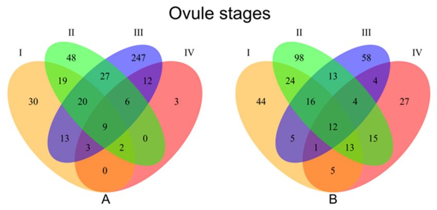
found to be significantly down- or up-regulated between apomictic versus sexual genotypes (Figure 2;
Venn A and Venn B, respectively). The distribution of up- and downregulated transcripts in apomicts
differed across the studied developmental stages I to IV (30, 48, 247, and 3 upregulated, 44, 98, 58,
and 27 downregulated for each stage respectively) (Figure 2).
Figure 2. Venn diagrams showing number of differentially expressed transcripts between apomictic
and sexual genotypes at each ovule developmental stage. A: Genes downregulated in the 6x apomicts
compared to 2x sexuals B: Genes upregulated in the 6x apomicts compared to 2x sexuals. I = premeiotic,
II = meiotic/aposporous, III = early embryo sac, IV = mature embryo sac.
Blast2Go analyses could be completed on only a small fraction of the differentially expressed
transcripts, the limiting step being the inability to identify significant homologies to the NCBI nucleotideGenes 2020, 11, 728 9 of 22
database(s). Of the number of genes which could be annotated (110 apomictically upregulated and 161
apomictically downregulated), only 59 (53.6%) upregulated and 78 (48.5%) downregulated transcripts
from apomicts could be assigned a gene ontology (GO) term (Table S1; Table 2). Hence, representation
analysis was not possible due to the significant bias introduced by insufficient GO categorization.
A full list of genes can be accessed at GenBank (BankIt2351891: MT624108 - MT624295).
Table 2. Numbers of differentially expressed transcripts at each of four ovule developmental stages.
Number of Contigs Number of Contigs, >300 bp Differentially Expressed Transcripts Annotated
462102 62102
Develop. stage Upregulated 1 Downregulated 1
I 120 (44) 96 (30) 72
II 195 (98) 131 (48) 107
III 113 (58) 337 (247) 156
IV 81 (3) 35 (3) 30
1data from 6x apomictic individuals compared to the normal expression in 2x sexuals; numbers in brackets represent
number of stage specific transcripts.
A clustering-based STEM analysis [51] was performed to detect significant changes in patterns of
transcript abundance across ovule development, by first grouping transcripts with similar expression
profiles through development for the sexual and apomictic array datasets separately (Figure S2).
In doing so, 46 and 62 transcripts could be assigned to three distinct patterns (STEM analysis, p < 0.01)
in the sexual and apomictic groups, respectively. A comparison of patterns between these transcript
sets identified eight transcripts as having significant differences (STEM analysis, p < 0.01) in the
corresponding reproductive form, and showed a general trend of expression increase in developmental
stage II followed by a sharp drop in stage III in apomicts (Figure S3). None of the eight genes could
be assigned a GO term, and three had a significant homology to the transposon mutator sub-class
protein (XP_006654086.1), salt overly sensitive 1b isoform 1 (EXC05020.1), and annexin-like protein
(XP_007042996.1), respectively.
3.2. Transcriptome Wide Signatures of Hybridization, Ploidy Variation and Parent of Origin Effects
In order to understand and classify gene expression patterns of the apomictic hybrid (R. carpaticola
× R. cassubicifolius) compared to the ancestral sexual parents (R. carpaticola and R. cassubicifolius),
304 differentially expressed transcripts were first identified in at least one developmental stage based
on minimum fold change and statistical significance (log2 > 2, p-value < 0.01, FDR < 0.05) between the
apomictic hybrids and the diploid sexuals (both groups having three genotypes each for statistical
comparison). Secondly, a stage-by-stage comparison was performed for the expression of these 304
transcripts in the second tetraploid sexual parent (R. cassubicifolius; for which sample size precluded
the first level statistical analysis—see Section 2) such that they could be classified into four different
expression states (i.e., the hybrid relative to each ancestral parent). Genes showing expression bias with
respect to the diploid or tetraploid parent were classified as showing a parent of origin effect, while those
significantly over- or under-expressed in the apomict compared to both parents were classified as
showing transgressive effects. Lastly, genes showing expression level changes in proportion to that of
ploidy (e.g., increasing expression with increase of ploidy) were classified as showing a ploidy effect.
Examination of sequence similarity between genes showing a parent of origin effect and a
high-quality SNP library (44) revealed only eight genes characterized by sufficient DNA sequence
read coverage in all individuals, and of those only one (dyad contig id: 368515, Dryad entry
doi:10.5061/dryad.nk151) showed 100% SNP similarity (for 16 SNPs) between the apomictic and the
tetraploid genotypes only, implying a potential origin from the putative tetraploid parent (Table S2).
The same procedure was applied to 19,116 genes that were not significantly differentially expressed
between the apomictic hexaploids and the sexual diploids in order to classify an additional state of
gene expression (2x-parent of origin; see methods). This approach was necessary considering that only
a single 4x sample was used, and allowed the comparison of gene expression of this one parent (4x) toGenes 2020, 11, 728 10 of 22
the hybrid apomict. This classification was further subdivided with regards to whether expression
of the hybrid and the diploid were higher or lower in comparison to the tetraploid. In total eight
different groups of genes were classified, with most being downregulated in apomictic ovules at stage
II (Figure 3; Table 3, Tables S3 and S4).
Figure 3. Box plots of expression distributions for genes which were significantly up- or downregulated
in at least one apomictic ovule stage, as grouped into transgressive, ploidy-mediated and parent of
origin patterns.Genes 2020, 11, 728 11 of 22
Table 3. Number of differentially expressed genes 1 associated with each ovule developmental stage
and expression class in diploid–polyploid comparisons (brackets show relative percentage compared
to the overall stage number of genes).
Expression Class 2 Develop. Stage Transgressive Effect Parent of Origin Effect Ploidy Effect Total
Apomictic 6x upregulated 3 I 5 (0.1) 27 (0.49) 23 (0.41) 55
II 15 (0.58) 10 (0.38) 1 (0.04) 26
III 16 (0.16) 44 (0.46) 36 (0.38) 96
IV 2 (0.4) 1 (0.2) 2 (0.4) 5
Total up 38 (0.21) 82 (0.45) 62 (0.34) 182
Apomictic 6x downregulated 3 I 6 (0.26) 7 (0.3) 10 (0.44) 23
II 18 (0.32) 37 (0.65) 2 (0.03) 57
III 9 (0.24) 11 (0.3) 17 (0.46) 37
IV 2 (0.4) 1 (0.2) 2 (0.4) 5
Total down 35 (0.29) 56 (0.46) 31 (0.25) 122
1for seq. names, GO terms, enzyme codes and statistics refer to Tables S1 and S5; 2 compared to normal sexual
diploid expression; 3 p < 0.001, FDR < 0.05, log2 change > 2.
Overall, most transcripts were expressed in a parent of origin pattern, followed by ploidy and then
transgressive patterns (Table 3; Table S3). The strongest effect was apparently due to parent of origin,
with a total of 82 upregulated and 138 downregulated apomictic-specific transcripts. Ploidy-mediated
patterns of expression were revealed in 62 and 93 apomictic specific upregulated and downregulated
transcripts, respectively. Transcripts showing transgressive patterns were the least abundant, with 38
and 73 upregulated and downregulated in apomicts, respectively (Table 3).
Of all transcripts showing such patterns, only 83 (49 and 34 up- and downregulated in apomicts
respectively) could be annotated (Table S5). A simple screening of these genes revealed only three
genes related to ovule development and reproduction. These three genes were coding for embryo sac
developmental arrest protein (EDA), gamete expressed protein (GEX3), and a gene of the argonaute
family (AGO) (e-value = 1.43 e-13, 3.28 e-32, 1.61 e-63, respectively and mean similarity of 47%,
70.5% and 84.4%, respectively; Table S5). Other annotated genes were related to expression of
retrotransoposons, transposon proteins, transcription factors, ribonucleases, kinases, among others
(Table S5).
3.3. qPCR Validation
Validation of the randomly selected genes showed concordance with the microarray analysis
from the five upregulated and five downregulated genes. According to the REST software all 10 genes
showed statistically significant up- and downregulation when measured with qRT-PCR (Table S6).
4. Discussion
Whether caused by a single “master gene”, or through a polygenic complex, apomixis has long
been a dilemma for scientists. Using different experimental approaches, the search for factor(s)
underlying the switch in reproduction has led to the identification of a number of gene candidates in
different species [22,54,55]. Such studies have also revealed a shift in gene expression through time for
dozens or hundreds of genes normally involved in sexual reproduction [31,56,57]. Omic approaches
have become useful for detecting expression shifts, although functional characterization and screening
of the many candidates from such an experiment is a formidable task. This is especially true when
dealing with wild species (as most apomicts are) in which annotated genomic information is not
available. Additionally, the effects of polyploidy and hybridization [58] are often associated with the
apomixis phenotype, adding an extra level of complexity to all analyses.
Despite hybridization and polyploidy introducing difficulties into data interpretations
(see Mau et al. [59], their association with apomixis potentially reflects its mechanism of induction [18,60].
The “genomic shock”, i.e., the genomic perturbation at both genetic and epigenetic levels produced
by the union of two different genomes [61,62], could trigger the cascade of spatial and timing
mis-expression of sex specific genes and lead to apomixis [20,63]. More recent studies suggest thatGenes 2020, 11, 728 12 of 22
polyploidy is not essential for expression of aposporous apomixis, because it appears in diploid
hybrids [37] and non-hybrids [64,65], albeit in low frequencies. These and other studies suggest that
origin of apomixis in wild populations probably starts in diploid populations and is just indirectly
enhanced by side-effects of polyploidy [66,67].
Continuing our ongoing work [41,44] with wild allopolyploid apomictic Ranunculus auricomus and
related sexuals, we wanted to shed light upon the molecular basis for apomixis in Ranunculus
and possible factors underlying the transition from sexuality to apomixis. We have analyzed
genome-wide gene expression through morphologically-defined ovule developmental stages in
both apomictic and sexual genotypes, and using a specific sampling strategy to maximize biological
diversity which enabled the identification of differences encompassing both reproductive mode
(e.g., apomictic versus sexual) and genetic background (e.g., different sample clusters) (see Figure 1).
Thus, our approach allowed us to disclose species’ specific transcriptional variation connected to
polyploidy and hybridity, and partition differentially expressed genes into patterns reflective of (a)
reproduction specific expression, (b) heterochronic expression across ovule development, and (c) the
expression of homologous genes in the apomicts and their ancestral (i.e., phylogenetic) parents.
4.1. Transcriptional Variation Reflect Contrasted Sexual vs. Apomictic Developments in the Ovule
In the Ranunculus cytotypes used here, Hojsgaard et al. [44] showed that the first stage of ovule
primordium development is undifferentiated between apomictic and sexual Ranunculus. In contrast,
diverse developmental irregularities accumulate during megasporogenesis, leading to perturbed and
non-functional meiosis in apomicts (i.e., arrested development and/or altered megaspore selection; [44]),
followed or accompanied by enlargement of a somatic cell that takes on the role of an aposporous
initial (AI) cell. Deviations between the sexual and apomictic pathways continue during gametogenesis
with subsequent development of the AI, parallel in timing with normal sexual gametogenesis in a
few cases where meiosis progressed. The resultant unreduced megagametophyte (arising from the
AI) then attains a similar multicellular structure to that of the sexuals at the final developmental
stage [44]. Our tissue sampling design covered such developmental stages preceding and leading up
to AI development till formation of female gametophytes (Table 1).
A reflection of the phenotypically-diverging development between pathways, gene expression
differences between sexual and apomictic ovules show fewer differences in the total number of
differentially expressed transcripts in the first and last stages of ovule sampling, whereas higher
numbers of differentially expressed transcripts were found during the second and third stages (Table 2
and Figure 1). These findings agree with overall findings in other apomictic systems displaying global
gene de-regulations in ovules at different developmental stages [68,69]. In our Ranunculus samples,
stages II encompass AI appearance, usually in parallel with meiotic depletion, whereby the MMC
did not enter meiosis or aborted during meiotic division. Stage III encompass the acquisition of a
gametogenesis program whereby the AI enlarges to squeeze the forming meiotic megagametophyte
(if any) and eventually take its place. Despite the relative weakness of any GO inference (see results),
we note that no particular GO classes distinguished differentially expressed genes (Table S1) at any
stage. Especially in stage II, the appearance of aposporous initials and parallel development/arrestment
of meiotic products (with almost equal frequencies between hexaploids [44]) may blur the distinction of
genes expressed between these two cell lineages. Later on, in stage III, the differences between sexual
and apomictic developments become most distinct (Figure 3) as further developmental competition
increase rates of abortion in the meiotic pathway (whether of meiotic products, functional megaspore or
early embryo sacs), and only unreduced gametophytes develop further. Stage IV might be principally
not so different in gene expression as there are no key developmental transitions as in stages II
(AI appearance, sporogenesis) and III (gametogenesis), and mature sexual and apomictic embryo sacs
do not differ phenotypically.Genes 2020, 11, 728 13 of 22
4.2. Apomixis Inheritance in Allopolyploid Ranunculus and Partitioned Gene-Expression Effects
Apomixis involves changes to a number of individual (meiotic recombination, purging of
mutations) and population (gene flow, adaptation) attributes which contribute to shape the life history
of species [17]. As complex as the genetic processes underlying apomixis can be, it is believed to be
controlled by one or few master regulatory genes [54]. In the 1980s, after a series of experimental
crossings and offspring analyses, Nogler published a seminal work on the genetic inheritance of
apomixis in Ranunculus. The association of apomixis and ovule gene de-regulation has been described
in many plant species [22], thus, here we focus on discussing the relevance of our present results in
the frame of Nogler’s ideas about apomixis inheritance, including more recent studies on different
Ranunculus species.
4.2.1. Homologous Parent of Origin Effects in the Apomictic Hybrid
As mentioned above, the hexaploid natural hybrid studied here originated during the last
glaciation from a cross between a Ranunculus carpaticola diploid and a R. cassubicifolius tetraploid [41,48].
Our comparative analyses of gene expression patterns between each putative parental cytotype and its
derivative hybrid lineage reveal taxon-related differences.
In the putative diploid parent, we found a relatively low number of transcripts with similar
expression levels as in hexaploids, and thus the bias in gene expression of hexaploids toward the
tetraploid parent suggests a parent of origin effect. Contributing to this effect is gene dosage, whereby the
hexaploid is composed of two haploid genomes from the diploid parent R. carpaticola [48]. In regard to
Nogler’s hypothesis, this observation suggests that A− is absent (as expected) in diploids [35]. However,
aposporous embryo sacs had been observed in very low frequencies in diploid R. carpaticola [44]
which poses the alternative explanation that A− is present in diploids but is not expressed, or that it
is part of a multigenic network influencing the trait. Sequence similarity analysis using high quality
single nucleotide polymorphisms (SNPs) mined from a previous study [41] revealed that only one
of the genes showing a parent of origin effect and sufficient DNA sequence read coverage in all
individuals showed 100% SNP similarity between alleles present in the apomictic and the tetraploid
genotypes, but not to the diploid parent, implying a potential origin from the putative tetraploid
parent. Considering the variability in read coverage between samples and the fact that the genotypes
analyzed are, sensu stricto, not those involved in the original hybridization event circa 80,000 years
before present [41], more cannot be inferred from the SNP dataset except that the diploid R. carpaticola
probably lacks the factor A− .
Regarding the putative tetraploid parent, over all developmental stages 220 genes were found
to be similarly expressed between 6x apomictic R. carpaticola × cassubicifolius and 4x R. cassubicifolius
plants, once again suggesting a parent of origin effect and genomic dosage (6x has expectedly four
putative haploid genomes from R. cassubicifolius [48]). Even though the relatively higher number of
genes displaying a tetraploid parent of origin pattern of expression in Ranunculus ovules is likely biased
by the lack of biological replicates, the result is concordant with similar studies in natural populations
and synthetic hybrids of cotton [70], maize [71], rice [72] and Senecio [73]. All such studies showed
similarly higher levels of parent of origin expression relative to transgressive effects, which tended to
decrease with the age of the hybrid.
Investigations into the causes of parental expression bias point to the effect of cis-trans regulation,
epigenetic changes, and introgression in organisms with different life histories like Cirsium arvense
(thistle) [74] and cotton [53]. In Ranunculus synthetic hybrids (between diploids Ranunculus carpaticola
× notabilis and tetraploid and diploid R. cassubicifolius × notabilis), apospory appeared spontaneously
in diploid and triploid F1 hybrids of sexual species that did not previously show any signs of apomixis;
however, functional apomictic seed were found in triploid F1 hybrids [44], and in diploid F2 hybrids [37],
both in low frequencies.
Even though Nogler’s [35,36] apospory-incurring A− factor is causal in the genomes of hybrids,
it is plausible that such a factor is present in one (or both) parental species but not expressed.Genes 2020, 11, 728 14 of 22
Previous morphological and genetic analyses of tetraploid R. cassubicifolius indicate that the cytotype is
sexual [35,39,47,48], and embryological studies failed to show any sign of AI formation [44]. However,
the high dosage of the wild-type A+ from the tetraploid sexual parent might explain the relatively
high frequencies of sexual ovule (69%) and seed formation (29%) in hexaploid apomicts [44] compared
to other polyploid apomicts having divergent evolutionary histories in which sexuality is found
at residual levels (±5%, e.g., [75,76]). Likewise, Ranunculus variabilis, a naturally-related tetraploid
apomict of the R. auricomus complex, showed overall higher frequencies of apospory and apomictic
seed formation than our hexaploid hybrids used here [46]. These results suggest that the dosage of
apomixis-factors depends on the parentage and evolutionary origin of plants, and does not necessarily
increase linearly with the level of ploidy. Considering this, the origin of apomixis in the hexaploids
could be explained in two ways. First, apospory could have appeared for the first time spontaneously in
natural, triploid hybrids between a diploid sexual R. carpaticola and a tetraploid sexual R. cassubicifolius
(as observed in 2x × 4x crosses mentioned above [44]). Subsequent polyploidization of the triploid
hybrid could result in an hexaploid genotype like that established in nature.
Second, A− could have already been present but not phenotypically expressed in tetraploid
R. cassubicifolius plants. This possibility was already discussed by Nogler [35] as a probable consequence
of dosage effects (see discussion below) and supported by observations on progenies from several
rounds of backcrossing to the sexual parent which show a progressive delay in the initiation of apomictic
development [35]. In many natural hybrids and polyploids, the correct spatial and temporal expression
of developmental programs is altered, and is hypothesized as a central point for the development of
apomixis [18,30,60,63]. Thus, the expression of apomixis in the hexaploids Ranunculus carpaticola ×
cassubicifolius analyzed here is likely associated with the observed heterochronic expression of genes
during sporogenesis and gametogenesis which could only arise after hybridization or polyploidization.
4.2.2. Ploidy Effect and Apomixis Expressivity in the Hybrid
Ploidy or additivity effects are due to the presence of multiple genome sets in pairs of
diploid–polyploid species. Levels of genome and gene dosage can have relevant effects on different
general plant attributes like heterosis [77], or reproductively related features like fitness [78]. However,
disaggregating contributions of polyploidy versus hybridity to such attributes is usually not possible
or require very specific experimental setups (e.g., using isogenic and hybrid genetic contexts [79]).
In our analysis, the contribution of differentially expressed genes displaying apparent ploidy effects
cannot be untangled from hybridity. Even though the relative contribution of ploidy effects to the
total number of differentially expressed genes is intermediate (below those grouped under a parent
of origin effect; Table 3), together with transgressive effects (hybridization) they contribute the most
genes to the molecular changes driven the emergence of apomixis in Ranunculus ovules. At this point,
the number of differentially expressed genes is not high enough to suggest global heterochrony rather
than a few dominant factors is the molecular basis for apomixis in this group. Therefore, we will focus
our discussion on possible genome dosage effects associated with ploidy and hybridity, and framed by
Nogler’s theoretical model of apomixis inheritance.
In his cytological and inheritance studies in Ranunculus, Nogler [36] further observed that in
experimental crossings increased dosage of the controlling factor for apomixis (A− ) relative to the wild
type sexual A+ allele resulted in higher levels of apomixis penetrance, as evidenced by the occurrence
of AI cells. Results of Barke et al. [37] on diploid hybrids also support an allelic dosage effect. Assuming
that A− is allelic (or epiallelic), it then becomes relevant to understand how dosage could vary (see [35])
between cytotypes. Tetraploids are fully sexual [44], implying either that they lack A− altogether (and
hence A− must have appeared in hybrids only), or that A− is present in tetraploids but because of
wrong timing (see above), low dosage (A− A+ A+ A+ genotypes) and/or lack of a hybrid background,
it is not expressed (i.e., low or lack of AI cells, as observed in natural diploid and tetraploid sexual
Ranunculus spp. and experimental hybrids [37,44]).Genes 2020, 11, 728 15 of 22
In hexaploid apomicts, one or more copies of A− in a hybrid background could result in
increased expression levels with respect to the diploid and tetraploid parents of lower ploidy. In fact,
both the frequencies of AI’s (67%, ranging between 50–87%) in hexaploids and observed variability
in expressivity of apomixis (71% ± 12%) [44] suggest variable dosage of factors directly or indirectly
related to apomixis (i.e., A− ). The observed variability in levels of sexuality in these facultative
hexaploids (mean value 69%, ranging between 56–96% of ovules with functional megaspores [44])
point to hexaploids being heterozygous for A− and the wild type factor A+ . Since Ranunculus plants
were grown under identical conditions, such variability may be due to a variable dosage for A−
and/or distinct genomic contexts. Multiple copies of A− can be acquired by gamete recombination
and formation of sexual offspring between facultative hexaploids. However, an increase in copies of
A− would be restricted by its dosage effects, as increasing expressivity (i.e., level of apomixis) will
concomitantly reduce levels of residual sexuality, thus making it increasingly impossible to attain
an obligate (100% expressivity) apomictic hexaploid individual via recombination. This explains
observations in Ranunculus, Paspalum and many other apomictic genera in which no fully apomictic
plants had been so far recovered ([46,65,80,81]; although, see [82] for cases of obligate apomixis in
Boechera). The opposite situation is also possible, with occasional recombinant individuals having no
copies of A− that have lost their capacity to reproduce apomictically. Even though it has not been
observed in natural apomictic hexaploid Ranunculus auricomus individuals [44–46,48], a reversal to
sex in an apomictic lineage has been rarely observed [81,83] but is theoretically expected in apomictic
populations [67,84,85].
From a phylogenetic viewpoint, in the species-rich Ranunculus genus most taxa have unique
biogeographic and phylogenetic histories [86]. Apomixis appeared at least twice independently [39].
Hence the origin of the A− allele in independent lineages can certainly be of diverse nature
(i.e., via vertical or horizontal transfer, through polyploidy and/or hybridization). Considering
our pedigree system of diploid and tetraploid parents and derivative hexaploid hybrid, we have
partial evidence that the A− factor originating apomixis in hexaploids was inherited from the putative
tetraploid parent. Yet, one cannot assume with certainty whether A− is absent in the diploid or
polyploid plants studied here, whereby they display apomixis elements but lack functional apomixis
(e.g., low levels of multiple embryo sacs where observed in diploid sexual R. carpaticola; [44]). In any
case, expression patterns of A− and its dosage would reflect ploidy effects, being absent/not expressed
in diploids, lowly expressed in triploid carriers and highly expressed in the hexaploid apomict
(as observed in Hojsgaard et al. [44]).
4.2.3. Transgressive Gene Expression in the Apomictic Hybrid
Transgressive segregation relates to the formation of extreme or transgressive phenotypes (falling
beyond the parental range) often observed in segregating populations mainly for traits influenced by
multiple quantitative loci (QTL) [87]. Studies on apomixis inheritance have demonstrated that apomixis
factors deviate from Mendelian segregation due to modifiers and epistasis associated with secondary
factors [32]. In our analysis, observed transgressive gene expression effects may be influencing the
dominance of A− (see discussion below), and the extent to which such transgressive expression
attenuates apomixis expression would require another experimental design.
Expression patterns of an apomixis factor A− and associated cofactors could, in addition to
the parent of origin and ploidy effects discussed above, be associated with cis and trans regulatory
dynamics that characterize hybrid genomes [88]. Aposporous initials have been observed in the
tetraploid R. megacarpus carrying the postulated A− factor [35], and in a low percentage in synthetic
diploid and triploid hybrids between sexual parents [37,44]. Although A− might control aposporous
embryo sac formation as have been described in other aposporous systems [89], A− cannot directly
control the timing of AI induction as it is dosage dependent [35]. Thus, since the timing of AI
induction is key for successful apomictic development [35], transgressive gene expression caused by
genome merging during hybridization seems to have a relevant role in Ranunculus. TransgressiveGenes 2020, 11, 728 16 of 22
gene expression effects might well cause shifts in timing of floral development and reproductive
programs underlying developmental heterochrony and the transition to apomixis in hybrids [18].
The identification of 35 and 38 homologs showing transgressive effects which were down- and
up-regulated in the apomictic, respectively, support this view. Such a relatively low number of genes
displaying transgressive expression could be a reflection of the old evolutionary age of the hexaploid
lineage (about 80,000 yo [41]), as suggested by Hegarty et al. [73] whereby transgressive effects caused
by genomic shock after hybridization are ameliorated by genetic mechanisms and evolutionary forces
acting upon neopolyploids during first generations post-hybridization and polyploidization.
Despite differentially expressed genes falling in the transgressive expression effects group
contributed the less to the overall gene dysregulation in hexaploid Ranunculus, the only three
genes related to plant reproduction out of all genes that could be annotated were from this group.
This also suggest transgressive rather than ploidy or parent of origin expression effects might play
a more active role on apomixis emergence. These genes are involved in megagametogenesis and
development, ones being the embryo sac developmental arrest protein (EDA), gamete expressed
protein (GEX3), and a gene of the argonaute family (AGO). Interestingly, Arabidopsis EDA mutants
show a series of defects during megagametogenesis, resulting in interrupted or abnormal meiotic
division [90]. Furthermore, GEX3 is essential for pollen tube guidance during double fertilization,
and its misregulation in Arabidopsis resulted in reduced seed set and undeveloped embryos [91].
If these genes had analogous phenotypic effects in Ranunculus, its transgressive expression in hexaploid
apomicts is likely modulating sexual vs. apomixis expression and seed formation, which might explain
observed developmental variability [44,46]. Finally, in Arabidopsis and Maize genes defective for AGO9
and AGO104 were shown to control female gamete formation via a small RNA pathway controlling
methylation and transcription of many targets in the ovary [92,93]. Mutants displayed apomixis-like
phenotypes including AI-like cells and unreduced gametes [92,93]. In Ranunculus in general and in
the hexaploid apomictic R. carpaticola × cassubicifolius plants, the AGO gene might also be associated
with a possible epigenetic control of apomixis and gamete formation. Likewise, observed variability of
proportions of facultative apospory under different light stress conditions in hexaploid clone-mates of
R. carpaticola × cassubicifolius, including residual levels in diploid and tetraploid cytotypes, suggests
an influence of epigenetic regulatory mechanisms on reproductive phenotypes [45,46]. In R. kuepferi,
differential cytosine-methylation patterns found in sexual and apomictic natural populations support
this hypothesis [94]. In other apomictic plant groups different genes had been found linked to the
apomixis phenotype or to individual components of apomixis (e.g., [95–101]). Even when these three
genes might have important roles in the functional expression of apomixis in Ranunculus, it seems
more likely they are being involved in the molecular cascade rather than being master genes associated
to apomeiosis, parthenogenesis or endosperm development.
Global shifts in gene expression patterns associated with hybridization and polyploidy [102]
have been hypothesized to underlie the switch from sex to apomixis [18]. Experimental support
for this hypothesis has been found in apomictic Boechera [31,103] and between sexual species of
Tripsacum [30]. The hexaploid apomictic Ranunculus lineage analyzed here had a hybrid origin around
the last glacial maximum [41,47,48]. Hybridization as a trigger of apomixis in this lineage is supported
by synthetically-derived hybrid Ranunculus mirroring the original hybridization event which show
formation of apomeiotic embryo sacs and low rates of functional apomixis in the first two generations
after hybridization [37,44]. This hypothesis is supported by our gene expression analyses in sexual and
apomictic ovules pointing to at least three genes likely associated with the expression of the apomixis
phenotype, with influences of hybridity and polyploidy reflected in dosage and transgressive effects.
Similar reproductive phenotypes have been observed in sexual mutants [92] and other experimental
hybrids in Sorghum and Antennaria [21]. Interestingly, despite having different forms of apomixis [104],
diplosporous Boechera [31] and aposporous Ranunculus similarly show a negative spike in differential
gene expression at similar stages of megaspore mother cell/aposporous initial cells progression during
ovule development in comparisons between sexual–apomictic ovaries. Even though such parallelismYou can also read