RIP1/RIP3-regulated necroptosis as a target for multifaceted disease therapy (Review)
←
→
Page content transcription
If your browser does not render page correctly, please read the page content below
INTERNATIONAL JOURNAL OF MOlecular medicine 44: 771-786, 2019

                      RIP1/RIP3-regulated necroptosis as a target for
                          multifaceted disease therapy (Review)
                     YUPING LIU1*, TING LIU2*, TIANTIAN LEI2*, DINGDING ZHANG2,3*, SUYA DU4*,
                     LEA GIRANI5*, DANDAN QI2, CHEN LIN2, RONGSHENG TONG5 and YI WANG1,5

            1
            Health Management Center, Sichuan Academy of Medical Science and Sichuan Provincial People's Hospital,
      Chengdu, Sichuan 610072; 2School of Medicine, University of Electronic Science and Technology of China, Chengdu,
       Sichuan 610054; 3Key Laboratory for Genetics of Human Disease, Sichuan Academy of Medical Science and Sichuan
       Provincial People's Hospital, Chengdu, Sichuan 610072; 4Department of Clinical Pharmacy, Sichuan Cancer Hospital
       and Institute, Sichuan Cancer Center, School of Medicine, University of Electronic Science and Technology of China,
      Chengdu, Sichuan 610054; 5Personalized Drug Therapy Key Laboratory of Sichuan Province, Department of Pharmacy,
      Sichuan Academy of Medical Science and Sichuan Provincial People's Hospital, Chengdu, Sichuan 610072, P.R. China

                                         Received December 24, 2018; Accepted June 11, 2019

                                                      DOI: 10.3892/ijmm.2019.4244

Abstract. Necroptosis is a type of programmed cell death                 potential to be used for therapeutic purposes. To date, research
with necrotic morphology, occurring in a variety of biological           has elucidated the suppression of RIP1/RIP3 via effective
processes, including inflammation, immune response, embry-               inhibitors and highlighted their potential application in
onic development and metabolic abnormalities. The current                disease therapy. The present review focused on the molecular
nomenclature defines necroptosis as cell death mediated by               mechanisms of RIP1/RIP3‑mediated necroptosis, explored
signal transduction from receptor‑interacting serine/threonine           the functions of RIP1/RIP3 in necroptosis, discussed their
kinase (RIP) 1 to RIP3 (hereafter called RIP1/RIP3). However,            potential as a novel therapeutic target for disease therapy, and
RIP3‑dependent cell death would be a more precise definition             provided valuable suggestions for further study in this field.
of necroptosis. RIP3 is indispensable for necroptosis, while
RIP1 is not consistently involved in the signal transduction.
Notably, deletion of RIP1 even promotes RIP3‑mediated                    Contents
necroptosis under certain conditions. Necroptosis was previ-
ously thought as an alternate process of cell death in case              1. Introduction
of apoptosis inhibition. Currently, necroptosis is recognized            2. Structural characteristics of RIP1/RIP3
to serve a pivotal role in regulating various physiological              3. Molecular mechanisms of RIP1/RIP3‑regulated necroptosis
processes. Of note, it mediates a variety of human diseases,             4. Functional features of RIP1/RIP3 in necroptosis
such as ischemic brain injury, immune system disorders and               5. Necroptosis, a timoneer of pathological mechanisms
cancer. Targeting and inhibiting necroptosis, therefore, has the         6. RIP1/RIP3 inhibition in necroptosis
                                                                         7.	Application potential of RIP1/RIP3 inhibition in disease
                                                                             therapy
                                                                         8.	Conclusion and perspectives

Correspondence to: Dr Yi Wang, Health Management Center,
Sichuan Academy of Medical Science and Sichuan Provincial                1. Introduction
People's Hospital, 32 West Ring Road, Chengdu, Sichuan 610072,
P.R. China
                                                                         With the rapid development of research in the field of cellular
E‑mail: w_yi@yahoo.com
                                                                         death, it is acknowledged that necrosis can also be regulated
Dr Rongsheng Tong, Personalized Drug Therapy Key Laboratory of           in a programmed manner via a specific signal transduction
Sichuan Province, Department of Pharmacy, Sichuan Academy of             pathway called necroptosis or programmed necrosis (1,2).
Medical Science and Sichuan Provincial People's Hospital, 32 West
                                                                         Necroptosis‑mediated cell rupture is morphologically charac-
Ring Road, Chengdu, Sichuan 610072, P.R. China
                                                                         terized by the loss of cell plasma membrane and the swelling of
E‑mail: tongrs@126.com
                                                                         organelles (particularly mitochondria). Nevertheless, necrop-
*
    Contributed equally                                                  tosis, a form of programmed cell death (PCD), and its upstream
                                                                         molecular signaling pathways are under strict control (3,4). The
Key words: necroptosis, receptor‑interacting serine/threonine            initiation of necroptosis requires several different stimuli, as
kinase 1, receptor‑interacting serine/threonine kinase 3, pathological   well as the kinase activity of receptor‑interacting serine/threo-
mechanisms, disease therapy                                              nine kinase 1 (RIP1) and receptor‑interacting serine/threonine
                                                                         kinase 3 (RIP3) (5).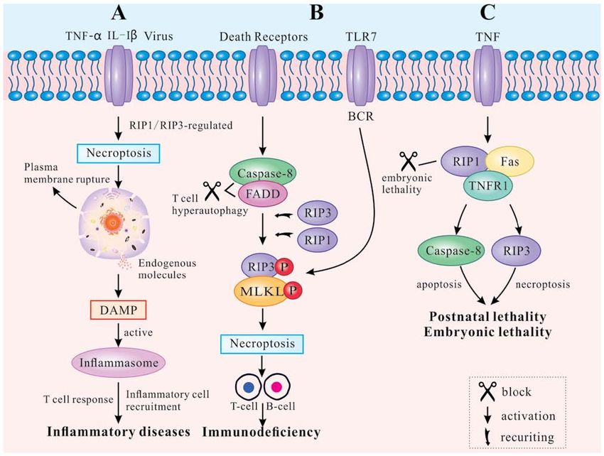
772                                         LIU et al: RIP1/RIP3 MEDIATES NECROPTOSIS

    The human RIP gene, located on chromosome 6p25.2,               work on necroptosis concerns studies of TNF signaling. TNF
encodes seven splicing isoforms: RIP1, RIP2, RIP3, RIP4, RIP5,      is a pleiotropic cytokine that has an essential role in inflam-
RIP6 and RIP7 (6). RIP1 was initially identified in 1995 as a       mation, tissue injury and cell death (22). In the TNF receptor
protein that interacted with the death domain (DD) of receptor      superfamily, researchers have found six human DRs, including
Fas (CD95) and elicited a characteristic programmed death           TNFR1, Fas (also known as CD95 or APO‑1), DR3 (also
response in susceptible cells (7). In 1997, RIP3 was discov-        known as TRAMP or APO‑3), TRAIL receptor 1 (TRAILR1,
ered as a protein attenuating both RIP1 and tumor necrosis          also known as DR4), TRAIL receptor 2 (TRAILR2, also
factor receptor 1 (TNFR1)‑induced NF‑κ B activation (8).            known as DR5, TRICK or KILLER) and DR6 (also known as
RIP1 and RIP3 are critical signaling molecules in necroptosis       CD358) (23‑26). However, the most prevalent pathway is the
and regulated by the caspase pathway and ubiquitination (9).        TNFR1‑mediated signal transduction, which can propel cell
Ubiquitination of RIP1 activates NF‑κB and mitogen‑activated        survival, apoptosis and necroptosis (27). The present review
protein kinases (MAPKs), leading to cell survival, while            focused on the three most dominant of those TNF‑mediated
deubiquitinated RIP1 induces the caspase‑8‑mediated apop-           pathways.
tosis pathway (3). When caspase‑8 is inhibited or deficient,            Different modifications of RIP1 can induce distinct
RIP1 combines with RIP3 via the C‑terminal RIP homotypic            outcomes of cell survival, apoptosis and necroptosis. Following
interaction motif (RHIM) domain to form the RIP1/RIP3               binding of TNF‑ α to TNFR1 at the plasma membrane,
complex (10). The complex initiates downstream signal trans-        TNF‑receptor‑associated death domain (TRADD) recruits
duction and triggers necroptosis (10). Excessive necroptosis        downstream proteins, namely RIP1, the E3 ubiquitin ligases
can cause embryonic lethality (11) and initiate multiple human      TNF‑receptor‑associated factor (TRAF) 2, TRAF5, and the
diseases, including, but not limited to, systemic inflammation,     cellular inhibitor of apoptosis (cIAP) 1 and cIAP2, to form
ischemic reperfusion injury and neurodegeneration (12).             the complex I (28,29). Then, the complex I mediates NF‑κ B
    The present review examined the functions of                    and MAPK signaling, contributing to cell survival or other
RIP1/RIP3‑regulated necroptosis on multifaceted pathological        non‑death functions (4,30,31). The K63‑linked ubiquitination
mechanisms. Furthermore, the importance of RIP1/RIP3 in             of RIP1 by cIAP1/2 promotes both the formation and activa-
determining the cell outcome was highlighted by the interactive     tion of the transforming growth factor‑activated kinase 1
molecular pathways noted among cell survival, apoptosis and         (TAK1)‑binding protein (TAB) complex and the inhibitor of
necroptosis. Then, the pivotal roles of RIP1/RIP3 in disease        NF‑κ B kinase (IKK) complex (consisting of NF‑κ B essential
treatment were discussed, highlighting their application            modulator, IKKα and IKKβ), supporting the NF‑κ B pathway
potential as new drug targets.                                      activation, and ultimately leading to cell survival (1) (Fig. 2).
                                                                        Complex I internalizes and transforms into a death‑inducing
2. Structural characteristics of RIP1/RIP3                          complex II following caspase‑8 activation (29). Two distinct
                                                                    types of complex II (IIa and IIb) can be distinguished based on
Structurally, RIP1 and RIP3 share nearly half of their amino acid   their composition and the activity of their proteins. After disso-
sequences and have very similar topology features. The RIP1         ciating from TNFR1, TRADD recruits Fas‑associated protein
protein consists of 671 amino acids (Homo sapiens). It contains     with death domain (FADD) and further promotes recruitment
a N‑terminal serine/threonine kinase domain, an intermediate        and activation of caspase‑8 to form the complex IIa (29,32).
domain (ID), a RHIM and a C‑terminal DD (13) (Fig. 1A). RIP3        Activation of caspase‑8 subsequently induces apoptosis
is composed of 518 amino acids (Homo sapiens), and contains         independently of RIP1 or its kinase activity (32). In certain
a N‑terminal kinase domain similar to that of RIP1, a RHIM          circumstances, including upon the absence of cIAP1/2, upon
domain and a unique C‑terminus without a DD (14) (Fig. 1A).         inhibition of IAP mediated by a small molecule mimic of
RIP1 acts as a multifunctional adaptor protein in response to       diablo IAP‑protein mitochondrial protein (33) and upon
the activated signal of death receptors (DRs), and its DD binds     auto‑degradation of cIAP1/2, RIP1 is released from complex I
to the DRs of TNFR1, Fas and TNF‑related apoptosis inducing         to form a caspase‑8‑activating complex (complex IIb) which
ligand (TRAIL) (15,16). It mediates prosurvival NF‑κ B activa-      mediates apoptosis (32). Complex IIb consists of RIP1, RIP3,
tion, caspase‑dependent apoptosis and RIP kinase‑dependent          FADD and the FLICE‑like inhibitory protein long form
necroptosis (17). The ID of RIP1 contains the RHIM that             (FLIPL)/caspase‑8 heterodimer, and it promotes RIP1‑ and
enables the protein to combine with RIP3. In contrast to RIP1,      caspase‑8‑dependent apoptosis (22). Hence, RIP1 is not
RIP3 is not directly required for DR‑induced cell survival or       an indispensable factor in apoptosis, although the process
death (18). RIP3 binds to RIP1 through its unique C‑terminal        is favored by its presence. Under physiological levels, the
segment to inhibit RIP1 and TNFR1‑mediated NF‑κ B acti-             caspase‑8/FLIPL heterodimer facilitates caspase‑8 oligomer
vation (19) (Fig. 1B). Experiments have revealed that tumor         assembly to trigger apoptosis. By contrast, high levels of
necrosis factor (TNF) induces the formation of an RIP1/RIP3         FLIPL and FLICE‑like inhibitory protein short form (FLIPS)
complex, indicating that RIP1 interacts with RIP3 through the       restrict caspase‑8/c‑FLIPL/S heterodimer activity, leading to
homotypic RHIM domain (20).                                         inhibition of apoptosis (34‑36). Inactivation of RIP1 and RIP3,
                                                                    mediated by cleavage via caspase‑8/FLIPL heterodimer in
3. Molecular       mechanisms      of   RIP1/RIP3‑regulated         Complex IIb, inhibits necroptosis (37,38) (Fig. 2).
necroptosis                                                             When caspase‑8 is inhibited by zVAD, RIP1 and RIP3 are
                                                                    combined via the RHIM, and form complex IIc, also known
Necroptosis can provide a substitute suicide mechanism in case      as the necrosome (39). Complex IIc is a crucial cytoplasmic
of malfunction of the classical apoptosis machinery (21). Most      signaling complex, which does not appear in the TNF‑induced
INTERNATIONAL JOURNAL OF MOlecular medicine 44: 771-786, 2019                                                      773

Figure 1. Structural diagrams of RIP1 and RIP3. (A) Schematic of functional domains of RIP1 and RIP3. (B) Protein tertiary structures of RIP1 and RIP3. RIP,
receptor‑interacting serine/threonine kinase; ID, intermediate domain; RHIM, RIP homotypic interaction motif; DD, death domain.

cell survival or apoptosis (10,40). Mitochondrial reactive                      to the host. The downstream MLKL signaling pathway of
oxygen species (ROS) oxidize RIP1 at three crucial cysteine                     RIP3 is indispensable for both TNFR1‑ and TLR‑induced
sites (C257, C268 and C586), and promote autophosphoryla-                       signaling (46), and caspase‑8 can block necroptosis that is
tion of RIP1 at Ser161. RIP1 autophosphorylation is pivotal                     directly initiated by the TIR domain‑containing interferon‑β
for the recruitment of RIP3 (41). In addition, CYLD lysine 63                   (TRIF)/RIP3/MLKL pathway (48,49). TLR4 and TLR3
deubiquitinase (CYLD), as a deubiquitinase removing                             are respectively activated by lipopolysaccharide (LPS) (50)
polyubiquitin chains from RIP1, facilitates the formation                       and polyinosine‑polycytidylic acid (I:C), a synthetic double
and activation of RIP1/RIP3 necrosomes by deubiquitylating                      stranded RNA (dsRNA) mimic (51). Thereafter, TLR3 and
RIP1. When CYLD is deficient, necrosomes promote a high                         TLR4 activate RIP3 and participate in ensuing necroptosis
level of ubiquitinated RIP1 and block phosphorylation of RIP1                   via TRIF or MyD88 (52‑54). The C‑terminal RHIM motif
and RIP3 (32,42). Furthermore, the RHIM is also required                        is required for RIP3 to interact with TRIF or MyD88. The
for the RIP1/RIP3 complex formation (19). After RIP1 and                        RIP3/TRIF signaling complex recruits and phosphorylates
RIP3 are combined, RIP1 gets phosphorylated by RIP3 (10).                       MLKL, inducing ROS accumulation and mediating TLR3‑
Intramolecular auto‑ and trans‑phosphorylation of RIP1/RIP3                     and TLR4‑induced necroptosis (46,47) (Fig. 3).
promotes recruitment of another key necroptosis‑signaling                           Increasing numbers of necroptotic stimuli have been
protein, the mixed lineage kinase domain like protein (MLKL).                   identified and divided into two groups: RIP1‑dependent and
MLKL is then phosphorylated by RIP3 to initiate necrop-                         RIP1‑independent (Fig. 3). RIP1‑dependent stimuli include
tosis (43). The phosphorylated MLKL is transferred from the                     TNF‑ α, Fas, TRAIL, interferon (IFN)‑ α and IFN‑ β. The
cytosol to the plasma and intracellular membranes via the                       primary death‑inducing signaling complex (DISC) is assem-
four helical bundle‑brace (4HBD‑BR) regions of MLKL (44).                       bled by stimulation of Fas or TRAILR at the plasma membrane,
The oligomerization of MLKL causes membrane pore forma-                         thereby activating caspase‑8 and triggering apoptosis indepen-
tion, resulting in the destruction of membrane integrity and                    dently of RIP1 (55). cIAP deficiency promotes the recruitment
eventually leading to necrotic death (45) (Fig. 2).                             of RIP1 and Fas when caspase‑8 is blocked, and enhances
    In addition to the classic TNFR1‑induced necroptosis                        the formation of the cytosolic ripoptosome complex which
pathway described above, toll‑like receptors (TLR) can also                     induces necroptosis (56). In bone‑marrow‑derived macro-
mediate necroptosis. The TLR signaling pathway is generally                     phages, type I IFNα and IFNβ bind to their cognate receptor
triggered by pathogen‑associated molecular patterns during                      IFNα/β receptor subunit 1 (IFNAR1) to activate Janus kinase 1
viral or microbial infection (46,47). TLR‑mediated necroptosis                  and form the IFN‑stimulated gene factor 3 (ISGF3) complex
results in the destruction of infected cells and is thus beneficial             (consisting of STAT1, STAT and IFN‑regulatory factor 9). The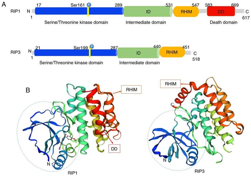
774                                               LIU et al: RIP1/RIP3 MEDIATES NECROPTOSIS

Figure 2. TNFR1‑mediated signaling pathways. After the binding of TNF to its receptor, TNFR1 undergoes a conformational change and recruits multiple
proteins to form complex I, consisting of TRADD, TRAF2/5, RIP1 and cIAP1/2. The K63‑linked ubiquitination of RIP1 by cIAP1/2 promotes the formation
and activation of the TAK1/TAB complex and the IKKα/IKKβ/NEMO complex, which induced the NF‑κ B pathway and cell survival. Destabilization of
complex I results in the formation of complex IIa, that contains TRADD, FADD and caspase‑8. When cIAPs are blocked and RIP1 deubiquitylated by CYLD,
complex IIb is formed. This consists of RIP1, RIP3, FADD, caspase‑8 and FLIPL. Both IIa and IIb can initiate apoptosis. When caspase‑8 is inhibited by
chemical caspase inhibitors, RIP1 binds to RIP3, resulting in the formation of RIP1/RIP3 by intramolecular auto‑ and trans‑phosphorylation. Then, RIP3
recruits and phosphorylates MLKL to form complex IIc, conventionally referred to as the necrosome. The phosphorylated MLKL then translocates from the
cytosol to the plasma and intracellular membranes. The oligomerization of MLKL results in membrane pore formation, causing membrane rupture and eventu-
ally necroptosis. TNFR1, TNF receptor 1; TNF, tumor necrosis factor; TRADD, TNF‑receptor‑associated death domain; TRAF, TNF‑receptor‑associated
factor; RIP, receptor‑interacting serine/threonine kinase; cIAP, cellular inhibitor of apoptosis 1; TAK1, transforming growth factor‑activated kinase 1;
TAB, TAK1‑binding protein; IKK, inhibitor of NF‑κ B kinase; NEMO, NF‑κ B essential modulator; FADD, Fas‑associated protein with death domain;
CYLD, CYLD lysine 63 deubiquitinase; FLIPL, FLICE‑like inhibitory protein long form; MLKL, mixed lineage kinase domain like protein.

ISGF3 complex promotes induction and activation of necro-                     RIP3 recruitment, leading to RIP3/RIP3 homo‑oligomeriza-
somes, and triggers necroptosis in a transcription‑dependent                  tion and RIP3 autophosphorylation. Phosphorylated RIP3
pathway (57). RIP1‑independent stimuli generally refer to                     recruits and phosphorylates MLKL, which promotes MLKL
LPS, dsRNA and viruses. DNA‑dependent activator of IFN                        oligomer‑executed necroptosis (59). However, the directorial
regulatory factors (DAI) can identify viral dsRNA, promote                    functions of RIP1 are not always positive for RIP3. Researchers
the recruitment of RIP3 to form necrosomes without RIP1,                      have constructed RIP3 dimers and RIP3 oligomers to analyze
and induce RIP3‑dependent necroptosis (58). Illuminating the                  how RIP1 induces the activation of RIP3 (60). The results
molecular mechanisms involved in necroptosis will elucidate                   demonstrated that the dimerization of RIP3 alone was insuf-
further the molecular biology underlying the pathology.                       ficient to induce the cell death. The RIP3 dimer seeded a
                                                                              RHIM‑dependent complex controlled by both caspase‑8
4. Functional features of RIP1/RIP3 in necroptosis                            and RIP1. On the other hand, without TNF stimulation and
                                                                              RIP1 activity, the oligomerization of RIP3 was sufficient to
RIP1 and RIP3 were recently delineated as two important                       induce necroptosis (60). These results indicated that RIP1 not
effectors in the cell death network, as they are cascade proteins             only activates RIP3 in response to TNF signaling, but also
responding to complex TNFR signaling and regulating                           participates in cytoprotection. TNFR1 promotes the activa-
cellular survival, apoptosis and necroptosis. It is important                 tion of the NF‑κ B pathway, p38α and its downstream effector
to note that RIP3 is indispensable for necroptosis, whereas                   MAPK‑activated protein kinase 2 kinase (MK2), thereby
RIP1 is not. TNF‑α‑induced RIP1/RIP3 interaction engages                      promoting cell survival (61). Recently, Jaco et al (62) reported
INTERNATIONAL JOURNAL OF MOlecular medicine 44: 771-786, 2019 775
Figure 3. Other stimuli leading to necroptosis. In addition to the TNF‑α‑mediated necroptosis pathway, multiple other necroptosis triggers have been identi-
fied. These involve the canonical pathway that requires RIP1 kinase activity, as well as the non‑canonical pathway that is dependent on either the TRIF
adaptor or the DAI sensor. RIP1‑dependent stimuli include TNF‑α, CD95L (also known as FASL), APO‑1L, TRAIL (also known as APO‑2L) and IFN‑α/β.
RIP1‑independent stimuli include viruses (such as CMV), viral DNA, LPS and polycytidylic acid. After various necroptotic stimuli induce necroptosis, a
necrosome is formed. Phosphorylated MLKL by RIP3 transfers from the cytosol to the plasma and intracellular membranes, causing destruction of membrane
integrity and eventually necrotic death. TNF, tumor necrosis factor; RIP, receptor‑interacting serine/threonine kinase; TRIF, TIR domain‑containing
interferon‑β; DAI, DNA‑dependent activator of IFN regulatory factors; FASL, Fas ligand; TRAIL, TNF‑related apoptosis inducing ligand; IFN, interferon;
CMV, cytomegalovirus; LPS, lipopolysaccharide; MLKL, mixed lineage kinase domain like protein.
that activated MK2 phosphorylated RIP1 at Ser321 to syner- necroptosis (66). These studies provided essential insight into
gize TNF‑induced cell rescue. Therefore, these results suggest the reciprocal regulation between RIP1/RIP3 and ROS in
that RIP1 serves as a positive checkpoint within TNF stimula- necroptosis.
tion that integrates cytokine production and cell survival. RIP1
is therefore thought as an inhibitor rather than an initiator of 5. Necroptosis, a timoneer of pathological mechanisms
RIP3‑induced necroptosis (63).
Furthermore, ROS participate in the regulation of Role of necroptosis in inflammation. Necroptosis, as a novel
necroptosis in many cell types, and enhance the formation of pathway of PCD, leads to release of endogenous moldecules
necrosomes induced by Smac mimetic bivalent 6 compound from disrupted dying cells, that subsequently triggers inflm-
(BV6)/TNFα (64). BV6/TNFα‑stimulated ROS generation mation and immune response (67). Necroptosis‑inducing
promotes the stabilization of the RIP1/RIP3 feedback loop. factors include TLR3 and TLR4 agonists [such as inter-
A previous study demonstrated that the metabolic enzymes leukin (IL)‑1β)], TNF, certain viral infections and T cell
glycogen phosphorylase (PYGL), glutamate dehydrogenase 1 receptors (22). TNF‑mediated and TLR‑mediated signaling
(GLUD1), and glutamate‑ammonia ligase (GLUL), are activated pathways are the primary pathways for necroptosis, both
by RIP3 (65). Enhancement of aerobic respiration is mediated regulated by RIP3 (10). RIP1/RIP3 or RIP3/TRIF signaling
by TNF‑induced ROS. However, the major mechanism of ROS complexes recruit and phosphorylate downstream MLKL,
and RIP1/RIP3 remains not completely understood. A recent causing rupture of the plasma membrane, as well as the
study demonstrated that mitochondrial ROS activated RIP1 release of endogenous molecules (45). These endogenous
autophosphorylation at Ser161 via oxidation of three crucial molecules are known as damage‑associated molecular patterns
cysteines in RIP1 (41). This phosphorylation accelerated RIP1 (DAMPs). They are also identified as part of the extended IL‑1
recruitment of the RIP3 aggregation and formed a necrosome, family (IL‑1α, IL‑1β, IL‑18, IL‑33, IL‑36α, β and γ) (68). The
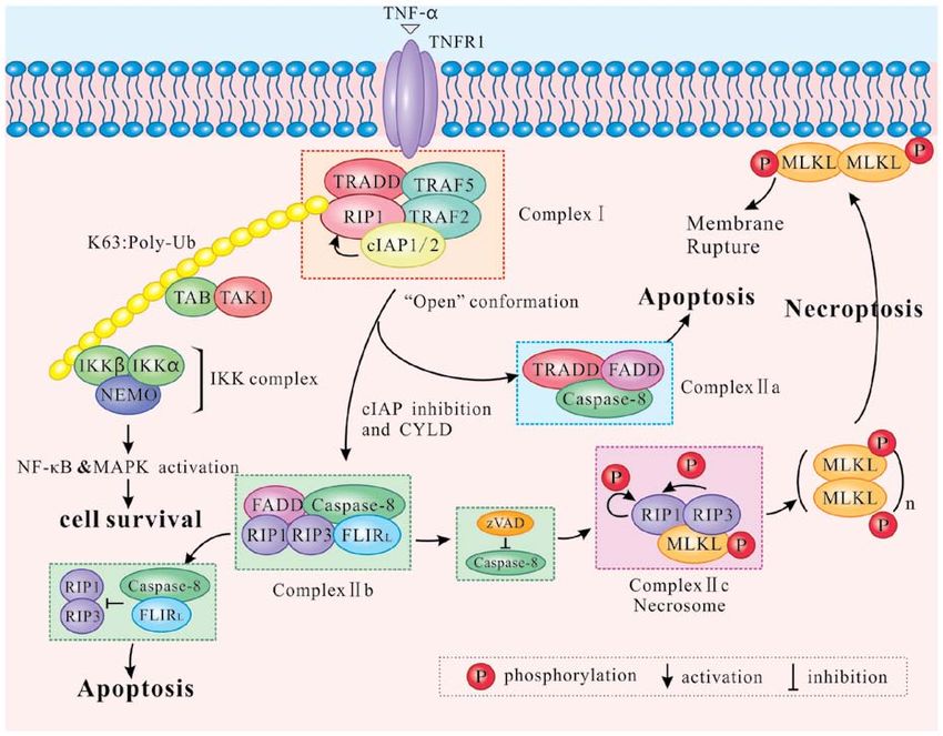
which then resulted in mitochondrial depolarization and cell leakage of DAMPs can activate inflammasomes, facilitate776 LIU et al: RIP1/RIP3 MEDIATES NECROPTOSIS
Figure 4. Necroptosis‑associated pathological mechanisms. (A) Inflammatory factors include TNF‑ α and IL‑1β. Certain viral infections can induce
RIP1/RIP3‑mediated necroptosis, which can result in the disruption of the plasma membrane and in the release of endogenous molecules, also known as
DAMPs. Inflammasomes are then activated by DAMPs, promoting inflammatory cell recruitment and virus‑specific T cell responses to induce inflammatory
diseases. (B) DRs can regulate the necroptosis of T and B cells. When either caspase‑8 or FADD is deficient, T cells undergo hyper‑autophagy and generate a
RIP1/RIP3‑mediated necrosome, triggering necroptosis. BCR mediates necroptosis via reaction with TLR7. Necroptosis of T lymphocytes and B lymphocytes
can cause immunodeficiency. (C) RIP1 prevents embryonic and postnatal lethality by blocking two different cell death pathways: FADD/caspase‑8‑medi-
ated apoptosis and RIP3‑mediated necroptosis. TNF, tumor necrosis factor; IL, interleukin; RIP, receptor‑interacting serine/threonine kinase; DAMPs,
damage‑associated molecular patterns; DR, death receptor; FADD, Fas‑associated protein with death domain; BCR, B cell receptor; TLR, toll‑like receptor;
MLKL, mixed lineage kinase domain like protein; TNFR1, TNF receptor 1.
inflammatory cell recruitment to the site of infection, and signaling molecules, including DNA damage and antigen
promote subsequent virus‑specific T cell responses to induce receptor ligation (73). Chronic necroptosis may be the basis
inflammation (69). Thus, regulating the mechanism of necrop- of human fibrotic and autoimmune disorders (74). Likewise,
tosis can result in both inhibition and promotion of the immune B cell receptor (BCR) mediates necroptosis via reaction with
response (Fig. 4A). TLR7 to prevent autoimmune diseases (75,76). The RIP1
inhibitor necrostatin‑1 (nec‑1) has been demonstrated to
Role of necroptosis in immunity. DRs regulate necroptosis in suppress B‑cell necroptosis, when pretreating B cells from
various cell types, such as B lymphocytes and T lymphocytes, patients with systemic lupus erythematosus (70). Fig. 4B
both of which are essential for immune homeostasis and toler- illustrates how programmed death can promote clonal loss
ance (48,70). Blocked DRs fail to clear activated lymphocytes of lymphocytes during infection and protect patients against
and unbalanced lymphoid homeostasis, ultimately leading autoimmune disease.
to autoimmune lymphoproliferative syndrome (ALPS) (71).
FADD, caspase‑8 and RIP kinases are indispensable for T cell Effects of necroptosis on animal development. Apoptosis and
clonal expansion, contraction and antiviral responses (48). necroptosis are closely associated with embryonic lethality
When caspase‑8 and FADD are deficient, T cells undergo and postnatal development (49). RIP1 determines cell survival
hyper‑autophagy and proliferative inhibition in response to or death by associating with TNFR1, TLRs and Fas (17,77).
antigenic stimulation. In addition, T cells generate necro- Embryonic lethality of RelA‑deficient mice has been demon-
somes, which induce caspase‑8‑independent necroptosis (72). strated to be mediated by apoptosis and necroptosis (78).
RIP1 and RIP3 are either recruited by DRs or by other cellular FADD functions as an adaptor to induce apoptosis byINTERNATIONAL JOURNAL OF MOlecular medicine 44: 771-786, 2019 777
recruiting and activating caspase‑8 (79). The characteristics of damage (89). The pan‑Aurora kinase inhibitor Tozasertib (also
FADD‑deficient embryos are high levels of RIP1 production known as VX‑680 and MK‑0457) was recently demonstrated
and massive necrosis. RIP1 ablation allows normal embryo- as a potent compound in inhibition of RIP1‑dependent necrop-
genesis in FADD‑deficient mice, but these mice usually die tosis and in the blockage of cytokinesis in cells (90). The food
around their first postnatal day (80). In addition, different and drug administration‑approved anticancer agents ponatinib
RIP1 kinase inactivating mutations have distinct effects on and pazopanib were demonstrated to be submicromolar
the embryogenesis of FADD‑deficient mice. For example, inhibitors of necroptosis through the targeting of components
RIP1K45A has been found not to prevent the embryonic upstream of MLKL (91). Ponatinib inhibits both RIP1 and
lethality of FADD‑deficient mice, while RIP1Δ (with an altered RIP3, while pazopanib preferentially targets RIPK1. Both
P‑loop in the kinase domain) does (81). If RIP1 was necessary drugs have potential values for the treatment of pathology
for the activation of RIP3, as aforementioned, FADD‑/‑ RIP1‑/‑ caused or aggravated by necroptotic cell death (91). Overall,
and FADD ‑/‑ RIP3‑/‑ mice should survive to adulthood (82). the aforementioned studies indicated that RIP1 kinase may
Nevertheless, a recent study revealed that RIP1‑deficient serve as a novel target for therapeutic drug development in
mice die soon after birth, leading to speculation for the posi- human disease therapy.
tive role of RIP1 in embryonic development and postnatal RIP3 is a critical regulator of necroptosis, however, very
life (83). RIP1 deletion enhances primary cell sensitivity to few specific inhibitors have yet been reported. B‑Raf (V600E)
FADD/caspase‑8‑mediated apoptosis induced by TNF. In inhibitors are generally considered as an important anticancer
addition, RIP1 deletion promotes RIP3/MLKL‑mediated drug in metastatic melanoma therapy (92). To date, the B‑Raf
necroptosis induced by TLR ligation (via TRIF) or inter- inhibitor dabrafenib was demonstrated to be a potent inhibitor of
feron (84). The perinatal lethality of RIP1‑/‑ mice can be RIP3 (92). Dabrafenib decreased RIP3‑mediated Ser358 phos-
rescued by a combination of additional mutations. A recent phorylation of MLKL and disrupted the interaction between
study indicated that haploid insufficiency of RIP3 improved RIP3 and MLKL (93). Results indicated that dabrafenib
the survival period of RIP1‑/‑ FADD‑/‑ double knockout mice could serve as a RIP3 inhibitor and as a potential preventive
beyond weaning age, while RIP1‑/‑ FADD ‑/‑ RIP3 ‑/‑ triple or therapeutic agent for RIP3‑involved necroptosis‑related
knockout (TKO) mice were significantly smaller in size and diseases (93). As expected, dabrafenib significantly reduced
weight (85). Furthermore, complete ablation of RIP3 further infarct lesion size and attenuated upregulation of TNF‑ α
prolonged the life span of TKO mice displaying normal size in mouse models of ischemic brain injury (94). In addition,
and weight (85). Therefore, it can be concluded that RIP1 murine cytomegalovirus (CMV) M45 contains a RHIM
may prevent postnatal lethality by blocking two different cell domain, and was confirmed to be a competitive inhibitor of
death pathways: FADD/caspase‑8‑mediated apoptosis and RIP3 (95). Human CMV blocks TNF‑induced necroptosis
RIPK3/MLKL‑mediated necroptosis. The effect of PCD on following RIP3 activation and MLKL phosphorylation (95),
animal development is highly complex and has not yet been leading to inhibition of the host defense mechanism.
thoroughly elucidated (Fig. 4C).
MicroRNA (miRNA)‑associated inhibition by targeting
6. RIP1/RIP3 inhibition in necroptosis RIP1/RIP3. As diagnostic and therapeutic strategies, miRNAs
provide a novel perspective for RIP1 inhibition. As afore-
Inhibitors of RIP1 and RIP3. The increasing discovery of mentioned, several inhibitors have been demonstrated to
inhibitors and drugs affecting the RIP1/RIP3 cascade pathway suppress the pro‑necroptosis function of RIP1 at the protein
already shows promise towards the treatment of various level; however, these do not function at the mRNA level.
diseases (Table I). Nec‑1 has been identified as a specific and miR‑155 represses cardiomyocyte progenitor cell necroptosis
potent small‑molecule and active inhibitor of RIP1 (2). Nec‑1 by targeting RIP1 rather than activating the Akt pro‑survival
is widely used in disease models to examine the contribution pathway (96), suggesting that miR‑155 might be a novel
of RIP1 to cell death and inflammation. Other necrostatins, approach in improving cell engraftment. Additionally, in a
including Nec‑3, Nec‑4 and Nec‑5, also stabilize RIP1 in an myocardial ischemia/reperfusion model, it was demonstrated
inactive conformation through interactions with hydrophobic that miR‑103/107, as a necrosis‑suppressor miRNA, directly
pockets and highly conserved amino acids (2,86). However, targeted FADD (97,98). FADD participates in hydrogen
the strongest inhibition of RIP1 has been observed with Nec‑1 peroxide‑induced necroptosis by influencing the formation
stable (Nec‑1s) (87). A previous study identified GSK2982772 of RIP1/RIP3 complex, suggesting that FADD‑targeting
(compound 5) as a novel inhibitor of RIP1 (88). GSK2982772 by miR‑103/107 might be a new approach for preventing
potently binds to RIP1 with exquisite kinase specificity and myocardial necrosis. Recent research has indicated that
has high activity in blocking TNF‑dependent necroptosis, miRNA dysregulation is involved in triple‑negative breast
as well as inflammation. Based on this previous study of cancer (TNBC) (100). Overexpression of miR‑182 inhibits
GSK2982772, Yoshikawa et al (27) designed and synthesized the CYLD action on the ubiquitin chains on RIP1, leading
a novel class of RIP1 kinase inhibitor, the compound 22 to caspase‑8‑dependent apoptosis in TNF‑α‑treated TNBC
[7‑oxo‑2,4,5,7‑tetrahydro‑6H‑pyrazolo(3,4‑c)pyridine], which cells (99). Additionally, miR‑145, which is downregulated
possesses moderate RIP1 kinase inhibitory activity and in TNBC, targeted cIAP1 and reduced the formation of the
P‑gp mediated efflux. Furthermore, using a mouse model of RIP1/FADD‑caspase‑8 complex (100). Therefore, miRNAs
systemic inflammatory response syndrome, it was demon- can be perceived as a novel approach for RIP1 regulation.
strated that compound 56 (RIPA‑56) targeted RIP1 directly, However, their potential effects in disease intervention requires
and reduced TNFα‑induced cell mortality and multi‑organ further research (Table II).778 LIU et al: RIP1/RIP3 MEDIATES NECROPTOSIS
Table I. Direct and indirect inhibitors of RIP1/RIP3.
Compound Targeted proteins Applications (Refs.)
Nec‑1 RIP1 Nec‑1 is the most active, compared with (2)
Nec‑3, Nec‑4 and Nec‑5
GSK2982772 (compound 5) RIP1 Phase 2a clinical studies for rheumatoid (88)
arthritis and ulcerative colitis
7‑oxo‑2,4,5,7‑tetrahydro‑6H‑ RIP1 Oral administration (10 mg/kg, twice a day) (27)
pyrazolo[3,4‑c]pyridine attenuated disease progression in a mouse
(compound 22) experimental autoimmune encephalomyelitis
model of multiple sclerosis
RIPA‑56 (compound 56) RIP1 For the treatment of systemic inflammatory (89)
response syndrome
Tozasertib (also called as pan‑Aurora kinase, RIP1 Protects against TNF‑induced necroptosis (90)
VX‑680 and MK‑0457)
Ponatinib RIP1, RIP3 Ponatinib can directly bind RIP1 and block (91)
RIP3 autophosphorylation
Pazopanib Preferentially targets RIP1 Similar to Nec‑1, RIP1 is the main target of (91)
pazopanib
Dabrafenib RIP3, B‑Raf (V600E) Alleviates acetaminophen‑induced liver injury (93)
Murine cytomegalovirus M45 RIP3 Binds to RIP3 through RHIM domain (95)
Kongensin A HSP90 (indirectly inhibits RIP3) Kongensin A covalently binds to HSP90 and (102)
inhibits the stability of RIP3/MLKL
Compound 17AAG HSP90 (indirectly inhibits RIP3) Promotes degradation of MLKL and RIP3 (104)
through the proteasome pathway
Alvespimycin (17‑DMAG) HSP90 (indirectly inhibits Destabilizes the necroptosis proteins RIP1 and RIP3 (105)
RIP1/RIP3)
Dyngo 4a TLR4 (indirectly inhibits RIP3) Blocks the internalization of TLR4 and prevents (108)
RIP3‑induced necroptosis of macrophages
RIP, receptor‑interacting serine/threonine kinase; Nec, necrostatin; TNF, tumor necrosis factor; RHIM, RIP homotypic interaction motif;
HSP90, heat shock protein 90; MLKL, mixed lineage kinase domain like protein; TLR4, Toll‑like receptor 4.
Table II. Applications of targeting miRNAs in necroptosis.
miRNA Targeted mRNAs Sequence (5'‑3') (Refs.)
miR‑155 RIP1 UUAAUGCUAAUUGUGAUAGGGG (96)
miR‑103 FADD AGCAGCAUUGUACAGGGCUAUGA (97,98)
miR‑107 FADD AGCAGCAUUGUACAGGGCUAUCA (98)
miR‑182 CYLD UUUGGCAAUGGUAGAACUCACACU (99)
miR‑145 cIAP1 GUCCAGUUUUCCCAGGAAUCCCUGGAUUCCUGGGAAAA CUGGACUU (100)
miRNA, microRNA; RIP, receptor‑interacting serine/threonine kinase; FADD, Fas‑associated protein with death domain; CYLD, CYLD
lysine 63 deubiquitinase; cIAP1, cellular inhibitor of apoptosis 1.
Indirect inhibition of RIP1/RIP3. Previously, Li et al (101) association of HSP90 and CDC37, leading to the inhibition of
revealed that the cochaperone complex of HSP90 and CDC37 RIP3‑dependent necroptosis (101,102). These results suggested
regulated the stability and function of RIP3 and MLKL, and that KA was an effective HSP90 inhibitor with a potential
participated in the RIP3 activation process during necrop- anti‑RIP3 effect in both RIP3‑dependent necroptosis and
tosis. The HSP90 inhibitor kongensin A (KA) disrupted the inflammation. In addition, the disruption of HSP90 functionINTERNATIONAL JOURNAL OF MOlecular medicine 44: 771-786, 2019 779
prevented necrosome formation, reduced MLKL phosphory- IR injury (115). In addition, in a mouse cardiac hypertrophy
lation and inhibited TNF‑induced necroptosis (103). The model established by transverse abdominal aortic constric-
compound 17AAG destabilized the interaction of MLKL and tion, both mRNA and protein expression levels of RIP1 and
RIP3 by inhibiting HSP90, resulting in degradation of MLKL RIP3 were increased significantly. Losartan downregulated
and RIP3 via the proteasome pathway (104). Alvespimycin the expression of RIP1/RIP3, resulting in the inhibition of
(17‑DMAG), an inhibitor of HSP90, facilitated the degrada- necroptosis and to the alleviation of cardiac hypertrophy (116).
tion of RIP3 following HSP90 inactivation (105). Therefore, RIP1/RIP3 may thus be an attractive target for future therapies
pharmacological modulation of RIP3‑induced necroptotic that aim to limit the adverse consequences of cardiac disease.
cell death through HSP90 could be a promising strategy for The quaternary nitrogen herbicide paraquat is a highly
therapy in several settings (Table I). toxic pro‑oxidant that triggers oxidative stress and multi‑organ
In TNFα‑induced necroptosis, as RIP1 and RIP3 form failure, including that of the heart. Recently, Zhang et al (117)
a protein complex through their common RHIM domain, revealed that Nec‑1 pretreatment prevented cardiac contractile
phosphorylation and activation of RIP3 and downstream dysfunction, reduced RIP1/RIP3 interaction, downregulated
MLKL occur (106). In addition, RHIM‑containing proteins, the RIP1/RIP3/MLKL signaling pathway, and dramatically
such as TLR, and interferon regulatory factors (Z‑DNA inhibited the production of ROS in paraquat‑challenged
binding protein 1, also known as DAI or DLM1) are known mice. Thus, the RIP1/RIP3/MLKL signaling cascade may
to activate RIP3 and further transduce necrosis signals to represent an innovative therapeutic direction for paraquat
MLKL (107). TLR3 or TLR4 directly activate necroptosis poisoning‑induced cardiac contractile dysfunctions (Table III).
through the RHIM‑dependent association of TRIF with RIP3.
This pathway proceeds independently of RIP1, but remains RIP1/RIP3‑dependent necroptosis in cancer therapy.
dependent on MLKL downstream of RIP3 kinase (46). Abundant research on RIP1/RIP3 has highlighted its role in
Dyngo 4a blocks the internalization of TLR4 and prevents cancer, which is due to its necroptosis‑inducing function (118).
RIP3‑induced necroptosis of macrophages (108). A previous Chen et al (119) have noted that necroptosis is a critical
study unveiled DAI as the RIP3 partner to interact with RIP3, cell‑killing mechanism in response to severe stress and blocked
mediating virus‑induced necrosis analogous to the RIP1/RIP3 apoptosis, and have proposed that it can serve as an alternative
complex controlling TNF‑induced necroptosis (109). Table I cell death program to prevent cancer. Previous studies have
summarizes the direct and indirect inhibitors of RIP3. indicated that increased RIP3 expression was correlated with
cancer development, including colon and lung cancers, naso-
7. Application potential of RIP1/RIP3 inhibition in disease pharyngeal carcinoma and non‑Hodgkin lymphoma (120‑122).
therapy The topoisomerase inhibitor SN38, an active metabolite of
irinotecan, was demonstrated to mediate cytotoxicity through
RIP1/RIP3‑dependent necroptosis in cardiovascular disease. the TNF/TNFR signaling pathway in a panel of colon cancer
Necroptosis participates in the development of several diseases, cells (123). SN38 also promoted the progression of necrop-
such as atherosclerosis cardiovascular disease, a leading cause tosis, inhibited cell proliferation and induced DNA damage
of mortality worldwide (110). Overexpression of RIP3 during accumulation (123). This suggested that the SN38‑induced
necroptosis of primary macrophages induced by oxidized activation of RIP1 and subsequent necroptosis may exert
LDL (ox‑LDL) facilitates the development of the disease (111). the therapeutic efficacy on colorectal carcinoma (123).
Furthermore, monoclonal antibodies can be detected in the Furthermore, Xin et al (124) reported that degradation of
core of atherosclerotic plaques, specifically recognizing the suppressor of cytokine signaling 1, a key negative regulator of
phosphorylation form of RIP3 at Ser232 (112). Notably, the IFN‑γ signaling, was prevented by TNF through RIP1/RIP3
mortality of apolipoprotein E/RIP3 double‑knockout mice signaling. The authors suggested that necroptotic inhibition
was delayed dramatically (112). These findings indicated that might be a novel strategy for the treatment of acute myeloid
RIP3‑mediated necroptosis in atherosclerotic plaques may leukemia through the combination of RIP1/RIP3 inhibitor
release pro‑inflammatory cytokines that exacerbate athero- with IFN‑γ. Recently, bufalin was demonstrated to increase
sclerosis. Of note, PS‑341, a potent and specific proteasome the expression of necroptosis mediators RIP1/RIP3 and ROS,
inhibitor, was demonstrated to impair macrophage necroptosis leading to poly(ADP‑ribose) polymerase (PARP)‑dependent
through stabilization of cIAPs and disruption of the formation tumor cell death and tumor growth inhibition in MCF‑7 and
of the RIP1/RIP3 complex (113). MDA‑MB‑231 human breast cancer cells (125). The promising
RIP1 inhibition leads to a reduction of infarct size, role of RIP1/RIP3‑dependent necroptosis in cancer therapy
implying a functional importance of necroptosis in myocar- warrants attention in future studies (Table III).
dial ischemia (MI) (114). Luedde et al (20) analyzed RIP3
expression in murine hearts and highlighted the potential RIP1/RIP3‑dependent necroptosis in metabolic diseases.
functional significance of RIP3‑dependent necroptosis in the Glucose and its metabolism have a crucial role in driving
modulation of post‑ischemic adverse remodeling in MI. A necroptosis4 (1). Larocca et al (126) found that hypergly-
previous study demonstrated that RIP3 was upregulated in cemia upregulated necroptosis and shifted from apoptosis to
murine hearts subjected to ischemia‑reperfusion (IR) injury, necroptosis, associated with increased expression of RIP1,
as well as in cardiomyocytes treated with LPS and hydrogen RIP3 and MLKL proteins. Sequentially, levels of RIP1
peroxide (115). This study further illustrated that upregulated and MLKL increased in cerebral tissue from hypergly-
RIP3 evoked endoplasmic reticulum (ER) stress, ultimately cemic neonatal mice that underwent hypoxia‑ischemia
resulting in cardiomyocyte necroptosis in the setting of cardiac brain injury (127). Current studies have documented that780 LIU et al: RIP1/RIP3 MEDIATES NECROPTOSIS
Table III. Potential of RIP1/RIP3‑regulated necroptosis in diseases therapy.
Disease Compound or treatment Pathological mechanism (Refs.)
Atherosclerosis PS‑341 Impairing macrophage necroptosis and inflammation (111‑113)
Myocardial ischemia Nec‑1 Reducing the infarct size induced by necroptosis (114)
Cardiac hypertrophy Losartan Inhibiting the RIP1/RIP3‑induced necroptosis of cardiac (116)
cell injury
Hyperglycemia Nec‑1s High glucose promotes necroptosis and the expression (126‑128)
of RIP1, RIP3, and MLKL proteins
Acetaminophen‑mediated Nec‑1 Inhibiting acetaminophen‑induced hepatic JNK (131)
liver injury phosphorylation and mitochondrial Bax translocation
Concanavalin A‑induced Nec‑1 or RIP3‑/‑ Inhibiting the necroptosis of hepatocytes (130)
autoimmune hepatitis
Alcoholic fatty liver RIP3‑/‑ Mice lacking RIP3 were protected from ethanol‑induced (134)
disease steatosis, hepatocyte injury, and expression of
proinflammatory cytokines
Nonalcoholic fatty liver RIP3‑/‑ Attenuating choline‑deficient diet‑induced liver injury, (135)
disease steatosis, inflammation, fibrosis and oxidative stress
CCl4‑induced liver Melatonin Preventing liver fibrosis by inhibiting necroptosis‑associated (136)
fibrosis inflammatory signaling
Renal ischemia‑reperfusion Nec‑1, RIP3‑/‑ Inhibiting the necroptosis of organ damage, independent (138)
injury of the immune system
Kidney inflammation, Nec‑1 Inhibiting the necroptosis of kidney injury induced by (140)
interstitial fibrosis unilateral ureteral obstruction
Colorectal carcinoma Topoisomerase inhibitor Promoting RIP1‑depended necroptosis of HT29 and (123)
SN38 (active metabolite HCT116 cell lines
of irinotecan)
Acute myeloid leukemia Nec‑1 combined with Combination treatment inactivates RIP1/RIP3‑mediated (124)
interferon‑γ necroptotic signaling
Breast cancer Bufalin Increasing the necroptosis mediators RIP1/RIP3 and (125)
reactive oxygen species in MCF‑7 and MDA‑MB‑231
human breast cancer cells
Retinal detachment Nec‑1, RIP3 ‑/‑
Preventing necroptosis and reducing oxidative stress (142)
Retinitis pigmentosa RIP3‑/‑ Necroptosis promotes cone photoreceptor degeneration (143)
Amyotrophic lateral Nec‑1s Necroptosis and inflammation exacerbate disease (147)
sclerosis (induced by progression
optineurin deficiency)
Hyperuricemia RIP3‑/‑ Reducing pro‑inflammatory cytokines and necroptosis (128)
of kidney cells
Donor organ injury Nec‑1, RIP3‑/‑ Improving renal function (149)
Paraquat‑induced cardiac Nec‑1 Downregulating the RIP1/RIP3/MLKL signaling pathway (117)
contractile dysfunction and preventing cardiac contractile dysfunction
Toxic epidermal necrolysis Dabrafenib Preventing RIP3‑mediated necroptosis (150)
RIP, receptor‑interacting serine/threonine kinase; Nec, necrostatin‑1; Nec‑1s, Nec‑1 stable.
the intensity of necroptosis is closely associated with high wild‑type mice, namely downregulation of circulating and
glucose levels. kidney pro‑inflammatory cytokines (IL‑1β, TNF‑α and IL‑6),
Hyperuricemia (HU) is closely related to metabolic a decrease of FADD, cleaved caspase‑8/‑3 and PARP expres-
syndrome. Wang et al (128) demonstrated that RIP3 was sion levels, and a decrease in TUNEL apoptotic staining in
strongly expressed in mice with HU, whereas RIP3 deficiency renal samples (128). These results suggested that RIP3 may
attenuated HU symptoms. Using RIP3‑knockout mice, various have a crucial role in HU, and it may serve as a novel target for
effects were observed following HU compared with RIP future therapeutic strategies (Table III).INTERNATIONAL JOURNAL OF MOlecular medicine 44: 771-786, 2019 781
RIP1/RIP3‑dependent necroptosis in liver injury. The death of tubulointerstitial injury. Inhibition of necroptosis reduced
hepatocytes initiates and aggravates chronic inflammation and the inflammatory response and interstitial fibrosis in renal
fibrosis during liver injury, ultimately leading to liver cirrhosis tissues (140). Therefore, the signaling pathways and the main
and hepatocellular carcinoma. Increasing evidence indicates regulators of necroptosis may serve as potential candidates for
that necroptosis has a key role in acute liver injury and chronic therapeutic strategies in kidney injury (Table III).
liver injury (129). Deutsch et al (130) indicated that RIP1 and
RIP3 have different roles in drug‑induced or immunological RIP1/RIP3‑dependent necroptosis in ocular disease.
acute liver injuries. In Concanavalin A (ConA)‑induced auto- Apoptosis was previously demonstrated to be a significant form
immune hepatitis, RIP3 deletion delayed hepatic injury, while of cell loss in photoreceptor death, but RIP‑mediated necrosis
RIP1 inhibition markedly exacerbated ConA‑induced hepa- was recently discovered to be a crucial mode of photoreceptor
titis (130). Conversely, in acetaminophen (APAP)‑mediated cell loss in an experimental model of retinal detachment (141).
liver injury, blockade of RIP1 or RIP3 ameliorated APAP Expression of RIP3 undergoes a 10‑fold increase after retinal
toxicity (130). Zhang et al (131) demonstrated that RIP1 was detachment (142). Nevertheless, Nec‑1 or RIP3 deficiency
strongly expressed and promoted acute liver failure in mice substantially prevent necroptosis and reduce oxidative stress
treated with APAP (300 mg/kg, intraperitoneally injected). of apoptosis‑inducing factor (142). Additionally, cone photore-
Further analysis demonstrated that Nec‑1, the inhibitor of RIP1, ceptor death in retinitis pigmentosa (RP) is widely considered
significantly inhibited APAP‑induced hepatic JNK phosphor- as a necroptotic mechanism, while rod photoreceptor death is
ylation and mitochondrial Bax translocation (131). Using the characterized by apoptotic features (143). Murakami et al (144)
APAP model in RIP3‑/‑ mice, Ramachandran et al (132) found reported that RIP3 expression was elevated in rd10 mouse
that RIP3 knockout significantly reduced hepatotoxicity after retinas in the cone phase, but not in rod degeneration, and thus
6 h of APAP treatment (200‑300 mg/kg), while the protective suggested that RIP3 may be a potential target in protecting
effect of RIP3 knockout on the liver was not obvious after 24 h cone photoreceptors. On the other hand, Sato et al (145) found
of APAP treatment. that RIP1/RIP3 accelerated both cone and rod photoreceptor
Chronic liver injury usually includes alcoholic fatty liver degeneration in interphotoreceptor retinoid‑binding protein
disease (AELD), nonalcoholic fatty liver disease (NAFLD), (Irbp)‑/‑ mice. It is worth noting that Irbp deficiency displays
liver fibrosis and cirrhosis. It has been reported that the severe early and progressive photoreceptor degeneration.
expression of RIP3 in the liver tissue of patients with AFLD is Based on these studies, RIP1/RIP3 may be regarded as a
abnormally elevated, and a series of pathological changes, such potential therapeutic target to prevent or delay photoreceptor
as hepatocyte lipid accumulation and elevated transaminase, are degeneration in patients with RP.
observed in the liver (133,134). Additionally, Afonso et al (135) In an animal model of retinal detachment (RD), cleaved
reported that the expression of RIP3 and phosphorylated MLKL IL‑1β was reported in infiltrated macrophages undergoing
was increased in the liver tissue of patients with NAFLD, RIP3‑dependent necroptosis, rather than dying photore-
while RIP3 knockout significantly reduced the steatosis and ceptors (146). In addition, Ito et al (147) suggested a direct
inflammatory response in mice with methionine‑choline diet. connection between amyotrophic lateral sclerosis induced
Choi et al (136) found that melatonin reduced the expression by optineurin deficiency and RIP1‑regulated necroptosis and
of RIP1, RIP3 and MLKL in rat fibrotic liver induced by inflammation. Because it promotes both inflammation and
CCl4, and inhibited the expression of high‑mobility group cell death, RIP1/RIP3 may be a common mediator of human
box 1 (HMGB1) and IL‑1α. This suggested that melatonin can degenerative diseases characterized by axonal degeneration
alleviate liver fibrosis by inhibiting the inflammatory signaling (Table III).
associated with programmed necrosis (136). Further studies are
required to fully elucidate the role of RIP1/RIP3‑dependent Other properties of RIP1/RIP3‑dependent necroptosis. Donor
necroptosis in liver injury (Table III). organ injury is invariably mentioned in transplantation, due
to immune responses related to IRI and alloimmune rejec-
RIP1/RIP3‑dependent necroptosis in kidney injury. Increasing tion (148). Lau et al (149) reported that in caspase‑inhibited
evidence has demonstrated that necroptosis has an important tubular epithelial cells (TECs), necroptosis was triggered
role in the pathogenesis of multiple types of kidney injury. in vitro, following inhibition by Nec‑1 or RIP3 ‑/‑ TEC.
Linkermann et al (137) first determined the presence of necrop- Following transplantation, recipients receiving RIP3‑/‑ kidneys
tosis in a murine model of renal ischemia‑reperfusion injury had longer survival and improved renal function (149). These
(IRI). The detection of RIP1 and RIP3 in whole‑kidney lysates results suggested that inhibition of RIP3‑dependent necroptosis
and freshly isolated murine proximal tubules revealed the and inflammatory injury in donor organs may provide clinical
contribution of necroptosis in renal injury (137). A subsequent benefit. Toxic epidermal necrolysis (TEN) is a severe adverse
study demonstrated that, in a mouse model of IRI, necroptosis drug reaction with a high mortality rate (150). Kim et al (151)
lead to primary organ damage, and RIP3‑knockout mice were detected upregulated expression of RIP3 and elevated phos-
protected from IRI (138). Another study used MLKL‑knockout phorylation of MLKL in skin sections from patients with
mice to investigate the role of necroptosis in acute kidney TEN. Dabrafenib notably prevented RIP3‑mediated MLKL
injury (139). Their results revealed the indispensable role phosphorylation and decreased necroptosis, by inhibiting
of MLKL in the necroptotic pathway (139). Additionally, RIP3 in a TEN model (151). Based on these findings, it can be
Xiao et al (140) reported that the effect of necroptosis and speculated that RIP may represent a potential target for treat-
the RIP1/RIP3/MLKL signaling pathway in renal inflam- ment of TEN. Table III presents a list of existing studies on the
mation and interstitial fibrosis was associated with primitive role of RIP1/RIP3‑mediated necroptosis in disease.782 LIU et al: RIP1/RIP3 MEDIATES NECROPTOSIS
8. Conclusion and perspectives Authors' contributions
It is well‑known that necroptosis, a physiologically relevant YW, RT and YL conceived and designed the review. DZ, TLiu,
form of cell death, is involved in pathological cell death resulting TLei, SD and LG wrote the manuscript. TLei, DQ and CL
from IRI. RIP1/RIP3 is a key cascade involved in necroptosis, prepared the figures. YW, RT and YL reviewed and edited the
which is regulated by caspase activation and ubiquitination. manuscript. All authors read and approved the final manuscript,
RIP1 regulates cell death and survival, while RIP3 mediates and agree to be accountable for all aspects of the research in
apoptosis and necroptosis. Both TLR and virus‑mediated ensuring that the accuracy or integrity of any part of the work
activation are recognized or identified by specific receptors are appropriately investigated and resolved.
or sensors placed either inside or on the surface of cells.
RIP1/RIP3 mediates the initiation of the necroptotic response Ethics approval and consent to participate
to different stimuli. Following ligand binding and receptor
activation, RIP3 enters complex II via interaction with RIP1, Not applicable.
which then activates MLKL participation in inflammation,
immune response, embryonic development and metabolic Patient consent for publication
abnormality.
In conclusion, an improved understanding of the molecular Not applicable.
events regulating necroptotic cell death would be extremely
beneficial for disease therapy, as these mechanisms are impli- Competing interests
cated in a variety of human illnesses. Further research in
this area can be expected to provide promising opportunities The authors declare that they have no competing interests.
regarding therapeutic exploitation of cell death programs. Due
to its crucial roles in organ growth and tumor cell prolifera- References
tion, RIP1/RIP3 may be a key potential therapeutic strategy in
multiple severe diseases. Therefore, by using small molecules 1. Vandenabeele P, Galluzzi L, Vanden Berghe T and Kroemer G:
that specifically target necroptosis, it may be possible to Molecular mechanisms of necroptosis: An ordered cellular
alleviate symptoms and prolong the lives of patients. In explosion. Nat Rev Mol Cell Biol 11: 700‑714, 2010.
2. Degterev A, Huang Z, Boyce M, Jagtap P, Mizushima N,
coming years, further research on this alternative cell death Cuny GD, Mitchison TJ, Moskowitz MA and Yuan J: Chemical
pathway may include identification of additional RIP1 and inhibitor of nonapoptotic cell death with therapeutic potential for
RIP3 substrates. RIP3 appears to be the foremost promoter of ischemic brain injury. Nat Chem Biol 1: 112‑119, 2005.
3. Christofferson DE and Yuan J: Necroptosis as an alternative
necroptosis. In the future, studies exploiting RIP3 inhibitors form of programmed cell death. Curr Opin Cell Biol 22: 263‑268,
may provide crucial insight into the diagnosis and treatment of 2010.
necroptosis‑associated diseases. 4. Ashkenazi A and Salvesen G: Regulated cell death: Signaling
and mechanisms. Annu Rev Cell & Dev Biol 30: 337‑356,
2014.
Acknowledgements 5. Zhang YY and Liu H: Connections between various trigger
factors and the RIP1/RIP3 signaling pathway involved in
necroptosis. Asian Pac J Cancer Prev 14: 7069‑7074, 2013.
Not applicable. 6. Mason AR, Elia LP and Finkbeiner S: The receptor‑interacting
serine/threonine protein kinase 1 (RIPK1) regulates progranulin
Funding levels. J Biol Chem 292: 3262‑3272, 2017.
7. Stanger BZ, Leder P, Lee TH, Kim E and Seed B: RIP: A novel
protein containing a death domain that interacts with Fas/APO‑1
This study was sponsored by the National Science Foundation (CD95) in yeast and causes cell death. Cell 81: 513‑523, 1995.
of China (grant no 81802504), the Sichuan National Science 8. Sun X, Lee J, Navas T, Baldwin DT, Stewart TA and Dixit VM:
RIP3, a novel apoptosis‑inducing kinase. J Biol Chem 274:
Research Funding (grant no. 2018JY0645), Sichuan Health and 16871‑16875, 1999.
Family Planning Commission Funding (grant no. 16ZD0253), 9. Newton K: RIPK1 and RIPK3: Critical regulators of inflamma-
Chengdu National Science Research Funding (grant tion and cell death. Trends Cell Biol 25: 347‑353, 2015.
10. Cho YS, Challa S, Moquin D, Genga R, Ray TD, Guildford M and
no. 2018‑YFYF‑00146‑SN), and funding from the Sichuan Chan FK: Phosphorylation‑driven assembly of the RIP1‑RIP3
Scientific Research Grant for Returned Overseas Chinese complex regulates programmed necrosis and virus‑induced
Scholars for Dr Yi Wang. This study was also supported by the inflammation. Cell 137: 1112‑1123, 2009.
National Key Research and Development Plan of China (grant 11. Kaiser WJ, Upton JW, Long AB, Livingston‑Rosanoff D,
Daley‑Bauer LP, Hakem R, Caspary T and Mocarski ES: RIP3
no. 2017YFC0113901), the Key Research and Development mediates the embryonic lethality of caspase‑8‑deficient mice.
Plan of Sichuan Science and Technology Bureau (grant Nature 471: 368‑372, 2011.
no. 2019YFS0278), the Health Care for government officials of 12. Zhou W and Yuan J: Necroptosis in health and diseases. Semin
Cell Dev Biol 35: 14‑23, 2014.
Sichuan Province and the Sichuan Health and Family Planning 13. Vandenabeele P, Declercq W, Van Herreweghe F and Vanden
Commission Funding (grant no. 120094) for Dr Yuping Liu. Berghe T: The role of the kinases RIP1 and RIP3 in TNF‑induced
necrosis. Sci Signal 3: re4, 2010.
14. Wu XN, Yang ZH, Wang XK, Zhang Y, Wan H, Song Y, Chen X,
Availability of data and materials Shao J and Han J: Distinct roles of RIP1‑RIP3 hetero‑ and
RIP3‑RIP3 homo‑interaction in mediating necroptosis. Cell
The datasets used and/or analyzed during the present study Death Differ 21: 1709‑1720, 2014.
15. Wen L, Zhuang L, Luo X and Wei P: TL1A‑induced NF‑kappaB
are available from the corresponding author on reasonable activation and c‑IAP2 production prevent DR3‑mediated apop-
request. tosis in TF‑1 cells. J Biol Chem 278: 39251‑39258, 2003.You can also read