Multiple cell death modalities and their key features (Review)
←
→
Page content transcription
If your browser does not render page correctly, please read the page content below
WORLD ACADEMY OF SCIENCES JOURNAL 2: 39-48, 2020

     Multiple cell death modalities and their key features (Review)
                                 GE YAN, MOHAMED ELBADAWI and THOMAS EFFERTH

                         Department of Pharmaceutical Biology, Institute of Pharmacy and Biochemistry,
                                  Johannes Gutenberg University, D‑55128 Mainz, Germany

                                     Received December 23, 2019; Accepted February 24, 2020

                                                      DOI: 10.3892/wasj.2020.40

Abstract. Cell death, as a final cellular decision which is            of apoptosis, as well as anoikis. Multiple mechanisms and
reached following complex communications, represents                   phenotypes compose programmed non‑apoptotic cell death,
a critical process with which to maintain organismic                   including vacuole‑presenting cell death (autophagy, entosis,
homeostasis. Different classifications and nomenclatures have          methuosis and paraptosis), mitochondrial‑dependent cell
brought considerable confusion to cell death determination.            death (mitoptosis and parthanatos), iron‑dependent cell death
In the present review article, the hallmarks of different cell         (ferroptosis), immune‑reactive cell death (pyroptosis and
death modes are systematically described and are fitted                NETosis), as well as other types, such as necroptosis. Finally,
into a simple classification system, where the cell death              necrosis represents a form of non‑programmed cell death.
entities are primarily categorized into programmed cell
death (PCD) or non‑PCD based on their signal dependency.
PCD can be further categorized as apoptotic cell death or              Contents
non‑apoptotic cell death. Programmed apoptosis consists
                                                                       1.   Introduction
                                                                       2.   Non‑programmed cell death
                                                                       3.   Programmed apoptotic cell death
Correspondence to: Professor Thomas Efferth, Department of             4.   Programmed non‑apoptotic cell death
Pharmaceutical Biology, Institute of Pharmacy and Biochemistry,        5.   Implications of cell death in human diseases
Johannes Gutenberg University, Staudinger Weg 5, D‑55128 Mainz,        6.   Conclusions and perspectives
Germany
E‑mail: efferth@uni‑mainz.de
                                                                       1. Introduction
Abbreviations: AIF, apoptosis‑initiating factor; Arf6, ADP
ribosylation factor 6; ATGs, autophagy‑related proteins; ATP,          Cell death, survival, proliferation and differentiation represent
adenosine triphosphate; BIM, BCL2‑like 11; BMF, BCL‑2 modifying        fundamental processes of life. Cell death plays a pivotal role
factor; DAI, DNA‑dependent activator of interferon; DRs, death
                                                                       in embryonic development, maintaining the homeostasis of
receptors; ECM, extracellular matrix; ER, endoplasmic reticulum;
GPX4, glutathione peroxidase 4; GSH, glutathione; IGF1R,
                                                                       the organism and eliminating damaged cells. Cell death was
insulin‑like growth factor 1 receptor; JNK, c‑JUN NH2‑terminal         initially divided into three types (1): Type I cell death (apop-
kinase; LAMP1, lysosomal‑associated membrane protein 1;                tosis), type II cell death (autophagy) and type III cell death
LC3, microtubule‑associated protein light chain 3; LDH, lactate        (necrosis). In recent years, multiple novel cell death modalities
dehydrogenase; LPS, lipopolysaccharide; MAPKs, mitogen‑activated       have been identified and characterized concerning their corre-
protein kinases; MLKL, mixed lineage kinase domain‑like protein;       sponding stimuli, molecular mechanisms and morphologies.
MOMP, mitochondrial outer membrane permeabilization; NET,              Some of these modalities share overlapping, but not identical
neutrophil extracellular trap; NLR, NOD‑like receptor; NOX4,           signal pathways and fail to be incorporated into the type I‑III
NADPH oxidase 4; PAD4, peptidylarginine deiminase 4; PAR,              categories. In 2018, the Nomenclature Committee on Cell
poly (ADP‑ribose); PARP, poly(ADP‑ribose)‑polymerase; PCD,             Death listed multiple cell death types in a molecule‑oriented
programmed cell death; PI3K, class III phosphoinositide 3‑kinase;
                                                                       manner (2). Tang et al also provided historical origins of
Rac1, Rac family small GTPase 1; RIPKs, receptor‑interacting protein
kinases; ROCK, Rho associated coiled‑coil containing protein kinase;
                                                                       items used during cell death research development and a
ROS, reactive oxygen species; TCR, T‑cell receptor; TIMM8a/DDP,        brief summary of molecular machinery involved in regu-
translocase of inner mitochondrial membrane 8a; TLRs, Toll‑like        lated cell death (3). However, the hierarchical association
receptors                                                              among different cell death types remained vague and the
                                                                       molecular interplays led to further confusion. Therefore, the
Key words: anoikis, apoptosis, autophagy, entosis, ferroptosis,        present review article aims to provide a simpler classification
methuosis, mitoptosis, necroptosis, necrosis, NETosis, paraptosis,     system and key features of different cell death modalities are
parthanatos, pyroptosis                                                abstracted.
                                                                           Cell death entities can be categorized into programmed or
                                                                       non‑programmed cell death based on their signal dependency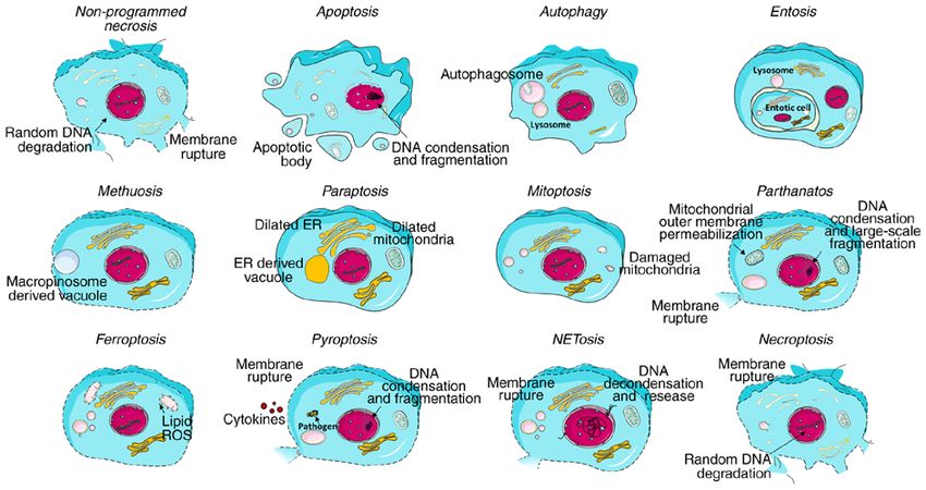
40                                              YAN et al: CELL DEATH MODALITIES

(Fig. 1). Programmed cell death (PCD) is driven by tightly          comet assay are able to detect the presence of fragmented
regulated intracellular signal transduction pathways. By            DNA. Annexin V in combination with cell‑impermeable
contrast, accidental cell death is referred to as non‑PCD as a      DNA staining dye is used to detect the outwards exposed
result of unexpected cell injury. Given the morphological char-     phosphatidylserine on cell membrane and cellular integrity.
acteristics and molecular mechanisms, PCD can be further            Alternatively, some assays evaluate the intermediate modula-
categorized into apoptotic cell death and non‑apoptotic cell        tors, e.g., caspase assay and poly‑ADP ribose polymerase
death. Apoptosis retains cell membrane integrity and occurs         (PARP) cleavage assay (20). Furthermore, specific apoptosis
in a caspase‑dependent manner. By contrast, non‑apoptotic           inhibitors, such as the pan‑caspase inhibitor, zVAD‑fmk, can
cell death is mostly characterized by membrane rupture and          also shed some light on the presence of apoptosis.
caspase‑independency. For simplicity, the present review
article focuses on the key features of the diverse cell death       4. Programmed non‑apoptotic cell death
modes and their assessment methods commonly utilized in
research (Table I), and refers the reader to specialized recent     Vacuole‑presenting cell death
review articles describing the processes of each cell death         Autophagy. Autophagic cell death is characterized by the
mode in further detail (4‑15).                                      appearance of large intracellular vesicles, plasma membrane
                                                                    blebbing, enlarged organelles and the depletion of cytoplasmic
2. Non‑programmed cell death                                        organelles in the absence of chromatin condensation (21)
                                                                    (Fig. 2). Noticeably, it functions as a lever in the cell process.
Non‑programmed necrosis. Non‑programmed necrosis is                 Autophagy is initiated upon cellular stress as a protective
stimulated by a number of external factors, e.g., infection,        response. Once the cellular stress is irreversible, the cell will be
toxins and physical injury, which lead to morphological altera-     committed to death also through excessive levels of autophagy.
tions, such as cytoplasmic swelling [oncosis, pre‑lethal phase      There are three forms of autophagy: Macro‑autophagy (Fig. 3),
caused by the disruption of ionic pumps such as Ca+ influx (16)],   micro‑autophagy and chaperone‑mediated autophagy (7). The
plasma membrane rupture and the subsequent loss of intracel-        macro‑autophagic process has been well documented (22‑24)
lular organelles without severe chromatin condensation, but         (Fig. 3). In micro‑autophagy, the cytoplasmic components are
randomly degraded DNA (17) (Fig. 2). Non‑programmed                 directly sequestrated into the lysosomes, where acidic hydro-
necrosis is often observed in ischemia, trauma and possibly         lases further mediate the degradation. Chaperone‑mediated
some forms of neurodegeneration. It is commonly consid-             autophagy selectively targets KFERQ motif (Lys‑Phe‑Glu‑Arg‑
ered as a passive process, which does not require de novo           Gln)‑containing proteins. These proteins can be recognized
macromolecular synthesis, but minimal energy (4).                   by chaperones, are subsequently hijacked into lysosomes and
    Based on the morphological features of necrosis, a number       eventually degraded (25). The specific degradation of the mito-
of methods, including lactate dehydrogenase (LDH) activity          chondria is referred to as mitophagy. The selective autophagy
detection and cell‑impermeable DNA binding dye, are                 of foreign pathogens is coined as xenophagy. There are also
commonly used to certify the cellular leakage and membrane          some other selective autophagy forms, such as lipophagy,
permeability (Table I).                                             aggrephagy and lysophagy (26).
                                                                        The detection methods are mostly developed for
3. Programmed apoptotic cell death                                  macro‑autophagy embodying direct measurement of autoph-
                                                                    agic activity (e.g., turnover of long‑lived proteins and LDH
Apoptosis. Apoptosis involves a series of tightly controlled        sequestration) and indirect analysis with autophagy specific
events and is characterized by cell shrinkage, membrane             antibodies through western blot‑based assay, fluorescence
blebbing, positional organelle loss, DNA condensation and           microscopy‑based assay and flow cytometry‑based assay (27)
fragmentation (Fig. 2). Three signaling pathways are known          (Table I).
to trigger apoptotic cell death: The extrinsic (death recep-            Entosis. Entosis (or cannibalism) is characterized by
tors) pathway, the intrinsic (mitochondrial) pathway and the        cell‑in‑cell formation (Fig. 2). Upon internalization, the
perforin/granzyme pathway (Fig. 3) (5).                             entotic cells remain viable for a short period of time. This
    Anoikis is a particular type of apoptosis, which essen-         process is frequently followed by lysosome‑mediated degra-
tially shares identical pathways as with apoptosis; however,        dation and non‑apoptotic cell death, while a fraction of the
is triggered by inadequate or inappropriate cell‑matrix             internalized cells can also extricate themselves or are expelled
interactions (18) (Fig. 3). The architectural state of the          from the host cell (28). Entosis is believed to be triggered by
cytoskeleton is expected to interfere with the function of          integrin‑extracellular matrix (ECM) detachment (29). Unlike
integrin, a pro‑survival effector (6). However, the connection      phagocytosis, the engulfment of entotic cells represents a
between cell architecture alteration and apoptosis remains          self‑control process through RhoA and the Rho‑associated
poorly identified. It has recently been indicated that c‑JUN        coiled‑coil containing protein kinases (ROCK). The entotic
NH2‑terminal kinase (JNK) signaling is required for efficient       cell and the host cell interact with each other through the
anoikis through a BAK/BAX‑dependent manner by increasing            E‑cadherin and α‑catenin cell junction interface. RhoA and
BCL2‑like 11 (BIM) expression and BCL‑2 modifying factor            ROCK in entotic cells lead to specific accumulation of actin
(BMF) phosphorylation (19).                                         and myosin complex (actomyosin) at the cell cortex opposite
    Apoptosis assessment methods have been rapidly devel-           to the junctional interface, which generates the unbalanced
oped over the past years (Table I). Terminal deoxynucleotidyl       contractile force driving cell‑in‑cell formation. However,
transferase dUPT nick‑end labeling (TUNEL) assay and                entosis is also observed in matrix‑attached epithelial cells.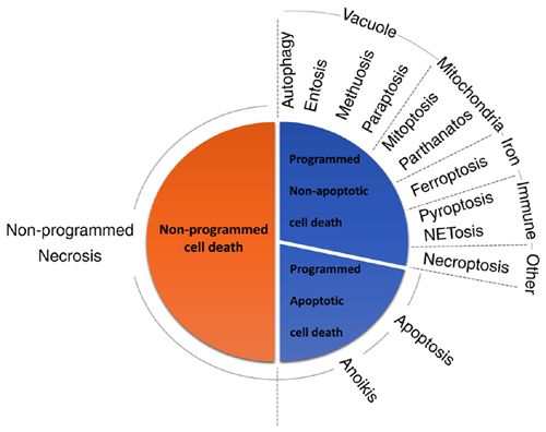
WORLD ACADEMY OF SCIENCES JOURNAL 2: 39-48, 2020                                                41

                                                                               death. Methuosis with its typical morphology, is often assessed
                                                                               by electron microscopy in research (36‑38) (Table I).
                                                                                   Paraptosis. The hallmark of paraptosis is the extensive
                                                                               cytoplasmic vacuolization derived from the dilated endo-
                                                                               plasmic reticulum (ER) or the mitochondria (39) (Fig. 2). It
                                                                               has been reported that the activation of insulin‑like growth
                                                                               factor 1 receptor (IGF1R) and its downstream signaling
                                                                               incorporating mitogen‑activated protein kinases (MAPKs)
                                                                               and JNK pathways can induce paraptosis, despite the fact
                                                                               that IGF1R is commonly considered as a pro‑survival modu-
                                                                               lator (40). A number of studies have indicated that paraptosis
                                                                               is associated with reactive oxygen species (ROS) generation
                                                                               and the accumulation of misfolded proteins in the ER, as
                                                                               well as mitochondrial Ca 2+ overload (10,41‑43), which exert
                                                                               an osmotic force to distend the ER lumen and mitochondria
                                                                               for vacuolization. In spite of the current available evidence,
Figure 1. Cell death classification. The cell death entities are categorized
                                                                               the molecular mechanisms underlying paraptosis have not yet
according to their signal‑dependency, morphological characteristics and        been fully addressed.
molecular mechanisms. The pie area in the figure does not represent the            Similar to entosis and methuosis, there is no specific assay
frequency of occurrence of each cell death.                                    available for the detection of paraptosis, at least to the best
                                                                               of our knowledge. It is mostly defined by the appearance of
                                                                               multiple single‑membraned cytoplasmic vacuoles, as detected
Wan et al proposed that the overactivation of myosin or                        by electron microscopy (44) (Table I).
unbalanced myosin activation through regulatory polarity
proteins between the contacting cells acted as the driving                     Mitochondrial‑dependent cell death
force for entosis in matrix‑attached epithelial cells (30). The                Mitoptosis. Unlike mitophagy (autophagic degradation of
engulfment is followed by lysosome‑mediated degradation,                       mitochondria), mitoptosis, also known as mitochondrial
which differs from autophagic cell death (31). The autophagic                  suicide, represents a process of programmed fission and
protein, microtubule‑associated protein light chain 3 (LC3),                   fusion of the mitochondria with the concomitant disruption of
does not participate to form the autophagosome. Instead, LC3                   the adenosine triphosphate (ATP) supply. As a consequence,
is directed to the single‑membrane vacuole in the host cell                    mitoptosis can be associated with both apoptosis (45) and
that harbors the engulfed cell through lipidation with the help                autophagy (46). The degraded mitochondria either become
of autophagy‑related protein (ATG)5, ATG7 and Vps34, and                       autophagosomes or mitoptotic bodies, which are extruded
promotes lysosome fusion followed by lysosome‑mediated                         from the cell. In this sense, mitoptosis itself is not a cell
degradation (8) (Fig. 3).                                                      death pathway, but a mitochondrial death pathway. However,
    However, there is as yet no specific assay available for the               the extensive mitochondrial fragmentation through elevated
detection of entosis, at least to the best of our knowledge. The               fission finally leads to cell death (47). Mechanically speaking,
presence of entosis is deduced from its typical cell‑in‑cell                   mitochondrial outer membrane permeabilization (MOMP)
structure, as detected by fluorescence imaging and electron                    induced by BAX/BAK triggers the release of a mitochon-
microscopy (32,33) (Table I).                                                  drial intermembrane space protein termed translocase of
    Methuosis. Methuosis represents a type of cell death                       inner mitochondrial membrane 8a (TIMM8a/DDP). DDP
characterized by the presence of the massive accumula-                         subsequently binds to DRP1 in the cytoplasm. The interaction
tion of large fluid‑filled single membrane vacuoles derived                    between DDP and DRP1 leads to the recruitment of DRP1 and
from macropinosomes, which is specifically accompanied                         retention in the mitochondria, which induces mitochondrial
with Ras hyper‑activation and apoptosis impairment.                            fission and finally, mitoptosis (48). Nevertheless, the process
Intriguingly, methuosis is not associated with the conven-                     remains poorly understood and is described mostly by its
tional Ras‑Raf‑MEK‑ERK axis or class III phosphoinositide                      morphological features.
3‑kinase (PI3K) signaling (34). The consequent morphology                          As a manner of mitochondrial suicide, the visualization
resembles necrosis in the manner of cell swelling and plasma                   of fragmented mitochondria with mitochondria‑specific dyes
membrane integrity loss. In methuosis, activated Ras stimu-                    (e.g., MitoTracker Green®) by utilizing fluorescence micros-
lates micropinocytosis through the downstream activation of                    copy and a close observation with electron microscopy provide
Rac family small GTPase 1 (Rac1). Coincidently, the reduc-                     certain clues on the presence of mitoptosis (45). Moreover,
tion of ADP ribosylation factor 6‑GTP (Arf6‑GTP) impedes                       specific antibodies against cytochrome c and TIMM8a/DDP
macropinosome recycling (35). The abnormal coalescence of                      are also utilized in research (48) (Table I).
nascent macropinosomes gives rise to massive cytoplasmic                           Parthanatos. Parthanatos represents a mitochon-
vacuolization. The vacuoles formed in the early stages of                      drial‑linked, but caspase‑independent cell death and is
methuosis are decorated with late endosomal markers [e.g.,                     characterized by the hyperactivation of PARP. PARP medi-
lysosomal‑associated membrane protein 1 (LAMP1) and                            ates the synthesis of poly(ADP‑ribose) (PAR), which further
Rab7] (9). The massive vacuoles, which are not able to be                      shuttles from the nucleus to the cytoplasm and binds to specific
recycled or merged with lysosomes, will finally lead to cell                   mitochondrial proteins followed by apoptosis‑inducing factor
42
Table I. Cell death modalities, their features and common detection methods.
Classification Cell death modality Key molecules Key morphology Detection methods
Non‑PCD Necrosis None Cell swelling; membrane rupture; loss of Lactate dehydrogenase activity detection; visualizing
organelle membrane integrity loss by cell‑impermeable DNA
binding dye
PCD‑apoptotic Apoptosis/anoikis DRs and their ligands, Cell shrinkage; membrane blebbing; loss Chromosome condensation detection; TUNEL assay;
Bax, Bak, AIF, caspase‑8, of positional organization of organelles Annexin V assay; caspase assay; PARP cleavage assay;
caspase‑3, caspase‑9 in the cytoplasm; DNA condensation and applying apoptosis inhibitors
fragmentation; nuclear membrane rupture
PCD‑vacuole Autophagy UKL1, PI3KIII, ATGs, LC3 Large intracellular vesicles; membrane Turnover of long‑lived proteins; LDH sequestration;
presenting blebbing; enlarged organelles; depletion western blot analysis with autophagy specific
of cytoplasmic organelles antibodies
Entosis RhoA, ROCKI/II, E‑cadherin, Cell‑in‑cell formation Morphology observation with fluorescence imaging
α‑catenin, actomyosin, LC3, and electron microscopy
ATGs
Methuosis Ras, Rac1, Arf6, LAMP1, Accumulation of large fluid‑filled single Morphology observation with electron microscopy
Rab7 membrane vacuoles; cell swelling;
membrane rupture
Paraptosis Unclear Accumulation of large fluid‑filled single Morphology observation with electron microscopy
membrane vacuoles; dilation of ER or
mitochondria
PCD‑mitochondria‑ Mitoptosis Bax, Bak, TIMM8a(DDP), Mitochondria disappearance; decomposition Morphology observation with fluorescence microscopy
dependent Drp1 of the mitochondrial reticulum to small and electron microscopy; western blot analysis with
YAN et al: CELL DEATH MODALITIES
spherical organelles mitoptosis‑specific antibodies
Parthanatos PARP, AIF Membrane rupture; mitochondrial outer Western blot analysis with parthanatos specific
membrane permeabilization; chromatin antibodies; Mitochondrial depolarization detection
condensation; DNA large‑scale fragmentation with fluorescent probe
PCD‑iron Ferroptosis System XC−, GPX4, Diminutive mitochondria with decreased Applying ferroptosis inhibitors; measuring lipid
dependent Lipid ROS cristae and collapsed and ruptured membrane peroxides e.g. malondialhyde and 4‑hydroxynonenal
quantification
PCD‑immune Pyroptosis NLRs, ALRs, caspase‑1, Cell swelling; membrane rupture; DNA Quantification of cytoplasmic LDH; visualizing
reactive caspase‑11 condensation and fragmentation membrane integrity loss by fluorescence microscopy;
western blot analysis with pyroptosis‑specific
antibodies
NETosis NOX4, PAD4 Chromatin decondensation; membrane Morphology observation with fluorescence
rupture microscopy; free‑cell DNA and DNA‑neutrophil
derived protein complex detection with fluorescent
probe and immunoblotWORLD ACADEMY OF SCIENCES JOURNAL 2: 39-48, 2020 43
(AIF) release. Free AIF is translocated from the mitochon-
PCD, programmed cell death; DRs, death receptors; PARP, poly(ADP‑ribose)‑polymerase; ULK1, unc‑51 like autophagy activating kinase 1; PI3K, class III phosphoinositide 3‑kinase; ATGs,
autophagy‑related proteins; LC3, microtubule‑associated protein light chain 3; ROCK, Rho associated coiled‑coil containing protein kinase; Rac1, Rac family small GTPase 1; Arf6, ADP ribosylation
factor 6; LAMP1, lysosomal‑associated membrane protein 1; ER, endoplasmic reticulum; TIMM8a/DDP, translocase of inner mitochondrial membrane 8a; AIF, apoptosis‑initiating factor; ROS, reac-
tive oxygen species; NLRs, NOD‑like receptors; ALRs, AIM2‑like receptors; NOX4, NADPH oxidase 4; PAD4, peptidylarginine deiminase 4; TLRs, toll‑like receptors; TCR, T‑cell receptor; RIPKs,
inhibitors; western blot analysis with necroptosis‑specific
dria into the nucleus. In the nucleus, AIF induces chromatin
depolarization detection; applying necroptosis specific
condensation and DNA breakage (49). Compared to the apop-
Visualizing membrane integrity loss; mitochondrial
totic process, intact PARP and its activation is required, rather
than PARP cleavage. Moreover, parthanatos cannot be inhib-
ited by broad‑spectrum caspase inhibitors (50), which proves
its independency of caspases. Parthanatos does not involve the
formation of apoptotic bodies. Furthermore, the DNA frag-
Detection methods
mentation is large‑scale rather than small‑to‑moderate scale,
as typically observed in apoptosis (11) (Fig. 2).
PAR accumulation, PARP‑1 activation and nuclear AIF
are practically used as biomarkers of parthanatos. The process
can be further confirmed with mitochondrial depolarization,
as detected with fluorescent probe staining (Table I).
Iron‑dependent cell death
Ferroptosis. Ferroptosis is normally associated with a
antibodies
normal‑appearing morphology, with an intact cell membrane
without blebbing and normal‑sized nucleus free of chromatin
condensation, although with diminutive mitochondria with
decreased cristae and collapsed and ruptured membranes (51)
(Fig. 2). It is initiated by the failure of the glutathione‑depen-
Cell swelling; membrane rupture; loss of
dent antioxidant defense through defects in system XC− or
glutathione peroxidase 4 (GPX4) (12). System XC− transports
extracellular cystine into the cell, which is then transformed
organelle; mitochondria swelling
into cysteine for glutathione (GSH) synthesis. GPX4 can
directly catalyze the reaction between glutathione and lipid
Key morphology
hydroperoxides to reduce the cellular level of lipid peroxida-
tion. Either the depletion of GSH or the inhibition of GPX4
results in lipid hydroperoxide accumulation. Free iron inter-
acts with lipid hydroperoxides through the Fenton reaction
and forms lipid ROS (Fig. 3). Excessive lipid ROS generation
finally leads to the cell death.
The induction of ferroptosis can be confirmed by
applying ferroptosis inhibitors (e.g., ferrostatin‑1 and
receptor‑interacting protein kinases; MLKL, mixed lineage kinase domain‑like protein.
liproxstatin‑1) and by measuring lipid peroxides (e.g.,
malondialhyde quantification and 4‑hydroxynonenal quan-
tification) (Table I).
DRs, TLRs, TCR, RIPKs,
Immune‑reactive cell death
Key molecules
Pyroptosis. Pyroptosis is an inflammatory form of programmed
cell death that commonly occurs upon the recognition of intra-
cellular pathogens in immune cells. The inflammation sensors
[e.g., NOD‑like receptors (NLRs)] of infected macrophages
MLMK
recognize the flagellin components of pathogens and initiate
the formation of multi‑protein complex inflammasomes, which
subsequently activate caspase‑1 (13) (Fig. 3). Upon activation,
caspase‑1 mediates the membrane pore formation through
the cleavage of gasdermin D, allowing the rupture of the cell
Cell death modality
membrane (52). The process is also accompanied by DNA
Necroptosis
condensation and fragmentation (Fig. 2). Moreover, caspase‑11
can be directly activated by bacterial lipopolysaccharide (LPS)
and induces pyroptosis (53).
Pyroptosis can be evaluated through the quantification of
released cytoplasmic LDH, the visualization of membrane
Table I. Continued.
integrity loss by fluorescence microscopy, the detection of
interleukin (IL)‑1β, caspase activation and gasdermin D
Classification
cleavage by western blot analysis (54) (Table I).
Other type
Neutrophil extracellular trap‑associated cell death
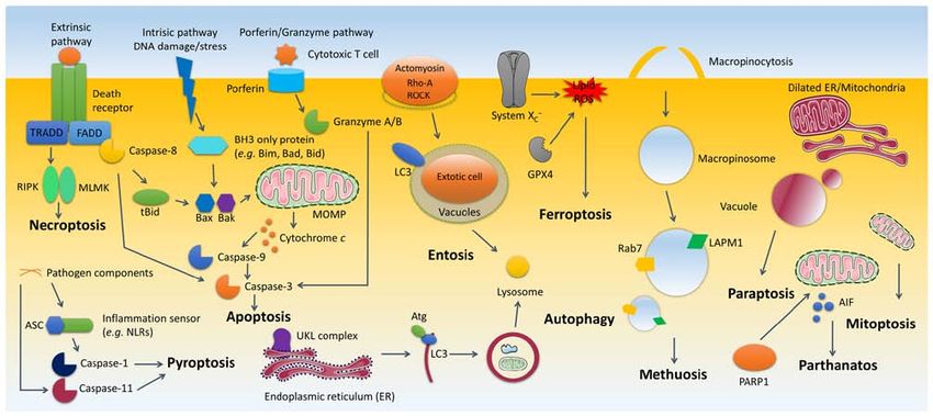
(NETosis). NETosis, a unique form of cell death, is initiated44 YAN et al: CELL DEATH MODALITIES Figure 2. Typical morphology of each cell death. The morphological alteration focuses on cell size, membrane integrity, chromatin density, organelle arrangement and presence of vacuoles. Figure 3. Synopsis of cell death processes. Ten cell death modalities (apoptosis, autophagy, entosis, methuosis, paraptosis, mitoptosis, parthanatos, ferroptosis, pyroptosis and necroptosis) are presented. Anoikis shares identical signaling pathways as apoptosis, apart from the fact that it is stimulated by inadequate or inappropriate cell‑matrix interactions. The cell death modalities (necrosis and NETosis) without elucidative mechanism were not included. Grey color indicates non‑functional molecules. Arrow direction indicates the causal association. RIPK, receptor‑interacting protein kinase; MLKL, mixed lineage kinase domain‑like protein; NLRs, NOD‑like receptors; MOMP, mitochondrial outer membrane permeabilization; LC3, microtubule‑associated protein light chain 3; ROCK, Rho associated coiled‑coil containing protein kinase; GPX4, glutathione peroxidase 4; ROS, reactive oxygen species; UKL complex, UKL1 in a complex with FIP200, ATG13 and ATG101. by the presence of pathogens or their components and mostly promoted through superoxide generated by NADPH oxidase 4 occurs in immune cells, particularly neutrophils. Upon (NOX4), autophagy and peptidylarginine deiminase 4 the recognition of pathogens within neutrophils, the cells (PAD4)‑dependent histone citrullination (56,57). However, undergo histone modification, chromatin decondensation and further research is expected to provide a clear molecular neutrophil extracellular trap [NET, comprising chromatin elucidation. and antimicrobial components including myeloperoxidase, The staining of co‑localized neutrophil‑derived proteins neutrophil elastase, cathepsin G, lysozyme and defensins (55)] and extracellular DNA, as well as citrullinated histones is release and this eventually leads to cell death. The process is utilized to evaluate NETosis. Moreover, cell‑free DNA and
WORLD ACADEMY OF SCIENCES JOURNAL 2: 39-48, 2020                                              45

DNA‑neutrophil derived protein complexes can be detected                 As for neurodegenerative diseases, the initial phase of cell
by PicoGreen® and ELISA. Both morphology and cell‑appen-             death in ischemia represents necrotic cell death, while delayed
dant NETosis components can be detected through flow                 cell death is apoptotic in nature due to the fact that the ischemic
cytometry (58) (Table I).                                            core tends to be necrotic and the penumbra region apop-
                                                                     totic (72). Autophagic cell death and parthanatos are linked to
Other types                                                          ischemia (11,73). In Parkinson's disease, apoptosis contributes
Necroptosis. Necroptosis, also known as programmed necrosis,         to the loss of nigral neurons due to the fact that almost every
is characterized by the activation of receptor‑interacting protein   Lewy body‑containing neuron (as a pathological feature of
kinases (RIPKs) through several signaling pathways (15).             Parkinson's disease) is positive for pro‑apoptotic modulator
RIPKs are activated upon recruitment to macromolecular               staining (74). Another study demonstrated that necrostatin‑1,
complexes from various cell‑surface receptors: Death recep-          an inhibitor of necroptosis, ameliorated neuronal loss in a
tors (DRs), Toll‑like receptors (TLRs), and the T‑cell receptor      model of Parkinson's disease (75), indicating that necroptosis
(TCR) (Fig. 3) (59,60). RIPK1 and RIPK3 function as the              may also play a role in Parkinson's disease. There is also
key components of necrosome (61). RIPK3 further activates            evidence suggesting the role of apoptosis in Huntington's
downstream molecule mixed lineage kinase domain‑like                 disease. However, its role in Alzheimer's disease remains
protein (MLKL) through phosphorylation (62,63), which leads          under debate (76).
to MLKL oligomerization. The oligomerized MLKL inserts                   Cell death modes, such as apoptosis, necrosis and autophagy
into and permeabilizes cellular membrane, which finally gives        in cardiac myocytes have been frequently reported to affect
rise to cell death (64). Moreover, RIP3‑dependent necroptosis        a variety of cardiovascular diseases, including myocardial
is also triggered by the cytosolic DNA sensor, DNA‑dependent         infarction, diabetic cardiomyopathy, ischemic cardiomyocyte
activator of interferon (DAI) regulatory factors, following viral    and congestive heart failure (77‑79). In addition, ferroptosis,
infection or the presence of double‑stranded viral DNA (65).         pyroptosis, as well as parthanatos are also documented to
Necroptosis reveals the necrotic morphology with membrane            contribute to ischemia/reperfusion injury (80). The other
rupture and loss of organelles (Fig. 2).                             cell death types have been studied to a much lesser extent as
    Necroptosis can be assessed by the loss of plasma membrane       compared to cardiovascular diseases. Likewise, apoptosis and
integrity by utilizing cell‑impermeable DNA binding dyes,            secondary necrosis are considered as major modes of cell death
the release of cellular contents, including LDH, high mobility       in systemic autoimmune diseases. Recent evidence indicates
group box 1 protein (HMGB1) and cyclophilin A by western             that NETosis accounts for certain immunological features in
blot analysis, mitochondrial potential by fluorescent probes         systemic lupus erythematosus (81).
and morphology by electron microscopy. The utilization
of necroptosis specific inhibitors, such as necrostatin‑1 and        6. Conclusions and perspectives
measuring key proteins in the pathway represent alternative
strategies (66) (Table I).                                           The cell death modes presented in the present review article are
                                                                     mostly distinguished by stimuli, molecules and morphologies.
5. Implications of cell death in human diseases                      Apart from non‑programmed necrosis, the other cell death
                                                                     modes are regulated in a signal‑dependent manner, despite
The dysregulation of cell death processes is highly relevant         the fact that a number of the pathways have not yet been fully
to tumorigenesis, as well as to the pathogenesis of a number         addressed. Some cell death modes are intensively interacting
of other diseases, such as degenerative, cardiovascular and          with others. For instance, the activation of tumor necrosis factor
autoimmune diseases. The association between cell death              receptor (TNFR) can stimulate both apoptosis and necroptosis;
and cancer is complex. The complexity is attributed to several       however, compromised apoptosis can shift the downstream
factors: On the one hand, there is more than one type of cell        pathway to necroptosis (82) and vice versa (83). Some processes
death endogenously engaged in cancer. On the other hand,             during cell death are connected; for instance, the occurrence
some types of cell death have dual and even opposing effects         of mitoptosis can turn out as autophagic cell death or apoptotic
on tumorigenesis. Firstly, apoptosis is involved in cancer.          cell death. In general, necrosis‑like cell death is associated with
Cancerous cells can evade apoptosis by downregulating or             membrane rupture. The consequent release of intracellular
blocking apoptosis signaling (67). Unexpectedly, apoptosis can       inflammatory factors can give rise to inflammation as observed
also drive tumor formation by promoting cell proliferation as a      in necrosis, necroptosis, NETosis and pyroptosis. By contrast,
compensation for cell loss (68). Secondly, necrosis is commonly      apoptotic cells do not stimulate inflammation, since they are
observed in tumors due to hypoxic microenvironments (67).            rapidly eliminated by phagocytes. However, if apoptotic cells
Thirdly, cancerous cells with defects in apoptosis tend to           are not properly processed, they can develop secondary necrosis.
utilize autophagy as a pro‑survival mechanism. Paradoxically,        These mutual connections indicate that different cell death types
impeded autophagy is also associated with tumorigenesis (69).        are not isolated from each other. The molecular links await to be
Fourthly, entosis represents tumor suppressive activity in           unveiled in greater detail. Their implications on diverse diseases
pancreatic cancer, whereas it promotes tumor progression in          are expected to be unraveled in the near future, since current
most other situations (70,71). Although the other cell death         studies on cell death modes involved in diseases are mostly
types are much less endogenously involved in cancer develop-         confined to the more classical cell death categories. Green (84)
ment, they are mostly utilized as anti‑cancer defense strategies     also addressed five quite interesting and inspiring questions
of the body and defects in their signaling plays an important        about the balance and context of cell death. In fact, much is still
role in drug resistance and clinical failures.                       unknown. Noticeably, this review article has primarily focused
46 YAN et al: CELL DEATH MODALITIES
on the features of pathological cell death and is limited to the 9. Maltese WA and Overmeyer JH: Methuosis. Nonapoptotic cell
death associated with vacuolization of macropinosome and
animal kingdom. However, there also exist physiologic cell endosome compartments. Am J Pathol 184: 1630‑1642, 2014.
death such as cornification (85) to form termination differentia- 10. Lee D, Kim IY, Saha S and Choi KS: Paraptosis in the anti‑cancer
tion and some cell death types are also similarly present in the arsenal of natural products. Pharmacol Ther 162: 120‑133, 2016.
11. Fatokun AA, Dawson VL and Dawson TM: Parthanatos:
plant kingdom (e.g., apoptosis‑like cell death) (86). Mitochondrial‑linked mechanisms and therapeutic opportuni-
ties. Br J Pharmacol 171: 2000‑2016, 2014.
Acknowledgements 12. Yu H, Guo P, Xie X, Wang Y and Chen G: Ferroptosis, a new
form of cell death, and its relationships with tumourous diseases.
J Cell Mol Med 21: 648‑657, 2017.
Not applicable. 13. Bergsbaken T, Fink SL and Cookson BT: Pyroptosis. Host cell
death and inflammation. Nat Rev Microbiol 7: 99‑109, 2009.
14. Neubert E, Meyer D, Rocca F, Günay G, Kwaczala‑Tessmann A,
Funding Grandke J, Senger‑Sander S, Geisler C, Egner A, Schön MP, et al:
Chromatin swelling drives neutrophil extracellular trap release.
The authors are grateful to PhD stipends given to GY (by the Nat Commun 9: 3767, 2018.
15. Vanlangenakker N, Vanden Berghe T and Vandenabeele P:
Chinese Scholarship Council) and to ME (by the German Many stimuli pull the necrotic trigger, an overview. Cell Death
Academic Exchange Service, DAAD). Differ 19: 75‑86, 2012.
16. Won SJ, Kim DY and Gwag BJ: Cellular and molecular pathways
of ischemic neuronal death. J Biochem Mol Biol 35: 67‑86, 2002.
Availability of data and materials 17. Weerasinghe P and Buja LM: Oncosis. An important non‑apop-
totic mode of cell death. Exp Mol Pathol 93: 302‑308, 2012.
Not applicable. 18. Frisch SM and Screaton RA: Anoikis mechanisms. Curr Opin
Cell Biol 13: 555‑562, 2001.
19. Girnius N and Davis RJ: JNK promotes epithelial cell anoikis
Authors' contributions by transcriptional and post‑translational regulation of BH3‑only
proteins. Cell Rep 21: 1910‑1921, 2017.
20. Muganda PM (ed): Apoptosis methods in toxicology. Humana
GY was responsible for the drafting of the manuscript and cell Press, New York, NY, 2016.
death information collection. ME was responsible for informa- 21. Liu Y and Levine B: Autosis and autophagic cell death: The dark
tion presentesst and figure construction. TE was responsible side of autophagy. Cell Death Differ 22: 367‑376, 2015.
22. Pajares M, Jiménez‑Moreno N, García‑Yagüe ÁJ, Escoll M,
for the initial conception of the study and for the revision of Ceballos ML de, van Leuven F, Rábano A, Yamamoto M, Rojo AI
the manuscript. All authors have read and approved the final and Cuadrado A: Transcription factor NFE2L2/NRF2 is a regu-
manuscript. lator of macroautophagy genes. Autophagy 12: 1902‑1916, 2016.
23. Mercer CA, Kaliappan A and Dennis PB: A novel, human Atg13
binding protein, Atg101, interacts with ULK1 and is essential for
Ethics approval and consent to participate macroautophagy. Autophagy 5: 649‑662, 2009.
24. Chen Y and Klionsky DJ: The regulation of autophagy‑unan-
swered questions. J Cell Sci 124: 161‑170, 2011.
Not applicable. 25. Mizushima N: A brief history of autophagy from cell biology to
physiology and disease. Nat Cell Biol 20: 521‑527, 2018.
Patient consent for publication 26. Hansen M, Rubinsztein DC and Walker DW: Autophagy as a
promoter of longevity: Insights from model organisms. Nat Rev
Mol Cell Biol 19: 579‑593, 2018.
Not applicable. 27. Orhon I and Reggiori F: Assays to monitor autophagy progres-
sion in cell cultures. Cells 6: pii: E20, 2017.
28. White E: Entosis: It's a cell‑eat‑cell world. Cell 131: 840‑842,
Competing interests 2007.
29. Ishikawa F, Ushida K, Mori K and Shibanuma M: Loss of
The authors declare that they have no competing interests. anchorage primarily induces non‑apoptotic cell death in a human
mammary epithelial cell line under atypical focal adhesion
kinase signaling. Cell Death Dis 6: e1619, 2015.
References 30. Wan Q, Liu J, Zheng Z, Zhu H, Chu X, Dong Z, Huang S and
Du Q: Regulation of myosin activation during cell‑cell contact
1. Green DR and Llambi F: Cell death signaling. Cold Spring Harb formation by Par3‑Lgl antagonism: Entosis without matrix
Perspect Biol 7: pii: a006080, 2015. detachment. Mol Biol Cell 23: 2076‑2091, 2012.
31. Garanina AS, Kisurina‑Evgenieva OP, Erok hina MV,
2. Galluzzi L, Vitale I, Aaronson SA, Abrams JM, Adam D, Smirnova EA, Factor VM and Onishchenko GE: Consecutive
Agostinis P, Alnemri ES, Altucci L, Amelio I, Andrews DW, et al: entosis stages in human substrate‑dependent cultured cells. Sci
Molecular mechanisms of cell death: Recommendations of Rep 7: 12555, 2017.
the nomenclature committee on cell death 2018. Cell Death 32. Sun Q and Overholtzer M: Methods for the study of entosis.
Differ 25: 486‑541, 2018. Methods Mol Biol 1004: 59‑66, 2013.
3. Tang D, Kang R, Berghe TV, Vandenabeele P and Kroemer G: 33. Huang H, Chen A, Wang T, Wang M, Ning X, He M, Hu Y,
The molecular machinery of regulated cell death. Cell Res 29: Yuan L, Li S, Wang Q, et al: Detecting cell‑in‑cell structures
347‑364, 2019. in human tumor samples by E‑cadherin/CD68/CD45 triple
4. Syntichaki P and Tavernarakis N: Death by necrosis. staining. Oncotarget 6: 20278‑20287, 2015.
Uncontrollable catastrophe, or is there order behind the chaos? 34. Kaul A, Overmeyer JH and Maltese WA: Activated Ras induces
EMBO Rep 3: 604‑609, 2002. cytoplasmic vacuolation and non‑apoptotic death in glioblastoma
5. Elmore S: Apoptosis: A review of programmed cell death. cells via novel effector pathways. Cell Signal 19: 1034‑1043,
Toxicol Pathol 35: 495‑516, 2007. 2007.
6. Paoli P, Giannoni E and Chiarugi P: Anoikis molecular pathways 35. Bhanot H, Young AM, Overmeyer JH and Maltese WA: Induction
and its role in cancer progression. Biochim Biophys Acta 1833: of nonapoptotic cell death by activated Ras requires inverse
3481‑3498, 2013. regulation of Rac1 and Arf6. Mol Cancer Res 8: 1358‑1374, 2010.
7. Ravanan P, Srikumar IF and Talwar P: Autophagy. The spotlight 36. Overmeyer JH, Young AM, Bhanot H and Maltese WA: A
for cellular stress responses. Life Sci 188: 53‑67, 2017. chalcone‑related small molecule that induces methuosis, a novel
8. Krishna S and Overholtzer M: Mechanisms and consequences of form of non‑apoptotic cell death, in glioblastoma cells. Mol
entosis. Cell Mol Life Sci 73: 2379‑2386, 2016. Cancer 10: 69, 2011.WORLD ACADEMY OF SCIENCES JOURNAL 2: 39-48, 2020 47
37. Trabbic CJ, Dietsch HM, Alexander EM, Nagy PI, Robinson MW, 58. Masuda S, Nakazawa D, Shida H, Miyoshi A, Kusunoki Y,
Overmeyer JH, Maltese WA and Erhardt PW: Differential Tomaru U and Ishizu A: NETosis markers: Quest for specific,
induction of cytoplasmic vacuolization and methuosis by novel objective, and quantitative markers. Clin Chim Acta 459: 89‑93,
2‑indolyl‑substituted pyridinylpropenones. ACS Med Chem 2016.
Lett 5: 73‑77, 2014. 59. Kaiser WJ, Sridharan H, Huang C, Mandal P, Upton JW,
38. Silva‑Pavez E, Villar P, Trigo C, Caamaño E, Niechi I, Pérez P, Gough PJ, Sehon CA, Marquis RW, Bertin J and Mocarski ES:
Muñoz JP, Aguayo F, Burzio VA, Varas‑Godoy M, et al: CK2 Toll‑like receptor 3‑mediated necrosis via TRIF, RIP3, and
inhibition with silmitasertib promotes methuosis‑like cell death MLKL. J Biol Chem 288: 31268‑31279, 2013.
associated to catastrophic massive vacuolization of colorectal 60. Weinlich R, Oberst A, Beere HM and Green DR: Necroptosis
cancer cells. Cell Death Dis 10: 73, 2019. in development, inflammation and disease. Nat Rev Mol Cell
39. Sperandio S, de Belle I and Bredesen DE: An alternative, Biol 18: 127‑136, 2017.
nonapoptotic form of programmed cell death. Proc Natl Acad 61. He S, Wang L, Miao L, Wang T, Du F, Zhao L and Wang X:
Sci USA 97: 14376‑14381, 2000. Receptor interacting protein kinase‑3 determines cellular
40. Sperandio S, Poksay K, de Belle I, Lafuente MJ, Liu B, Nasir J necrotic response to TNF‑alpha. Cell 137: 1100‑1111, 2009.
and Bredesen DE: Paraptosis: Mediation by MAP kinases and 62. Sun L, Wang H, Wang Z, He S, Chen S, Liao D, Wang L, Yan J,
inhibition by AIP‑1/Alix. Cell Death Differ 11: 1066‑1075, Liu W, Lei X and Wang X: Mixed lineage kinase domain‑like
2004. protein mediates necrosis signaling downstream of RIP3 kinase.
41. Yoon MJ, Lee AR, Jeong SA, Kim YS, Kim JY, Kwon YJ and Cell 148: 213‑227, 2012.
Choi KS: Release of Ca2+ from the endoplasmic reticulum and 63. Wang H, Sun L, Su L, Rizo J, Liu L, Wang LF, Wang FS and
its subsequent influx into mitochondria trigger celastrol‑induced Wang X: Mixed lineage kinase domain‑like protein MLKL
paraptosis in cancer cells. Oncotarget 5: 6816‑6831, 2014. causes necrotic membrane disruption upon phosphorylation by
42. Gandin V, Pellei M, Tisato F, Porchia M, Santini C and RIP3. Mol Cell 54: 133‑146, 2014.
Marzano C: A novel copper complex induces paraptosis in colon 64. Cai Z, Jitkaew S, Zhao J, Chiang HC, Choksi S, Liu J, Ward Y,
cancer cells via the activation of ER stress signalling. J Cell Mol Wu LG and Liu ZG: Plasma membrane translocation of trimer-
Med 16: 142‑151, 2012. ized MLKL protein is required for TNF‑induced necroptosis.
43. Ghosh K, De S, Das S, Muk herjee S and Sengupta Nat Cell Biol 16: 55‑65, 2014.
Bandyopadhyay S: Withaferin a induces ROS‑mediated parap- 65. Maelfait J, Liverpool L, Bridgeman A, Ragan KB, Upton JW
tosis in human breast cancer cell‑lines MCF‑7 and MDA‑MB‑231. and Rehwinkel J: Sensing of viral and endogenous RNA by
PLoS One 11: e0168488, 2016. ZBP1/DAI induces necroptosis. EMBO J 36: 2529‑2543, 2017.
44. Kessel D: Apoptosis, paraptosis and autophagy: death and 66. Vanden Berghe T, Grootjans S, Goossens V, Dondelinger Y,
survival pathways associated with photodynamic therapy. Krysko DV, Takahashi N and Vandenabeele P: Determination of
Photochem Photobiol 95: 119‑125, 2019. apoptotic and necrotic cell death in vitro and in vivo. Methods 61:
45. Lyamzaev KG, Nepryakhina OK, Saprunova VB, Bakeeva LE, 117‑129, 2013.
Pletjushkina OY, Chernyak BV and Skulachev VP: Novel mecha- 67. Messmer MN, Snyder AG and Oberst A: Comparing the effects
nism of elimination of malfunctioning mitochondria (mitoptosis). of different cell death programs in tumor progression and immu-
Formation of mitoptotic bodies and extrusion of mitochondrial notherapy. Cell Death Differ 26: 115‑129, 2019.
material from the cell. Biochim Biophys Acta 1777: 817‑825, 68. Labi V and Erlacher M: How cell death shapes cancer. Cell
2008. Death Dis 6: e1675, 2015.
46. Jangamreddy JR and Los MJ: Mitoptosis, a novel mitochon- 69. Mathew R, Karantza‑Wadsworth V and White E: Role of
drial death mechanism leading predominantly to activation of autophagy in cancer. Nat Rev Cancer 7: 961‑967, 2007.
autophagy. Hepat Mon 12: e6159, 2012. 70. Durgan J and Florey O: Cancer cell cannibalism: Multiple
47. Youle RJ and Karbowski M: Mitochondrial fission in apoptosis. triggers emerge for entosis. Biochim Biophys Acta Mol Cell
Nat Rev Mol Cell Biol 6: 657‑663, 2005. Res 1865: 831‑841, 2018.
48. Arnoult D, Rismanchi N, Grodet A, Roberts RG, Seeburg DP, 71. Wang X, Li Y, Li J, Le Li, Zhu H, Chen H, Kong R, Wang G,
Estaquier J, Sheng M and Blackstone C: Bax/Bak‑dependent Wang Y, Hu J and Sun B: Cell‑in‑cell phenomenon and its rela-
release of DDP/TIMM8a promotes Drp1‑mediated mitochon- tionship with tumor microenvironment and tumor progression:
drial fission and mitoptosis during programmed cell death. Curr A review. Front Cell Dev Biol 7: 311, 2019.
Biol 15: 2112‑2118, 2005. 72. Nitatori T, Sato N, Waguri S, Karasawa Y, Araki H, Shibanai K,
49. David KK, Andrabi SA, Dawson TM and Dawson VL: Kominami E and Uchiyama Y: Delayed neuronal death in the
Parthanatos, a messenger of death. Front Biosci (Landmark CA1 pyramidal cell layer of the gerbil hippocampus following
Ed) 14: 1116‑1128, 2009. transient ischemia is apoptosis. J Neurosci 15: 1001‑1011,
50. Yu SW, Wang H, Poitras MF, Coombs C, Bowers WJ, 1995.
Federoff HJ, Poirier GG, Dawson TM and Dawson VL: 73. Uchiyama Y, Koike M and Shibata M: Autophagic neuron death
Mediation of poly(ADP‑ribose) polymerase‑1‑dependent cell in neonatal brain ischemia/hypoxia. Autophagy 4: 404‑408,
death by apoptosis‑inducing factor. Science 297: 259‑263, 2002. 2008.
51. Latunde‑Dada GO: Ferroptosis. Role of lipid peroxidation, 74. Lev N, Melamed E and Offen D: Apoptosis and Parkinson's
iron and ferritinophagy. Biochim Biophys Acta Gen Subj 1861: disease. Prog Neuropsychopharmacol Biol Psychiatry 27:
1893‑1900, 2017. 245‑250, 2003.
52. Liu X, Zhang Z, Ruan J, Pan Y, Magupalli VG, Wu H and 75. Iannielli A, Bido S, Folladori L, Segnali A, Cancellieri C,
Lieberman J: Inflammasome‑activated gasdermin D causes Maresca A, Massimino L, Rubio A, Morabito G, Caporali L, et al:
pyroptosis by forming membrane pores. Nature 535: 153‑158, Pharmacological inhibition of necroptosis protects from dopami-
2016. nergic neuronal cell death in parkinson's disease models. Cell
53. Lacey CA, Mitchell WJ, Dadelahi AS and Skyberg JA: Rep 22: 2066‑2079, 2018.
Caspase‑1 and caspase‑11 mediate pyroptosis, inflammation, 76. Chi H, Chang HY and Sang TK: Neuronal cell death mecha-
and control of brucella joint infection. Infect Immun 86: pii: nisms in major neurodegenerative diseases. Int J Mol Sci 19: pii:
e00361‑18, 2018. E3082, 2018.
54. den Hartigh AB and Fink SL: Pyroptosis induction and detec- 77. Clarke M, Bennett M and Littlewood T: Cell death in the cardio-
tion. Curr Protoc Immunol: Jul 20, 2018 (Epub ahead of print). vascular system. Heart 93: 659‑664, 2007.
55. Branzk N and Papayannopoulos V: Molecular mechanisms regu- 78. Lee Y and Gustafsson AB: Role of apoptosis in cardiovascular
lating NETosis in infection and disease. Semin Immunopathol 35: disease. Apoptosis 14: 536‑548, 2009.
513‑530, 2013. 79. Chiong M, Wang ZV, Pedrozo Z, Cao DJ, Troncoso R,
56. Remijsen Q, Vanden Berghe T, Wirawan E, Asselbergh B, Ibacache M, Criollo A, Nemchenko A, Hill JA and Lavandero S:
Parthoens E, De Rycke R, Noppen S, Delforge M, Willems J and Cardiomyocyte death: Mechanisms and translational implica-
Vandenabeele P: Neutrophil extracellular trap cell death requires tions. Cell Death Dis 2: e244, 2011.
both autophagy and superoxide generation. Cell Res 21: 290‑304, 80. Del Re DP, Amgalan D, Linkermann A, Liu Q and Kitsis RN:
2011. Fundamental mechanisms of regulated cell death and implica-
57. Wang Y, Li M, Stadler S, Correll S, Li P, Wang D, Hayama R, tions for heart disease. Physiol Rev 99: 1765‑1817, 2019.
Leonelli L, Han H, Grigoryev SA, et al: Histone hypercitrul- 81. Darrah E and Andrade F: NETs: The missing link between cell
lination mediates chromatin decondensation and neutrophil death and systemic autoimmune diseases? Front Immunol 3: 428,
extracellular trap formation. J Cell Biol 184: 205‑213, 2009. 2013.48 YAN et al: CELL DEATH MODALITIES
82. Vanden Berghe T, Kaiser WJ, Bertrand MJ and Vandenabeele P: 86. Emanuele S, Oddo E, D'Anneo A, Notaro A, Calvaruso G,
Molecular crosstalk between apoptosis, necroptosis, and survival Lauricella M and Giuliano M: Routes to cell death in animal
signaling. Mol Cell Oncol 2: e975093, 2015. and plant kingdoms. From classic apoptosis to alternative ways to
83. Ali M and Mocarski ES: Proteasome inhibition blocks necrop- die‑a review. Rend Lincei Sci Fis 29: 397‑409, 2018.
tosis by attenuating death complex aggregation. Cell Death
Dis 9: 346, 2018.
84. Green DR: The coming decade of cell death research: Five This work is licensed under a Creative Commons
riddles. Cell 177: 1094‑1107, 2019. Attribution-NonCommercial-NoDerivatives 4.0
85. Eckhart L, Lippens S, Tschachler E and Declercq W: Cell death International (CC BY-NC-ND 4.0) License.
by cornification. Biochim Biophys Acta 1833: 3471‑3480, 2013.You can also read