Neuropeptide S: A Neuropeptide Promoting Arousal and Anxiolytic-like Effects
←
→
Page content transcription
If your browser does not render page correctly, please read the page content below
Neuron, Vol. 43, 487–497, August 19, 2004, Copyright 2004 by Cell Press
Neuropeptide S: A Neuropeptide Promoting
Arousal and Anxiolytic-like Effects
Yan-Ling Xu,1,6 Rainer K. Reinscheid,1,6,* tides such as hypocretin/orexin (Hcrt/Ox) (Sutcliffe and
Salvador Huitron-Resendiz,4 Stewart D. Clark,1 de Lecea, 2002), neuropeptide Y (Silva et al., 2002), ga-
Zhiwei Wang,1 Steven H. Lin,1 lanin (Bing et al., 1993; Holmes et al., 2003; Saper et
Fernando A. Brucher,2 Joanne Zeng,1 al., 2001), or nociceptin/orphanin FQ (Reinscheid and
Nga K. Ly,1 Steven J. Henriksen,4 Civelli, 2002) are also modulators of arousal and/or anxi-
Luis de Lecea,5 and Olivier Civelli1,3 ety. Anatomically, the dorsolateral pontine tegmental
1
Department of Pharmacology region is one of the important areas that have been
2
Department of Psychiatry and Human Behavior implicated in both sleep regulation and stress-related
3
Department of Developmental and Cell Biology behaviors. The dorsolateral tegmental region contains
University of California Irvine several distinct nuclei, such as Barrington’s nucleus and
Irvine, California 92697 the locus coeruleus (LC), and also comprises unidenti-
4
Department of Neuropharmacology fied neurons outside of the LC proper, such as the peri-
5
Department of Molecular Biology LC region (Rizvi et al., 1994; Sutin and Jacobowitz, 1988).
The Scripps Research Institute The LC is the primary source of noradrenergic input to
La Jolla, California 92037 the cortex, and the NA-LC system plays important roles
in regulating arousal and anxiety (Berridge and Water-
house, 2003; Swanson and Hartman, 1975). The firing
Summary of LC neurons correlates with vigilance states. Tonic
discharge of LC neurons is virtually absent during rapid
Arousal and anxiety are behavioral responses that in- eye movement (REM) sleep, low during slow wave sleep
volve complex neurocircuitries and multiple neuro- (SWS stages 1 and 2), and highest during wakefulness
chemical components. Here, we report that a neuro- (Foote et al., 1980; Hobson et al., 1975). Barrington’s
peptide, neuropeptide S (NPS), potently modulates
nucleus, the pontine micturition reflex center, expresses
wakefulness and could also regulate anxiety. NPS acts
corticotrophin-releasing factor (CRF) as its peptidergic
by activating its cognate receptor (NPSR) and inducing
neurotransmitter (Sutin and Jacobowitz, 1988; Swanson
mobilization of intracellular Ca2ⴙ. The NPSR mRNA is
et al., 1983; Valentino et al., 1995).
widely distributed in the brain, including the amygdala
In addition to these known neurotransmitters and neu-
and the midline thalamic nuclei. Central administration
rocircuitries that are involved in arousal and anxiety,
of NPS increases locomotor activity in mice and de-
there could be other important regulators and structures
creases paradoxical (REM) sleep and slow wave sleep
in the CNS that have not yet been uncovered. Novel
in rats. NPS was further shown to produce anxiolytic-
neurotransmitters or modulators can be found by using
like effects in mice exposed to four different stressful
orphan G protein-coupled receptors (GPCRs) as targets.
paradigms. Interestingly, NPS is expressed in a pre-
viously undefined cluster of cells located between the Orphan GPCRs are cloned receptor proteins whose en-
locus coeruleus (LC) and Barrington’s nucleus. These dogenous ligands have not yet been identified. Identifi-
results indicate that NPS could be a new modulator cation of the natural ligands (deorphanization) of orphan
of arousal and anxiety. They also show that the LC GPCRs leads to the discovery of novel neurotransmit-
region encompasses distinct nuclei expressing differ- ters or modulators. Using orphan GPCRs, several novel
ent arousal-promoting neurotransmitters. neuropeptides have recently been discovered which ul-
timately have shed new insights on our understanding
Introduction of particular brain functions and helped to reveal novel
therapeutic targets for mental disorders (Civelli et al.,
Sleep disorders and anxiety affect millions of people. 2001; Wilson et al., 1998).
Identifying and understanding the molecular regulators We describe here the physiological functions of such
and neurocircuitries that are involved in sleep/wake a newly deorphanized GPCR system, neuropeptide S
cycles or arousal and anxious states are keys to the (NPS), and its cognate GPCR. The sequence of the GPCR
development of therapeutic targets for these diseases. (GenBank accession numbers BD183774, BD183814,
Neurochemically, it has been shown that classical neu- BD183773) was first disclosed in a patent published in
rotransmitters such as noradrenaline (NA) (Aston-Jones April 2002 (WO 02/31145 A1; Sato et al., 2002). The
et al., 1991a; Berridge and Waterhouse, 2003), acetycho- patent also reported the isolation of its endogenous
line (Jones, 1991; Millan, 2003), serotonin (Millan, 2003; peptide ligand without providing further information
Ursin, 2002), glutamate (Chojnacka-Wojcik et al., 2001; about pharmacological characteristics or physiological
Jones, 2003), and GABA (Gottesmann, 2002) are impor- functions. Here, we report that NPS is a novel neuropep-
tant transmitters of arousal systems and also play im- tide that potently modulates arousal and could also reg-
portant roles in regulating emotional states as they relate ulate anxiety-related behavior. We further analyze the
to anxiety-like behavior. In addition, various neuropep- distribution of the NPS precursor mRNA expression and
describe the existence of a previously uncharacterized
*Correspondence: rreinsch@uci.edu population of cells that are adjacent to the noradrenergic
6
These authors contributed equally to this work. LC neurons.Neuron
488
[Ca2⫹]i were 9.4 ⫾ 3.2 nM, 3.2 ⫾ 1.1 nM, and 3.0 ⫾ 1.3
nM for human, rat, and mouse NPS, respectively.
Since position 10 of NPS is not conserved among the
different species, we decided to substitute the corre-
sponding amino acid with tyrosine (Y) in order to develop
an analog suitable for radioiodination. Human Y10-NPS
Figure 1. Primary Structures of Neuropeptide S from Human, Chim- retains full agonist activity with an EC50 of 6.7 ⫾ 2.4 nM
panzee, Rat, Mouse, Dog, and Chicken (data not shown). The monoiodinated form of Y10-NPS
Amino acids divergent from the human sequence are shown in bold was used as a radioligand in receptor binding experi-
type. Sequences were deduced from GenBank entries BD168686
ments. Binding of [125I] Y10-hNPS to CHO cells stably
(human), BD168712 (rat), BD168690 (mouse), BU293859 (chicken),
and genome sequencing traces 231487919 (chimpanzee) and
expressing hNPSR is saturable with high affinity (Kd ⫽
250468833 (dog). 0.33 ⫾ 0.12 nM; Bmax ⫽ 3.2 ⫾ 0.4 fmol/150.000 cells,
Figure 2B) and displaceable by increasing concentra-
tions of human NPS (IC50 ⫽ 0.42 ⫾ 0.12 nM) (Figure 2C).
No specific binding was detected in mock-transfected
Results
CHO cells. These results demonstrate that NPS binds
and activates its cognate receptor with high potency
Evolutionary Conservation of NPS
and specificity.
Primary Structures
The human, rat, and mouse NPS precursor proteins con-
tain a hydrophobic signal peptide and a pair of basic Distribution of NPS Precursor and Receptor
amino acid residues preceding the unprocessed pep- mRNA Expression
tide. Searching public DNA databases, we identified a We next examined the sites of synthesis of the NPS
chicken EST clone and partial genomic sequences for precursor and receptor mRNA in rats. Quantitative RT-
the chimpanzee and canine precursor proteins. Align- PCR shows that NPS and its receptor are expressed in
ment of the deduced primary structures of the mature various tissues, the highest levels being found in brain,
peptide shows that the amino-terminal residue in all thyroid, salivary, and mammary glands (Figure 3).
species is a conserved serine (Figure 1). According to Since both NPS and NPS receptor mRNA are ex-
the nomenclature that has been used most recently (Shi- pressed highly in CNS among all the tissues examined,
momura et al., 2002), we propose to name this peptide we next studied the localization of NPS and its receptor
“Neuropeptide S” (NPS). mRNA in rat brains by in situ hybridization. These experi-
ments revealed that the rat NPS precursor mRNA is
Pharmacological Profiles of NPS expressed discretely in a few brain areas, with strongest
and NPS Receptor expression in the LC area (Figure 4B), principle sensory
Cell lines stably expressing human NPS receptor (NPSR) 5 nucleus, and lateral parabrachial nucleus (Figures 5B
in both Chinese hamster ovary (CHO) cells and human and 5C). Moderate expression was also found in a few
embryonic kidney 293 T cells (HEK293 T) were used scattered cells of the dorsomedial hypothalamic nucleus
to define the pharmacological characteristics of NPS. (Figures 5E and 5F) and the amygdala (Figures 5H
Human, rat, and mouse NPS induce dose-dependent and 5I).
elevations in intracellular [Ca2⫹]i in both HEK293 T (Fig- To describe the NPS-expressing neurons in the LC
ure 2A) and CHO (data not shown) cell lines, indicating area more precisely, double in situ hybridization with
that the NPS receptor couples to Gq proteins. Half-maxi- antisense probes for NPS precursor and tyrosine hy-
mal effective concentrations (EC50) for mobilization of droxylase (TH) was carried out. As shown in Figures
Figure 2. Pharmacological Characterization of the Human NPS Receptor
(A) Dose response curve of [Ca2⫹]i mobilization induced by human, rat, and mouse NPS in an HEK cell line stably expressing human NPS receptor.
(B) Saturation binding of [125I] Y10-NPS (4 pM to 1.7 nM) to CHO cells stably expressing human NPS receptor.
(C) Displacement of 0.15 nM [125I] Y10-NPS by increasing concentrations of unlabeled human NPS. Data from triplicate experiments are shown
as means ⫾ SEM.NPS Promotes Arousal and Anxiolytic Effects
489
Figure 3. Tissue Distribution of NPS Precur-
sor and NPS Receptor mRNA in Rat Tissues
Quantitative RT-PCR was used to measure
transcript levels of NPS precursor (left) and
NPS receptor mRNA (right) in 45 rat tissues.
Transcript levels were normalized to -actin.
pbl, peripheral blood leucocytes.
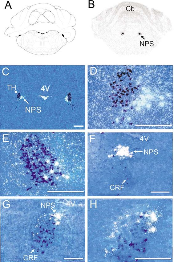
4C–4E, NPS does not colocalize with TH. The majority of cleus, paraventricular thalamic nucleus, and subiculum
NPS-positive cells were observed at midpontine levels, (Figures 6K and 6L). High levels of expression were also
ventromedial to the noradrenergic LC neurons. Few observed in cortical regions. Motor cortex 2 and retro-
NPS-expressing neurons were found intermingled with splenial agranular cortex are distinct areas in cortex that
TH-positive cells at the ventral pole of LC proper. We show strong expression of NPS receptor mRNA (Figures
conclude that the NPS-expressing neurons in the LC 6E, 6H, 6K, 6M, and 6O). Medium levels of expression
area form a cluster of cells that do not produce NA and are also found in dispersed neurons in other cortical
intermingle with LC proper neurons along the medial regions, such as somatosensory cortex (Figure 6N). High
and ventral border of LC, extending just medially into levels of expression was found in multiple nuclei of the
the peri-LC area. hypothalamus (Figures 6H and 6K). Moderate NPSR ex-
Within that area and ventromedial to the LC lies Bar- pression was also found in midbrain. Pons and medulla
rington’s nucleus, the micturition reflex center, which are brain regions that express NPSR mRNA only weakly
is a well-studied ovoid-shaped nucleus located at the (data not shown).
rostral pole of LC. It has been shown that Barrington’s These data suggest that NPS could be involved in a
nucleus is negative for TH and choline acetyltransferase variety of brain functions. Interestingly, NPS receptor
and most of its neurons express CRF (Rizvi et al., 1994; mRNA is not detected in LC area. However, significant
Valentino et al., 2000). Double in situ hybridization with NPSR expression is also found in thalamic midline nu-
NPS and CRF antisense riboprobes revealed that NPS clei, such as central medial thalamic nucleus, interanteri-
does not colocalize with CRF (Figures 4F–4H). At the omedial thalamic nucleus, reuniens and rhomboid thala-
level of highest NPS neuron density, only a few scattered mic nucleus (Figure 6E), which relay extensive inputs
neurons were found expressing CRF that were located from brain stem reticular formation to diffuse cortical
ventrally to the NPS-expressing neurons. At a more ros- fields and are involved in the regulation of arousal and
tral level, densely packed CRF-positive neurons were wakefulness (Van der Werf et al., 2002).
observed as the ovoid-shaped Barrington’s nucleus.
Only a few NPS-expressing neurons were found along NPS Increases Locomotor Activity
the dorsal border of Barrington’s nucleus at this level. and Promotes Wakefulness
We conclude that the NPS-expressing neurons lie cau- In view of the NPSR sites of expression and the promi-
dally to Barrington’s nucleus and at the mid-level of LC. nent expression of the NPS precursor in LC area, we
They extend ventromedially from the LC proper, caudo- hypothesized that NPS may be involved in arousal and
dorsally to Barrington’s nucleus. This unique anatomical anxiety. To start this investigation, we tested the effects
pattern of NPS-expressing neurons defines a previously of NPS on locomotor activity in both naive and habitu-
unrecognized population of cells located in-between the ated mice (Figure 7A). NPS (0.1 nmole or 1 nmole) admin-
noradrenergic LC proper and Barrington’s nucleus. istered intracerebroventricularly (i.c.v.) caused a signifi-
The NPSR mRNA is widely expressed in many brain cant increase in locomotor activity in both naive and
regions. The strongest expression signals were found habituated mice (p ⬍ 0.01) during the 60 min observation
in several discrete nuclei or regions, such as anterior period, while 10 pmoles NPS did not. The total distance
olfactory nucleus (Figures 6B and 6C), dorsal and ventral traveled, percentage of time moving, number of rearing
endopiriform nucleus (Figures 6B, 6C, 6E, 6H, 6I, and events, and center entries were also significantly in-
6K), amygdala (Figures 6H and 6I), precommissural nu- creased in mice injected with 0.1 and 1 nmole NPSNeuron
490
Figure 4. Expression of NPS Precursor
mRNA in the Pontine Area of the Rat Brain
(A) Schematic drawing of the section shown
in (B). The level is at bregma ⫺9.80 mm (Pax-
inos and Watson, 1997, reprinted with per-
mission from Elsevier). (B) Representative
autoradiogram of NPS mRNA expression in
LC area. (C–E) Dark-field images of double
in situ hybridization of NPS precursor mRNA
(white) and TH mRNA (dark blue) in LC area.
(D) Higher magnification of the area indicated
by an arrow in (C). (E) Higher magnification
of a more caudal section. (F–H) Dark-field im-
ages of double in situ hybridization of NPS
precursor mRNA (white) and CRF mRNA (dark
blue) at mid-level of LC area (F) and rostral
LC (G). (H) Higher magnification of the area
indicated by an arrow in (G). TH, tyrosine
hydroxylase; NPS, neuropeptide S; CRF, cor-
ticotropin-releasing factor. Landmarks: Cb,
cerebellum; 4V, fourth ventricle. Scale bar,
500 m in (C), 250 m in all other pictures.
(data not shown). The elevation of locomotor activity group. Interestingly, the increase in wakefulness during
in habituated animals indicates that NPS may produce the first hour post-NPS injection was followed by a re-
behavioral arousal independent of novelty or stress. bound in the amount of non-REM sleep at the second
The effects of NPS on locomotor activity suggest a hour (20% increase versus saline (F2,21 ⫽ 5.44; p ⬍ 0.01))
possible role of NPS in modulating sleep-wake patterns. and fourth hour (48% increase compared to saline-
Rats were implanted with a standard set of electrodes treated animals [F2,21 ⫽ 12.22; p ⬍ 0.01]). Together, these
and electroencephalograms (EEG) and electromyo- data show that NPS can promote arousal and might be
grams (EMG) were recorded after i.c.v. administration of involved in the induction of wakefulness or suppression
NPS. Polygraphic recordings of vigilance states indicate of sleep.
that rats treated with 0.1 nmole and 1.0 nmole of NPS
spent up to 69% and 87%, respectively, of the first hour NPS Attenuates Anxiety-like Behavior
of recording in wakefulness, compared to 45% for saline The expression of NPS receptor in several brain regions
treatment (F2,21 ⫽ 16.80; p ⬍ 0.01) (Figure 7B). In contrast, that are known to be involved in anxiety, such as amyg-
the amount of slow wave sleep stage 1 (SWS1) (F2,21 ⫽ dala, thalamus, and hypothalamic regions, indicates that
9.69; p ⬍ 0.01), stage 2 (SWS2) (F2,21 ⫽ 11.859; p ⬍ 0.01), the NPS system could also play a role in the behavioral
and REM sleep (F2,13 ⫽ 12.29; p ⬍ 0.01) in NPS-treated response to stress (Charney and Deutch, 1996; Red-
rats was significantly reduced compared with saline- mond and Huang, 1979; Sah et al., 2003). Naive mice
treated animals. The increase in wakefulness was due were tested in the open field, a paradigm of free explor-
to a significant increase in the mean duration of the atory behavior in a novel environment. It was found that
episodes (F2,21 ⫽ 7.22; p ⬍ 0.01) compared to the saline NPS significantly increased the number of entries in theNPS Promotes Arousal and Anxiolytic Effects 491 Figure 5. Distribution of NPS Precursor mRNA Expression in Rat Brain (A, D, and G) Drawings of the sections illustrated in (B) and (C) (Bregma ⫺9.68 mm), (E) and (F) (Bregma ⫺2.80 mm), and (H) and (I) (Bregma ⫺3.14 mm), respectively (Paxinos and Watson, 1997). (B, C, E, F, H, and I) Dark-field images of NPS precursor mRNA expression in coronal sections of rat brain. (E and H) Expression of NPS precursor mRNA in boxed regions in (D) and (G), respectively. (C, F, and I) Higher magnification of the area indicated by an arrow in (B), (E), and (H), respectively. Arrows in (F) and (I) indicate single cells showing hybridization signals for NPS precursor mRNA. LPB, lateral parabrachial nucleus; Pr5, principle sensory 5 nucleus; DMH, dorsomedial hypothalamic nucleus; Amg, amygdala. Landmarks: Cb, cerebellum; 3V, third ventricle; opt, optic tract. Scale bar, 500 m. central zone during the first 10 min, which could indicate a higher number of transitions from closed to the open an anxiolytic-like effect (p ⬍ 0.05; Figure 8A). However, arms (p ⬍ 0.05–0.01). The average number of transitions the same doses of NPS also increased ambulations in between the two closed arms of the elevated plus maze the outer zones of the open field, consistent with the (closed-closed transitions) was increased at all doses arousal-promoting effect of the peptide. In order to fur- but did not reach statistical significance. Closed-closed ther study NPS effects on stress as it relates to anxiety, transitions are a measure of general activity in this be- two additional tests were performed that were based on havioral paradigm, so our data indicate that in the ele- the natural aversion of rodents to open or unprotected vated plus maze NPS may not produce significant hyper- spaces: the light-dark box and the elevated plus maze locomotion. Together, the increased number of entries (Figures 8B and 8C). Mice injected with NPS exhibited and prolonged time spent in the unprotected zones of a dose-dependent reduction in anxiety-like behavior in both paradigms (open arm/light area) suggest that cen- both paradigms. tral administration of NPS produces an anxiolytic-like In the light-dark box, mice injected with NPS at a dose effect. However, consistent with the hyperlocomotor ef- range of 0.03–3 nmole, but not at 0.01 nmole, spent a fect of NPS as described above, these NPS doses (⬎0.1 prolonged time in the light area (p ⬍ 0.05–0.01, Figure nmole) also significantly increased the total activity in 8B) and showed a higher percentage of entries in the both tests. light area (data not shown). The latency until the first exit Most anxiolytic drugs increase exploratory activity in from the protected dark compartment was significantly the open field, light-dark box, or elevated plus maze reduced by NPS at doses between 0.3 and 3 nmole. paradigms. However, compounds stimulating locomo- General activity was also enhanced as the number of tion could produce false-positive effects in these tests transitions between the two compartments significantly because the enhanced exploration could be secondary increased at doses between 0.1 and 3 nmole. In the to the increase in general activity. In order to validate elevated plus maze, mice injected with 0.1 and 1 nmole the observed anxiolytic-like effects of NPS, we tested NPS, but not at 0.01 nmole, spent significantly more increasing doses of NPS in the marble-burying para- time on the open arms (p ⬍ 0.05, Figure 8C) and showed digm. Mice tend to bury objects such as glass marbles
Neuron 492 Figure 6. Distribution of NPS Receptor mRNA Expression in Rat Brain (A, D, G, and J) Schematic drawings of the sections shown in (B) and (C) (Bregma, 3.20 mm), (E) and (F) (Bregma ⫺1.80 mm), (H) and (I) (Bregma ⫺2.80 mm), and (K) and (L) (Bregma ⫺4.52 mm), respectively (Paxinos and Watson, 1997). (B, E, H, and K) Autoradiograms of NPSR mRNA expression in coronal rat brain sections. Arrows in (B), (E), (H), and (K) indicate endopiriform nucleus (En). Arrowheads in (E), (H), and (K) refer to secondary motor cortex (M2), retrosplenial agranular cortex (RSA)/M2, and RSA, respectively. (C, F, and I) Dark-field images of boxed regions in (B), (E), and (H), respectively. (L) Dark field image of midline thalamic regions of section (K). (M and N) Dark-field image of cortical regions in section (E). Arrows in (N) indicate scattered cells expressing NPSR mRNA in somatosensory cortex. (O) Dark-field image of cortical and subicular regions in section (K). AON, anterior olfactory nucleus; DEn, dorsal endopiriform nucleus; CM, central medial thalamic nucleus; IAM, interanteromedial thalamic nucleus; Rh, rhomboid thalamic nucleus; Re, reuniens thalamic nucleus; Amg, amygdala; Hyp, hypothalamus; S, subiculum; Prc, precommissural nucleus; PVP, paraventricular thalamus nucleus, posterior; PH, posterior hypothalamus. Landmarks: aca, anterior commissure, anterior part; pt, paratenial thalamic nuclei; opt, optic tract; D3V, dorsal third ventricle; 3V, third ventricle; Hip, hippocampus. Scale bar, 500 m. present in their environment. Anxiolytic drugs such as with anxiolytic-like activity (Njung’e and Handley, 1991). benzodiazepines reduce the number of marbles buried As shown in Figure 8D, mice injected with saline covered over a fixed period of time. It has been suggested that about 50% of the marbles during the 30 min observation the inhibition of marble-burying behavior is correlated period (total of 18 marbles per cage). NPS dose-depen-
NPS Promotes Arousal and Anxiolytic Effects
493
Figure 7. Central Administration of NPS Produces Behavioral Arousal and Wakefulness
(A) Hyperlocomotion effects of NPS in naive and habituated mice. Naive mice were new to the test chamber, while habituated animals were
acclimatized for 1 hr prior to the injection. In naive mice, 0.1 and 1 nmole NPS induce significant hyperlocomotion (F3,324 ⫽ 92.83, p ⬍ 0.0001,
two-way ANOVA for repeated measures). The same doses of NPS also produced significant effects in habituated animals (F3,336 ⫽ 135.59,
p ⬍ 0.0001).
(B) Arousal promoting effects of NPS in rats. NPS increases the amount of wakefulness and decreases SWS1, SWS2, and REM sleep in rats
(n ⫽ 8 for each dose). **p ⬍ 0.01, 0.1 nmole and 1.0 nmole compared with saline; *p ⬍ 0.01, 1.0 nmole compared with saline (ANOVA followed
by Scheffe’s post hoc test).
dently reduced the number of marbles buried. NPS- neurons, since orexin 1 receptors are found to be colo-
injected mice were actively exploring the marbles and calized with TH in LC neurons and electrophysiological
eventually engaged in burying them, however, at signifi- recordings from LC neurons show excitatory effects of
cantly lower numbers as compared to mice injected with exogenously applied Hcrt/Ox (Bourgin et al., 2000).
saline (p ⬍ 0.05–0.01). In summary, the combined results However, the arousal-promoting effect of NPS is unlikely
of all four paradigms measuring anxiety-like behavior mediated by direct activation of noradrenergic systems,
suggest that NPS might produce anxiolytic-like effects since our anatomical data show that NPS-expressing
in the presence of increased arousal. neurons do not produce NA and no NPSR mRNA was
detected in LC neurons. However, we cannot rule out
an indirect activation of noradrenergic systems. Electro-
Discussion
physiological recording will be necessary to confirm a
possible link between NPS and monoaminergic trans-
We provide evidence that the neuropeptide NPS is a mitter systems that have been implicated in the neuro-
novel modulator of arousal and possibly anxiety-related chemistry of wakefulness and arousal.
behavior. The effects of NPS on inducing wakefulness One unexpected outcome of this study is the discov-
are rapid (during the first hour after injection) and potent, ery of a cluster of NPS-expressing neurons that do not
since low doses of NPS are sufficient to reduce all sleep produce NA or CRF and are localized in close proximity
stages, such as REM, SWS1, and SWS2, suggesting a to the LC proper and Barrington’s nucleus. The cluster
profound change in sleep architecture. Recently, the of NPS-expressing neurons is likely to be a previously
neuropeptide hypocretin 1/orexin A (Hcrt/Ox) has also uncharacterized population of cells in the peri-LC area.
been demonstrated to induce arousal, and genetic anal- Noteworthy, it has been reported before that a large
ysis has provided compelling evidence that the absence number of uncharacterized neurons are found in the
of Hcrt/Ox or its receptor(s) produces narcolepsy in peri-LC area (Aston-Jones et al., 1991b; Rizvi et al.,
mice, dogs, and humans (Sutcliffe and de Lecea, 2002). 1994), and our present data suggest that the NPS neu-
Single i.c.v. injection of Hcrt/Ox produces arousal lasting ronal cluster could be a subset of these neurons.
for 2–3 hr (Bourgin et al., 2000; Hagan et al., 1999), It is well documented that the noradrenergic LC is
whereas comparable NPS administrations show a more involved in the regulation of an aroused state of wake-
short-term effect within the first hour postinjection. Both fulness (Berridge and Waterhouse, 2003). On the other
peptides appear to increase wakefulness while sup- hand, several studies found no major disruption of EEG
pressing REM sleep and deep sleep (SWS stage 2) activity after selective cytotoxic lesions of TH-positive
(Bourgin et al., 2000), although one study could not de- LC neurons or genetic ablation of the noradrenaline-
tect a significant effect of Hcrt/Ox on deep sleep dura- synthesizing enzyme dopamine -hydroxylase (Cirelli et
tion (Hagan et al., 1999). Hctr/Ox appears to exert its al., 1996; Hunsley and Palmiter, 2003), underscoring the
effects partially by directly activating noradrenergic LC fact that arousal is modulated by multiple neuronal sys-Neuron 494 Figure 8. Anxiolytic-like Effects of NPS in Mice NPS produces dose-dependent anxiolytic-like effects in C57Bl/6 mice exposed to the open field (A), light-dark box (B), elevated plus maze (C), and marble burying paradigm (D). Doses and groups: all doses are in nmole per animal; open field (n ⫽ 8 for each dose); light-dark box (PBS, n ⫽ 10; 0.01 nmole, n ⫽ 5; 0.03 nmole, n ⫽ 5; 0.1 nmole, n ⫽ 5; 0.3 nmole, n ⫽ 11; 1 nmole, n ⫽ 5; 3 nmole, n ⫽ 8); elevated plus maze (n ⫽ 5 for all doses); marble burying (PBS and 0.01 nmole, n ⫽ 10; 0.1 and 1 nmole, n ⫽ 9). **p ⬍ 0.01, *p ⬍ 0.05 compared to PBS control, ANOVA followed by Dunnett’s test for multiple comparisons. All data are presented as means ⫾ SEM. tems. Our present data provide evidence that NPS could Central administration of NPS produces anxiolytic- be a previously uncharacterized arousal-modulating like effects but also increases locomotor activity at simi- transmitter system. Interestingly, the close vicinity of lar doses. In the open field, elevated plus maze, and NPS-producing neurons and the noradrenergic neuronal light-dark box paradigms, increases in exploration are cluster in LC indicate that this brainstem area might generally interpreted as an anxiolytic effect, but the in- contain two independent transmitter systems that regu- terpretation might be confounded by hyperlocomotion. late vigilance states. Factor analysis, however, has shown that the behavioral
NPS Promotes Arousal and Anxiolytic Effects
495
parameters monitored in these tests can be divided into GATTAGCTCAGTAAAACTCAA-3⬘ and 5⬘-GCAGAATTCGTCATGAT
two components—an activity component (total distance TTTGCTCTTTGAAAGG-3⬘. The cloned DNAs were sequenced on
both strands.
traveled, number of transitions) and an anxiety compo-
nent (number of entries in unprotected zone, time spent
Cell Transfection and Intracellular Ca2ⴙ Measurement
in unprotected zone) (Rodgers and Johnson, 1995)—and
HEK293 T cells and CHO dhfr(⫺) cells were transfected with the
that these two components show poor correlation. For human NPS receptor cDNA cloned into pcDNA 3.1(⫹) using Lipofec-
example, both the psychostimulants amphetamine and tAmine. Stable clones were selected with 800 mg/l G418 and tested
cocaine produce hyperlocomotion yet increase anxiety- for mobilization of intracellular Ca2⫹ with 100 nM NPS (generous gift
like behavior, i.e., they are anxiogenic (Hascoet and of Phoenix Pharmaceuticals, Belmont, CA). Changes in intracellular
Bourin, 1998; Paine et al., 2002). On the other hand, the Ca2⫹ were measured in a fluorometric imaging plate reader system
(FLIPR, Molecular Devices) as described before (Saito et al., 1999).
wake-promoting neuropeptide Hcrt/Ox enhances arousal
Dose response curves for agonist activation were calculated from
and hyperlocomotion and suppresses REM sleep but has peak fluorescence values of triplicate incubations, and EC50 values
no effect on anxiety-like behavior in rodents (Hagan et were calculated with Prism software (GraphPad, San Diego, CA).
al., 1999). Moreover, typical anxiolytic drugs, such as
benzodiazepines, have either no effect or reduce loco- Radioligand Binding Assay
motor activity, depending on the doses used (Chaouloff Y10-NPS was labeled with 125I using the chloramine T method and
et al., 1997). Therefore, although we acknowledge the purified by reversed-phase HPLC in a collaboration with NEN Perkin
possible confounding effects of hyperlocomotion, we Elmer (Boston, MA). CHO cells stably expressing human NPSR were
seeded into 24-well plates and cultured for 48 hr. For saturation
suggest that the increased exploratory activity observed
binding experiment, [125I] Y10-NPS at concentrations from 4 pM to
in mice after NPS administration may indicate an anxio- 1.7 nM were used. For displacement binding, increasing concentra-
lytic-like profile in these three paradigms. To further tions of unlabeled human NPS (1 pM to 3 M) were used to compete
investigate the possible anxiolytic-like effects of NPS, with 0.15 nM [125I] Y10-NPS. Nonspecific binding was determined in
we used the marble burying test as an alternative behav- the presence of 1 M unlabeled human NPS. The binding assay
ioral paradigm. In this test, the selective suppression of was carried out as described (Sakurai et al., 1998). In brief, cells
were washed with PBS first and then incubated with radioligand
marble burying behavior is suggested to correlate with
with or without unlabeled NPS peptide in DMEM medium containing
anxiolytic activity, in contrast to the other three para- 0.1% bovine serum albumin at 20⬚C for 1.5 hr. Cells were washed
digms where increases of natural behaviors are an index five times with cold PBS and lysed with 1 N NaOH. Bound radioactiv-
of anxiolytic-like effects. Numerous drugs clinically ef- ity was counted in a MicroBeta liquid scintillation counter (EG&G
fective in the treatment of anxiety disorders, such as Wallac, Gaithersburg, MD) and corrected for counting efficiency.
benzodiazepines or selective serotonin reuptake inhibi- Data from triplicate incubations were analyzed using PRISM.
tors, reduce marble burying behavior in rodents (Borsini
et al., 2002). Our data demonstrate that NPS also inhibits Quantitative Real-Time PCR
Tissue was collected from male and female adult Sprague-Dawley
this natural behavior at doses which increase locomo-
rats, and RNA was extracted with Trizol. PolyA⫹ RNA was prepared
tion. Altogether, central administration of NPS reduces using OligoTex (Qiagen) and converted to cDNA using oligo dT and
behavioral signs of anxiety in four different anxiety tests. random primers with Superscript reverse transcriptase (Invitrogen).
These findings indicate that NPS could be involved in Primers for NPSR (5⬘-TGCAGGGAGCAAAGATCACA-3⬘ and 5⬘-AAT
modulating anxiety responses. CTGCATCTCATGCCTCTCA-3⬘), NPS precursor (5⬘-TGTCGCTGTCC
In conclusion, we have characterized the neuropep- ACAATGCAT-3⬘ and 5⬘-AATCAGATTTTCCAGACACCTTAGAAG-3⬘),
and -actin (5⬘-CACGGCATCGTCACCAACT-3⬘ and 5⬘-AGCCACAC
tide NPS. Central administration of NPS produces a
GCAGCTCATTG-3⬘) were predicted using ABI Prism Primer Select
unique behavioral profile by increasing locomotor ac- software and tested for linearity of amplification using cloned cDNAs
tivity and wakefulness in rodents. NPS could also exert as template. Quantitative real-time PCR was performed in an ABI
anxiolytic-like effects. In addition, we identify a pre- Prism 7000 using SYBR Green PCR Master Mix (Applied Bio-
viously undescribed group of neurons adjacent to the systems).
noradrenergic LC that express NPS. The discovery of
this previously uncharacterized transmitter system that In Situ Hybridization
modulates sleep-wake cycles and anxiety might help to A 326 bp fragment of the rat NPS receptor (corresponding to nt
further our understanding of sleep disorders, such as 408–734) was amplified by PCR and subcloned into pBluescript SK.
A fragment of the rat NPS precursor (corresponding to nt 92–276)
insomnia, and pathological states of anxiety. It should was cloned into the same vector. Sense and antisense riboprobes
be noted that excessive anxiety and disruption of sleep were labeled with 35S-UTP. Rat tyrosine hydroxylase (TH) cDNA was
patterns are often observed in patients suffering from a gift from Dr. Francis Leslie (UCI) and cloned in pBluescript. Rat
depression (Ohayon et al., 1998). corticotropin-releasing factor (CRF) cDNA was a gift from Dr. Chris-
tine Gall (UCI) and cloned in the same vector. For double in situ
Experimental Procedures hybridization, antisense TH riboprobes or CRF riboprobes were la-
beled with digoxigenin using DIG RNA labeling kit (Roche Applied
Molecular Cloning of Human NPS Receptor Science). In situ and double in situ hybridization to 20 m coronal
and Rat NPS Precursor sections of adult Sprague-Dawley rat brains was carried out as
Human NPS receptor was cloned into pcDNA3.1(⫹) from human described before (Clark et al., 2001).
brain cDNA (Clontech, Carlsbad, CA) using nested PCR. Primers
were 5⬘-AGGAGCAAGGACAGTGAGGCTCAA-3⬘ and 5⬘-TGCCCAA Behavioral Studies
GCAGGTGACAAGGACCT-3⬘ for first round amplification and 5⬘- Male C57Bl/6 mice (National Cancer Institute, Bethesda, MD), age
ATACTCGAGCCATGCCAGCCAACTTCACAGAGGGCA-3⬘ and 5⬘-GCT 10–14 weeks, were group housed (four animals per cage) under
TCTAGAGCTCAGCCTAGCACTGGCACTGCCCTA-3⬘ for the second controlled conditions (temperature 21⬚C ⫾ 2⬚C; relative humidity
round. Rat NPS precursor cDNA was cloned into pBluescript from 50%–60%; 12 hr light-dark cycle, lights on 6:00 AM) with free access
a rat total brain cDNA library (Clontech). First round amplification to food and water. Prior to drug injections, mice were briefly anesthe-
primers were 5⬘-CAGATTTTGGGAAGTCCA-3⬘ and 5⬘-AGATTAATT tized with halothane. NPS was dissolved in phosphate-buffered sa-
CCCCGAGTC-3⬘; second round primers were 5⬘-GTTTCTAGAAAT line (PBS, pH 7.4) and injected i.c.v. (total volume: 2 l) as describedNeuron
496
before (Laursen and Belknap, 1986). Mice were allowed to recover Chojnacka-Wojcik, E., Klodzinska, A., and Pilc, A. (2001). Glutamate
for 5 min and then placed in the observation apparatus. receptor ligands as anxiolytics. Curr. Opin. Investig. Drugs 2, 1112–
For sleep studies, adult male Sprague-Dawley rats (250-300 g) 1119.
were implanted under halothane anesthesia (1%–2%) with a stain- Cirelli, C., Pompeiano, M., and Tononi, G. (1996). Neuronal gene
less steel cannula for i.c.v. administration and a standard set of expression in the waking state: a role for the locus coeruleus. Sci-
stainless steel screw electrodes for chronic sleep recordings as ence 274, 1211–1215.
reported previously (Bourgin et al., 2000). Rats were injected with
Civelli, O., Nothacker, H.P., Saito, Y., Wang, Z., Lin, S.H., and
NPS or vehicle (5 l) at the beginning of the light cycle, and cortical
Reinscheid, R.K. (2001). Novel neurotransmitters as natural ligands
activity was recorded over 6 hr. All animal experiments had been
of orphan G-protein-coupled receptors. Trends Neurosci. 24,
approved by the local IACUC committee and were done in accor-
230–237.
dance with federal regulations and guidelines on animal experimen-
tation. Clark, S.D., Nothacker, H.P., Wang, Z., Saito, Y., Leslie, F.M., and
Locomotion was monitored in rectangular plexiglass boxes (60 ⫻ Civelli, O. (2001). The urotensin II receptor is expressed in the cholin-
40 ⫻ 50 cm). Horizontal activity was measured over 60 min by 18 ⫻ ergic mesopontine tegmentum of the rat. Brain Res. 923, 120–127.
12 infrared sensors placed 2 cm above the floor. A second row of Foote, S.L., Aston-Jones, G., and Bloom, F.E. (1980). Impulse activity
sensors at 8 cm above the floor was used to record rearing events. of locus coeruleus neurons in awake rats and monkeys is a function
The imaginary central zone was defined as a 30 ⫻ 20 cm rectangle of sensory stimulation and arousal. Proc. Natl. Acad. Sci. USA 77,
in the middle of each observation area. Data were collected using 3033–3037.
MatLab (Mathworks, Natick, MA). Experimental procedures for open Gottesmann, C. (2002). GABA mechanisms and sleep. Neuroscience
field, elevated plus maze, and light-dark box were described pre- 111, 231–239.
viously (Köster et al., 1999). Marble burying was measured in mice
Hagan, J.J., Leslie, R.A., Patel, S., Evans, M.L., Wattam, T.A.,
placed individually in polypropylene cages (30 ⫻ 18 ⫻ 12 cm) con-
Holmes, S., Benham, C.D., Taylor, S.G., Routledge, C., Hemmati,
taining 18 glass marbles (1.5 cm diameter) evenly spaced on 5 cm
P., et al. (1999). Orexin A activates locus coeruleus cell firing and
deep rodent bedding (bed-o’cob, The Andersons Inc., Maumee, OH)
increases arousal in the rat. Proc. Natl. Acad. Sci. USA 96, 10911–
(Njung’e and Handley, 1991). No food or water was present during
10916.
the observation period. Cages were covered with a metal grid, and
the number of marbles covered at least two-thirds was counted Hascoet, M., and Bourin, M. (1998). A new approach to the light/dark
after 30 min. test procedure in mice. Pharmacol. Biochem. Behav. 60, 645–653.
Hobson, J.A., McCarley, R.W., and Wyzinski, P.W. (1975). Sleep
Acknowledgments cycle oscillation: reciprocal discharge by two brainstem neuronal
groups. Science 189, 55–58.
This work was supported in part by grants from NIH (R.K.R., O.C.) Holmes, A., Kinney, J.W., Wrenn, C.C., Li, Q., Yang, R.J., Ma, L.,
and the Stanley Medical Research Institute (O.C.). We thank Hans- Vishwanath, J., Saavedra, M.C., Innerfield, C.E., Jacoby, A.S., et al.
Peter Nothacker for helpful discussions; Christine Gall for critical (2003). Galanin GAL-R1 receptor null mutant mice display increased
review of in situ hybridization results; Gary Lynch for help with anxiety-like behavior specific to the elevated plus-maze. Neuropsy-
locomotor experiments; and Valerie Jackson, Hua Zeng, and Alanna chopharmacology 28, 1031–1044.
Pei Sun for technical assistance.
Hunsley, M.S., and Palmiter, R.D. (2003). Norepinephrine-deficient
Received: March 16, 2004 mice exhibit normal sleep-wake states but have shorter sleep la-
Revised: July 6, 2004 tency after mild stress and low doses of amphetamine. Sleep 26,
Accepted: July 30, 2004 521–526.
Published: August 18, 2004 Jones, B.E. (1991). The role of noradrenergic locus coeruleus neu-
rons and neighboring cholinergic neurons of the pontomesencepha-
References lic tegmentum in sleep-wake states. Prog. Brain Res. 88, 533–543.
Jones, B.E. (2003). Arousal systems. Front. Biosci. 8, s438–s451.
Aston-Jones, G., Chiang, C., and Alexinsky, T. (1991a). Discharge
Köster, A., Montkowski, A., Schulz, S., Stübe, E.M., Knaudt, K.,
of noradrenergic locus coeruleus neurons in behaving rats and mon-
Jenck, F., Moreau, J.L., Nothacker, H.P., Civelli, O., and Reinscheid,
keys suggests a role in vigilance. Prog. Brain Res. 88, 501–520.
R.K. (1999). Targeted disruption of the orphanin FQ/nociceptin gene
Aston-Jones, G., Shipley, M.T., Chouvet, G., Ennis, M., van Bocks- increases stress susceptibility and impairs stress adaptation in
taele, E., Pieribone, V., Shiekhattar, R., Akaoka, H., Drolet, G., Astier, mice. Proc. Natl. Acad. Sci. USA 96, 10444–10449.
B., et al. (1991b). Afferent regulation of locus coeruleus neurons:
Laitinen, T., Polvi, A., Rydman, P., Vendelin, J., Pulkkinen, V., Salmi-
anatomy, physiology and pharmacology. Prog. Brain Res. 88, 47–75.
kangas, P., Makela, S., Rehn, M., Pirskanen, A., Rautanen, A., et
Berridge, C.W., and Waterhouse, B.D. (2003). The locus coeruleus- al. (2004). Characterization of a common susceptibility locus for
noradrenergic system: modulation of behavioral state and state- asthma-related traits. Science 304, 300–304.
dependent cognitive processes. Brain Res. Brain Res. Rev. 42,
Laursen, S.E., and Belknap, J.K. (1986). Intracerebroventricular in-
33–84.
jections in mice. Some methodological refinements. J. Pharmacol.
Bing, O., Moller, C., Engel, J.A., Soderpalm, B., and Heilig, M. (1993). Methods 16, 355–357.
Anxiolytic-like action of centrally administered galanin. Neurosci.
Millan, M.J. (2003). The neurobiology and control of anxious states.
Lett. 164, 17–20.
Prog. Neurobiol. 70, 83–244.
Borsini, F., Podhorna, J., and Marazziti, D. (2002). Do animal models
Njung’e, K., and Handley, S.L. (1991). Evaluation of marble-burying
of anxiety predict anxiolytic-like effects of antidepressants? Psy-
behavior as a model of anxiety. Pharmacol. Biochem. Behav. 38,
chopharmacology (Berl.) 163, 121–141.
63–67.
Bourgin, P., Huitron-Resendiz, S., Spier, A.D., Fabre, V., Morte, B.,
Criado, J.R., Sutcliffe, J.G., Henriksen, S.J., and de Lecea, L. (2000). Ohayon, M.M., Caulet, M., and Lemoine, P. (1998). Comorbidity of
Hypocretin-1 modulates rapid eye movement sleep through activa- mental and insomnia disorders in the general population. Compr.
tion of locus coeruleus neurons. J. Neurosci. 20, 7760–7765. Psychiatry 39, 185–197.
Chaouloff, F., Durand, M., and Mormede, P. (1997). Anxiety- and Paine, T.A., Jackman, S.L., and Olmstead, M.C. (2002). Cocaine-
activity-related effects of diazepam and chlordiazepoxide in the rat induced anxiety: alleviation by diazepam, but not buspirone, dimen-
light/dark and dark/light tests. Behav. Brain Res. 85, 27–35. hydrinate or diphenhydramine. Behav. Pharmacol. 13, 511–523.
Charney, D.S., and Deutch, A. (1996). A functional neuroanatomy of Paxinos, G., and Watson, C. (1997). The Rat Brain in Stereotaxic
anxiety and fear: implications for the pathophysiology and treatment Coordinates, Compact Third Edition (San Diego: Academic Press).
of anxiety disorders. Crit. Rev. Neurobiol. 10, 419–446. Redmond, D.E., Jr., and Huang, Y.H. (1979). Current concepts. II.NPS Promotes Arousal and Anxiolytic Effects 497 New evidence for a locus coeruleus-norepinephrine connection with Note added in proof anxiety. Life Sci. 25, 2149–2162. Reinscheid, R.K., and Civelli, O. (2002). The orphanin FQ/nociceptin During the preparation of this manuscript a mutant form of the NPS knockout mouse: a behavioral model for stress responses. Neuro- receptor was identified as a candidate gene for asthma and termed peptides 36, 72–76. GPRA (G protein-coupled receptor for asthma susceptibility) (Laiti- Rizvi, T.A., Ennis, M., Aston-Jones, G., Jiang, M., Liu, W.L., Behbe- nen et al., 2004). hani, M.M., and Shipley, M.T. (1994). Preoptic projections to Bar- rington’s nucleus and the pericoerulear region: architecture and terminal organization. J. Comp. Neurol. 347, 1–24. Rodgers, R.J., and Johnson, N.J. (1995). Factor analysis of spatio- temporal and ethological measures in the murine elevated plus- maze test of anxiety. Pharmacol. Biochem. Behav. 52, 297–303. Sah, P., Faber, E.S., Lopez De Armentia, M., and Power, J. (2003). The amygdaloid complex: anatomy and physiology. Physiol. Rev. 83, 803–834. Saito, Y., Nothacker, H.P., Wang, Z., Lin, S.H., Leslie, F., and Civelli, O. (1999). Molecular characterization of the melanin-concentrating- hormone receptor. Nature 400, 265–269. Sakurai, T., Amemiya, A., Ishii, M., Matsuzaki, I., Chemelli, R.M., Tanaka, H., Williams, S.C., Richarson, J.A., Kozlowski, G.P., Wilson, S., et al. (1998). Orexins and orexin receptors: a family of hypothala- mic neuropeptides and G protein-coupled receptors that regulate feeding behavior. Cell 92, 573–585. Saper, C.B., Chou, T.C., and Scammell, T.E. (2001). The sleep switch: hypothalamic control of sleep and wakefulness. Trends Neurosci. 24, 726–731. Sato, S., Shintani, Y., Miyajima, N., and Yoshimura, K. (2002). Novel G protein-coupled receptor protein and DNA thereof. World Patent Application. WO 02/31145 A1. Shimomura, Y., Harada, M., Goto, M., Sugo, T., Matsumoto, Y., Abe, M., Watanabe, T., Asami, T., Kitada, C., Mori, M., et al. (2002). Identification of neuropeptide W as the endogenous ligand for or- phan G-protein-coupled receptors GPR7 and GPR8. J. Biol. Chem. 277, 35826–35832. Silva, A.P., Cavadas, C., and Grouzmann, E. (2002). Neuropeptide Y and its receptors as potential therapeutic drug targets. Clin. Chim. Acta 326, 3–25. Sutcliffe, J.G., and de Lecea, L. (2002). The hypocretins: setting the arousal threshold. Nat. Rev. Neurosci. 3, 339–349. Sutin, E.L., and Jacobowitz, D.M. (1988). Immunocytochemical lo- calization of peptides and other neurochemicals in the rat laterodor- sal tegmental nucleus and adjacent area. J. Comp. Neurol. 270, 243–270. Swanson, L.W., and Hartman, B.K. (1975). The central adrenergic system. An immunofluorescence study of the location of cell bodies and their efferent connections in the rat utilizing dopamine-beta- hydroxylase as a marker. J. Comp. Neurol. 163, 467–505. Swanson, L.W., Sawchenko, P.E., Rivier, J., and Vale, W.W. (1983). Organization of ovine corticotropin-releasing factor immunoreactive cells and fibers in the rat brain: an immunohistochemical study. Neuroendocrinology 36, 165–186. Ursin, R. (2002). Serotonin and sleep. Sleep Med. Rev. 6, 55–69. Valentino, R.J., Pavcovich, L.A., and Hirata, H. (1995). Evidence for corticotropin-releasing hormone projections from Barrington’s nu- cleus to the periaqueductal gray and dorsal motor nucleus of the vagus in the rat. J. Comp. Neurol. 363, 402–422. Valentino, R.J., Kosboth, M., Colflesh, M., and Miselis, R.R. (2000). Transneuronal labeling from the rat distal colon: anatomic evidence for regulation of distal colon function by a pontine corticotropin- releasing factor system. J. Comp. Neurol. 417, 399–414. Van der Werf, Y.D., Witter, M.P., and Groenewegen, H.J. (2002). The intralaminar and midline nuclei of the thalamus. Anatomical and functional evidence for participation in processes of arousal and awareness. Brain Res. Brain Res. Rev. 39, 107–140. Wilson, S., Bergsma, D.J., Chambers, J.K., Muir, A.I., Fantom, K.G., Ellis, C., Murdock, P.R., Herrity, N.C., and Stadel, J.M. (1998). Orphan G-protein-coupled receptors: the next generation of drug targets? Br. J. Pharmacol. 125, 1387–1392.
You can also read