Experimental chronic sleep fragmentation alters seizure susceptibility and brain levels of interleukins 1β and 6
←
→
Page content transcription
If your browser does not render page correctly, please read the page content below
RESEARCH PAPER
                                               Acta Neurobiol Exp 2021, 81: 96–109
                                               DOI: 10.21307/ane‑2021‑010

                                                                Experimental chronic sleep fragmentation
                                                               alters seizure susceptibility and brain levels
                                                                          of interleukins 1β and 6
                                                         Željko Grubač1, Nikola Šutulović1, Djudja Jerotić2, Sonja Šuvakov2, Aleksandra Rašić‑Marković1,
                                                                    Djuro Macut3, Tatjana Simić2, Olivera Stanojlović1 and Dragan Hrnčić1*

                                                1
                                                    Laboratory of Neurophysiology, Institute of Medical Physiology “Richard Burian”, Belgrade University Faculty of Medicine, Belgrade, Serbia,
                                                                     2
                                                                       Institute of Clinical and Medical Biochemistry, Belgrade University Faculty of Medicine, Belgrade, Serbia,
                                                           3
                                                             Clinic of Endocrinology, Diabetes and Metabolic Disease, CCS, Belgrade University Faculty of Medicine, Belgrade, Serbia,
                                                                                                         * Email: dragan.hrncic@mfub.bg.ac.rs

                                                     Brain hyperexcitability in sleep apnea is believed to be provoked by hypoxemia, but sleep fragmentation can also play a significant
                                                     role. Sleep fragmentation can trigger inflammatory mechanisms. The aim of this research was to investigate the effects of chronic
                                                     sleep fragmentation on seizure susceptibility and brain cytokine profile. Chronic sleep fragmentation in male rats with implanted EEG
                                                     electrodes was achieved by the treadmill method. Rats were randomized to: treadmill control (TC); activity control (AC) and sleep
                                                     fragmentation (SF) group. Convulsive behavior was assessed 14 days later by seizure incidence, latency time and seizure severity
                                                     during 30 min following lindane administration. The number and duration of EEG ictal periods were determined. Levels of IL‑1β and
                                                     IL‑6 were measured in the animals’ serum and brain structures (hippocampus, thalamus and cerebral cortex), in separate rat cohort
                                                     that underwent the same fragmentation protocol except lindane administration. Incidence and severity of seizures were significantly
                                                     increased, while latency was significantly decreased in SF+L compared with TC+L group. Seizure latency was also significantly decreased
                                                     in SF+L compared to AC+L group. The number and duration of ictal periods were increased in the SF+L compared to the AC+L group.
                                                     IL‑1β was significantly increased in the thalamus, cortex and hippocampus in the SF compared to the AC and TC groups. IL‑6 was
                                                     statistically higher only in the cortex of SF animals, while in the thalamic or hippocampal tissue, no difference was observed between
                                                     the groups. It could be concluded that fourteen‑day sleep fragmentation increases seizure susceptibility in rats and modulates brain
                                                     production of IL‑1β and IL‑6.

                                                     Key words: chronic sleep fragmentation, brain hyperexcitability, cytokines, neuroinflammation, sleep apnea

                                                                       INTRODUCTION                                        and social jetlag caused by modern lifestyles are ac‑
© 2021 by Acta Neurobiologiae Experimentalis

                                                                                                                           companied by quantitative and qualitative sleep alter‑
                                                   Sleep fragmentation is defined as a series of brief                     ations. An altered sleep pattern has been already rec‑
                                               arousals repeated consecutively throughout the night                        ognized as a cause of behavioral changes, but recent
                                               (Bonnet and Arand, 2003). This type of fractured sleep                      evidences suggest a bidirectional relationship between
                                               causes excessive daytime sleepiness, impaired cogni‑                        the psychiatric illnesses, disrupted circadian rhythm
                                               tion and numerous neurobehavioral deficits (Bonnet                          and disrupted sleep (Wulff et al., 2010). Since total
                                               and Arand, 2003; Vijayan, 2012). Clinical and transla‑                      sleeping time in fragmented sleep is insignificantly
                                               tional studies suggested certain lifestyle factors may                      changed, most scientists tend to attribute these conse‑
                                               be important modulators of epilepsy, elucidating those                      quences to periodic interruptions and stress (Devinsky
                                               related to sleep as particularly important. Shift work                      et al., 1994).

                                               Received 21 July 2020, accepted 9 February 2021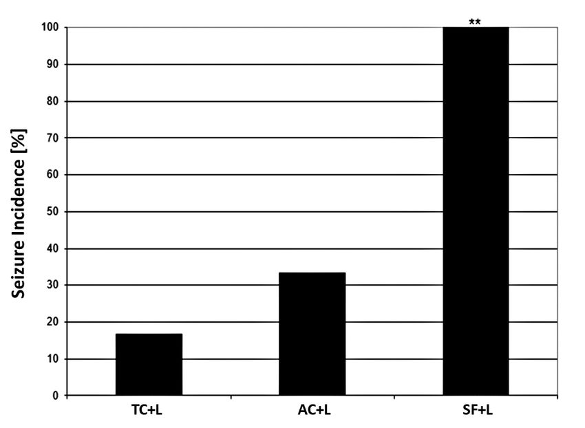
Acta Neurobiol Exp 2021, 81:
                         81 96–109                              Sleep fragmentation related seizures and neuroinflammation   97

    Patients with wide variety of diseases, particular‑                                  METHODS
ly those with obstructive sleep apnea (OSA) often suf‑
fer from fragmented sleep (Day et al., 1999). Treating            Animals and housing
sleep apnea can be crucial to epilepsy prevention and
treatment (Malow et al., 2000; Chihorek et al., 2007).                Two months old male Wistar albino rats were used.
It is believed that the main cause of hyperexcitability           The animals were obtained from the Military Medical
in patients with OSA is hypoxemia (Manni and Tartara,             Academy breeding laboratory (Belgrade, Serbia). Rats
2000; Seyal et al., 2010), but many researchers in the            were housed in separate transparent plastic cages
field are of the opinion that sleep fragmentation, as an          (55 cm × 35 cm × 15 cm), they were given 25‑30 g of
accompanying phenomenon, should also be considered                food (Purina rat chow) per day and had free access to
as a powerful contributing factor (Ferrillo et al., 2000;         water (ad libitum access) continuously. Ambient condi‑
Parrino et al., 2000; Manni et al., 2005; Seyal et al., 2010;     tions were set up to an optimal breeding conditions,
Foldvary‑Schaefer et al., 2012).                                  i.e., 22‑24°C, 50±5% relative humidity, 12:12 h light/
    Numerous uncertainties exist on mechanisms of in‑             dark cycle with light switched on from 8:00 h to 20:00 h.
creased neuronal excitability upon sleep alterations.             One week was the period of adaptation to these envi‑
It is known that sleep fragmentation can trigger                  ronmental conditions upon rat arrival in the laborato‑
low‑grade chronic inflammatory mechanisms (Mulling‑               ry. All animals were not used more than once in the
ton et al., 2009; Tang et al., 2009). Moreover, systemic or       experimental procedures.
central nervous system inflammation exerts the mod‑                   All experimental procedures were approved by the
ulation of synaptic plasticity via neuroinflammatory              Ethical Committee of the Faculty of Medicine, Univer‑
mediators, thus influencing brain neuronal networks               sity of Belgrade and were in full compliance with the
functioning (Di Filippo et al., 2013; Hrncic et al., 2018;        European Council Directive (2010/63/EU).
Šutulović et al., 2019). An increased production of dif‑
ferent cytokines, especially IL‑1β, TNF‑α and IL‑6 in ac‑
tivated astrocytes and microglia during epileptogene‑             Implantation of intracranial electrodes
sis, has been already proven in several studies (Ravizza          for electroencephalographic (EEG) recordings:
et al., 2008; Vezzani and Friedman, 2011). Indeed, these          EEG registration and analysis
cytokines play various roles through multiple path‑
ways and mediate complex connections between the                      Three gold‑plated screws were implanted into the
immune system and epilepsy (Li et al., 2011).                     skull of the animals for EEG recordings. Prior to the
    Since epilepsy nowadays presents a major society              procedure rats were anaesthetized with intraperitone‑
burden, with the prevalence of 0.5–1.0% (Hauser et al.,           al injection (i.p.) of sodium pentobarbital (50 mg/kg),
1991) and 30% of affected individuals having continu‑             and positioned in a stereotaxic apparatus afterwards.
ous seizures despite antiepileptic drugs (AEDs) (Sand‑            Recording electrodes were implanted over frontal
er, 1993). The reduction of factors which may promote             (2 mm rostrally to bregma and 2 mm from the median
seizures, such as sleep fragmentation (Rajna and Veres,           line), parietal (2 mm rostrally to lambda and 2 mm lat‑
1993; Frucht et al., 2000), could lead to new and more            erally to median line) and occipital (2 mm caudally to
effective treatment strategies. In this study we used             lambda) cortices.
the lindane model of experimentally induced general‑                  Upon 7‑day recovery period, rats had 24 h habitua‑
ized seizures, as described earlier (Vučević et al., 2008;        tion to the EEG recording settings with a full cable set‑
Hrnčić et al., 2016), because lindane induced seizures            up attached.
are refractory to numerous classical antiepileptic drugs              A classical 8‑channel EEG apparatus (RIZ, Zagreb,
and therefore believed to be suitable for exploration of          Croatia), with a modified output degree enabling the
refractory epilepsy (Vučević et al., 2008).                       transfer of output signals to the input circuit of 12‑byte
    The objective of this study was to investigate the            AD card PCL‑711B (Advantech Co. Ltd.) and sampling
effects of chronic sleep fragmentation on seizure sus‑            frequency of 512 Hz/channel installed into a comput‑
ceptibility using an experimental model of lindane‑in‑            er, was used. The signals were digitized using a SCB‑68
duced seizures and a high frequency sleep disruption              data acquisition card (National Instruments Co, Austin,
model of sleep fragmentation characteristic for ob‑               Texas, USA). Data acquisition and signal processing
structive sleep apnea. We also wanted to examine the              were performed with the Lab VIEW platform software
effect of sleep fragmentation on the IL‑1beta and IL‑6            developed in the NeuroSciLaBG Laboratory (Stanojlović
levels in the hippocampus, thalamus and cortex of rat’s           et al., 2009; Hrnčić et al., 2011). Following parameters
brain tissue, and see if they could bridge the missing            of signal processing were applied: 50 Hz notch filter for
link between these disorders.                                     ambient noise removal, and 0.3 Hz and 100 Hz the cut‑
98 Grubac et al. Acta Neurobiol Exp 2021, 81: 96–109
off frequencies for the high‑pass and low‑pass filters, each protocol received only DMSO, comprising the SFc,
respectively. All EEG recordings sessions lasted 30 min ACc and TCc groups (n=6 per group), which served as
and were visually monitored. Criteria for EEG ictal pe‑ control groups to chemoconvulsant.
riod identification were: spontaneous and generalized The remaining third of the animals from the SF, AC
spiking activity; lasting more than 1s., and amplitude and TC protocol (SF, AC, TC, n=6 per group) were imme‑
of at least twice the background EEG activity. diately sacrificed to obtain serum and brain structure
We analyzed the number and duration of ictal peri‑ isolation for determination of cytokines.
ods using left frontal‑right parietal cortex lead, since The study design time‑flow chart is presented in
recorded electrographic seizures were generalized Fig. 1.
with simultaneous and comparable ictal activity in all
leads. All ictal periods were detected visually.
Convulsive behavior
Chronic sleep fragmentation Convulsive behavior parameters were observed
30 min upon lindane injection and were defined as fol‑
A forced awakening‑protocol was used in order to ex‑ low: incidence – number of convulsing rats in group,
perimentally fragment sleep over the period of 14 con‑ expressed as percentage; latency period – time between
secutive days. Sleep fragmentation was achieved by an lindane administration and first convulsion and seizure
established methodology as previously described (McK‑ severity – modified descriptive scale with grades from
enna et al., 2007; Hrnčić et al., 2016; Grubac et al., 2019) 0 to 4. The grades were defined as in our previous stud‑
in details using the first 6 h of light phase of the light/ ies (Stanojlović et al., 2009): 0 – no convulsive behav‑
dark cycle as a fragmentation period. This method uti‑ ior; grade 1 – head nodding, lower jaw twitching; grade
lizes the protocol based on treadmill apparatus for small 2 – myoclonic body jerks (hot plate reaction), bilateral
animals, which was applied to the control group, addi‑ forelimb clonus with full rearing (Kangaroo position);
tional control locomotor activity group and the study 3 – progression to generalized clonic convulsions fol‑
group subjected to sleep fragmentation. Briefly, tread‑ lowed by tonic extension of fore and hind limbs and tail
mill activity was defined by ON mode (working mode, and grade 4 prolonged severe tonic‑clonic convulsions
belt moving) at the speed of 0.02 m/s and OFF mode lasting over 5 min (status epilepticus).
(stop mode) at the speed of 0.00 m/s. The treadmill was
programmed to work alternately for 30 s ON and 90 s
OFF every 2 min during the entire period of 6 h in or‑ Determination of interleukin levels
der to achieve sleep fragmentation (sleep fragmentation
protocol SF, 180 consecutive cycles in total; n=18) similar Blood samples were collected from the tail vein, and
to one in patients with OSA. The corresponding activity centrifuged at 1575 g for 10 min and frozen at ‑20°C
control protocol was also included in the study to avoid until assayed for cytokine levels. Duplicate serum al‑
any confounding effects of the movement itself, i.e., the iquots for all analyses were used. Brain levels of IL‑1
total movement of the rats was equal to the SF group, beta and IL‑6 were determined in rats from the SF, AC
but without interruption of their sleep during longer and TC groups (n=6). These groups have undergone the
periods of time (10 min ON and 30 min OFF, activity con‑ same protocol of sleep fragmentation like SF+L, AC+L
trol protocol, AC; n=18). The treadmill control consist‑ and TC+L and SFc, ACc and TCc, but were sacrified on
ed of rats staying in the treadmill apparatus at moving day 15 th, at 8 th a.m. by decapitation (so it wouldn’t in‑
speed of 0 m/s and conditions equivalent to those in the terfere with the IL levels), instead of being treated with
cages (treadmill control protocol: TC, consecutive cycles lindane or DMSO, respectively. The isolation of brain
in total over 6 h, n=18). After each treadmill session, the and dissection of the hippocampus, thalamus and fron‑
rats from the SF, AC and TC protocols were transferred to to‑parietal cerebral cortex was performed in the pre‑
home cages and the procedure was repeated in the same viously described method (Paxinos and Watson, 2007).
manner the next day for fourteen consecutive days. All tissue samples were homogenized on ice in the com‑
On the 15 th day, one third of the animals from each mercial RIPA buffer (Sigma‑Aldrich, USA) supplement‑
protocol received intraperitoneal injection of subcon‑ ed with protease inhibitory cocktail (#P8340, Sigma‑Al‑
vulsive dose of lindane (L, 4 mg/kg) according to our drich, USA) and stored at ‑80°C until further usage.
previous study (Hrnčić et al., 2016), comprising the The IL‑1β and IL‑6 concentrations were assayed in the
SF+L, AC+L and TC+L group (n=6 per group). Lindane above‑mentioned brain structures using the commer‑
was dissolved in dimethylsulfoxide (DMSO) immedi‑ cial ELISA kit (Rat IL‑1β ELISA Kit, Sigma‑Aldrich, USA
ately prior to use. The other third of the animals from and Rat IL‑6 ELISA Kit, Sigma‑Aldrich, USA) accordingActa Neurobiol Exp 2021, 81:
81 96–109 Sleep fragmentation related seizures and neuroinflammation 99
Fig. 1. Schematic illustration of experimental design time flow. Upon animal arrival, acclimatization to laboratory conditions was allowed for 7 days.
Sleep fragmentation was achieved by procedure using treadmill for small laboratory animals. Prior to fragmentation all animals were adapted to the
moving of the treadmill track for 2 days in sessions of 1 h (5 min ON and 5 min OFF working regime, treadmill belt speed ON=0.02 m/s, OFF=0 m/s).
For the sleep fragmentation procedure (its beginning is denoted as day 1 at time line), animals (n=54) were subjected to one of the three protocols
(n=18 in each) in the treadmill during 6 h of the light period (between 8 a.m. – 2 p.m.) for 14 consecutive days: sleep fragmentation protocol (SF:
30 s OFF: 90 s ON regime, 30 sleep interruptions per h), activity control protocol (AC: 10 min ON: 30 min OFF regime, 1.5 sleep interruptions per
h), treadmill control protocol (TC: constantly OFF regime, 0 sleep interruptions per h). On the 15th day, one third of the animals from each protocol
received lindane (L) comprising the SF+L, AC+L and TC+L groups (n=6 per group). The other third of the animals from each protocol received only
vehicle, comprising the SFc, ACc and TCc groups (n=6 per group), which served as control groups to chemoconvulsant. The remaining third of the
animals (n=6 per group) form each protocol (SF, AC and TC group) were immediately sacrificed to obtain serum and brain structure isolation for
determination of cytokines.
to manufacturer’s instructions which did not require of the data on interleukin levels has been proven by
any specific protein isolation procedure apart from the the Kolmogorov‑Smirnov test and one‑way ANOVA,
aforementioned homogenization. Briefly, sandwich as‑ with LSD post hoc testing used to evaluate differences
say procedure included addition of samples/standards in interleukin levels between groups. For parametric
into antibody pre‑coated wells with an overnight at 4°C variables, data were presented with means ± standard
incubation after which biotin‑labelled detection anti‑ error, while non‑parametric variables were presented
body was added. Concentrations of IL‑1β and IL‑6 were as medians with 25 th and 75 th percentiles. Significance
calculated based on standard curve plot. was set as p100 Grubac et al. Acta Neurobiol Exp 2021, 81: 96–109
exact P=0.0076, Fig. 2). There was no significant differ‑
ence in incidence between the TC+L and AC+L group
(33.33% vs. 16.67%, Fisher’s exact P=0.5000, Fig. 2).
The next parameter of convulsive behavior re‑
sponse that we analyzed was latency time to first sei‑
zure sign upon lindane administration. Kruskal‑Wal‑
lis ANOVA showed significant (H 2=12.15, PActa Neurobiol Exp 2021, 81:
81 96–109 Sleep fragmentation related seizures and neuroinflammation 101
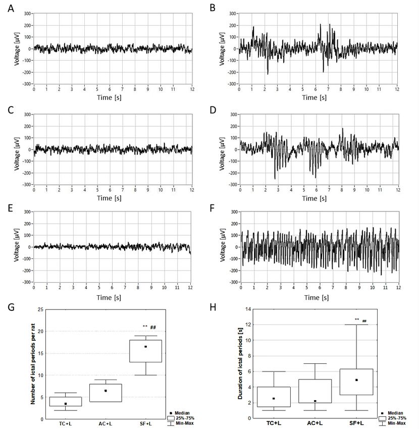
EEG analysis Rats from groups receiving lindane developed ictal activi‑
ty in EEG consisting of sporadic, smaller groups or series of
The EEG activity in rats from the control SFc, ACc and high‑amplitude spikes (EEG ictal periods, Fig. 4B, D, F). We
TCc groups showed no signs of ictal activity (Fig. 4A, C, E). analyzed the number and duration of these ictal periods.
Fig. 4. Representative EEG tracings recorded in TCc, ACc and SFc groups (A, C, E) and TC+L, AC+L and SF+L groups (B, D, F). The number (G)
and duration (H) of EEG ictal periods upon lindane administration. Representative EEG tracings recorded in control groups (TCc, ACc and
SFc, A, C, E) showing baseline activity without signs of epileptiform discharges. Ictal activity has been recorded upon lindane administration
in TC+L, AC+L and SF+L group (B, D, F) in form of spike bursts of different duration and number. Lead: left frontal–right parietal cortex. The
number (G) and duration (H) of ictal periods was measured during 30 min EEG recordings upon lindane administration. The significance
of the differences between the groups was estimated by Kruskal‑Wallis ANOVA and Mann‑Whitney U test (**p102 Grubac et al. Acta Neurobiol Exp 2021, 81: 96–109
According to Kruskal‑Wallis ANOVA, number of ic‑ DISCUSSION
tal periods was significantly different (H2=13.28, PActa Neurobiol Exp 2021, 81:
81 96–109 Sleep fragmentation related seizures and neuroinflammation 103
Fig. 5. Interleukin levels in rat brain structures – cortex, thalamus and hippocampus. (C) demonstrated significantly higher values in SF group compared
to others (**p104 Grubac et al. Acta Neurobiol Exp 2021, 81: 96–109
Fig. 6. Serum levels of IL‑1β and IL‑6. Levels IL‑1β and IL‑6 in animals’ serum were measured using commercial ELISA kit according to the instructions of the
manufacturer. Difference in both, IL‑1β (A) and IL‑6 (B) levels among the three groups was not statistically significant in serum (p>0.05 vs. both TC and AC).
but inducing both behavioral and EEG epileptic activity cial effect of CPAP on epilepsy was shown in about 40%
after administration of lindane in subconvulsive dose. of treated patients with OSA (Vaughn et al., 1996; Beran
Our present data also showed significant increase of et al., 1999; Sonka et al., 2000), with approximately 50%
IL‑1β levels in the thalamus, cerebral cortex and hip‑ seizure reduction in treated apneic patients (Pornsrin‑
pocampal tissue in rats undergoing protocol of chronic iyom et al., 2014). Indeed, hypoxia has been already es‑
sleep fragmentation compared to the corresponding tablished as a proconvulsive factor (Jensen et al., 1991;
controls. In addition, the levels of IL‑6 were significant‑ 1992) by facilitating glutaminergic neurotransmission
ly increased in the cortex of sleep fragmented animals, (Rubaj et al., 2003; Yang et al., 2013), while CPAP can re‑
but no statistical difference was found in the thalamus sult in new onset of frontal lobe seizures if applied in‑
or hippocampus. correctly (Miano et al., 2008). Based on our results, we
Sleep fragmentation and intermittent hypoxia are could hypothesize that sleep fragmentation also could
hallmarks of OSA in which reduction in seizure thresh‑ represent the mechanism responsible for lowering the
old has been reported (Malow et al., 2003). Unanswered threshold to seizure in sleep apnea conditions.
question in the contemporary literature is to which Epileptogenesis is a complex process characterized
extent sleep fragmentation contribute to this seizure by an enduring predisposition to generate epileptic
threshold reduction in apneic patient. Our study as‑ seizures and it has been found to be associated with
sessed this question from the viewpoint of chronic changes in immunological profile (Vezzani et al., 2002;
sleep fragmentation modeled in experimental con‑ Lehtimäki et al., 2007; Rosa et al., 2016). In our cur‑
ditions since intermittent hypoxia and sleep disrup‑ rent study, the levels of IL‑1β in the SF group showed
tion are very difficult to study separately in clinical significant increase in the thalamus, cortex and hip‑
settings because they occur simultaneously in apneic pocampus when compared to the AC and TC groups.
patients. Our current study has proven that long‑term There was no statistical difference between the AC and
sleep fragmentation, modeled according to severe form TC groups, but levels of IL‑1β were higher in the activ‑
of OSA, aggravates seizures. This is in agreement with ity control group compared to sedentary controls, so
line that seizures are very sensitive to alterations in we cannot completely rule out physical activity as the
sleep pattern. Namely, good quality sleep is import‑ confounding factor for this increase. As for the levels
ant for optimal human functioning, but it is particu‑ of IL‑6, statistically higher levels were only found in
larly essential in patients with epilepsy who may find the cortex of fragmented animals, and no difference
themselves in a cycle of worsening seizures, further between the groups was observed in the thalamic or
sleep disruption, and intractable epilepsy (Bazil, 2017). hippocampal tissue.
Moreover, this study could actually give the answer to Relationship between epilepsy and IL‑1β levels are
the question why continuous positive airway treatment not completely clear in clinical studies, i.e., in some
(CPAP) is not sufficient treatment of OSA in terms of link has been proven and in some not (Tilgen et al.,
seizure control (Malow et al., 2003; Bazil, 2017). Benefi‑ 2002; Virta et al., 2002; Kanemoto et al., 2003; OzkaraActa Neurobiol Exp 2021, 81:
81 96–109 Sleep fragmentation related seizures and neuroinflammation 105
et al., 2006). On the other hand, basic experimental This mechanism has been supported by exploring
studies give us better opportunity to establish a link anticonvulsive effects of IL‑1R in bicuculline induced
between elevated levels of IL‑1β and enhanced pro‑ seizures in rodents (Vezzani et al., 2002). In our current
pensity for seizure activity. Vezzani group (1999) was study, increased levels of IL‑1β in the cerebral cortex,
the first to report that IL‑1β enhances focal electro‑ hippocampus and thalamus found in the SF group of
graphic seizures induced by kainate via glutamatergic rats are congruent with theories about proconvulsive
signaling. and neurotoxic effects of IL‑1β. Proconvulsive role
Inflammatory alterations in the rats during the could be achieved through several mechanisms: IL‑1β
acute epileptic events are characterized by rapid in‑ acts as a direct proconvulsive agent through inhibition
crease of IL‑1β in activated microglia and astrocytes of GABA A receptors and K + efflux (Wang et al., 2000);
(Fabene et al., 2008). Moreover, this elevated Il‑1β lev‑ inhibits glial uptake of excitatory neurotransmitters
el persisted even upon seizure cessation, what adds to (Hu et al., 2000) and causes intracellular calcium ion
the presumption on IL‑1β involvement in the mecha‑ surge, thus resulting in modification of voltage‑depen‑
nisms underlying the onset of spontaneous seizures dent ion channels and reduction of seizure threshold
(Allan et al., 2005; Fabene et al., 2008). The activa‑ (Viviani et al., 2003; Xu et al., 2013). Other mechanisms
tion of IL‑1β signaling occurs through nontranscrip‑ could also be involved, for example IL‑1β is known to
tion‑dependent enzymatic pathway which includes increase nitric oxide production in the brain (Meini et
IL‑1R1–mediated activation of the neutral sphingo‑ al., 2000), as well as to stimulate glutamatergic neu‑
myelinase (Sanchez‑Alavez et al., 2006) and the sub‑ rotransmission pathway and evoke neuronal hyper‑
sequent production of ceramide, which in turn acti‑ excitability (Zhu et al., 2006). Contrary to our results,
vates the tyrosine kinase protein, Src (Sanchez‑Ala‑ Sayyah et al. (2005) reported an anticonvulsive effect of
vez et al., 2006). IL‑1β than enhances calcium influx intraventriculary injected IL‑1β in amygdala‑kindling
into hippocampal pyramidal cells exposed to N‑meth‑ mice, which could potentially be explained with IL‑1β
yl‑d‑aspartate (NMDA) by phosphorylation (via Src ki‑ ability to modulate intracellular concentration of Ca 2+
nase) of the NR2B subunit of the NMDA receptor com‑ in dose‑dependent manner, thus balancing excitato‑
plex, which is responsible for regulating its calcium ry and inhibitory neurotransmission (Zhu et al., 2006;
permeability properties (Viviani et al., 2003). When Li et al., 2011). For schematic illustration of proposed
released, IL‑1β binds with high affinity to the type mechanisms see Fig. 7.
1 IL‑1 receptor (IL‑1R1) that is expressed by hippo‑ Beside IL‑1β, epileptogenesis is also related to alter‑
campal neurons (Ravizza and Vezzani, 2006). In con‑ ations in IL‑6, however its role is still unclear and giv‑
trast, when seizures evoke rapid production of IL‑1β, en research results are contradictory (D’Arcangelo et
IL‑1RA is upregulated to a far lower extent and with al., 2000). After seizures, IL‑6 mRNA is rapidly induced
a several hour delay (De Simoni et al., 2000; Ravizza in the hippocampus, cortex, dentate gyrus, amygdala,
and Vezzani, 2006). This finding suggests that the and meninges, whereas up‑regulation of IL‑6‑receptor
brain is much less effective than the periphery in in‑ mRNA seems to be limited to the hippocampus (Le‑
ducing a crucial mechanism for rapidly terminating htimäki et al., 2003). Several clinical studies demon‑
the effects of a sustained rise in endogenous IL‑1β. strated rapid and transient post‑ictal increase of IL‑6,
Seizures are often accompanied by extensive neuro‑ which peaked at 12 h and remained for 24 h after sei‑
nal injury, which may account for IL‑1 production by zures. However, some patients with chronic epilepsy
microglia (Sanchez‑Alavez et al., 2006). At the cellu‑ also had higher levels of IL‑6 in basal conditions (Virta
lar level, Toll‑like receptors (TLRs), activated by sei‑ et al., 2002; Lehtimäki et al., 2004, 2007; Alapirtti et
zure‑induced heath shock proteins (HSPs), induce the al., 2009). Different types of epilepsy showed differ‑
activation of nuclear factor kappa B (NF‑κB), which ent patterns in IL‑6 elevation. Taking into account the
finally results in IL‑1β production (Hanke and Kielian, lower post‑ictal levels of IL‑6 in some serious forms of
2011). TLR3 contributes to hippocampal excitability epilepsy, it can presumably act as a neuroprotective
through its upregulation of pro‑inflammatory cyto‑ agent (Bauer et al., 2009). In contrast to this finding,
kines such as IFN‑β (Costello and Lynch, 2013). FOX3P there is also a report about exogenous IL‑6 attenuat‑
is also expressed by microglia to downregulate in‑ ed seizures in rats (Fukuda et al., 2007). Studies using
flammatory processes through modulation of NF‑κB, transgenic animals reflect the double‑edged nature of
a key inflammatory transcription factor (Chung et al., IL‑6 with IL‑6 deficient mice more prone to seizures,
2010). Involvement of these mechanisms in the ele‑ and those with over expression of IL‑6 showing low‑
vation of Il‑1β observed in our current study is still er threshold to spontaneous and NMDA‑induced sei‑
open question that should be addressed in one of the zures, probably due to reduced GABA‑mediated inhi‑
following studies. bition (De Sarro et al., 2004).106 Grubac et al. Acta Neurobiol Exp 2021, 81: 96–109
Fig. 7. Schematic representation of the proposed mechanism of IL‑1β activation and subsequent production of seizure.
In our study elevated levels of IL‑1β and IL‑6 were deed, upregulated IL‑6 has been revelled in conditions
found in the cortex, which is always involved in epilep‑ of higher neuronal excitability (Uludag et al., 2013),
tic discharge (Alapirtti et al., 2018). which is presumably consequence of sleep fragmenta‑
IL‑1β has been recognized as a fully proconvul‑ tion in our study.
sive and proinflammatory cytokine, as it is obvious There are several limitations of this study. The
from our discussion above. On the other hand, IL‑6 study has been performed in male rats having in
is a pleiotropic cytokine that has both pro‑ and an‑ mind their stability regarding hormonal status. How‑
ti‑inflammatory actions with predominantly anticon‑ ever, further studies on female rats should be also
vulsive nature (Erta et al., 2012). We found increased done, in diestrus phase of their estrus cycle due to
IL‑1β levels in the cortex, hippocampus and thalamus, possible influence of sex hormones (Scharfman and
with IL‑6 level was increased only in the cortex upon MacLusky, 2014). Namely, there are important sex‑de‑
chronic sleep fragmentation. We could speculate that pendent differences that influence seizures and epi‑
IL‑1β may contribute to epileptogenesis in our study lepsy. Studies in which pooling male and female are
by modulating or blocking IL‑6 signal transduction in likely to obtain confusing and different results unless
the hippocampus and thalamus, since such a crosstalk experimental conditions are fully controlled (Tan and
between these two cytokines has been demonstrated Tanb, 2001). We focused herein on sleep fragmenta‑
in other organs (Deon et al., 2001). Namely, antagonism tion effects on IL‑1β and IL‑6 production in different
may occur at the level of signal transduction, i.e., IL‑1β brain structures, while finding the exact cell type
has been shown to suppress Janus kinase‑STAT signal‑ (astrocytes, microglia, neurons) as a source of these
ling by IL‑6 and antagonize IL‑6 induction of tissue in‑ cytokine production should be addressed in further
hibitor of metalloproteases 1 expression (Deon et al., immunohistochemical studies.
2001). Neuroinflammation depends on cytokines, glial
and neuronal signalling, as well as on extracellular ma‑
trix, and all of the components being interconnected CONCLUSIONS
(Hrncic et al., 2018). Additionally, increased IL‑6 level
in the cortex could be compensatory response in or‑ In summary, we have demonstrated herein that
der to reestablish dysregulated cytokine balance. In‑ a 14‑day chronic sleep fragmentation increases sei‑Acta Neurobiol Exp 2021, 81:
81 96–109 Sleep fragmentation related seizures and neuroinflammation 107
De Sarro G, Russo E, Ferreri G, Giuseppe B, Flocco MA, Di Paola ED,
zure susceptibility in rats, which is objectively prov‑
De Sarro A (2004) Seizure susceptibility to various convulsant stim‑
en by convulsive behavioral response and EEG anal‑ uli of knockout interleukin‑6 mice. Pharmacol Biochem Behav 77:
ysis and altered brain production of inflammatory 761–766.
cytokines IL‑1β and IL‑6, which could be potential De Simoni MG, Perego C, Ravizza T, Moneta D, Conti M, Marchesi F,
explanation for increased seizure susceptibility. For De Luigi A, Garattini S, Vezzani A (2000) Inflammatory cytokines and
related genes are induced in the rat hippocampus by limbic sta‑
sure, other mechanisms may also be involved in the
tus epilepticus: Cytokines in status epilepticus. Eur J Neurosci 12:
development of brain hyperexcitability. Further stud‑ 2623–2633.
ies are needed with IL‑1β and IL‑6 antagonists, as well Deon D, Ahmed S, Tai K, Scaletta N, Herrero C, Lee IH, Krause A, Ivashkiv LB
as modifications of IL‑1β and IL‑6 genes, in order to (2001) Cross‑talk between IL‑1 and IL‑6 signaling pathways in rheuma‑
confirm the proexcitatory role of these cytokines in toid arthritis synovial fibroblasts. J Immunol 167: 5395–5403.
Devinsky O, Ehrenberg B, Barthlen GM, Abramson HS, Luciano D (1994)
sleep fragmentation and find exact cellular source of
Epilepsy and sleep apnea syndrome. Neurology 44: 2060–2064.
its production. Di Filippo M, Chiasserini D, Gardoni F, Viviani B, Tozzi A, Giampà C, Costa C,
Tantucci M, Zianni E, Boraso M, Siliquini S, Iure A de, et al. (2013) Effects
of central and peripheral inflammation on hippocampal synaptic plas‑
ACKNOWLEDGMENT ticity. Neurobiol Dis 52: 229–236.
Erta M, Quintana A, Hidalgo J (2012) Interleukin‑6, a major cytokine in the
central nervous system. Int J Biol Sci 8: 1254–1266.
This study has been supported by the Ministry of Fabene PF, Mora GN, Martinello M, Rossi B, Merigo F, Ottoboni L, Bach S,
Education, Science and Technological Development of Angiari S, Benati D, Chakir A, Zanetti L, Schio F, et al. (2008) A role for
Serbia (grant #175032). leukocyte‑endothelial adhesion mechanisms in epilepsy. Nat Med 14:
1377–1383.
Ferrillo F, Beelke M, Nobili L (2000) Sleep EEG synchronization mechanisms
and activation of interictal epileptic spikes. Clin Neurophysiol 111:
REFERENCES S65‑73.
Foldvary‑Schaefer N, Andrews ND, Pornsriniyom D, Moul DE, Sun Z,
Alapirtti T, Lehtimäki K, Nieminen R, Mäkinen R, Raitanen J, Moilanen E, Bena J (2012) Sleep apnea and epilepsy: who’s at risk? Epilepsy Behav
Mäkinen J, Peltola J (2018) The production of IL‑6 in acute epileptic sei‑ 25: 363–367.
zure: A video‑EEG study. J Neuroimmunol 316: 50–55. Frucht MM, Quigg M, Schwaner C, Fountain NB (2000) Distribution
Alapirtti T, Rinta S, Hulkkonen J, Mäkinen R, Keränen T, Peltola J (2009) of seizure precipitants among epilepsy syndromes. Epilepsia 41:
Interleukin‑6, interleukin‑1 receptor antagonist and interleukin‑1beta 1534–1539.
production in patients with focal epilepsy: A video–EEG study. J Neurol Fukuda M, Morimoto T, Suzuki Y, Shinonaga C, Ishida Y (2007) Interleukin‑6
Sci 280: 94–97. attenuates hyperthermia‑induced seizures in developing rats. Brain Dev
Allan SM, Tyrrell PJ, Rothwell NJ (2005) Interleukin‑1 and neuronal injury. 29: 644–648.
Nature Rev Immunol 5: 629–640. Grubac Z, Sutulovic N, Ademovic A, Velimirovic M, Rasic‑Markovic A,
Baud MO, Magistretti PJ, Petit J‑M (2015) Sustained sleep fragmentation Macut D, Petronijevic N, Stanojlovic O, Hrncic D (2019) Short‑term sleep
induces sleep homeostasis in mice. Sleep 38: 567–579. fragmentation enhances anxiety‑related behavior: The role of hormonal
Bauer S, Cepok S, Todorova‑Rudolph A, Nowak M, Köller M, Lorenz R, alterations. PLoS One 14: e0218920.
Oertel WH, Rosenow F, Hemmer B, Hamer HM (2009) Etiology and site Hanke ML, Kielian T (2011) Toll‑like receptors in health and disease in the
of temporal lobe epilepsy influence postictal cytokine release. Epilepsy brain: mechanisms and therapeutic potential. Clin Sci 121: 367–387.
Res 86: 82–88. Hauser WA, Annegers JF, Kurland LT (1991) Prevalence of epilepsy in
Bazil C (2017) Sleep and epilepsy. Semin Neurol 37: 407–412. Rochester, Minnesota: 1940‑1980. Epilepsia 32: 429–445.
Beran RG, Plunkett MJ, Holland GJ (1999) Interface of epilepsy and sleep Hrnčić D, Grubač Ž, Rašić‑Marković A, Šutulović N, Šušić V, Bjekić‑Macut J,
disorders. Seizure 8: 97–102. Stanojlović O (2016) Sleep disruption increases seizure susceptibility:
Bonnet MH, Arand DL (2003) Clinical effects of sleep fragmentation versus Behavioral and EEG evaluation of an experimental model of sleep ap‑
sleep deprivation. Sleep Med Rev 7: 297–310. nea. Physiol Behav 155: 188–194.
Chihorek AM, Abou‑Khalil B, Malow BA (2007) Obstructive sleep apnea is Hrnčić D, Rašić‑Marković A, Djuric D, Šušić V, Stanojlović O (2011) The Role
associated with seizure occurrence in older adults with epilepsy. Neu‑ of nitric oxide in convulsions induced by lindane in rats. Food Chem Tox‑
rology 69: 1823–1827. icol 49: 947–954.
Chung HS, Lee JH, Kim H, Lee HJ, Kim SH, Kwon HK, Im SH, Bae H (2010) Hrncic D, Sutulovic N, Grubac Z, Rasic‑Markovic A, Stanojlovic O (2018)
Foxp3 is a novel repressor of microglia activation: repression of microg‑ The central nervous system is not imunoprivileged: Inflammation and
lia activation by Foxp3. Glia 58: 1247–1256. epileptogenesis. Vojnosanitetski Pregl 75: 820–825.
Costello DA, Lynch MA (2013) Toll‑like receptor 3 activation modulates hip‑ Hu S, Sheng WS, Ehrlich LC, Peterson PK, Chao CC (2000) Cytokine effects
pocampal network excitability, via glial production of interferon‑β: TLR3 on glutamate uptake by human astrocytes. Neuroimmunomodulation
activation induces interictal activity in hippocampus. Hippocampus 23: 7: 153–159.
696–707. Jensen FE, Applegate CD, Holtzman D, Belin TR, Burchfiel JL (1991) Epilep‑
D’Arcangelo G, Tancredi V, Onofri F, D’Antuono M, Giovedì S, Benfena‑ togenic effect of hypoxia in the immature rodent brain. Ann Neurol 29:
ti F (2000) Interleukin‑6 inhibits neurotransmitter release and the 629–637.
spread of excitation in the rat cerebral cortex. Eur J Neurosci 12: Jensen FE, Holmes GL, Lombroso CT, Blume HK, Firkusny IR (1992) Age‑de‑
1241–1252. pendent changes in long‑term seizure susceptibility and behavior after
Day R, Gerhardstein R, Lumley A, Roth T, Rosenthal L (1999) The behav‑ hypoxia in rats. Epilepsia 33: 971–980.
ioral morbidity of obstructive sleep apnea. Prog Cardiovasc Dis 41: Kanemoto K, Kawasaki J, Yuasa S, Kumaki T, Tomohiro O, Kaji R,
341–354. Nishimura M (2003) Increased frequency of interleukin‑1beta‑511T al‑108 Grubac et al. Acta Neurobiol Exp 2021, 81: 96–109 lele in patients with temporal lobe epilepsy, hippocampal sclerosis, and Ravizza T, Vezzani A (2006) Status epilepticus induces time‑dependent neu‑ prolonged febrile convulsion. Epilepsia 44: 796–799. ronal and astrocytic expression of interleukin‑1 receptor type I in the rat Lehtimäki KA, Keränen T, Huhtala H, Hurme M, Ollikainen J, Honkaniemi J, limbic system. Neuroscience 137: 301–308. Palmio J, Peltola J (2004) Regulation of IL‑6 system in cerebrospinal fluid Rosa DV, Rezende VB, Costa BS, Mudado F, Schütze M, Torres KC, and serum compartments by seizures: the effect of seizure type and Martins LC, Moreira‑Filho CA, Miranda DM, Romano‑Silva MA (2016) Cir‑ duration. J Neuroimmunol 152: 121–125. culating CD4 and CD8 T cells expressing pro‑inflammatory cytokines in Lehtimäki KA, Keränen T, Palmio J, Mäkinen R, Hurme M, Honkaniemi J, a cohort of mesial temporal lobe epilepsy patients with hippocampal Peltola J (2007) Increased plasma levels of cytokines after seizures in sclerosis. Epilepsy Res 120: 1–6. localization‑related epilepsy. Acta Neurol Scand 116: 226–230. Rubaj A, Zgodziński W, Sieklucka‑Dziuba M (2003) The epileptogenic effect Lehtimäki KA, Peltola J, Koskikallio E, Keränen T, Honkaniemi J (2003) of seizures induced by hypoxia: the role of NMDA and AMPA/KA antag‑ Expression of cytokines and cytokine receptors in the rat brain after ka‑ onists. Pharmacol Biochem Behav 74: 303–311. inic acid‑induced seizures. Mol Brain Res 110: 253–260. Sanchez‑Alavez M, Tabarean IV, Behrens MM, Bartfai T (2006) Ceramide Li G, Bauer S, Nowak M, Norwood B, Tackenberg B, Rosenow F, Knake S, mediates the rapid phase of febrile response to IL‑1beta. PNAS 103: Oertel WH, Hamer HM (2011) Cytokines and epilepsy. Seizure 20: 2904–2908. 249–256. Sander JW (1993) Some aspects of prognosis in the epilepsies: a review. Lucey BP, Leahy A, Rosas R, Shaw PJ (2015) A new model to study sleep Epilepsia 34: 1007–1016. deprivation‑induced seizure. Sleep 38: 777–785. Sayyah M, Beheshti S, Shokrgozar MA, Eslami‑far A, Deljoo Z, Malow BA, Levy K, Maturen K, Bowes R (2000) Obstructive sleep apnea Khabiri AR, Haeri Rohani A (2005) Antiepileptogenic and anticon‑ is common in medically refractory epilepsy patients. Neurology 55: vulsant activity of interleukin‑1 beta in amygdala‑kindled rats. Exp 1002–1007. Neurol 191: 145–153. Malow BA, Weatherwax KJ, Chervin RD, Hoban TF, Marzec ML, Martin C, Scalise A, Desiato MT, Gigli GL, Romigi A, Tombini M, Marciani MG, Binns LA (2003) Identification and treatment of obstructive sleep apnea Izzi F, Placidi F (2006) Increasing Cortical Excitability: A Possible Ex‑ in adults and children with epilepsy: a prospective pilot study. Sleep planation For The Proconvulsant Role Of Sleep Deprivation. Sleep Med 4: 509–515. 29: 1595–1598. Manni R, Tartara A (2000) Evaluation of sleepiness in epilepsy. Clin Neuro‑ Scharfman HE, MacLusky NJ (2014) Sex differences in the neurobiology of physiol 111: S111–114. epilepsy: A preclinical perspective. Neurobiol Dis 72: 180–192. Manni R, Zambrelli E, Bellazzi R, Terzaghi M (2005) The relationship be‑ Seyal M, Bateman LM, Albertson TE, Lin T‑C, Li C‑S (2010) Respiratory tween focal seizures and sleep: an analysis of the cyclic alternating pat‑ changes with seizures in localization‑related epilepsy: Analysis of periic‑ tern. Epilepsy Res 67: 73–80. tal hypercapnia and airflow patterns: respiratory change with seizures. McKenna JT, Tartar JL, Ward CP, Thakkar MM, Cordeira JW, McCarley RW, Epilepsia 51: 1359–1364. Strecker RE (2007) Sleep fragmentation elevates behavioral, electro‑ Shrivastava D, Jung S, Saadat M, Sirohi R, Crewson K (2014) How to inter‑ graphic and neurochemical measures of sleepiness. Neuroscience 146: pret the results of a sleep study. J Community Hosp Intern Med Perspect 1462–1473. 4: 24983. Medic G, Wille M, Hemels ME (2017) Short‑ and long‑term health conse‑ Sonka K, Juklícková M, Pretl M, Dostálová S, Horínek D, Nevsímalová S quences of sleep disruption. Nat Sci Sleep 9: 151–161. (2000) Seizures in sleep apnea patients: occurrence and time distribu‑ Meini A, Benocci A, Frosini M, Sgaragli G, Pessina G, Aldinucci C, Youmbi GT, tion. Sb Lek 101: 229–232. Palmi M (2000) Nitric oxide modulation of interleukin‑1[beta]‑evoked Stanojlović O, Rašić‑Marković A, Hrnčić D, Šušić V, Macut D, Radosavljević T, intracellular Ca2+ release in human astrocytoma U‑373 MG cells and Djuric D (2009) Two Types of seizures in homocysteine thiolactone‑treat‑ brain striatal slices. J Neurosci 20: 8980–8986. ed adult rats, behavioral and electroencephalographic study. Cell Mol Miano S, Pelliccia A, Evangelisti M, Pagani J, Villa MP (2008) Role of contin‑ Neurobiol 29: 329–339. uous positive airway pressure therapy on the pathogenesis of sleep‑re‑ Šutulović N, Grubač Ž, Šuvakov S, Jovanović Đ, Puškaš N, Macut Đ, lated frontal lobe epilepsy in a child with obstructive sleep apnea syn‑ Marković AR, Simić T, Stanojlović O, Hrnčić D (2019) Chronic prostatitis/ drome. J Child Neurol 23: 124–128. chronic pelvic pain syndrome increases susceptibility to seizures in rats Mullington JM, Haack M, Toth M, Serrador J, Meier‑Ewert H (2009) Cardio‑ and alters brain levels of IL‑1β and IL‑6. Epilepsy Res 153: 19–27. vascular, inflammatory and metabolic consequences of sleep depriva‑ Tan M, Tanb U (2001) Sex Difference in Susceptibility to Epileptic tion. Prog Cardiovasc Dis 51: 294–302. Seizures in Rats: Importance of Estrous Cycle. Int J Neurosci 108: Ozkara C, Uzan M, Tanriverdi T, Baykara O, Ekinci B, Yeni N, Kafadar A, 175–191. Buyru N (2006) Lack of association between IL‑1beta/alpha gene poly‑ Tang Y, Preuss F, Turek FW, Jakate S, Keshavarzian A (2009) Sleep depriva‑ morphisms and temporal lobe epilepsy with hippocampal sclerosis. Sei‑ tion worsens inflammation and delays recovery in a mouse model of zure 15: 288–291. colitis. Sleep Med 10: 597–603. Parrino L, Smerieri A, Spaggiari MC, Terzano MG (2000) Cyclic alternating Tilgen N, Pfeiffer H, Cobilanschi J, Rau B, Horvath S, Elger CE, Propping P, pattern (CAP) and epilepsy during sleep: how a physiological rhythm Heils A (2002) Association analysis between the human interleukin modulates a pathological event. Clin Neurophysiol 111: S39‑46. 1beta (‑511) gene polymorphism and susceptibility to febrile convul‑ Paxinos G, Watson C (2007) The Rat Brain in Stereotaxic Coordinates. Else‑ sions. Neurosci Lett 334: 68–70. vier, Amsterdam; Boston. Uludag IF, Bilgin S, Zorlu Y, Tuna G, Kirkali G (2013) Interleukin‑6, inter‑ Pornsriniyom D, Kim H won, Bena J, Andrews ND, Moul D, leukin‑1 beta and interleukin‑1 receptor antagonist levels in epileptic Foldvary-Schaefer N (2014) Effect of positive airway pressure therapy seizures. Seizure 22: 457–461. on seizure control in patients with epilepsy and obstructive sleep apnea. Vaughn BV, D’Cruz OF, Beach R, Messenheimer JA (1996) Improvement of Epilepsy Behav 37: 270–275. epileptic seizure control with treatment of obstructive sleep apnoea. Rajna P, Veres J (1993) Correlations between night sleep duration and sei‑ Seizure 5: 73–78. zure frequency in temporal lobe epilepsy. Epilepsia 34: 574–579. Vezzani A, Conti M, De Luigi A, Ravizza T, Moneta D, Marchesi F, De Ravizza T, Gagliardi B, Noé F, Boer K, Aronica E, Vezzani A (2008) Innate and Simoni MG (1999) Interleukin‑1β immunoreactivity and microglia are adaptive immunity during epileptogenesis and spontaneous seizures: enhanced in the rat hippocampus by focal kainate application: function‑ Evidence from experimental models and human temporal lobe epilep‑ al evidence for enhancement of electrographic seizures. J Neurosci 19: sy. Neurobiol Dis 29: 142–160. 5054–5065.
Acta Neurobiol Exp 2021, 81:
                         81 96–109                                              Sleep fragmentation related seizures and neuroinflammation              109

Vezzani A, Friedman A (2011) Brain inflammation as a biomarker in epilep‑           lindane‑induced experimental epilepsy in rats. Canadian J Physiol Phar‑
   sy. Biomarkers Med 5: 607–614.                                                   macol 86: 173–179.
Vezzani A, Moneta D, Richichi C, Aliprandi M, Burrows SJ, Ravizza T,              Wang S, Cheng Q, Malik S, Yang J (2000) Interleukin‑1β Inhibits γ‑Amino‑
   Perego C, De Simoni MG (2002) Functional role of inflammatory cyto‑              butyric acid type A (GABAA) receptor current in cultured hippocampal
   kines and antiinflammatory molecules in seizures and epileptogenesis.            neurons. J Pharmacol Exp Ther 292: 497–504.
   Epilepsia 43: 30–35.                                                           Wulff K, Gatti S, Wettstein JG, Foster RG (2010) Sleep and circadian rhythm
Vijayan VK (2012) Morbidities associated with obstructive sleep apnea. Ex‑          disruption in psychiatric and neurodegenerative disease. Nature Rev
   pert Rev Respir Med 6: 557–566.                                                  Neurosci 11: 589–599.
Virta M, Hurme M, Helminen M (2002) Increased frequency of interleu‑              Xu D, Miller SD, Koh S (2013) Immune mechanisms in epileptogenesis.
   kin‑1beta (‑511) allele 2 in febrile seizures. Pediatr Neurol 26: 192–195.       Front Cell Neurosci 7: 195.
Viviani B, Bartesaghi S, Gardoni F, Vezzani A, Behrens MM, Bartfai T,             Yang Q, Wang Y, Feng J, Cao J, Chen B (2013) Intermittent hypoxia from
   Binaglia M, Corsini E, Di Luca M, Galli CL, Marinovich M (2003) Inter‑           obstructive sleep apnea may cause neuronal impairment and dysfunc‑
   leukin‑1beta enhances NMDA receptor‑mediated intracellular calcium               tion in central nervous system: the potential roles played by microglia.
   increase through activation of the Src family of kinases. J Neurosci 23:         Neuropsychiatr Dis Treat 9: 1077–1086.
   8692–8700.                                                                     Zhu G, Okada M, Yoshida S, Mori F, Ueno S, Wakabayashi K, Kaneko S
Vučević D, Hrnčić D, Radosavljević T, Mladenović D, Rašić‑Marković A,               (2006) Effects of interleukin‑1β on hippocampal glutamate and GABA
   Lončar‑Stevanović H, Djurić D, Macut D, Šušić V, Stanojlović O (2008)            releases associated with Ca2+‑induced Ca2+ releasing systems. Epilepsy
   Correlation between electrocorticographic and motor phenomena in                 Res 71: 107–116.
You can also read