Melatonin Signaling Controls Circadian Swimming Behavior in Marine Zooplankton
←
→
Page content transcription
If your browser does not render page correctly, please read the page content below
Melatonin Signaling Controls
Circadian Swimming Behavior
in Marine Zooplankton
Maria Antonietta Tosches,1,* Daniel Bucher,1,2 Pavel Vopalensky,1 and Detlev Arendt1,2,*
1European   Molecular Biology Laboratory, Developmental Biology Unit, Meyerhofstrasse 1, 69117 Heidelberg, Germany
2Centre  for Organismal Studies (COS), University of Heidelberg, 69120 Heidelberg, Germany
*Correspondence: tosches@embl.de (M.A.T.), arendt@embl.de (D.A.)
 http://dx.doi.org/10.1016/j.cell.2014.07.042
This is an open access article under the CC BY license (http://creativecommons.org/licenses/by/3.0/).

SUMMARY                                                               in cnidarians, and in dinoflagellates (Balzer and Hardeland, 1991;
                                                                      Hardeland and Poeggeler, 2003; Roopin and Levy, 2012). The
Melatonin, the ‘‘hormone of darkness,’’ is a key                      ubiquitous presence of melatonin (Hardeland and Poeggeler,
regulator of vertebrate circadian physiology and                      2003) has been linked to its chemical properties, which make it
behavior. Despite its ubiquitous presence in Meta-                    one of the most powerful radical scavengers known in nature
zoa, the function of melatonin signaling outside ver-                 (Tan et al., 2007). Beyond that, the presence of melatonin recep-
tebrates is poorly understood. Here, we investigate                   tors in all animal lineages, except sponges (Feuda et al., 2012),
                                                                      indicates that melatonin acquired an additional signaling func-
the effect of melatonin signaling on circadian swim-
                                                                      tion early in animal evolution.
ming behavior in a zooplankton model, the marine                         It has been speculated that the role of melatonin as the ‘‘hor-
annelid Platynereis dumerilii. We find that melatonin                 mone of darkness’’ has evolved directly from its antioxidant
is produced in brain photoreceptors with a verte-                     properties: in a system with constant melatonin synthesis, the
brate-type opsin-based phototransduction cascade                      reduction of melatonin levels during the day due to its light-
and a light-entrained clock. Melatonin released at                    dependent oxidation would make it a suitable signal for darkness
night induces rhythmic burst firing of cholinergic                    (Hardeland et al., 1995). The newly evolving melatonin receptors
neurons that innervate locomotor-ciliated cells. This                 might thus have recognized melatonin as a ‘‘chemical indicator
establishes a nocturnal behavioral state by modu-                     of darkness’’ for the circadian regulation of some physiological
lating the length and the frequency of ciliary arrests.               process and/or behavior. However, the nature of such mela-
Based on our findings, we propose that melatonin                      tonin-controlled behavior has so far remained elusive, as the
                                                                      role of melatonin signaling has been poorly investigated outside
signaling plays a role in the circadian control of ciliary
                                                                      vertebrates. In few cases, it has been demonstrated that mela-
swimming to adjust the vertical position of zoo-
                                                                      tonin has modulatory (often inhibitory) effects on locomotion
plankton in response to ambient light.                                (Anctil et al., 1991; Bentkowski et al., 2010; Tanaka et al.,
                                                                      2007; Tilden et al., 2003); yet it is not clear whether these effects
INTRODUCTION                                                          are linked to circadian rhythms and how they relate to the ancient
                                                                      role of melatonin signaling in animals.
In vertebrates, melatonin and its signaling through G-protein-           To broaden our perspective on the function of melatonin
coupled receptors (GPCRs) play a fundamental role in the              signaling in metazoans, we investigated its possible role in the
circadian modulation of physiology and behavior. Melatonin is a       day/night control of zooplankton locomotion. Primary larvae
highly diffusible hormone secreted at night by the pineal organ,      forming most of the zooplankton are part of the life cycle in the
which in nonmammalian vertebrates is directly light sensitive         majority of animal phyla. They swim using locomotor cilia, which
(Lamb, 2013). As a ‘‘hormone of darkness,’’ melatonin levels          are either distributed over the entire body surface or concen-
change according to circadian, lunar, and seasonal cycles (Reiter,    trated in specialized ciliary bands. In the ocean, almost all of
1993). A conserved function of melatonin in vertebrates, from fish    these larvae show a pronounced rhythmic behavior, known as
to mammals, is the regulation of sleep (Dollins et al., 1994; Zhda-   diel vertical migration (DVM), which generally consists of an up-
nova et al., 2001). In mammals, this occurs through direct modu-      ward swimming phase at dusk and a downward displacement
lation of neuronal excitability in the suprachiasmatic nucleus, the   phase throughout the night and/or at dawn (Alldredge and
brain circadian pacemaker (Jiang et al., 1995), and in the thal-      King, 1980; Forward, 1988; Rudjakov, 1970). The control of ver-
amus, where sleep is initiated (Ochoa-Sanchez et al., 2011).          tical migration in the oceans has been linked to the origin of an-
   Melatonin is one of the oldest biologically active molecules       imal circadian rhythms (Gehring and Rosbash, 2003; Pittendrigh,
in nature, present in a nearly ubiquitous range of organisms,         1993), as a mechanism to escape damaging UV irradiation dur-
including bacteria and plants. Its rhythmic production has been       ing the daytime (Calkins and Thordardottir, 1980; Rhode et al.,
reported not only in vertebrates, but also in various protostomes,    2001). We thus considered it an attractive hypothesis that

46 Cell 159, 46–57, September 25, 2014 ª2014 The Authors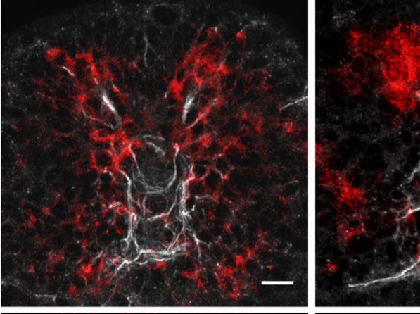
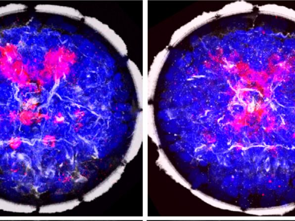
Figure 1. Coexpression of Opsins, Clock
A B 48hpf C 48hpf Genes, and hiomt in the Dorsal Larval Brain
cPRCs (A) SEM of a Platynereis trochophore larva,
episphere showing the position of the developing apical ner-
vous system (episphere) and the prototroch (image
ptrn
courtesy of H. Hausen).
prototroch * * (B) Z-projection of the brain axonal scaffold at 48
A D hpf, stained with an anti-acetylated tubulin anti-
vcc
body, apical view. Main landmarks are indicated.
R L R L hiomt + L-cry + CNGAα
cPRCs, ciliary photoreceptor cells (yellow arrows);
hiomt + L-cry + CNGAα + c-opsin1
P V prototroch ptrn, prototroch ring nerve; vcc, ventral cerebral
commissure. Scale bar, 20 mm.
D 34hpf E 34hpf F 34hpf (C) Average gene coexpression at 48 hpf obtained
with Profiling by Image Registration (PrImR). Blue,
coexpression of hiomt, L-cry, CNGAa, and c-
opsin1; magenta, coexpression of hiomt, L-cry,
and CNGAa; gray, average axonal scaffold.
* * * (D–F) Expression of hiomt (D), CNGAa (E), and
L-cry (F) at 34 hpf. All images are Z-projections of
the entire episphere (apical views, dorsal up). Blue,
DAPI; red, gene expression; gray, axonal scaffold.
hiomt CNGAα L-cry (G–K) Expression of hiomt (G), CNGAa (H), L-cry (I),
peropsin (J), and CNGAb (K) at 48 hpf. All images
G 48hpf H 48hpf I 48hpf are 10 mm Z-projections of the extended cPRCs
region (apical views, dorsal up). Red, gene
expression; gray, axonal scaffold. Arrows, cilia of
cPRCs; asterisks, apical organ. Scale bar, 10 mm.
(L) Schematic representation of the larval brain; the
region harboring expression of opsins, CNG-based
* * * phototransduction, clock genes, and melatonin
synthesis markers is highlighted.
See also Figures S1 and S2.
hiomt CNGAα L-cry
opsins
J 48hpf K 48hpf L CNG-based phototransduction
circadian clock
genomes (Figure S1 available online).
melatonin synthesis
cPRCs Therefore, in order to identify the Platy-
ptrn nereis melatonin-producing cells, we per-
* formed whole-mount in situ hybridization
* *
D
vcc
(WMISH) with probes detecting tran-
R L
scripts of Platynereis hiomt and of marker
genes for phototransduction and circa-
peropsin CNGAβ V
prototroch dian clock (Figure 1). Expression was de-
tected in the episphere, where the larval
brain differentiates (Figures 1A and 1B),
melatonin signaling may play a role in the diurnal control of ciliary starting at 34 hr postfertilization (hpf) in dorsomedial cells (Fig-
swimming. To elucidate the possible interplay between ambient ures 1D–1F) and covering the extended ciliary photoreceptor
light detection, melatonin signaling, and circadian larval swim- region at 48 hpf (dashed circles in Figures 1G–1J). This area
ming activity in an invertebrate zooplankton larva, we chose comprises the previously described ciliary photoreceptors
the annelid Platynereis dumerilii, a marine animal model amen- expressing c-opsin1 (Arendt et al., 2004). In vertebrate ciliary
able to molecular and behavioral studies, which has conserved photoreceptors, phototransduction involves the G-alpha sub-
vertebrate-type ciliary photoreceptors (Arendt et al., 2004; unit Gt, a member of the Gi family, and nonselective cationic
Jékely et al., 2008). cyclic nucleotide-gated (CNG) channels, which lead to an OFF
response (hyperpolarization) in the presence of light. The same
RESULTS phototransduction components were expressed specifically in
the Platynereis dorsal brain (Figures S2A–S2C, 1E, 1H, and
Markers of Melatonin Synthesis, Phototransduction, 1K). Intriguingly, markers of the circadian clock, like bmal (Arendt
and Circadian Clock Are Coexpressed in the Dorsal et al., 2004), vrille, and L-cry, the ortholog of the Drosophila light-
Brain of Platynereis Larvae sensitive cryptochrome, were expressed in the same region (Fig-
As an entry point, we found that the most specific marker of ures S2D, 1F, and 1I; see also Zantke et al., 2013). Coexpression
melatonin synthesis, the gene hydroxyindole-O-methyltransfer- of these genes was apparent from the similar position of stained
ase (hiomt) (Nowak et al., 1993), is conserved in several bilaterian cells adjacent to the photoreceptor cilia (arrows in Figure 1) and
Cell 159, 46–57, September 25, 2014 ª2014 The Authors 47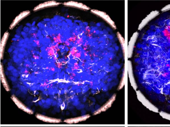
A B Figure 2. Global Changes of Gene Expres-
ZT 5 - 7 .5hp f
p
tr-cry
11 2. f
5h f
period
ZT 5 - 6 3.5h
- 7 5hp
2.5 sion during the Light-Dark Cycle
ZT - 5 pf
ZT - 6 f
ZT .5 - hpf
pf
20 7hp
1.5
relative expr.
relative expr.
ZT - 48 f
ZT - 5 f
hp
14 hp
17 4h
6
7
23 0
2.0 (A–D) Gene expression profiling of Platynereis
ZT - 45
1.0
1.5 larvae between 45 and 75 hpf with Fluidigm Dy-
3.
8.
5
8
ZT
0.5 1.0
namic Arrays. (A) Hierarchical clustering of scaled
0 0.5
gene expression levels. Sampling stages and
CNGAβ
45 50 55 60 65 70 75 45 50 55 60 65 70 75 circadian times indicated at the top. High and
vtn
time (hpf) time (hpf)
NPY-4 low normalized expression levels are indicated
neurops with green and magenta, respectively. (B and C)
r-ops4
CNGB
C Expression profiles from the Fluidigm screen.
CNGAα peropsin CNGAα Gene expression levels are plotted as relative to
aanat 6 the expression at 45 hpf. Each point is the average
relative expr.
relative expr.
mao 5 4
4
of the two technical replicates (see the Extended
ddc 3
hiomt 3 Experimental Procedures for details). Error bars
2
FMRF 2 indicate SD. (B) Expression of the clock genes tr-
L-cry 1 1
TPH
cry and period between 45 and 75 hpf. The red
45 50 55 60 65 70 75 45 50 55 60 65 70 75
not lines indicate relative expression in siblings raised
time (hpf) time (hpf)
bmal under an inverted light-dark cycle (the red solid
gch
hiomt
bars below indicate the night phase of the inverted
perops sert
YFamide 4
10 cycle). (C) Examples of day-night gene expression
relative expr.
relative expr.
PDF 8 dynamics. CNGAa and hiomt are upregulated
vmat 3 6
bsx
at night; sert is developmentally regulated; and
2 4
r-ops3 peropsin is both night upregulated and devel-
phc2 2
1 opmentally regulated. (D) Schematic of the sero-
MTNR
FVRI
45 50 55 60 65 70 75 45 50 55 60 65 70 75 tonin-melatonin biosynthesis pathway. The genes
time (hpf) time (hpf)
syt significantly upregulated at night are shown in
WLDamide green. Hpf, hours post fertilization; ZT, zeitgeber
RYamide
DLamide time.
FVamide D See also Figure S3.
FLamide
sert
r-ops1 Gch Vmat
tr-cry TPH Ddc
clock Trp 5HTP serotonin
period Aanat tive PCR (qPCR) screen (Figures 2 and
c-ops1
Hiomt S3). To this aim, Platynereis larvae were
Sert
melatonin sampled between 45 and 75 hpf (with
the night phase between 56 and 64 hpf).
-1 0 1
The circadian system is already active
Normalized expression and entrainable at these larval stages:
the expression levels of clock genes
confirmed with the Profiling by Image Registration (PrImR) tech- (such as clock, period, and tr-cry; Zantke et al., 2013) oscillated
nique (Tomer et al., 2010), which revealed coexpression of Platy- throughout the light-dark cycle, and in sibling larvae raised in an
nereis hiomt, L-cry, CNGAa, and c-opsin1 in a few cells of the inverted light-dark cycle (shifted by 12 hr), the phase of clock
dorsal brain, including the ciliary photoreceptors (blue in Fig- gene expression was likewise shifted by 12 hr (Figure 2B). Clus-
ure 1C). Interestingly, the expression of melatonin synthesis, tering analysis of gene expression changes over our entire data
phototransduction, and clock markers was broader than the set retrieved two major groups: genes with expression levels
highly restricted c-opsin1 expression (magenta in Figure 1C) constantly increasing throughout development (reflecting the
and overlapped with that of other opsin family representatives, steady increase in the number of differentiated cells at these
such as neuropsin (Figure S2E) and peropsin (Figure 1J; stages) and genes upregulated specifically during the night (Fig-
compare extended red coexpression region in summary Fig- ures 2A, 2C, and S3). The first group included neuronal differen-
ure 1L). Therefore, the Platynereis dorsal brain is a complex tiation markers, such as synaptotagmin (syt) and prohormone
site of nonvisual light sensitivity. Our data suggest that photo- convertase 2 (phc2), several neuropeptides (Conzelmann et al.,
sensitivity, circadian entrainment, and melatonin release are 2011), and rhabdomeric opsins expressed in the eyes (r-ops1
directly coupled, at the cellular level, in the Platynereis dorsal and r-ops3; Randel et al., 2013). The second group included
brain, just as in pineal photoreceptors. several genes expressed in the hiomt+ cells, such as peropsin,
neuropsin, and CNG channel subunits, indicating a night-depen-
Melatonin-Synthesis and Phototransduction Genes Are dent change of light sensitivity. Moreover, all genes involved in
Specifically Upregulated at Night the melatonin synthesis and degradation pathway (TPH, ddc,
The first experimental evidence for such coupling was obtained mao, aanat, and hiomt) were upregulated at night, whereas those
from a quantification of gene expression dynamics during the exclusively involved in serotonin signaling (vmat, sert) were not
light-dark cycle. We measured simultaneously the expression (Figures 2C and 2D). These findings suggested that melatonin it-
levels of clock genes, neuropeptides, opsins, and genes of the self is produced and released during the night, as reported in
melatonin synthesis pathway using a high-throughput quantita- other invertebrate species (Hardeland and Poeggeler, 2003),
48 Cell 159, 46–57, September 25, 2014 ª2014 The Authors
and that melatonin release is directly controlled by light and/or by     choline (ACh) significantly increased ciliary closure time,
the circadian clock.                                                     whereas the acetylcholine receptor antagonist mecamylamine
                                                                         was able to suppress the ACh effect and reduce ciliary arrests
Melatonin Signaling Establishes a Nocturnal                              (Figure 4A). Notably, mecamylamine specifically suppressed
Behavioral State                                                         the long closures that are regulated by melatonin signaling (Fig-
To identify and characterize a possible effect of nocturnal mela-        ure 4B). Acetylcholinesterase staining confirmed direct innerva-
tonin release on larval behavior, we assayed larval ciliary swim-        tion of the prototroch by cholinergic axons (Figure 4C). In partic-
ming. In Platynereis, the larval brain controls ciliary locomotion       ular, the ventral cholinergic neurons, some of which directly
by direct innervation of the prototroch, an equatorial girdle of         innervate the prototroch (cfr MN3l/MN3r in Randel et al., 2014),
cells equipped with multiple motile cilia (Figures 1A and 1B).           qualified as candidate melatonin-responsive neurons, as indi-
Changes in the ciliary beating frequency (CBF) and in ciliary            cated by the coexpression of the Platynereis melatonin receptor
closure time (i.e., length and frequency of ciliary arrests) differen-   (MTNR; Figure S2) and the cholinergic marker ChAT (Figure 4D).
tially affect swimming speed, depth, and direction (Conzelmann
et al., 2011; Jékely et al., 2008). Thus, we assayed CBF and            Nocturnal Melatonin Signaling Induces Rhythmic
ciliary arrests at 52 hpf, comparing larvae in their midday and          Activity of Cholinergic Ciliomotor Neurons
midnight (ZT8 and ZT20; ZT, zeitgeber time; Figure 3A). Although         To test whether melatonin signaling modulates neuronal activity,
CBF did not differ significantly between day and night (Figure 3B),      we performed two-photon imaging on larvae expressing the
we found a significant increase in overall ciliary closure time at       genetically encoded calcium indicator GCaMP6s. To facilitate
night (Figure 3C). Treatment at night with the melatonin receptor        the identification of the ventral cholinergic neurons, we took
antagonist luzindole dramatically decreased closure time (Fig-           advantage of the fact that these cells are located at the interface
ure 3D), indicating that the nocturnal increase in ciliary closure       between the two halves of the larval brain developing from the AB
time depends on melatonin signaling. Consistently, daytime               and CD blastomeres (Figure S4). Therefore, we expressed
treatment with 100 mM melatonin was sufficient to almost double          GCaMP6s and H2B-RFP in the CD lineage by single-blastomere
ciliary closure time (Figure 3E). Finally, we found that absence of      injections (Figure 5A). In control conditions, we detected sparse,
light during daytime was not sufficient by itself to increase ciliary    arrhythmic small-amplitude calcium transients in these neurons
closure time (Figure 3F). Our data are thus consistent with the          and along their projections. Unexpectedly, the application of
possibility that the circadian clock contributes to the regulation       melatonin elicited enhanced neuronal activity in the most ventral
of the melatonin rhythm (as in vertebrates; Falcón et al., 2010).       cell of the cholinergic cluster. This heightened neuronal activity
   Next, we reasoned that the ciliary closure time depends on two        also revealed a direct contralateral projection to the prototroch
parameters: the frequency and the length of the closure events.          cells through the ventral cerebral commissure (Figure 5B). Mela-
Therefore, we dissected how these parameters change during               tonin robustly induced a sustained rhythmic activity pattern,
day and night. At night, the frequency of ciliary arrests is signifi-    characterized by the appearance of GCaMP6s peaks of regular
cantly higher compared to daytime (Figure 3G). Moreover, the             amplitude and frequency (mean frequency: 0.43 Hz; Figure 5C;
bimodal distribution of ciliary closure lengths (Figure 3H) indi-        Movie S1). The melatonin-induced calcium peaks were readily
cated the existence of two distinct kinds of arrests, the ‘‘short’’      suppressed by subsequent addition of the melatonin receptor
and ‘‘long’’ closures (with an average length of 0.2 and 2.2 s,          antagonist luzindole (Figure 5C). Moreover, we detected this ac-
respectively; Figure 3H). The day-night transition involves a sig-       tivity pattern specifically in the prototroch-projecting ciliomotor
nificant increase of the proportion of long closures (Figures 3I         neuron and never in any of the neighboring CD cells (Figure 5D).
and 3I’). The melatonin receptor antagonist luzindole applied at         To test whether the melatonin-induced activity recapitulates the
night recapitulated the diurnal condition: the overall frequency         nocturnal endogenous activity, we performed the same experi-
of arrests was reduced (Figure 3J) and the long closures were            ments at night. In untreated larvae, we found spontaneous rhyth-
suppressed (Figures 3K and K0 ). Conversely, treatment with              mic calcium peaks reminiscent of the melatonin-induced activity
melatonin during the day increased the proportion of long clo-           (mean frequency: 0.31 Hz; Figure 5E). These peaks were sup-
sures to night levels (Figures 3L and 3L0 ). Taken together, our         pressed by application of luzindole (Figure 5E), indicating that
data indicate the existence of two alternative behavioral states         at night this rhythmic activity is induced by endogenous mela-
during day and night, which correlate with the day-night differ-         tonin release. Analysis of GCaMP6s peak frequency and tempo-
ences in gene expression profile. Melatonin signaling is neces-          ral distribution confirmed that daytime administration of mela-
sary and sufficient to increase ciliary closure frequency and            tonin mimics the spontaneous nocturnal activity (Figures 5F
length, establishing the nocturnal behavioral state.                     and 5G). Notably, the rhythmic activity of the cholinergic neuron
                                                                         proved insensitive to mecamylamine treatment (Figure S5), indi-
Cholinergic Neurons Control Ciliary Arrests                              cating that the effect of cholinergic agonists and antagonists on
To dissect the mechanism of ciliary arrest, we set out to identify       ciliary arrests occurs downstream of these neurons (i.e., at the
the ‘‘ciliomotor neurons’’ driving ciliary arrests and to test           cholinergic ciliomotor synapses on the prototroch cells).
whether they are responsive to melatonin signaling. Given that
the prototroch cells express the acetylcholine receptor a9/10            Melatonin-Induced Bursting Enhances
(Jékely et al., 2008), the best candidates were the early devel-        Synaptic Transmission
oping cholinergic neurons previously identified in the larval brain      To better understand the neurophysiological processes that un-
(Jékely et al., 2008). Consistent with this, application of acetyl-     derlie the observed rhythmic firing and the nocturnal change in

                                                                              Cell 159, 46–57, September 25, 2014 ª2014 The Authors 49
A B C D E
behavioral 20 *** *
fertilization
assay
***
40 25 * 40
closures (% of time)
closures (% of time)
closures (% of time)
night 15
30 20 30
(ZT20)
CBF (beats/s)
10 15
20 20
day 10
(ZT8)
5 10 10
5
0 24 48 0 0 0 0
50μM 100μM 100μM
developmental stage (hpf) day night day night control control
luzindole mel
F G H I night I’
**
relative frequency of closures / min short day 40
% long closures
*
15 1.0
40
number of closures / min
number of closures / min
20
0.8
2
closures (% of time)
30
0.6
10 0
long day night
0.4
20 1
5
0.2
10
0.0
0
0 0
dark 0.05 0.20 1 5 20 0.05 0.20 1 5 20
day day night
adapted closure length (s) closure length (s)
J K control K’ L control L’
luzindole 40 melatonin *
*** ***
% long closures
% long closures
40
15
3 20 2
number of closures / min
number of closures / min
20
number of closures / min
10
2 0 0
100μM 100μM
control control
luzind. 1 melat.
5 1
0 0 0
100μM 0.05 0.20 1 5 20 0.05 0.20 1 5 20
control
luzindole closure length (s) closure length (s)
Figure 3. Melatonin Signaling Modulates Nocturnal Locomotor Activity
(A) Design of the behavioral experiments. Larvae were assayed for locomotor activity at 52 hpf (red arrows), corresponding to ZT8 (midday) or ZT20 (midnight).
(B) Ciliary beating frequency (CBF; beats/s) during day and night. n = 59 and 25 larvae. p = 0.44, unpaired t test.
(C) Ciliary closure time (% of total time) during day and night. p < 0.001, unpaired t test. n = 72 and 55 larvae.
(D) Ciliary closure time (% of total time) at night, in larvae treated with 50 or 100 mM of the melatonin receptor antagonist luzindole in DMSO (gray). Control = 0.1%
DMSO (blue). p < 0.05 and p < 0.001, unpaired t test. n = 30, 12, and 16 larvae.
(E) Ciliary closure time (% of total time) during daytime; larvae were treated with 100 mM of melatonin in NSW (gray). Control = NSW (yellow). p < 0,05, unpaired t
test. n = 28 and 31 larvae.
(F) Ciliary closure time (% of total time) at ZT8; larvae were imaged under normal daylight or in complete darkness after 1 hr of dark adaptation. n = 27 and 23
larvae.
(G) Frequency of ciliary arrests (number of closures/min) during day and night. n = 58 and 55 larvae, respectively. p < 0.05, unpaired t test.
(H) Probability density plot of ciliary closure lengths (on the logarithmic scale) during daytime, showing a bimodal distribution with distinct long and short arrests.
(I) Normalized distribution of ciliary closure lengths during day (yellow) and night (blue). n = 58 and 55 larvae.
(I0 ) Percentage of long ciliary arrests during day and night; data are from (I). Long arrests are 33% and 42% of total arrests, respectively. p < 0.01, chi-square test.
(J) Frequency of ciliary arrests (number of closures/min) in controls (0.1% DMSO, blue) and in larvae treated with 100 mM luzindole (gray) during the night. n = 30
and 31 larvae. p < 0.001, unpaired t test.
(K) Normalized distribution of ciliary closure lengths in controls (0.1% DMSO, blue) and in larvae treated with 100 mM luzindole (gray). n = 30 and 31 larvae.
(K0 ) Percentage of long ciliary arrests in controls (0.1% DMSO) and in larvae treated with 100 mM luzindole at night; data are from (K). Long closures are 29% and
2% of total, respectively. p < 0.001, chi-square test.
(L) Normalized distribution of ciliary closure lengths in controls (NSW, yellow) and in larvae treated with 100 mM melatonin (gray). n = 24 and 23 larvae.
(L’) Percentage of long ciliary arrests in controls and in larvae treated with 100 mM melatonin during the day; data are from (L). Long closures are 33% and 42% of
total, respectively. p < 0.05, chi-square test. In (B)–(G) and (J), data are shown as mean ± SEM, and error bars indicate SEM. NSW, natural seawater.
ciliary arrests, we performed intracellular sharp electrode re- potential recordings (Conzelmann et al., 2011); however, intra-
cordings from prototroch cells. Ciliary arrests coincided with cellular recordings with simultaneous imaging of ciliary beating
electrical activity, as reported previously from extracellular field revealed that these arrests coincide with depolarizations that
50 Cell 159, 46–57, September 25, 2014 ª2014 The Authors
A                                 B                             B’                  tonin was reversible with application of the melatonin receptor
                                                                                    antagonist luzindole (Figure 6E). Moreover, application of meca-
                                                                                    mylamine after melatonin treatment completely eliminated excit-
                                                                                    atory synaptic transmission, indicating that this activity was
                                                                                    exclusively cholinergic (Figure 6E). These results indicate that
                                                                                    each nocturnal peak of GCaMP6s fluorescence corresponds to
                                                                                    a burst of action potentials in the presynaptic cholinergic cells,
                                                                                    increasing the likelihood of prototroch spiking. To further assess
                                                                                    the effect of melatonin-induced bursting on synaptic transmis-
                                                                                    sion, we quantified EPSP amplitude before and after application
                                                                                    of melatonin. This revealed a significant increase of the mean
                                                                                    EPSP amplitude, indicating that the number of synaptic vesicles
C                                         D                                         released per action potential more than doubles during the mela-
                                                                                    tonin-induced bursting (Figures 6F and 6G). At the same time,
                                                                                    the mean resting membrane potential of the prototroch cells
                                                                                    (which do not express MTNR) was not significantly altered in
                                                                                    presence of melatonin (from 70.45 to 70.39 mV, n = 7, ns).
                                                                                    Therefore, we conclude that at night melatonin signaling directly
                                                                                    controls the excitability of presynaptic cholinergic neurons, that
                                                                                    it induces rhythmic bursting and potentiates synaptic transmis-
                                                                                    sion at the ciliomotor-prototroch cells synapses, and that this
                                                                                    augmented release of acetylcholine enhances the frequency
                                                                                    and duration of ciliary arrests.

Figure 4. Ciliary Arrests Are Triggered by Cholinergic Transmission                 DISCUSSION
(A) Ciliary closure time (% of total time) during daytime, in controls (NSW,
yellow), and in larvae treated with 1 mM ACh (red), with 1 mM of the cholinergic
antagonist mecamylamine or with a mixture of 1 mM of mecamylamine and               Our combination of expression profiling, neuronal activity imag-
1 mM ACh (gray). One-way ANOVA with post hoc Holm adjustment, *p < 0.05,            ing, and intracellular recordings in a zooplankton model reveals
***p < 0.001. n = 15, 19, 22, and 17 larvae, respectively. Error bars represent     a role of melatonin signaling in the circadian control of ciliary
SEM.                                                                                swimming. In Platynereis, melatonin is necessary and sufficient
(B) Normalized distribution of ciliary closure lengths in controls (0.95% EtOH,     to establish a nocturnal behavioral state characterized by
yellow) and in larvae treated with 100 mM mecamylamine (gray). n = 17 and 20
                                                                                    enhanced ciliary arrests. We can distill the underlying circuit ar-
larvae.
(B0 ) Percentage of long closures in controls (0.95% EtOH) and in larvae treated    chitecture into two major components (red and blue in Figure 7).
with 100 mM mecamylamine during the day; data are from (B). Long closures           The first is the sensory-neuromodulatory component, consti-
are 35% and 14% of total, respectively. p < 0.001, chi-square test.                 tuted by melatonin-releasing, ambient light-detecting photore-
(C) Acetylcholinesterase (AChE) staining of a 52 hpf Platynereis trochophore        ceptors that harbor a circadian clock. The second is the effector
larva (apical view), showing signal in the prototroch ring nerve (ptrn). The blue   component, represented by the cholinergic ciliomotor neurons,
arrowheads indicate the ventral brain cholinergic neurons and projections to
                                                                                    which are direct targets of nocturnal melatonin signaling and
the ptrn through the ventral cerebral commissure (vcc).
(D) Coexpression (white) of melatonin receptor (MTNR, red; see also Figure S2)
                                                                                    respond by rhythmic bursting. This system represents a minimal-
and the cholinergic marker ChAT (green) in the 48 hpf larval brain, after image     istic example of integration of sensory-neuromodulatory and
registration. Arrowheads, ventral brain cholinergic neurons.                        motor circuits in animal nervous systems. Comparative evidence
                                                                                    indicates that at least parts of these circuits are of more wide-
                                                                                    spread occurrence in animals and may thus be evolutionary
arise from an intrinsic electrical excitability of the prototroch cell              ancient.
(Figure 6A). Consistent with excitatory cholinergic transmission,
application of acetylcholine induced prototroch depolarization,                     Complex Integration of Ambient Light in the ‘‘Apical
and this effect was suppressed by concurrent mecamylamine                           Nervous System’’
application (Figure S6). Application of melatonin caused a signif-                  The shared key feature of the dorsal brain neurons described
icant increase in prototroch spike frequency (Figures 6A–6C), as                    here is the coexpression of hiomt with markers of opsin-based
well as a decrease in the distribution of interspike intervals (Fig-                phototransduction and the circadian clock. The circadian clock
ure 6D), reflecting the sustained electrical activity that would be                 is already entrained in early larvae, and several genes expressed
expected given the effect melatonin has on the length of ciliary                    in these cells, including hiomt itself, are upregulated at night,
arrests.                                                                            suggesting that ambient light detection and the circadian clock
   Intracellular recordings also revealed the presence of excit-                    control melatonin synthesis. A coupling of these three processes
atory postsynaptic potentials (EPSPs). In agreement with the cal-                   in the same cells is also observed in the pineal photoreceptors
cium imaging data, melatonin changed prototroch EPSPs from                          of lower vertebrates (Lamb, 2013), and intriguingly, the Platyner-
single sparse events to regular periodic bursts, each character-                    eis ciliary photoreceptors (previously compared to pineal and
ized by a duration of 0.25 ± 0.08 s (Figure 6E). The effect of mela-                retinal photoreceptors; Arendt et al., 2004) are part of the

                                                                                         Cell 159, 46–57, September 25, 2014 ª2014 The Authors 51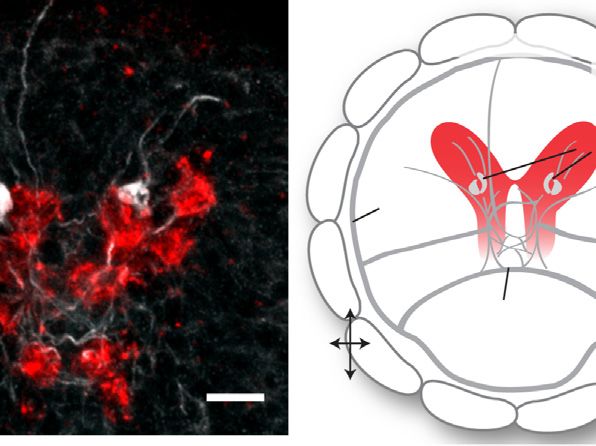
A                                                     B

         C

         D                                  E

                          F                                G

                                                                   (legend on next page)

52 Cell 159, 46–57, September 25, 2014 ª2014 The Authors
clock-controlled, melatonin-releasing system. The expression of                          In Platynereis larvae, the melatonin-sensitive cholinergic sys-
multiple opsins in nonvisual photoreceptors of the most anterior                      tem regulates distinct, long-lasting diurnal and nocturnal behav-
(or apical) part of the larval nervous system has been docu-                          ioral states. At the level of the ciliomotor neurons, this regulation
mented in other protostomes and in deuterostomes (Ooka                                involves the switch from sparse firing to rhythmic bursting. This
et al., 2010; Passamaneck et al., 2011). A shared feature of this                     finding is especially interesting as it may shed light on the evolu-
so-defined larval ‘‘apical nervous system’’ (specified by                             tionary origin of circadian behavioral states in animals. Circadian
conserved developmental patterning mechanisms; Marlow                                 behavioral states, such as sleep-wake cycles, are widespread in
et al., 2014) is the presence of multiple sensory cells that project                  the animal kingdom, indicating that the day-night regulation of
into a neurosecretory neuropil and release hormones and neuro-                        activity rhythms is crucial for homeostasis and survival (Allada
modulators (Tosches and Arendt, 2013).                                                and Siegel, 2008). A defining and conserved feature of sleep,
   The expression of different opsins in subsets of hiomt+ cells in                   which makes it distinct from rest, is the disconnection of the
Platynereis hints at a sophisticated responsiveness of the apical                     brain from the sensory environment and hence the increase of
nervous system to light. Opsins responding to different wave-                         arousal threshold. The function of this sensory disconnection is
lengths would allow dissecting the spectral composition of light                      still mysterious. Platynereis larvae can respond to a variety of
at different depths or during dusk and dawn and may also                              sensory cues, which would ultimately affect ciliary arrests
contribute to moonlight detection for the regulation of circalunar                    through the same ciliomotor system. For example, ciliomotor
rhythms, which in adult worms has been attributed to the brain re-                    neurons are innervated by interneurons of the larval visual sys-
gion harboring the ciliary photoreceptors (Zantke et al., 2013).                      tem, indicating that they respond to visual stimuli, together
The upregulation of peropsin, neuropsin, and CNG channels                             with other motoneurons (Randel et al., 2014). But could any sen-
during the dark phase might then prepare the larvae to sense                          sory cue be relayed in presence of rhythmic bursting? If not, the
moonlight. The expanding array of functional tools available for                      nocturnal rhythmic burst firing might be a mechanism that filters
Platynereis (Zantke et al., 2014) will allow testing these hypothe-                   incoming sensory information and reduces responsiveness to
ses and unraveling the contributions of individual opsins and neu-                    sensory stimuli. In a similar way, in mammals, the switch of
ropeptides to the annelid ambient light-dependent behaviors.                          thalamic relay neurons to rhythmic burst firing is responsible
                                                                                      for filtering sensory information during sleep (McCormick and
A Cholinergic Ciliomotor System Mediating a Circadian                                 Feeser, 1990) and can be induced directly by melatonin
Behavioral State Switch                                                               (Ochoa-Sanchez et al., 2011). It will be interesting to determine
On the effector side, we identify a pair of cholinergic ciliomotor                    whether these functional analogies correspond to conserved
neurons as regulators of ciliary arrests. These neurons belong                        molecular mechanisms, for instance, to conserved targets
to a larger cholinergic system that innervates larval ciliary bands,                  downstream of melatonin signaling. Promising candidates are
as revealed by acetylcholinesterase staining; within this system,                     Shaker voltage-gated potassium channels, key determinants
they are the only neurons expressing the melatonin receptor and                       of neuronal excitability, which are regulated by melatonin
responding to melatonin treatments. Cholinergic innervation of                        (Yang et al., 2011) and are necessary for sleep in flies and mam-
ciliary bands has been observed in most protostome and                                mals (Cirelli et al., 2005; Douglas et al., 2007).
deuterostome larvae, and a general requirement of cholinergic
transmission for ciliary arrests has been also documented (La-                        Early Evolution of Melatonin Signaling in the Marine
calli and Gilmour, 1990; Lacalli et al., 1990), indicating that the                   Environment
cholinergic ciliomotor system described here is of broader rele-                      In marine zooplankton, the most prominent behavior associ-
vance for understanding zooplankton neurobiology.                                     ated with day-night cycles is DVM (see Introduction). We

Figure 5. Melatonin-Dependent Rhythmic Activity in Ventral Cholinergic Neurons
(A) Left: expression of GCaMP6s (green) and H2B-RFP (red) in the brain of larvae injected with GCaMP6s and H2B-RFP mRNAs at two-cell stage (Z-projection of
the episphere, apical view). Scale bar, 30 mm. Middle and right: magnification of the most ventral cells of the CD lineage labeled by GCaMP6s and H2B-RFP.
These cells correspond to ventral brain cholinergic neurons (compare Figure S4). Scale bar, 10 mm.
(B) SD of GCaMP6s fluorescence (apicoventral view) after melatonin treatment in a 2-min-long recording, showing that the most ventral neuron of the CD domain
(compare cell number 5 in A) responds to melatonin with dynamic activity (high SD) and projects to the prototroch (pr, dashed line) through the ventral cerebral
commissure (vcc). Scale bar, 10 mm.
(C) GCaMP6s fluorescence of a representative prototroch-projecting cholinergic neuron (cell number 5 in A) during daytime, in control conditions (left), after
treatment with 100 mM melatonin (middle), and after subsequent addition of 100 mM of the melatonin receptor antagonist luzindole (right). Upper row: GCaMP6s
signal normalized with a global F0; lower row: GCaMP6s signal normalized with a local F0 (moving average over a window of 0.8 s), to highlight changes in
fluorescence over a small timescale (see the Experimental Procedures for details on the analysis).
(D) GCaMP6s fluorescence of the cells 1–4 (A) after treatment with melatonin. Data normalized with the moving average approach.
(E) GCaMP6s fluorescence of a representative prototroch-projecting cholinergic neuron at night, in control conditions (left), and after treatment with 100 mM
luzindole (right).
(F) Frequency of GCaMP6s peaks (number of peaks/min) in untreated larvae during daytime (yellow), after melatonin treatment during daytime (gray), and in
untreated larvae during the night (blue). n = 10, 8, and 4 larvae, respectively. One-way ANOVA with post hoc Holm adjustment, *p < 0.05, ***p < 0.001.
(G) Bean plot showing the distribution (on the logarithmic scale) of the peak-to-peak intervals (s) in untreated larvae during daytime (yellow), after melatonin
treatment during daytime (gray), and in untreated larvae at night (blue). Thin white lines indicate single data points; black horizontal lines indicate means. n = 10, 8,
and 4 larvae, respectively.
See also Figures S4 and S5 and Movie S1.

                                                                                             Cell 159, 46–57, September 25, 2014 ª2014 The Authors 53
A day, untreated 10mV E day, untreated 2mV
2 sec 0.5s
day, melatonin
membrane potential
kymograph
day, melatonin + luzindole
B day, melatonin 10mV
2 sec
day, melatonin + mecamylamine
membrane potential
kymograph
C D 5000 F G **
inter-spike interval (ms)
*** 2000
EPSP amplitude (mV)
1000
6
5
spike frequency
melat.
5
500
4
(spikes / s)
4
1mV
3
200
3
1ms
ctrl
2
2
100
1
1
50
0
0
ctrl melat. control melatonin ctrl melat.
Figure 6. Melatonin Induces Rhythmic Bursting of Cholinergic Prototroch Presynaptic Neurons and an Enhanced Release of ACh
(A) Intracellular recording from a prototroch cell during daytime (top), and kymograph of ciliary beating imaged simultaneously (bottom; black bars: ciliary arrests),
showing the correlation of prototroch spikes with ciliary arrests (100 mM).
(B) Same as (A), in a melatonin-treated larva (100 mM).
(C) Average prototroch spike frequency in controls (NSW) and in melatonin-treated larvae. p < 0.001, n = 7 larvae each. Error bars, SEM.
(D) Bean plot showing the distribution (on the logarithmic scale) of interspike intervals (ms) in controls (NSW, yellow) and in melatonin-treated larvae (100 mM,
gray). Thin white lines indicate single data points; black horizontal lines indicate means. n = 7 larvae each.
(E) EPSPs recorded from prototroch cells. Traces (from top to bottom) represent EPSPs in untreated larvae and EPSPs after treatment with melatonin (100 mM),
melatonin plus subsequent addition of luzindole (100 mM), and melatonin plus subsequent addition of mecamylamine (100 mM).
(F) Average EPSP responses measured from prototroch cells in absence (yellow) and the presence of melatonin (gray). n = 7 larvae.
(G) Average EPSPs amplitude (mV) in controls (yellow) and melatonin-treated larvae (gray). n = 7 larvae, p < 0.01, paired t test. Error bars, SEM.
See also Figure S6.
propose that the day-night changes in ciliary closure time oxidative stress (Gehring and Rosbash, 2003; Pittendrigh,
evoked by melatonin signaling in Platynereis correspond to 1993). Light-sensitive cryptochromes, present in all basal meta-
different phases of DVM in the field. In Platynereis, a long ciliary zoan phyla, including sponges (Rivera et al., 2012), might have
closure time correlates with a low vertical position (Conzel- represented the first link between ambient light detection and
mann et al., 2011); therefore, the nocturnal behavior would clock entrainment. We now extend this reasoning with the idea
lead to the slow descent of larvae in the water column. In that melatonin signaling has been added to this system as an
line with this conclusion, field studies have shown that plank- efficient paracrine signal, ensuring better coordination of the
tonic annelids reach their highest position in the water column organismal response and whole-body physiology and becoming
at dusk and then sink throughout the whole night (Alldredge a perfect ‘‘chemical indicator of darkness’’ after its original
and King, 1980). Other zooplankton species show different radical scavenger role. Because melatonin receptors and opsins
temporal DVM regimes that often involve muscular rather arose from the same duplication of an ancestral G-protein-
than ciliary swimming, suggesting a more complex regulation coupled receptor (Feuda et al., 2012), we further hypothesize
of locomotor activities downstream of melatonin signaling as that melatonin signaling and opsin-based phototransduction
an adaptation to variable ecological constraints (Lampert, evolved in the same DVM context. As photopigment, opsin out-
1989). performed cryptochrome by increased signaling speed, signal
Interestingly, it has been proposed that circadian rhythms and amplification, coupling with different transduction cascades
ambient light detection evolved in the context of DVM, an ad- and an evolvable spectral tuning that, after repeated duplica-
vantageous behavior facilitating escape from light-induced tions, allowed perception of colors at different depths and times
54 Cell 159, 46–57, September 25, 2014 ª2014 The Authors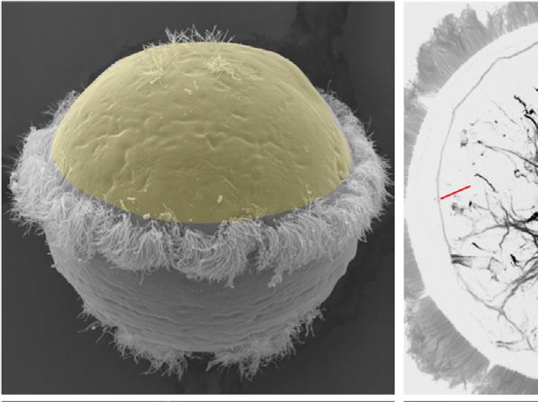
Figure 7. Day-Night Modulation of Platyne-
  DAY                                                       NIGHT                                                    reis Ciliary Swimming by Melatonin Signaling
             L-cry,                 opsins,                            L-cry,                 opsins,                In the extended ciliary photoreceptor region (red),
           circadian      ?          CNG                             circadian        ?        CNG                   the circadian clock and various opsins with a
             clock                 channels                            clock                 channels                CNG-based phototransduction cascade regulate
                                                                                                                     rhythmic release of melatonin. During the day (left),
                                                                                                                     ventral cholinergic ciliomotor neurons (blue) fire
                       melatonin                                                 melatonin                           sporadically and arrhythmically. Basal acetylcho-
                                                                                                                     line release at the ciliomotor-prototroch synapses
                                                                                                                     ensures a low frequency and duration of ciliary
                                                                                                                     arrests. At night (right), high melatonin levels
                                                                                      melatonin                      directly affect the electrical activity of the cholin-
                                                                                                                     ergic ciliomotor neurons, which express the
                                                                                                                     melatonin receptor (MTNR). Therefore, the cho-
                                                                                                                     linergic neurons switch to a rhythmic bursting
                                                                                                                     mode, which boosts the release of acetylcholine at
                                   MTNR                                                       MTNR                   the ciliomotor-prototroch synapses (i.e., increase
                                                                                                                     of EPSPs amplitude). This increases the frequency
                              ACh                                                         ACh
                                                                                                                     of spiking in prototroch cells, causing longer and
                                                                                                                     more frequent ciliary arrests.

                                                                                            and rps9 were used to obtain the normalized dCq
                                                                                            values, and for each time point the two dCqs from
                 sparse firing                             rhythmic burst firing            the two PreAmps were averaged. Expression rela-
                                                                                            tive to the first time point (45 hpf) was calculated as
        low release probability of ACh               high release probability of ACh
                                                                                            2ddCq. The expression values of all the genes
                                                                                            (except the housekeeping genes) were used for
              less ciliary arrests                         more ciliary arrests             clustering analysis. Clustering was performed
                                                                                            with the ‘‘average’’ method using the Euclidean
                                                                                            distance. For clustering, expression values were
                                                                                            scaled to the same range in order to compare
of the day (Nilsson, 2013). This way the full system involving similar trends regardless of the amplitude of expression changes. Comparable
opsin-based ambient light detection, circadian clock, and mela- results were obtained from three biological replicas.
tonin signaling became a key regulator of circadian behavior and
persisted in the dorsal brain of annelids and in the pineal and                           Behavioral Experiments
retinal photoreceptors of vertebrates.                                                    Behavioral experiments were typically performed between 51 and 55 hpf. The
                                                                                          following drugs were used: L-cis-diltiazem (Enzo Life Sciences) in natural sea
                                                                                          water (NSW), luzindole (Tocris), stock in DMSO, mecamylamine (Sigma),
EXPERIMENTAL PROCEDURES                                                                   stock in 95% EtOH or NSW, and melatonin (Sigma) and acetylcholine (Sigma)
                                                                                          solutions freshly prepared in NSW. Drugs were diluted to their final concen-
Whole-Mount In Situ Hybridizations, Image Registration, and                               tration in NSW; corresponding concentrations of vehicle were used as con-
Acetylcholinesterase Staining                                                             trols. Ciliary beating and arrests were imaged as described (Conzelmann
Platynereis genes were either obtained from expressed sequence tag libraries              et al., 2011), with a DMK 21BF04 camera (The Imaging Source) and a frame
or cloned with rapid amplification of cDNA ends (RACE) PCRs and/or RT-PCR                 rate of 60 or 15 frames/s, respectively. A 750-nm-long pass filter was always
using gene specific primers (PCR primers are listed in the Extended Experi-               interposed between the light source and larvae. Ciliary closure time was
mental Procedures). In situ hybridizations and acetylcholinesterase stainings             measured from 1-min-long movies. Behavioral data were analyzed in R. All
(AChE) were performed and imaged following established protocols, as                      the experiments were repeated at least twice with larvae from different em-
detailed in the Extended Experimental Procedures.                                         bryonic batches.

qRT-PCR Screen with Fluidigm Dynamic Arrays                                               Two-Photon Calcium Imaging with GCaMP6s
Platynereis larvae were fertilized at ZT8 (midday) and raised in an 18 C incu-           Platynereis CD blastomeres were injected with GCaMP6s (Chen et al., 2013)
bator, under a 16L:8D cycle. Nine groups were sampled at different time                   and H2B-RFP mRNAs (final concentration of 250 mg/ml each). Injected larvae
points, as indicated in Figures 2 and S3. At night, animals were collected under          at the 48–52 hpf stage were mounted in 3% low-melting agarose on glass-bot-
dim red light. For the experiments with the inverted cycle conditions, immedi-            tom culture dishes (MatTek) and maintained in NSW. Imaging was performed
ately after fertilization, sibling larvae were raised under an inverted light cycle       using a Zeiss LSM780 microscope, with a 32-Ch GaAsP detector and a two-
(12 hr shift).                                                                            photon light source (Chamaleon, Coherent) set at 910 nm. Two-photon imag-
   cDNA was synthesized from 100 ng of total RNA; 1.25 ml of each cDNA was                ing was chosen to avoid any interference of light with behavioral responses.
used as a template for a preamplification reaction (PreAmp) of target genes.              Movies (248 3 250 pixels) were acquired under a 403 oil-immersion objective
Diluted PreAmps were loaded on a 48.48 Fluidigm BioMark Dynamic Array                     and with a temporal resolution between 8.26 and 16 Hz. Responses to drugs
chip (Spurgeon et al., 2008). The chip was run following manufacturer’s in-               were imaged 5–20 min after application to the bath and without any change of
structions (see the Extended Experimental Procedures for further details on               imaging settings. GCaMP6s movies were analyzed using FIJI and R, as
the protocol and the list of primers used). Cq values were obtained with the Flu-         described in the Extended Experimental Procedures. The same region of inter-
idigm Real-Time PCR Analysis software. Fluidigm data were analyzed with the               est (ROI) was used to quantify fluorescence before and after drug application.
Bioconductor HTqPCR package (Dvinge and Bertone, 2009). The genes cdc5                    Data are presented as DF/F0.

                                                                                                Cell 159, 46–57, September 25, 2014 ª2014 The Authors 55
Electrophysiology Bentkowski, P., Markowska, M., and Pijanowska, J. (2010). Role of melatonin
Sharp electrode recordings with simultaneous high-speed imaging were per- in the control of depth distribution of Daphnia magna. Hydrobiologia 643,
formed on 40–60 hpf Platynereis larvae. Holding pipettes were made from bo- 43–50.
rosilicate glass (Science Products) with an outer diameter (od) of 1 mm and Calkins, J., and Thordardottir, T. (1980). The ecological significance of solar UV
were fire polished to minimize damage to the larvae. Recording electrodes radiation on aquatic organisms. Nature 283, 563–566.
were made from pipettes with an od of 1.5 mm, filled with 3 M KCl, and showed
Chen, T.-W., Wardill, T.J., Sun, Y., Pulver, S.R., Renninger, S.L., Baohan, A.,
resistances between 15–25 mU. To facilitate electrode placement, larvae were
Schreiter, E.R., Kerr, R.A., Orger, M.B., Jayaraman, V., et al. (2013). Ultrasen-
digested with 46.7 mg/ml of Proteinase K for 10–15 min.
sitive fluorescent proteins for imaging neuronal activity. Nature 499, 295–300.
Electrophysiological recordings were performed on a multiclamp 700A
amplifier. Signals were acquired at 20 kHz and analyzed using Clampfit 10.3 Cirelli, C., Bushey, D., Hill, S., Huber, R., Kreber, R., Ganetzky, B., and Tononi,
(Molecular Devices). Input resistances of prototroch cells were monitored by G. (2005). Reduced sleep in Drosophila Shaker mutants. Nature 434, 1087–
delivering small hyperpolarizing currents via the recording electrode, and 1092.
only prototroch cells that displayed resting potentials between 65 to Conzelmann, M., Offenburger, S.-L., Asadulina, A., Keller, T., Münch, T.A., and
80 mV and input resistances between 10–25 mU were used for analysis. Jékely, G. (2011). Neuropeptides regulate swimming depth of Platynereis
Simultaneous high-speed (20 Hz) imaging of ciliary beating was performed larvae. Proc. Natl. Acad. Sci. USA 108, E1174–E1183.
on an Andor Neo S-CMOs camera and analyzed using FIJI.
Dollins, A.B., Zhdanova, I.V., Wurtman, R.J., Lynch, H.J., and Deng, M.H.
(1994). Effect of inducing nocturnal serum melatonin concentrations in daytime
ACCESSION NUMBERS on sleep, mood, body temperature, and performance. Proc. Natl. Acad. Sci.
USA 91, 1824–1828.
The new sequences have been deposited in GenBank (accession numbers:
Douglas, C.L., Vyazovskiy, V., Southard, T., Chiu, S.-Y., Messing, A., Tononi,
KM199642 to KM199652 and KM393192 to KM393194).
G., and Cirelli, C. (2007). Sleep in Kcna2 knockout mice. BMC Biol. 5, 42.
Dvinge, H., and Bertone, P. (2009). HTqPCR: high-throughput analysis and
SUPPLEMENTAL INFORMATION visualization of quantitative real-time PCR data in R. Bioinformatics 25,
3325–3326.
Supplemental Information includes Extended Experimental Procedures, six
Falcón, J., Besseau, L., Manganou, E., Sauzet, S., Fuentès, M., and Boeuf, G.
figures, and one movie and can be found with this article online at http://dx.
(2010). The pineal organ of fish. In Biological Clock in Fish, E. Kulczykowska,
doi.org/10.1016/j.cell.2014.07.042.
W. Popek, and B.G. Kapoor, eds. (Boca Raton, FL: CRC Press).
ACKNOWLEDGMENTS Feuda, R., Hamilton, S.C., McInerney, J.O., and Pisani, D. (2012). Metazoan
opsin evolution reveals a simple route to animal vision. Proc. Natl. Acad. Sci.
The work was supported by the European Molecular Biology Laboratory (to USA 109, 18868–18872.
M.A.T., D.B., P.V., and D.A.), by a European Research Council grant (EA- Forward, R.B. (1988). Diel vertical migration: zooplankton photobiology and
AdG-2011-294810_BrainEvoDevo) (to D.A., M.A.T., and P.V.), and by Deut- behaviour. Oceanogr. Mar. Biol. Ann. Rev. 26, 361–393.
sche Forschungsgemeinschaft grants (AR 387/2-1 to D.A; SFB488-A14 to Gehring, W., and Rosbash, M. (2003). The coevolution of blue-light photore-
D.A. and D.B.). We thank the EMBL Advanced Light Microscopy Facility and ception and circadian rhythms. J. Mol. Evol. 57 (Suppl 1), S286–S289.
Genomic Core Facility, with special thanks to P. Collier and V. Benes for the
Hardeland, R., and Poeggeler, B. (2003). Non-vertebrate melatonin. J. Pineal
Fluidigm experiments, H. Bading for generous access to the resources of
Res. 34, 233–241.
the Department of Neurobiology, IZN, Heidelberg, C.P. Bengtson for assis-
tance during electrophysiology experiments, G. Jekely for the initial analysis Hardeland, R., Balzer, I., Poeggeler, B., Fuhrberg, B., Urı́a, H., Behrmann, G.,
of MTNR and CNGAa expression and critical feedback, K. Tessmar-Raible Wolf, R., Meyer, T.J., and Reiter, R.J. (1995). On the primary functions of mela-
for insightful discussions, H. Martinez-Vergara for producing the GCaMP6s tonin in evolution: mediation of photoperiodic signals in a unicell, photooxida-
mRNA, R. Tomer for averaging gene expression patterns for PrImR, H. Roebert tion, and scavenging of free radicals. J. Pineal Res. 18, 104–111.
for the initial efforts to clone the hiomt gene, and the Arendt lab for constructive Jékely, G., Colombelli, J., Hausen, H., Guy, K., Stelzer, E., Nédélec, F., and
feedback. Arendt, D. (2008). Mechanism of phototaxis in marine zooplankton. Nature
456, 395–399.
Received: March 13, 2014 Jiang, Z.G., Nelson, C.S., and Allen, C.N. (1995). Melatonin activates an out-
Revised: June 7, 2014 ward current and inhibits Ih in rat suprachiasmatic nucleus neurons. Brain
Accepted: July 25, 2014 Res. 687, 125–132.
Published: September 25, 2014
Lacalli, T.C., and Gilmour, T.H.J. (1990). Ciliary reversal and locomotory con-
trol in the pluteus larva of Lytechinus pictus. Philos. Trans. R. Soc. London.
REFERENCES
Ser. B 330, 391–396.
Allada, R., and Siegel, J.M. (2008). Unearthing the phylogenetic roots of sleep. Lacalli, T.C., Gilmour, T.H.J., and West, A.E. (1990). Ciliary band innervation in
Curr. Biol. 18, R670–R679. the bipinnaria larva of Piaster ochraceus. Philos. Trans. R. Soc. London. Ser. B
330, 371–390.
Alldredge, A.L., and King, J.M. (1980). Effects of moonlight on the vertical
migration patterns of demersal zooplankton. J. Exp. Mar. Biol. Ecol. 44, Lamb, T.D. (2013). Evolution of phototransduction, vertebrate photoreceptors
133–156. and retina. Prog. Retin. Eye Res. 36, 52–119.
Anctil, M., Pani, A.K., and Ali, M.A. (1991). Modulation of rhythmic contractions Lampert, W. (1989). The adaptive significance of diel vertical migration of
by melatonin via cyclic GMP in the coelenterate Renilla koellikeri. J. Comp. zooplankton. Funct. Ecol. 3, 21–27.
Physiol. B 161, 569–575. Marlow, H., Tosches, M.A., Tomer, R., Steinmetz, P.R., Lauri, A., Larsson, T.,
Arendt, D., Tessmar-Raible, K., Snyman, H., Dorresteijn, A.W., and Wittbrodt, and Arendt, D. (2014). Larval body patterning and apical organs are conserved
J. (2004). Ciliary photoreceptors with a vertebrate-type opsin in an invertebrate in animal evolution. BMC Biol. 12, 7.
brain. Science 306, 869–871. McCormick, D.A., and Feeser, H.R. (1990). Functional implications of burst
Balzer, I., and Hardeland, R. (1991). Photoperiodism and effects of indole- firing and single spike activity in lateral geniculate relay neurons. Neuroscience
amines in a unicellular alga, Gonyaulax polyedra. Science 253, 795–797. 39, 103–113.
56 Cell 159, 46–57, September 25, 2014 ª2014 The AuthorsNilsson, D.-E. (2013). Eye evolution and its functional basis. Vis. Neurosci. 30, Roopin, M., and Levy, O. (2012). Melatonin distribution reveals clues to its bio-
5–20. logical significance in basal metazoans. PLoS ONE 7, e52266.
Nowak, J.Z., Szyman ska, B., Zawilska, J.B., and Bia1ek, B. (1993). Hydroxyin- Rudjakov, J.A. (1970). The possible causes of diel vertical migrations of plank-
dole-O-methyltransferase activity in ocular and brain structures of rabbit and tonic animals. Mar. Biol. 6, 98–105.
hen. J. Pineal Res. 15, 35–42. Spurgeon, S.L., Jones, R.C., and Ramakrishnan, R. (2008). High throughput
Ochoa-Sanchez, R., Comai, S., Lacoste, B., Bambico, F.R., Dominguez-Lo- gene expression measurement with real time PCR in a microfluidic dynamic
pez, S., Spadoni, G., Rivara, S., Bedini, A., Angeloni, D., Fraschini, F., et al. array. PLoS One 3, e1662.
(2011). Promotion of non-rapid eye movement sleep and activation of reticular Tan, D.X., Manchester, L.C., Terron, M.P., Flores, L.J., and Reiter, R.J. (2007).
thalamic neurons by a novel MT2 melatonin receptor ligand. J. Neurosci. 31, One molecule, many derivatives: a never-ending interaction of melatonin with
18439–18452. reactive oxygen and nitrogen species? J. Pineal Res. 42, 28–42.
Ooka, S., Katow, T., Yaguchi, S., Yaguchi, J., and Katow, H. (2010). Spatiotem- Tanaka, D., Furusawa, K., Kameyama, K., Okamoto, H., and Doi, M. (2007).
poral expression pattern of an encephalopsin orthologue of the sea urchin Melatonin signaling regulates locomotion behavior and homeostatic states
Hemicentrotus pulcherrimus during early development, and its potential role through distinct receptor pathways in Caenorhabditis elegans. Neuropharma-
in larval vertical migration. Dev. Growth Differ. 52, 195–207. cology 53, 157–168.
Passamaneck, Y.J., Furchheim, N., Hejnol, A., Martindale, M.Q., and Lüter, C. Tilden, A.R., Brauch, R., Ball, R., Janze, A.M., Ghaffari, A.H., Sweeney, C.T.,
(2011). Ciliary photoreceptors in the cerebral eyes of a protostome larva. Evo- Yurek, J.C., and Cooper, R.L. (2003). Modulatory effects of melatonin on
devo 2, 6. behavior, hemolymph metabolites, and neurotransmitter release in crayfish.
Brain Res. 992, 252–262.
Pittendrigh, C.S. (1993). Temporal organization: reflections of a Darwinian
Tomer, R., Denes, A.S., Tessmar-Raible, K., and Arendt, D. (2010). Profiling by
clock-watcher. Annu. Rev. Physiol. 55, 16–54.
image registration reveals common origin of annelid mushroom bodies and
Randel, N., Bezares-Calderón, L.A., Gühmann, M., Shahidi, R., and Jékely, G. vertebrate pallium. Cell 142, 800–809.
(2013). Expression dynamics and protein localization of rhabdomeric opsins in
Tosches, M.A., and Arendt, D. (2013). The bilaterian forebrain: an evolutionary
Platynereis larvae. Integr. Comp. Biol. 53, 7–16.
chimaera. Curr. Opin. Neurobiol. 23, 1080–1089.
Randel, N., Asadulina, A., Bezares-Calderón, L.A., Verasztó, C., Williams, E.A., Yang, X.-F., Miao, Y., Ping, Y., Wu, H.-J., Yang, X.-L., and Wang, Z. (2011).
Conzelmann, M., Shahidi, R., and Jékely, G. (2014). Neuronal connectome of a Melatonin inhibits tetraethylammonium-sensitive potassium channels of rod
sensory-motor circuit for visual navigation. eLife, e02730. ON type bipolar cells via MT2 receptors in rat retina. Neuroscience 173, 19–29.
Reiter, R.J. (1993). The melatonin rhythm: both a clock and a calendar. Expe- Zantke, J., Ishikawa-Fujiwara, T., Arboleda, E., Lohs, C., Schipany, K., Hallay,
rientia 49, 654–664. N., Straw, A.D., Todo, T., and Tessmar-Raible, K. (2013). Circadian and circa-
Rhode, S.C., Pawlowski, M., and Tollrian, R. (2001). The impact of ultraviolet lunar clock interactions in a marine annelid. Cell Reports 5, 99–113.
radiation on the vertical distribution of zooplankton of the genus Daphnia. Na- Zantke, J., Bannister, S., Rajan, V.B.V., Raible, F., and Tessmar-Raible, K.
ture 412, 69–72. (2014). Genetic and genomic tools for the marine annelid Platynereis dumerilii.
Rivera, A.S., Ozturk, N., Fahey, B., Plachetzki, D.C., Degnan, B.M., Sancar, A., Genetics 197, 19–31.
and Oakley, T.H. (2012). Blue-light-receptive cryptochrome is expressed in a Zhdanova, I.V., Wang, S.Y., Leclair, O.U., and Danilova, N.P. (2001). Melatonin
sponge eye lacking neurons and opsin. J. Exp. Biol. 215, 1278–1286. promotes sleep-like state in zebrafish. Brain Res. 903, 263–268.
Cell 159, 46–57, September 25, 2014 ª2014 The Authors 57You can also read