The Australian Registry of Wildlife Health is committed to the preservation of Australia's biodiversity through increased understanding of the ...
←
→
Page content transcription
If your browser does not render page correctly, please read the page content below
The Australian Registry of Wildlife Health
  is committed to the preservation of
Australia's biodiversity through increased
understanding of the interaction among
 animals, the environment, and disease
              causing agents.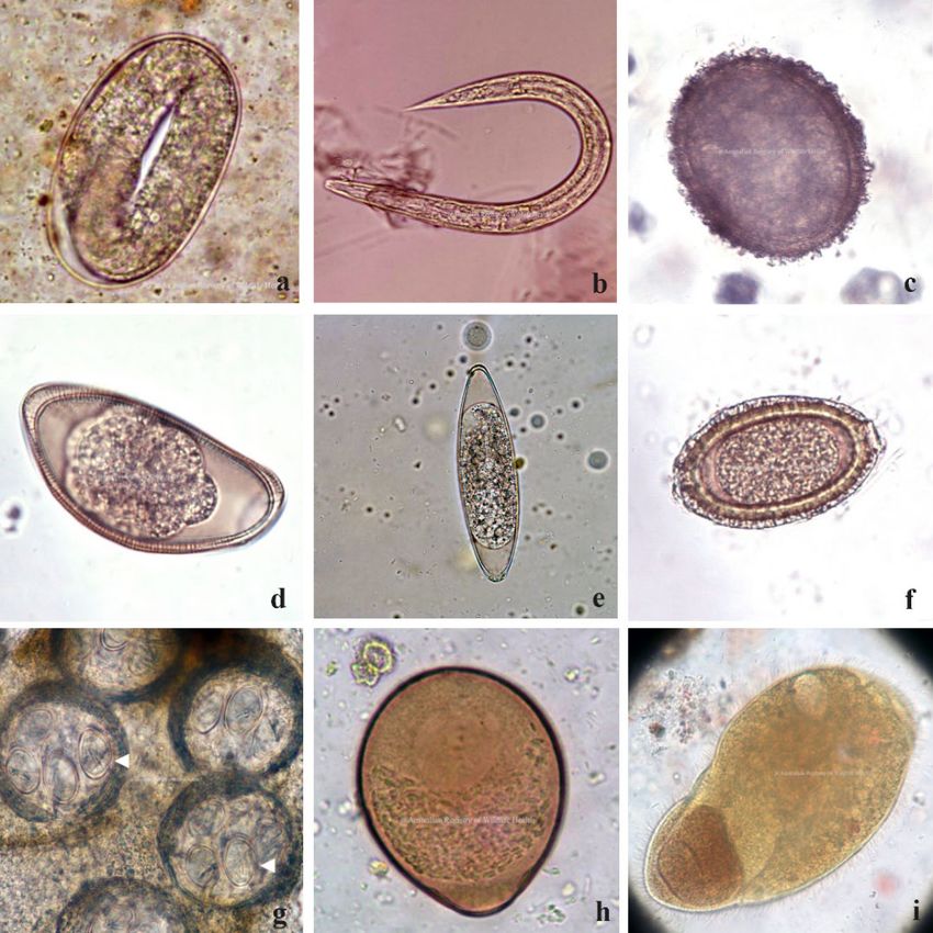
Production of this document was made possible by: Wildlife Rescue and Rehabilitation – an
Australian Government initiative

Cite this document as: Hall, J. and Rose, K. 2021. Common Diseases of Urban Wildlife: Reptiles.
Taronga Conservation Society Australia, Sydney.

All Images are subject to Copyright©

The information and materials contained in this section of the site are subject to copyright and are
for individual educational use only. Authorisation should be sought from the Registry for any other
use of these materials.

The views expressed in this document are those of the authors, and not necessarily of their
organisations. The Registry makes every effort to verify the information contained within this
document, but the accuracy and completeness of the information cannot be guaranteed. The
reader assumes all risk in using information provided. This document contains images of sick and
dead wildlife. These images are included for the sole purpose of improving wildlife care and
welfare. If you have any concerns regarding information contained in this document, please
contact the Registry directly.

                                                                                         1|Page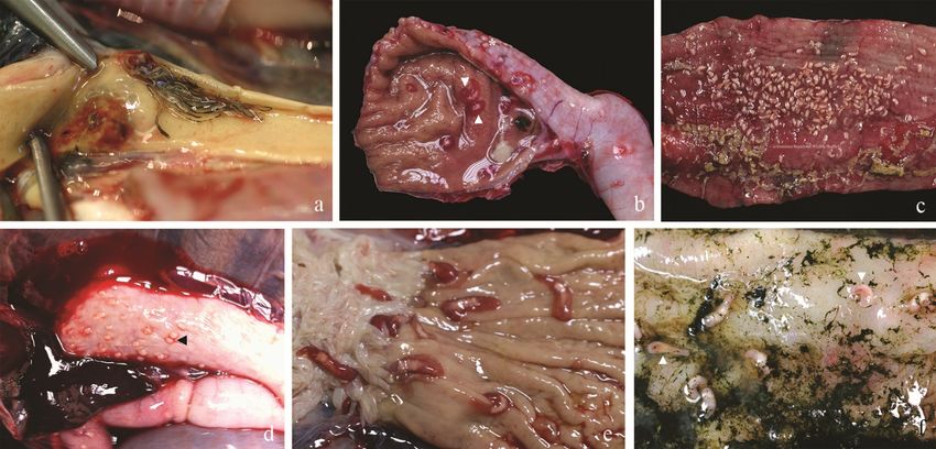
Contents
1     List of Images .................................................................................................................................. 4
2     Introduction .................................................................................................................................... 6
3     Parasitic Disease.............................................................................................................................. 6
    3.1      Ectoparasites ........................................................................................................................... 6
    3.2      Endoparasites.......................................................................................................................... 8
      3.2.1          Gastrointestinal parasites ............................................................................................... 8
      3.2.2          Blood parasites.............................................................................................................. 10
      3.2.3          Protozoa ........................................................................................................................ 11
4     Bacterial diseases .......................................................................................................................... 13
    4.1      Salmonellosis ........................................................................................................................ 13
    4.2      Mycobacteriosis .................................................................................................................... 14
    4.3      Enterococcus lacertideformus in skinks and geckos ............................................................. 15
    4.4      Dermatophilosis .................................................................................................................... 16
5     Viral diseases................................................................................................................................. 16
    5.1      Nidoviruses ........................................................................................................................... 16
      5.1.1          Bellinger River Virus ...................................................................................................... 16
      5.1.2          Bobtail flu (Shingleback nidovirus-1) ............................................................................ 17
      5.1.3          Snake nidovirus ............................................................................................................. 17
    5.2      Adenoviruses......................................................................................................................... 18
    5.3      Paramyxovirus....................................................................................................................... 18
      5.3.1          Sunshine virus ............................................................................................................... 18
6     Fungal diseases ............................................................................................................................. 18
    6.1      Mycotic and other dermatoses ............................................................................................. 18
      6.1.1          Nannizziopsis ................................................................................................................. 19
7     Nutritional Disease........................................................................................................................ 20
    7.1      Nutritional Osteodystrophy (metabolic bone disease)......................................................... 20
      7.1.1          Severe emaciation in chelonians .................................................................................. 21
8     Traumatic Injury ............................................................................................................................ 22
    8.1      Shock ..................................................................................................................................... 22
    8.2      Soft Tissue Injury including bite wounds .............................................................................. 22
    8.3      Cloacal or penile prolapse ..................................................................................................... 23
    8.4      Shell Injury ............................................................................................................................ 23
    8.5      Tail injuries ............................................................................................................................ 24
9     Diseases of Unknown Aetiology.................................................................................................... 24
    9.1      Neoplasia............................................................................................................................... 24

                                                                                                                                        2|Page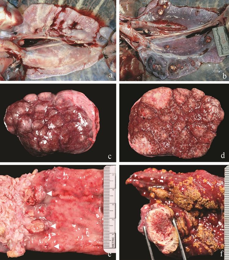
10   Species mentioned in text......................................................................................................... 25
11   References ................................................................................................................................ 27

                                                                                                                                 3|Page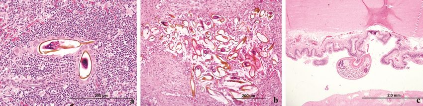
1 List of Images
Figure 1 a) Central netted dragon cloacal mites (Image: P Thompson), b) orange mites clustered
around toenails of a wild Asian house gecko, and c) orange mites under scales along the ventral tail
of a wild Asian house gecko .................................................................................................................... 7
Figure 2 a) Engorged adults and nymphs, Amblyomma moreliae, causing b) anaemia in a wild eastern
water dragon, and c) camouflaged ticks on the ventral surface of a lace monitor ................................ 7
Figure 3 a, b) A ball of leeches in the axillary area of a Bellinger River Snapping Turtle (b, arrows), and
b) invertebrate eggs (unknown species) glued over the surface of the plastron and underside of
carapace (asterisk) .................................................................................................................................. 7
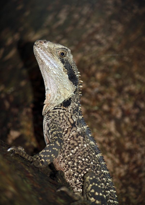
Figure 4 Various reptile faecal parasites a) strongyle-type ova, b) strongyle larvae, c) ascarid ovum, d)
oxyurid ovum, e) oxyurid ovum, f) capillaria ovum, g) cestode ovum (note characteristic hooklets), h)
encysted Nyctotherus sp. ciliate, i) Nyctotherus sp. ciliate. Images courtesy of P. Thompson. ............. 8
Figure 5 Eastern water dragon with a) profound anaemia caused by a severe co-infection of ticks and
(b,c) Abbreviata physignathi nematodes in the stomach ....................................................................... 9
Figure 6 Marine turtles with a) aortic aggregate of adult trematodes, b) adult trematodes (arrow)
and serosal granulomas (asterisk) in small intestine, c) adult trematodes along mucosal surface of
small intestine, d) serosal granulomas, e) adult trematodes in stomach (black arrow), and f) adult
trematodes in small intestine (arrows)................................................................................................. 10
Figure 7 Marine turtle histopathology (H&E) a) trematode ova in spleen, b) trematode ova in aorta,
and c) adult trematode in small intestinal lumen................................................................................. 10
Figure 8 Trematodes seen a) in a faecal float, b) on a wet preparation of a serosal surface scrape of
small intestine, c) excysting trematodes on serosal surface scrape of small intestine, and d) adult
trematodes viewed under a dissecting microscope ............................................................................. 10
Figure 9 Haemogregarine parasite within a red cell identified on blood smear, Diamond Python.
Image courtesy of P. Thompson. .......................................................................................................... 11
Figure 10 Microsporidia, central knob-tail gecko ................................................................................. 11
Figure 11 Cryptosporidia spp. Faecal sample, Inland Taipan. Modified Zeihl-Neilsen stain................. 12
Figure 12 a) unsporulated and b) sporulated coccidia (Isospora) in faeces, and c) unsporulated
Schellackia-like coccidian and d) sporulated Schelackia-like coccidian in green turtle faeces ............ 13
Figure 13 Monocytes containing Schellackia-like coccidian zoites (black arrows), blood film of a green
turtle ..................................................................................................................................................... 13
Figure 14 Desert death adder with lesions throughout the liver caused my Mycobacterial infection 14
Figure 15 a,b) Mycobacterium chelonae in green turtle lungs, c,d) Mycobacterium marinum
granulomas in enlarged spleen of a hawksbill turtle (whole, and cut surface), and d,e)
gastrointestinal granulomas caused by Mycobacterium marinum in a hawksbill turtle ...................... 15
4|Page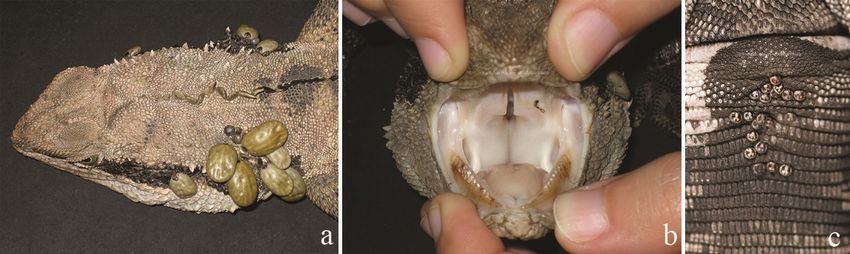
Figure 16 a) emaciated Asian house gecko with facial and tail lesions, Enterococcus lacertideformus,
b) Lister's Gecko with expanded spectacle and facial lesions, E. lacertideformus, c) Blue-tailed skink
with open wound, E. lacertideformus ................................................................................................... 16

Figure 17 Bellinger River snapping turtles with progressing eye lesions from a) unilateral mild (left
eye) swelling, b) bilateral swelling and c) severe bilateral swelling and ulceration ............................. 17

Figure 18 Mycotic dermatosis a) juvenile saltwater crocodile, Rhizomucor variabilis, b) juvenile
saltwater crocodile, Fusarium solani, c) eastern water dragon, Paecilomyces lilacinus, d) Arafura file
snake, Trichophyton verrucosum .......................................................................................................... 19

Figure 19 A severely emaciated eastern water dragon with proliferative yellow skin lesions over the
legs, ventral abdomen and tail caused by Nannizziopsis barbatae ...................................................... 20

Figure 20 Emydura spp. kept in unfavourable housing for unknown period with a) plastron
dismemberment from carapace and b) costal bone separation from marginal scutes and bones of the
carapace, c) severely emaciated wild green turtle with dissolution of connective tissues of plastron
resulting in bone instability and exposure............................................................................................ 21

Figure 21 Eastern blue-tongue skink with traumatic bite wounds to lower lateral abdomen (a) and
tail (b) leading to sepsis. ....................................................................................................................... 23

Figure 22 Propeller injuries of green turtles a) four centrally aligned linear shell fractures, b) three
linear shell fractures to the upper right quadrant, c) multiple right side shell fractures and
evisceration of internal organs (arrow) ................................................................................................ 24

Figure 23 a, b) diamond python, retrolobular carcinoma, c) diamond python, granulocytic round cell
tumour, d) northwestern carpet python, kidney sarcoma ................................................................... 25

Figure 24 a) blotched blue-tongue lizard, oviductal sarcoma, b) pygmy mulga monitor, metastatic
chondroblastic osteosarcoma, c) shingleback, multisystemic lymphosarcoma with multifocal liver
lesions ................................................................................................................................................... 25

                                                                                                                                           5|Page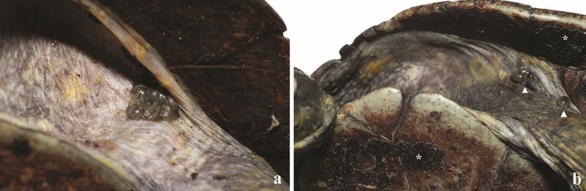
2      Introduction
A variety of diseases have been recognised within free ranging Australian reptiles. The purpose of this
document is to review the diseases that occur often within particular species or taxonomic groups of
reptiles. We hope that this information assists with the timely recognition of common parasites,
microbes, intoxicants and injuries to accelerate the appropriate care and welfare of wild animals.
Throughout the text we offer advice towards achieving a diagnosis. As best-practice wildlife
treatments change rapidly over time, treatment of reptiles in a rehabilitation situation should be made
in consultation with a veterinary professional.

    A notifiable disease is one that must be reported to agricultural authorities. If you suspect or can
    confirm that an animal is showing symptoms of one of the diseases listed as reportable, you must
    report it to:
        • your local vet or
        • Wildlife Health Australia State Coordinator,
             www.wildlifehealthaustralia.com.au/AboutUs/ContactDetails.aspx
        • your state or territory's department of primary industries or agriculture by phoning the
             Emergency Animal Disease Watch Hotline on 1800 675 888.

3      Parasitic Disease
3.1     Ectoparasites

    Zoonotic: may be vectors for other pathogens
    Species affected: All
    Similar presentation to: viruses (pox virus), fungal dermatopathies

Reptiles may be infested with a wide variety of ectoparasites, primarily mites and ticks. Little is known
about the relationship between haematophagous arthropods and their hosts, despite Australia having
more reptile-specific ticks than any other region (Natusch, et al., 2018). Ectoparasites are capable of
transmitting viruses, bacteria, protozoa and microfilaria.

Mites and ticks are usually evident upon close visual inspection between the scales in the region of
the head and neck. Mites are eliminated through a combination of environmental decontamination,
and treatment of the reptile with suitable parasiticides.

The snake mite, Ophionyssus natricis, is an introduced species recently reported in a variety of captive
and wild snakes and lizards in south-eastern and southern Australia (Norval, et al., 2020). This mite is
of significance to both animal and human health, with the mite reported to cause abnormal shedding,
anaemia, and death in snakes and lizards, and dermatitis in people (Norval, et al., 2020). The mites
may easily be transferred to new locations via movement of their hosts such as via translocation,
rehabilitation and release, or by introduction of infested animals to a naïve or mixed species
environment in a captive setting. Mites often go undetected as they burrow under the scales, and
accidental introduction could happen quite easily, therefore reptiles should be kept separate during
rehabilitation, good hygiene practices should be maintained, animals should always be released at
point of capture when possible, and on occasions where this is not possible animals should be treated
for mites prior to release.

                                                                                             6|Page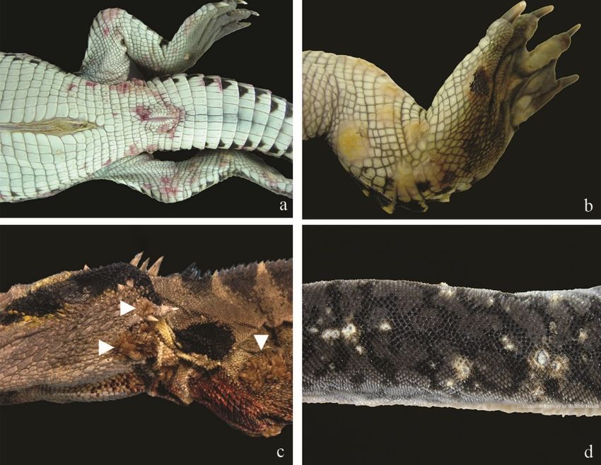
Figure 1 a) central netted dragon cloacal mites (Image: P Thompson), b) orange mites clustered around toenails of a wild
Asian house gecko, and c) orange mites under scales along the ventral tail of a wild Asian house gecko

Ticks of the genera Amblyomma and Aponomma are most commonly found infesting reptiles
(McCracken, 1994). Large tick burdens may result in anaemia. Treatment of tick infestation is usually
accomplished by manual removal of the tick. Alternatively, antiparasitic agents may be used to treat
tick infestations.

Figure 2 a) Engorged adults and nymphs, Amblyomma moreliae, causing b) anaemia in a wild eastern water dragon, and c)
camouflaged ticks on the ventral surface of a lace monitor

Leeches may also be commonly found in the axilla and inguinal area of many turtles. The impact of
these parasites and their capacity to act as a vector of disease are unknown, however, their
presence is generally considered incidental with little impact on the host.

Figure 3 a, b) A ball of leeches in the axillary area of a Bellinger River Snapping Turtle (b, arrows), and b) invertebrate eggs
(unknown species) glued over the surface of the plastron and underside of carapace (asterisk)

                                                                                                                  7|Page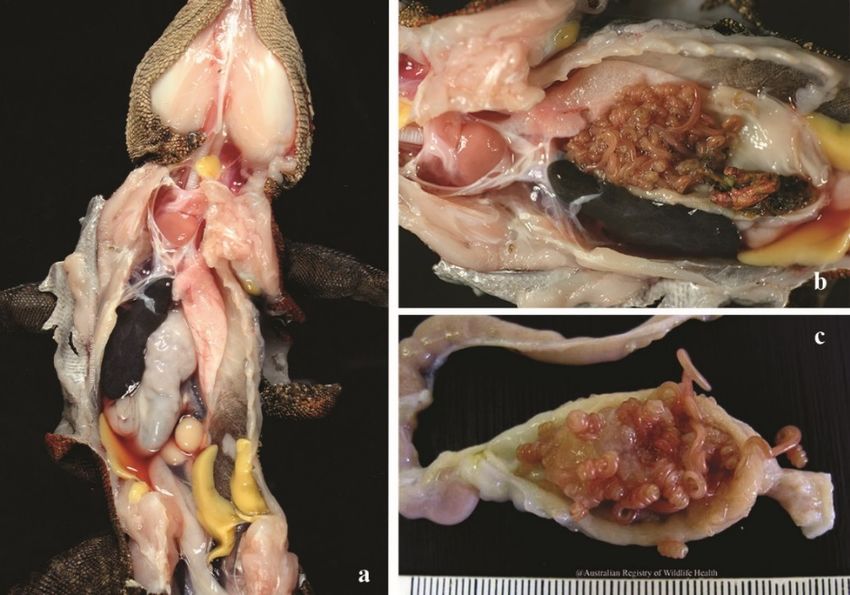
3.2     Endoparasites

3.2.1     Gastrointestinal parasites

Many endoparasites infect reptiles. The interpretation of faecal floatation’s in reptiles must be
undertaken with care, since parasite ova of prey are often found, including invertebrate parasites of
insectivorous species.

In most cases, the presence of intestinal parasites is incidental and low levels of parasites are
expected in all free-ranging wildlife. There are occasions however, where parasite burdens may
become severe enough to cause clinical disease including anaemia, melena, thrombosis or blockage
of the gastrointestinal tract. Treatments are best undertaken after parasite identification, and aimed
at reducing rather than eliminating parasites. Retaining natural host-parasite relationships should be
a consideration for animals in care and for health maintenance during translocation or other
conservation action.

Strongylurus paronai is a common gastric roundworm of Eastern bluetongue skinks, Eastern water
dragons and frilled lizards (Griffiths, et al., 1998). In dead animals, this parasite often crawls into the
pharynx and oral cavity.

Figure 4 Various reptile faecal parasites a) strongyle-type ova, b) strongyle larvae, c) ascarid ovum, d) oxyurid ovum, e)
oxyurid ovum, f) capillaria ovum, g) cestode ovum (note characteristic hooklets), h) encysted Nyctotherus sp. ciliate, i)
Nyctotherus sp. ciliate. Images courtesy of P. Thompson.

Nematodes of the Abbreviata and Spinicauda genus’ are common, species-specific parasites of the
Eastern water dragon, and can be found within 100 km of the coast mainly, perhaps exclusively, in

                                                                                                                8|Page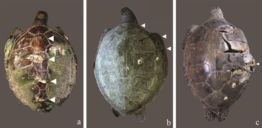
NSW (Jones, 2007). These parasites are commonly found in tight coils in the stomach, but can
migrate to the oesophagus and stomach after the host’s death.

Figure 5 Eastern water dragon with a) profound anaemia caused by a severe co-infection of ticks and (b,c) Abbreviata
physignathi nematodes in the stomach

3.2.1.1 Sparganosis
Reptiles are the secondary intermediate host for the plerocercoid cestode Spirometra sp. which
needs a minimum of three intermediate hosts before infecting the definitive host; canids, felids and
other mammals, including humans. Crustaceans and copepods are the primary intermediate host. In
Australia, Spirometra eranacei is often reported from wild short-beaked echidnas which are,
presumably, like humans, accidental hosts. While this parasite causes little disease for the
intermediate host, people who are infected through ingestion of contaminated meat or water may
experience prolonged inflammation and tissue damage as the parasite migrates through the body.

3.2.1.2    Spirorchid flukes (trematodes) in sea turtles
  Zoonotic: No
  Species affected: Loggerhead turtle, hawksbill turtle, green turtle, flatback turtle, leatherback
  turtle
  Similar presentation to: bacterial infection, coccidiosis

Flukes of the Spirorchiidae family cause morbidity and mortality in marine turtles, especially green
and loggerhead turtles (Marchiori, et al., 2017). In Australia, approximately 50% of green turtles are
infected with spirorchid flukes (Gordon, et al., 1998). Flukes may be found in blood vessels, or within
the alimentary tract of infected turtles depending on the species of fluke found. Small flukes are
sometimes present in the lumen of the urinary bladder of marine turtles. Adult flukes may cause
aortic blockages, aneurisms, ulceration of the intestinal mucosa or urinary bladder, while eggs may
cause disseminated granulomas (inflammatory nodules) throughout various tissues, most notable
along the serosal surface of the small intestine (Gordon, et al., 1998).

                                                                                                            9|Page
Figure 6 Marine turtles with a) aortic aggregate of adult trematodes, b) adult trematodes (arrow) and serosal granulomas
(asterisk) in small intestine, c) adult trematodes along mucosal surface of small intestine, d) serosal granulomas, e) adult
trematodes in stomach (black arrow), and f) adult trematodes in small intestine (arrows)
Gross and microscopic observation of adult trematodes or ova in blood vessels, tissue, and faeces
are suitable diagnostic tools, however identification of species using morphological features is
difficult and molecular techniques are more reliable and readily used (Marchiori, et al., 2017).
Figure 7 Marine turtle histopathology (H&E) a) trematode ova in spleen, b) trematode ova in aorta, and c) adult trematode
in small intestinal lumen
Figure 8 Trematodes seen a) in a faecal float, b) on a wet preparation of a serosal surface scrape of small intestine, c)
excysting trematodes on serosal surface scrape of small intestine, and d) adult trematodes viewed under a dissecting
microscope
3.2.2 Blood parasites
Haemoprotozoa and microfilaria are common incidental findings in reptiles. Haemogregarina species,
Trypanosoma spp., Haemoproteus spp. and Plasmodium spp. are frequently found during
haematological examinations.
10 | P a g eHaemogregarine parasites have been identified
within the pulmonary parenchyma and red blood
cells of numerous snakes and monitors seized by
Australian customs service officials upon illegal
entry into Australia. Mosquitos and mites are the
arthropod hosts most likely to transmit
haemogregarines; however, leeches, ticks and
other haematophagous arthropods may act as
intermediate hosts. These intermediate hosts
release sporozoites during a blood meal.
Sporozoites enter erythrocytes and undergo
schizogony in various tissues throughout the
body. Merozoites and gametocytes are also
found within the erythrocyte and are ingested by Figure 9 Haemogregarine parasite within a red cell identified
haematophagous insects to allow subsequent on blood smear, Diamond Python. Image courtesy of P.
Thompson.
transmission of the parasite.
3.2.3 Protozoa
Zoonotic: Yes
Species affected: yellow-bellied sea snake, desert death adder, eastern water dragon, central
knob-tailed gecko, inland bearded dragon, red-bellied black snakes, Stimson’s python, inland
taipan, tiger snake, frilled lizard, freshwater crocodile
Similar presentation to: bacterial infection, other parasitic infections, viral infections
Microsporidia are protozoal parasites that have been
detected within various reptiles and may or may not
represent clinical pathology (Sokolova, et al., 2016). Infection
has been associated with necrotising lesions of the
musculature, bone pathology in sea snakes (Gillett, et al.,
2016), granulomatous lesions in the ovary of an eastern
water dragon and central knob-tailed gecko (Reece & Hartley,
1994), and granulomatous multi-organ inflammation in
inland bearded dragon (Sokolova, et al., 2016). Overseas, the
microsporidian Encephalitozoon pogonae has been reported Figure 10 Microsporidia, central knob-tail
to cause weight loss and death in numerous captive inland gecko
bearded dragons in Austria, the USA, and Japan (Shibasaki, et
al., 2017). Microsporidia appear as clusters of basophilic oval to round bodies when viewed in tissue
sections stained with haematoxylin and eosin. Microsporidia are gram positive. Mature spores are
acid fast, and contain a polar mass/body that stains positively with periodic acid-Schiff (PAS) staining
protocols. Traditionally, morphology has been used to identify microsporidia however this is
challenging, and molecular techniques are required to definitively identify the parasite.
Immunocompromised humans are also susceptible to infection, and microsporidia have been
reported to infect the eyes of otherwise healthy people, therefore care must be taken when handling
infected reptiles (Gillett, et al., 2016).
11 | P a g eCryptosporidia spp. are coccidian parasites that can
cause hypertrophic gastritis (inflamed stomach lining)
in snakes and lizards. In Australia, cryptosporidiosis
has been identified in captive red-bellied black
snakes, Stimson’s python, inland taipan, tiger snake,
inland bearded dragon, and a wild frilled lizard (Oros,
et al., 1998; Koehler, et al., 2020). Clinical signs
associated with cryptosporidiosis include weight loss,
regurgitation, diarrhoea and death. Infection occurs
via the faecal-oral route and is more prominent in
Figure 11 Cryptosporidia spp. Faecal sample, Inland animals with concurrent disease (Wildlife Health
Taipan. Modified Zeihl-Neelsen stain Australia, 2018). The parasite undergoes asexual and
sexual reproduction within the host cell cytoplasm
along the mucosal brush border. The oocysts sporulate in situ, resulting in continuous self-infection,
however oocysts are shed in faeces or regurgitate intermittently and repeat testing may be required
to confirm infection (Wildlife Health Australia, 2018). Ante-mortem diagnosis can be achieved through
demonstration of oocysts within modified acid fast stained faecal smears. The sensitivity of faecal
staining tests is increased through serial testing and centrifugation techniques that concentrate the
oocysts. DNA sequencing, PCR, histopathology, and direct fluorescence antibody testing are also used
in the laboratory setting. Molecular detection and identification are preferred. There is no known
effective treatment for cryptosporidiosis.
There is a risk of zoonotic infection from reptiles that have consumed prey infected with non-reptilian
Cryptosporidia sp. such as C. parvum and C. muris (Wildlife Health Australia, 2018).
3.2.3.1 Coccidiosis in green turtles
Zoonotic: No
Species affected: Green turtles
Similar presentation to: trauma, trematodiasis, septicaemia
While coccidia can potentially be found in all reptiles without causing clinical disease, there are
instances where coccidia can cause morbidity and mortality. Systemic coccidiosis in green turtles is
one such example.
Repeated epizootics of neurologic dysfunction and mortality in subadult and adult green turtles have
been identified along the east coast of NSW and Queensland on several occasions. Affected turtles
are often found circling in estuaries, rolling in the surf, or stranded on the beach moribund or with a
head tilt. These epizootics have been attributed to disseminated coccidiosis, characterised by the
presence of necrosis and non-suppurative inflammation in the intestinal tract, renal interstitium,
thyroid gland interstitium, and throughout the parenchyma of the brain. Grossly visible
inflammatory changes in the intestinal tract, kidney, thyroid gland, or brain may or may not be
evident (Rose, et al., 2003).
Mortalities were thought to be caused by a coccidian parasite Caryospora cheloniae, however, Ban
de Gouvea Pedroso, et al. (2020) used molecular characterisation to identify the presence of
protozoa 98.8% similar to Schellackia orientalis. They found that mortality outbreaks were more
likely to occur in larger animals (curved carapace length >68cm), in warmer months particularly
October, during or one month prior to El Nino like events and that disease occurrence can be
expected to occur anywhere between the southern end of Fraser Island, Qld to Sydney, NSW (Ban de
Gouvea Pedroso, et al., 2020). Coccidiosis was not detected north of Fraser Island.
12 | P a g eCoccidia harvested from the intestinal tract or faeces can be cultivated in filtered seawater, where
they develop into the stellate sporulation pattern. A diagnosis of systemic coccidiosis can also be
made when sporozoites are identified within circulating monocytes within a blood film or buffy coat
preparation.
Figure 12 a) unsporulated and b) sporulated coccidia (Isospora) in faeces, and c) unsporulated Schellackia-like coccidian and
d) sporulated Schelackia-like coccidian in green turtle faeces
Figure 13 Monocytes containing Schellackia-like coccidian zoites (black arrows), blood film of a green turtle
4 Bacterial diseases
Ideally treatment of bacterial infection is based upon isolation of the organism within lesions, and
antimicrobial sensitivity testing. Treatment without consultation and confirmation of the infectious
agent may lead to ineffective treatment, and antimicrobial resistance. Some of these organisms are
potentially zoonotic. Sound hygiene protocols for reptiles in rehabilitation will protect both reptiles
and their rehabilitators.
4.1 Salmonellosis
Zoonotic: Yes
Species affected: All
Similar presentation to: may be no signs of disease, other bacterial infections, trauma, parasites
Salmonella spp. can be found readily in wild and captive reptiles, however, infection in reptiles is not
often associated with clinical disease. Salmonella does have the potential to become a severe,
possibly fatal, zoonotic pathogen, therefore good hygiene practices are recommended to anyone
handling reptiles, especially young children and immunocompromised people. Various studies have
found that snakes are more likely to carry Salmonella spp. than other reptiles, but lizards and turtles
can also be carriers, and while captive reptiles are significantly more likely to carry Salmonella, those
in the wild can also be a reservoir (Wildlife Health Australia, 2018; McWhorter, et al., 2021).
Clinical disease is often the result of an insult that disrupts the barrier between the gastrointestinal
system and the body cavity such as trauma, parasites, or other disease, or from an increase in
general stress such as handling, overcrowding, or suboptimal environmental conditions (Wildlife
Health Australia, 2018). Salmonella may also be transmitted thorough ingestion of contaminated
food such as feeder mice (in a captive setting), native rodents, and insects (McWhorter, et al., 2021).
13 | P a g eWhile many animals may be asymptomatic, clinical disease may present as anorexia, regurgitation,
depression, respiratory disease, dehydration, or diarrhoea, and in turtles lesions of the plastron or
carapace (Wildlife Health Australia, 2018).
Microbiological culture or PCR are diagnostic however repeat testing may be required due to
intermittent shedding in faeces. Cloacal swabs are preferable over faecal samples for testing.
Treatment of asymptomatic reptiles that are otherwise healthy is discouraged, while supportive
treatment with possible antibiotic treatment of sick reptiles is preferred. However, some isolates of
Salmonella spp. are antibiotic resistant and treatment without susceptibility studies should be
avoided (McWhorter, et al., 2021). Recovery from clinical infection may be slow. Good hygiene
practices and minimising stress is the best method for limiting clinical disease, while good hygiene
practices for any people handling either wild or captive reptiles are recommended.
4.2 Mycobacteriosis
Zoonotic: Yes
Species affected: All
Similar presentation to: other bacterial infections, fungal infections
Reptiles, like any animal, have the potential to suffer significant disease through infection with
Mycobacterium spp. bacteria. A wide variety of Mycobacterium spp. have been isolated from all
classes of reptiles, including painted dragons, knob-tailed geckos, carpet pythons (centralian and
north-western), desert death adders, Arafura file snakes, freshwater crocodiles, Jardine River turtles,
eastern long-necked turtles, hawksbill turtles, loggerhead turtles, flatback turtles and green turtles.
Figure 14 Desert death adder with lesions throughout the liver caused my Mycobacterial infection
Mycobacteria are able to survive in soil for many years, but appear to be most common in aquatic
environments and are pathogens of concern in the rehabilitation setting. Clinical signs may vary and
are often non-specific, dependant on the site of infection. On gross examination, infection is often
associated with granulomatous lesions of liver, spleen, lungs (especially in marine turtles), gut, bone
and skin. Granulomatous centres are often necrotic therefore sample collection at the margin of the
lesion is recommended.
Diagnosis requires culture of the organism; however acid-fast bacteria can often be seen on Zeihl-
Neelsen stain of impression smears of the lesion. Treatment is generally not recommended due to
zoonotic potential and generally poor response to treatment. Mycobacteria also appear to have a
high resistance to chemical disinfectants and UV (Wildlife Health Australia, 2013). For this reason, it
is recommended that turtles undergoing rehabilitation should be kept in isolated/individual water
bodies for the duration of their treatment. Water filtration systems should contain a sterilisation
element to prevent mycobacteria from proliferating in water and in biological filtration systems.
14 | P a g eFigure 15 a,b) Mycobacterium chelonae in green turtle lungs, c,d) Mycobacterium marinum granulomas in enlarged spleen
of a hawksbill turtle (whole, and cut surface), and d,e) gastrointestinal granulomas caused by Mycobacterium marinum in a
hawksbill turtle
4.3 Enterococcus lacertideformus in skinks and geckos
Zoonotic: No
Reportable: please report to Australian Registry of Wildlife Health
Species affected: Christmas Island blue-tailed skink, Christmas Island Lister’s gecko, Asian house
gecko, common four-clawed gecko
Similar presentation to: neoplasia, trauma, sparganosis, other bacterial infections
In 2014 on the remote Australian Territory of Christmas Island some of the critically endangered,
conservation dependent Lister’s Geckos developed unusual facial swellings and those animals
became emaciated and died. At the same time, similar lesions were noted in some of the feral
geckos found on the island. These lesions were found to be caused by a novel bacterium, which has
now been identified as Enterococcus lacertideformus (Agius, et al., 2021). E. lacertideformus has
been identified to cause fatal infections in 3 species of gecko and one skink species on Christmas
Island, with probable reports also recorded in reptiles in the USA, Poland and Malaysia. The
biosecurity risk to Australian native fauna is significant, making examination of reptiles with
ulcerative or proliferative lesions extremely important.
15 | P a g eInfection is thought to be spread by biting or via the oral cavity and animals present most commonly
with gingival swelling leading to gelatinous subcutaneous nodules of the head and face, and finally
multisystemic expansile nodules throughout multiple tissues whereby this biofilm coated bacteria
begin to replace bone and soft tissues (Rose, et al., 2017; Agius, et al., 2021). There is little
inflammatory response from the host and animals go on to die, often from emaciation. Clinical
presentation in geckos is much more dramatic as the soft tissues of the head and face are contorted
with swelling and nodules, however in skinks presentation may be much more subtle, and ante-
mortem signs may not be easily appreciated. In skinks, infection is often associated with open, non-
healing wounds.
Figure 16 a) emaciated Asian house gecko with facial and tail lesions, Enterococcus lacertideformus, b) Lister's Gecko with
expanded spectacle and facial lesions, E. lacertideformus, c) Blue-tailed skink with open wound, E. lacertideformus
4.4 Dermatophilosis
Dermatophilosis, caused by Dermatophilus congolensis, is a common invader of the skin of reptiles
and an important zoonotic disease that can cause serious skin lesions in humans. D. congolensis has
mistakenly been referred to as a fungal pathogen in the past, however it is a filamentous bacterium
that causes crusty scabs in a variety of animals, and ‘brown spot disease’ in farmed crocodiles
(Shilton, 2019). Proliferative crusty skin lesions may be superficial, but these sometimes obscure
deeper abscesses. As with fungal infection, this disease is typically associated with animals kept in
damp or humid conditions, or under cool conditions where there is insufficient access to heat
sources.
Dermatophilosis can be identified within gram stained skin scrapings or biopsies. The organisms can
appear as cocci or as beaded, branching double chains of cocci. D. congolensis requires extended
periods (up to 14 days) to grow in anaerobic culture. The infection is often treated with topical
iodine preparations and parenteral long-acting, broad spectrum antibiotics, along with improvement
of husbandry and reduction in environmental stressors.
5 Viral diseases
5.1 Nidoviruses
5.1.1 Bellinger River Virus
Zoonotic: No
Reportable: please report to Australian Registry of Wildlife Health
Species affected: Bellinger River snapping turtle
Similar presentation to: trauma, ranavirus
A large scale mortality event in 2015, which resulted in the loss of approximately 90% of the
population of wild Bellinger River turtles, was likely caused by a novel nidovirus commonly called
Bellinger River virus (Zhang, et al., 2018). The virus affected predominantly adult and subadult
animals which either presented dead, or with bilateral swelling of the eyelids which became
16 | P a g eulcerated in severely affected animals, and in some cases tan coloured lesions on the skin of the
ventral thighs.
Gross and microscopic examination showed necrosis of the kidneys, spleen and lacrimal glands of
the eyes. Real-time PCR is available for molecular confirmation of infection with Bellinger River virus.
Infection with Bellinger River Virus was uniformly fatal in animals found exhibiting lesions; however
clinically health animals may return recurrent positive PCR ocular swabs.
Figure 17 Bellinger River snapping turtles with progressing eye lesions from a) unilateral mild (left eye) swelling, b) bilateral
swelling and c) severe bilateral swelling and ulceration
Despite extensive work in the field it is not known where this virus originated or if it is present in
other waterways, highlighting the need for vigilance when observing any freshwater turtles coming
into, or being released from, rehabilitation care.
5.1.2 Bobtail flu (Shingleback nidovirus-1)
A syndrome commonly referred to as ‘bobtail flu’ has been identified in both wild and captive
shingleback lizards from Western Australia. This syndrome is most likely due to infection of the
upper respiratory tract with a novel nidovirus (Shingleback nidovirus-1), however a causal
association between the syndrome and viral infection has not been proven (O'Dea, et al., 2016).
Clinically, shinglebacks may show a loss of condition, lethargy, depression, pale mucous membranes,
increased mucous in the naso-oral cavities that may bubble from the eyes or nostrils, sneezing, or
watery or swollen eyes (O'Dea, et al., 2016). Diagnosis is based on PCR from oral/tracheal and/or eye
swabs and infection appears to be highly contagious highlighting the need for good hygiene during
rehabilitation of injured or ill shinglebacks. Early reports suggest that the same virus may be present
in centralian, eastern and northern blue-tongued skinks across multiple states (Shilton, et al., 2019).
This highlights the importance of biosecurity control around the movement of reptiles for
conservation or for the global pet trade.
5.1.3 Snake nidovirus
In 2017 an experimental infection trial confirmed Nidoviruses were responsible for a disease seen
previously in boids and pythons in Australia, USA and Europe (Shilton, et al., 2019). In these species,
clinical symptoms were primarily respiratory and included anorexia, reddened mucosa in the mouth,
oral ulceration, excessive mucous in the mouth and/or lungs, or sudden death (Shilton, et al., 2019).
Immunohistochemistry and in situ hybridisation testing of lung, intestine, oral swabs, and faeces
accurately confirm nidovirus infection. The mode of transmission for this virus is unknown.
As this is an emerging disease, little is known about infection in Australian snakes, however initial
work has detected nidovirus in a number of species across most states (Shilton, et al., 2019) and as
such, increased awareness about the symptoms of this disease is necessary for early detection and
control especially where animals may be kept in rehabilitation without strict quarantine and either
mixed with other species or released into the wild.
17 | P a g e5.2 Adenoviruses
Adenoviruses have been identified in numerous species of lizards, snakes, chelonians and crocodiles
globally including bearded dragons, central netted dragon, and eastern blue-tongue skinks
(Hyndman & Shilton, 2011; Hyndman, et al., 2019). Adenoviruses are generally species specific.
Infection can result in anorexia and weight loss, diarrhoea, weakness, neurological signs, or sudden
death, especially in young animals, however infection in adult animals may be sub-clinical and these
individuals may become carriers of the virus. Concurrent parasitic infections are reported (Hyndman
& Shilton, 2011; Hyndman, et al., 2019). Transmission is believed to be via the faecal-oral route, and
there may be vertical transmission from mother to offspring however this is unconfirmed.
Microscopic examination of liver in affected animals may reveal hepatocellular degeneration to
necrosis, and large eosinophilic to basophilic intranuclear inclusion bodies. Diagnosis can be
confirmed by PCR testing of oral and cloacal swabs.
5.3 Paramyxovirus
5.3.1 Sunshine virus
Sunshine virus is an emerging disease of captive Australian pythons associated with outbreaks of
neurological and respiratory disease (Shilton, et al., 2019). The effect of this virus on wild pythons is
unknown. Clinical signs may include lethargy, anorexia, abnormal skin sloughing, and various
neurological signs, however in red-tailed boas infected with sunshine virus in Thailand, neurological
signs have not been reported (Shilton, et al., 2019).
Transmission is via direct contact, but may also be vertical from infected parent to egg/offspring.
PCR of oral/cloacal swabs is diagnostic in live pythons however animals may be asymptomatic for
several years before exhibiting clinical signs, therefore serial sampling is recommended (Shilton, et
al., 2019). In deceased pythons, brain is preferred for PCR however lung, kidney and liver may also
be tested.
6 Fungal diseases
6.1 Mycotic and other dermatoses
Zoonotic: Yes
Species affected: All
Similar presentation to: bacterial infection (particularly Dermatophilus spp.), viral infection,
trauma, nutritional deficiency (vitamin A, Zinc)
Mycotic infection of the skin is common in reptiles, especially lizards. Organisms are considered
opportunistic as they are often present in soil or substrate and come into contact with damaged skin
or immunocompromised animals. Infections may be more common in animals from habitats or
enclosures that are damp and have limited sunlight. Typical infections include Basidiobolus spp.
Geotrichium spp., Paecilomyces spp., Trichophyton spp., Aspergillus spp., Fusarium spp.,
Acremonium spp., Chrysosporium spp., and Nannizziopsis spp. (McCracken, 1994; Shilton, 2019;
Peterson, et al., 2020). Although most common in captive reptiles, free-ranging animals in unsuitable
habitats or exposed to concurrent disease or other stressors may suffer infection with opportunistic
fungi. Nannizziopsis and similar fungal genera are considered primary fungal pathogens that are
capable of infecting otherwise healthy reptiles.
18 | P a g eFungal infections may be more common in animals with dysecdysis, or abnormal skin shedding. This
can be easily demonstrated in eastern blue-tongue skinks where dysecdysis can lead to strangulation
and sloughing of the digits or feet with secondary infection by the fungus Trichophyton terrestre.
Figure 18 Mycotic dermatosis a) juvenile saltwater crocodile, Rhizomucor variabilis, b) juvenile saltwater crocodile,
Fusarium solani, c) eastern water dragon, Paecilomyces lilacinus, d) Arafura file snake, Trichophyton verrucosum
6.1.1 Nannizziopsis
Zoonotic: No
Reportable: please report to Australian Registry of Wildlife Health
Species affected: crocodilians, eastern water dragon, eastern blue-tongue skink, centralian blue-
tongue skink, tommy roundhead dragon, shingleback lizard
Similar presentation to: other fungal infections, pox virus, trauma, Dermatophilus spp. Infection,
nutritional deficiency (vitamin A, Zinc)
Skin or systemic infection with the fungus Nannizziopsis spp., is an emerging disease which has been
described as causing fatal disease in a number of both captive and wild reptile species. Previous
reports of Chrysosporium spp. in farmed crocodilians may actually be attributed to Nannizziopsis
crocodili; however this is unconfirmed (Shilton, 2019).
Nannizziopsis barbatae has been ascribed as the cause of a significant event in wild eastern water
dragons in Qld and has also been recorded in wild eastern bluetongue skink, centralian blue-tongue
skink, tommy roundhead dragon, and shingleback lizard (Peterson, et al., 2020).
Skin lesions can differ depending on species but generally, infection is characterised by thickening of
the skin, proliferative yellow/tan crusting lesions, inflammation, necrosis, and ulceration of skin, and
19 | P a g esevere emaciation (Peterson, et al., 2020). Infection is contagious and capable of causing significant
disease which inevitably leads to death. There is no known effective treatment. Infection is
confirmed using histopathology, culture, PCR and sequence analysis.
Figure 19 A severely emaciated eastern water dragon with proliferative yellow skin lesions over the legs, ventral abdomen
and tail caused by Nannizziopsis barbatae
7 Nutritional Disease
Malnutrition, other than emaciation, is rare in free ranging wildlife. When malnutrition does occur in
free ranging wildlife, it is primarily the result of inappropriate supplemental feeding by humans.
Intestinal parasites, other infections, and inappropriate housing of captive reptiles can result in altered
nutrient absorption.
7.1 Nutritional Osteodystrophy (metabolic bone disease)
Nutritional osteodystrophy, or metabolic bone disease, is characterised by either failure to mineralise
a growing skeleton, or demineralisation of a mature skeleton. This condition is seen in native reptiles
that have been collected from the wild and held as pets or in prolonged rehabilitation. The disease is
rarely seen in free-ranging reptiles, other than those that are suspected to be released pets.
Nutritional osteodystrophy occurs primarily in reptiles that have been on a long-term diet deficient in
calcium or containing excessively high concentrations of phosphorus. Osteodystrophy may also occur
when reptiles have had insufficient dietary vitamin D3 and no exposure to the ultraviolet rays required
to produce metabolically active vitamin D.
Plant derived vitamin D2 (ergocalciferol) is not considered to be metabolically active in reptiles. The
active form of vitamin D in reptiles is vitamin D3 (cholecalciferol). Vitamin D3 must be supplied in the
food, or the animal allowed access to ultraviolet light to convert vitamin D2 to vitamin D3 in the skin
(Boyer, 1996).
Ideally, reptile diets should contain a 2:1 ration of calcium to phosphorus. The inappropriate nature
of some common reptile dietary items are illustrated by lean beef meat that contains a ratio of
approximately 1:16 Ca:P, and beef heart that contains approximately 1:38 Ca:P. Feeding insects a
calcium rich diet two to three days prior to feeding them to reptiles prevents nutritional
osteodystrophy in insectivorous species. Additionally, insects may be dusted with a supplement
containing calcium carbonate immediately prior to being fed to reptiles.
Reptiles with nutritional osteodystrophy have soft, misshapen bones and shells. The lower jaw may
be shortened when muscle traction draws in the soft bones. Lizards with osteodystrophy have an
abnormal posture, since they are unable to support their body effectively. The animal may have a
hunched spine, a swayed back, or a sideways curve of the spine, or vertebral compression fractures.
If spinal cord injury accompanies vertebral fracture, the reptile will have rear limb paresis or paralysis.
20 | P a g eThe long bones, particularly the femurs, are often very swollen due to periosteal thickening around a
thin, weakened cortex. Radiographically, affected lizards have a diffusely reduced bone density. The
cortical shadow may appear thin, or may be very thick, due to fibrous tissue proliferation. Folding
fractures of the long bones or compression fractures of the spine may also be evident on radiographic
examination.
Chelonians with nutritional osteodystrophy have a soft, misshapen shell with upturned marginal
scutes. The lesions are most severe if they occur in a young reptile that has not yet mineralised its
skeleton. The carapace may sag centrally, scutes become uneven, and the shell may be too small in
comparison with the rest of the body. Radiographically, these chelonians have a reduced bone density
and porous shell, and may have pathological long bone fractures.
Nutritional osteodystrophy is diagnosed by visual inspection and palpation of the skeleton in
conjunction with radiographic examination. Serum calcium and phosphorus concentrations are often
normal.
Due to extensive bony deformity and the extensive time in a captive environment, some reptiles with
osteodystrophy will not be suitable for release. When the lesions are mild, reptiles are often treated
with parenteral calcium and possibly also vitamin D3. The diet must be corrected to include a 2:1 ratio
of calcium to phosphorus. Ultraviolet light should be provided through exposure to sun, or a broad-
spectrum artificial light placed within 60 cm of the reptile, without any filtration through glass or
plastic (Boyer, 1996). Response to therapy should be monitored through radiographic examination
every four to six weeks.
Internal fixation of pathological fractures most often results in further splitting of the fracture site,
especially in small reptiles. Cage rest and an external splint will often usually result in satisfactory
resolution of fractures in reptiles with osteodystrophy. Treatment of severely affected reptiles should
be undertaken with care as the prognosis for return to function may be guarded and the advanced
condition is extremely painful.
7.1.1 Severe emaciation in chelonians
In chelonians, severe nutritional imbalance and emaciation can lead to complete breakdown of the
connective tissues of the carapace and plastron causing bone to collapse. This syndrome has been
recorded in both wild marine and freshwater turtles. The cause of initial debility is generally due to a
chronic disease process leading to profound emaciation and nutritional loss. In many cases both
disease and shell instability are too severe to respond to treatment. In freshwater turtles, this may
occur when the animal is already significantly debilitated upon entry into brumation and is
accelerated once activity is resumed.
Figure 20 Emydura spp. kept in unfavourable housing for unknown period with a) plastron dismemberment from carapace
and b) costal bone separation from marginal scutes and bones of the carapace, c) severely emaciated wild green turtle with
dissolution of connective tissues of plastron resulting in bone instability and exposure
21 | P a g e8 Traumatic Injury
Lizards, snakes and turtles are commonly admitted to urban wildlife care centres. These reptiles are
almost uniformly admitted due to traumatic injury. Underlying infectious disease and clinically
apparent parasitic diseases in urban reptiles presented with trauma are likely to be under-reported.
8.1 Shock
Many animals that have suffered a serious injury or are debilitated by disease are found in a state of
shock. Shock is defined as acute circulatory failure that results in multisystemic decrease in blood flow
and therefore low oxygenation of tissues. Clinical signs of shock are often related to low blood
pressure. The mucous membranes of an animal in shock may be pale or muddy and the peripheral
blood vessels are collapsed or provide a weak pulse. The heart rate may be weak and rapid. Animals
in a state of shock are often weak, depressed, have rapid breathing and reduced urate output. Animals
suffering from endotoxic shock, may have bright red mucosa.
Dehydration often contributes to the lack of peripheral perfusion and oxygenation. An animal is
severely dehydrated when the eyes are sunken, the capillary refill time is very slow, the mucous
membranes are dry and tacky, and the skin has lost its elasticity.
The neuroendocrine cascade that is initiated during shock is initially protective, but over time energy
reserves are depleted and peripheral vasoconstriction contributes to hypoperfusion of tissues. The
heart, lungs, liver, gastrointestinal tract, pancreas, and central nervous system are most susceptible
to damage induced by hypoxia.
Pulmonary effects of shock can include consolidation of tissue, and increased risk of bacterial
infection. The effects of shock on the lung can be highly species specific. Some species experience
“Acute Respiratory Distress Syndrome”, also known as shock lung, which is manifested as pulmonary
oedema and decreased activity of alveolar macrophages.
Cells exposed to hypoxia initially undergo degenerative change, but once cell death has taken place,
the changes induced may be irreversible. Acute necrosis of the proximal renal tubules and periacinar
(centrolobular) regions of the liver occur under conditions of low oxygenation. Mucosal ulceration and
decreased mobility occur when the gastrointestinal tract is deprived of oxygen. These gastrointestinal
lesions can allow bacteria or bacterial toxins to enter the blood stream. Animals that are treated in
the early phase of shock may respond to initial fluid therapy, but succumb to acute renal tubular
necrosis (urate nephrosis and visceral gout), gastrointestinal ulceration or sepsis three to five days
later. If reduced blood flow continues, pancreatic damage can result in the release of vasoactive
substances and myocardial depressant factor. Ultimately, reduced blood and oxygen flow to the brain
causes nerve cell death.
8.2 Soft Tissue Injury including bite wounds
Soft tissue injury in reptiles is often inflicted by predators, lawnmowers, or vehicles. Careful
examination is required to assess the degree of damage sustained and identify secondary infection of
wounds. Bite wounds are heavily contaminated with bacteria and many victims will quickly become
septicaemic. Healed wounds should be monitored to ensure that the scar tissue does not impede
normal skin shedding.
22 | P a g eFigure 21 Eastern blue-tongue skink with traumatic bite wounds to lower lateral abdomen (a) and tail (b) leading to sepsis.
Predation is an everyday occurrence in wildlife. Bite wounds inflicted by feral or domestic pets account
for a large proportion of the animals admitted to wildlife care centres. Bite wounds caused by canine
and feline predators are most often centred over the neck, shoulders, and dorsal thoracic region.
Puncture wounds caused by feline predators are often very fine. These wounds can be difficult to see,
particularly in reptiles, where the skin is tight and often multi-coloured. Canid-inflicted bite wounds
do not necessarily break the skin. The mild outward appearance of predator-induced lesions often
masks very serious internal injuries. Feline bite wounds can puncture deep into the tissues, and felids
have the potential to break bones or reduce the underlying muscle to pulp. Canine bite wounds are
most often associated with circular subcutaneous contusions and crushing injury to the chest. Canine
bite wounds often cause extensive pulmonary contusion and fractured ribs. Measuring the distance
between paired puncture wounds can be used to estimate the inter-canine tooth distance, which can
help to differentiate wounds inflicted by cats or foxes (18-22 mm inter-canine distance) from those
inflicted by large dogs (>25 mm inter-canine distance).
Feline bite wounds are often heavily contaminated with Pasteurella multocida, and other bacteria,
and sepsis is a very common sequela. Canine bite wounds may be contaminated with a wide variety
of gram negative and anaerobic bacteria. The prognosis for any animal receiving predator bite
wounds, however, is most often guarded to poor.
8.3 Cloacal or penile prolapse
Chelonians and lizards that experience severe blunt trauma to the lumbosacral region may develop
cloacal, colonic, urinary bladder, oviductal or penile prolapse. Prolapse of the hemipenes is also seen
in snakes that receive crushing injuries to the caudal body. Prolapse of these organs in captive reptiles
usually occurs secondary to enteritis, urinary calculi formation, or inflammation within the
reproductive tract.
8.4 Shell Injury
Shell abrasions and erosions are a common finding in debilitated or injured chelonians. These wounds
may be the result of traumatic injury, such as predation by canids, or infection. Shell erosions are often
covered by a tan or green exudate. Swabs from the wound should be collected for direct microscopic
examination, cytologic examination, and microbial culture. When the shell is damaged, any necrotic
scutes and underlying necrotic bone should be debrided (Barten, 1996). Old shell injuries may have
an exposed core of necrotic bone. Although the bone may appear normal, the deeper tissues lining
the necrotic bone are often infected.
23 | P a g eYou can also read