8 A guide to the treatment and control of equine gastrointestinal parasite infections - ESCCAP
←
→
Page content transcription
If your browser does not render page correctly, please read the page content below
A guide to the treatment and control of
8     equine gastrointestinal parasite infections
ESCCAP Guideline 08 Second Edition – March 2019

                                                    1
ESCCAP
                   Malvern Hills Science Park, Geraldine Road, Malvern,
                       Worcestershire, WR14 3SZ, United Kingdom

                    First Edition Published by ESCCAP in August 2018
                          Second Edition Published in March 2019

                                  © ESCCAP 2018–2019

                                     All rights reserved

    This publication is made available subject to the condition that any redistribution or
     reproduction of part or all of the contents in any form or by any means, electronic,
         mechanical, photocopying, recording or otherwise is with the prior written
                                     permission of ESCCAP.

    This publication may only be distributed in the covers in which it is first published
                   unless with the prior written permission of ESCCAP.

        A catalogue record for this publication is available from the British Library.

                                 ISBN: 978-1-907259-75-3

2
TABLE OF CONTENTS

1. Background                                                                                 6

2. Introduction                                                                               6

3. General factors: age, husbandry, use, weather and climate                                  7

4. Specific information and recommendations for control measures against selected equine      8
   gastrointestinal parasites (key biological factors, life cycle, epidemiology/prevalence,
   clinical signs, diagnosis, drug treatment/resistance)

   a. Non-migratory strongylids (commonly named “small strongyles”)                           8

   b. Migratory strongylids (commonly named “large strongyles”)                               10

   c. Roundworms (Parascaris equorum and Parascaris univalens)                                13

   d. Tapeworms (Anoplocephala perfoliata, Anoplocephala magna
      and Paranoplocephala mamillana)                                                         15

   e. Bot flies (Gasterophilus spp.)                                                          16

   f. Threadworm (Strongyloides westeri)                                                      17

   g. Pinworm (Oxyuris equi)                                                                  18

5. Measures against free-living environmental stages                                          20

6. General treatment strategies for foals, yearlings, adults and mares                        21
   (tabulated specific treatment recommendations in an annual context)

   6.1. Selective treatment approach                                                          21

   6.2. Strategic treatment approach                                                          22

7. Training of the practice team, guidance for the horse owner                                25

8. Diagnosis of worm infections and anthelmintic resistance                                   25

   8.1. Diagnosis of worm infections                                                          25

   8.2. Diagnosis of anthelmintic resistance                                                  26

9. Supplement
   Minor species: liver fluke (Fasciola hepatica), lungworm (Dictyocaulus arnfieldi),         26
   stomach worms (Habronema spp., Draschia megastoma and Trichostrongylus axei)

10. Glossary                                                                                  29

           A guide to the treatment and control of
  8        equine gastrointestinal parasite infections
 ESCCAP Guideline 08 Second Edition – March 2019

                                                                                                   3
FIGURES

    Figure 1:    Small strongyle/cyathostomin life cycle                                                           8

    Figure 2:    Development of cyathostomins in the intestine                                                     8

    Figure 3:    Large intestine of a horse with multiple encysted small strongyle larvae in the mucosa            9

    Figure 4:    Gastrointestinal strongyle eggs in horse faeces, egg marked with L1 contains first stage larva    9

    Figure 5:    Horse faeces with typically red-coloured cyathostomin stages                                      9

    Figure 6:    Anterior end of large strongyles depicting the buccal capsule, leaf crown and tooth-like         10
                 structures at base of the buccal capsule

    Figure 7:    Strongylus vulgaris life cycle                                                                   11

    Figure 8:    Development and migration of Strongylus vulgaris larvae                                          11

    Figure 9:    Aorta at cranial mesenteric artery junction showing several L4/pre-adult stages                  12
    		           of Strongylus vulgaris

    Figure 10:   Parascaris equorum/Parascaris univalens life cycle                                               13

    Figure 11:   Small intestinal infection with Parascaris spp.                                                  14

    Figure 12:   Anoplocephala perfoliata life cycle                                                              15

    Figure 13:   Head section of adult Anoplocephala perfoliata                                                   15

    Figure 14a: Oxyuris equi (pinworm) adults                                                                     18

    Figure 14b: Anterior end of Oxyuris equi adult with typical hourglass-shaped oesophagus                       18

    Figure 15:   Oxyuris equi life cycle                                                                          18

    Figure 16a: Oxyuris equi infection with itching and dermatitis of the tail root, tail rubbing,                19
    		          broken/matted hair (“rat-tail”)

    Figure 16b: Massive Oxyuris equi egg excretion with cream-coloured, dried egg clusters                        19

4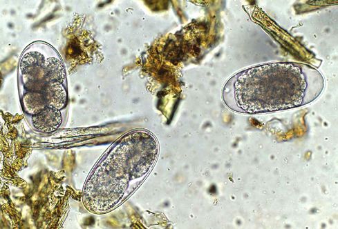
TABLES
Table 1: List of selected equine endoparasite species, their localisation and the drug classes 7
of which effective compounds are registered for treatment in European countries
Table 2: Age-specific scheme for a treatment plan of grazing foals 23
Table 3: Age-specific scheme for a treatment plan of grazing yearlings and young horses 23
(up to and including four years old)
Table 4: Age-specific scheme for a strategic treatment plan of grazing adult horses 24
Table 5: Schedule and key procedures for selective treatment of small strongyle 24
(cyathostomin) infections in adult horses
ACKNOWLEDGEMENT
ESCCAP would like to thank Hubertus Hertzberg PD Dr. med. vet., Dipl. EVPC of the Institute of Parasitology,
University of Zürich for his assistance during the guideline writing process.
IMAGE ACKNOWLEDGEMENTS
ESCCAP would like to thank the following for permitting their images to be reproduced within this guideline:
Institute of Animal Pathology, Freie Universität, Berlin
Jakub Gawor (Witold Stefański Institute of Parasitology, Polish Academy of Sciences, Warsaw, Poland)
K. Seidl, Institute for Parasitology and Tropical Veterinary Medicine, Freie Universität, Berlin
5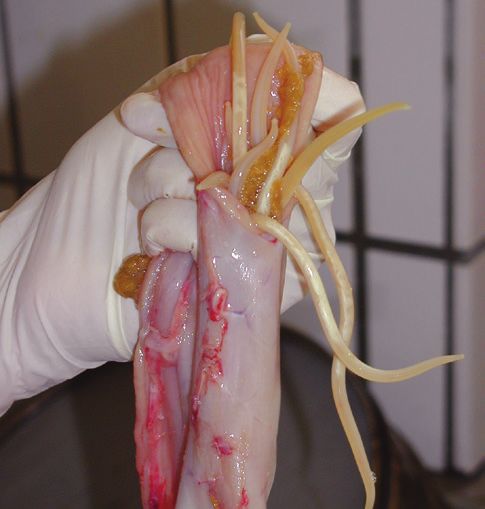
1. Background

    The European Scientific Counsel for Companion Animal Parasites (ESCCAP) was founded in the UK in 2005
    and has since then seen the creation of 11 affiliated ESCCAP national associations representing 16 European
    countries. The key objective of ESCCAP is to provide veterinary professionals with practical, independent
    and research-based advice on how best to protect companion animals from parasitic infection and
    disease, at the same time providing guidance on how to limit the potential for zoonotic parasitic infections.
    To this end, several specific guidelines addressing ecto- and endoparasitic infections in dogs and cats have
    already been published. This is the first equine guideline on this subject and it follows the format of previous
    ESCCAP guidelines.

    2. Introduction

    As grazing animals, horses can be infected by a broad range of gastrointestinal parasites. It must be accepted
    that every horse with access to pasture will be repeatedly exposed to infection with several species of
    gastrointestinal parasites during its lifetime. This may also apply to horses which are always or mostly kept
    indoors or in non-grass enclosures; these animals may be exposed to infection with gastrointestinal worms
    such as roundworms or pinworms. Consequently, the prevention, treatment and control of parasitic infections
    in horses is an ongoing task for equine veterinarians, horse farm managers and horse owners.

    Thanks to the ready availability and frequent use of effective and well-tolerated antiparasitic drugs for most
    of the major gastrointestinal parasites, cases of clinical disease have now become much less prevalent.
    However, since no parasite species has been eradicated and no protective vaccine is available for any equine
    parasite species, routine control and surveillance are required to maintain equine health.

    It is beyond the scope of this guideline to cover all equine gastrointestinal parasites, therefore those most
    prevalent in Europe and only those with the highest clinical relevance will be discussed. These are listed in
    Table 1.

    The aim of this guideline is to provide equine practitioners with concise information and practical advice
    concerning the most important gastrointestinal parasites in horses. An updated overview of these parasites
    under prevailing epidemiological conditions in Europe is provided. The focus of this guideline is to offer
    recommendations which will greatly assist in the prevention or minimisation of parasite infections and thus
    avoid clinical disease in horses. This includes diagnostic and preventive management measures (i.e.
    prophylactic and metaphylactic measures) in the context of the specific needs of the different horse age
    groups, forms of husbandry and types of horse use.

6
Table 1: List of selected equine endoparasite species, their localisation and the drug classes of which effective compounds are
registered for treatment in European countries.

    Parasite species                  Localisation                      Morphological characters             Available1 (selection)
    Anoplocephala perfoliata          Small intestine/caecum            4–8 cm long, flat, segmented         PZQISO, (PYRPY, only partially
    and others                                                                                               effective at 2–3 x increased
                                                                                                             dosage)
    Cyathostomins                     Large intestine                   0.5–2 cm long, thin, small buccal    IVMML, MOXML, FBZBZ,
    (small strongyles)                                                  capsule                              PYRPY, PIPVO
                                      Mucosal/encysted stages                                                MOXML, (FBZBZ)
    Dictyocaulus arnfieldi            Lung                              2.5–8.5 cm long, round               IVMML, MOXML, FBZBZ
    Fasciola hepatica                 Liver                             Up to 5 x 1 cm, flat, leaf-like      None registered
                                                                                                             (reclassification of TCBZBZ)
    Gasterophilus spp.,               Mouth, oesophagus, stomach,       L3 1.5–2 cm long, barrel-shaped,     IVMML, MOXML
    bot fly larvae                    intestines                        two mouth hooks
    Habronema spp.,                   Stomach                           1.0–2.5 cm, thin, hair-like          IVMML, MOXML
    Draschia megastoma
    Oxyuris equi (pinworms)           Large intestine/rectum            ♀ 4–15 cm and with tapering tail,    IVMML, MOXML, FBZBZ, PYRPY
                                                                        ♂ 0.9–1.2 cm
    Parascaris equorum,               Small intestine                   ♀ 16–50 cm, ♂ 15–28 cm, round,       IVMML, MOXML, FBZBZ,
    P. univalens (roundworms)                                           mouth opening with three lips        PYRPY, PIPVO
                                      Lung stages                                                            IVMML
    Strongyloides westeri             Small intestine                   0.8 cm, very thin                    IVMML, MOXML, FBZBZ
    Trichostrongylus axei             Stomach                           0.4 cm, as fine as hair              IVMML, MOXML
    Strongylus vulgaris,              Large intestine                   1–5 cm long, thin, large buccal      IVMML, MOXML, FBZBZ,
    Strongylus equinus,                                                 capsule                              PYRPY, PIPVO
    Strongylus edentatus
    (large strongyles)                Migrating/somatic stages                                               IVMML, MOXML, (FBZBZ only
                                                                                                             partial efficacy against
                                                                                                             S. vulgaris and S. edentatus)
1
      Drugs and drug classes: benzimidazoles (BZ), fenbendazole (FBZ), isoquinoline (ISO), ivermectin (IVM), macrocyclic lactones
      (ML), moxidectin (MOX, cave: only use moxidectin in horses >4 months old), piperazine (PIP), pyrimidines (PY), pyrantel (PYR),
      triclabendazole (TCBZ), praziquantel (PZQ) and various other (VO). Those marked in red indicate when cases of anthelmintic
      resistance have been published for the respective drug class and parasite species in Europe.

3. General factors: age, husbandry, use, weather and climate

For effective and sustainable parasite control in horses, it is important to apply all the available knowledge on
preventive management measures designed and adapted according to the specific needs of the type of horse
and the conditions under which the animal is being kept.

Some parasite infections, like roundworms, lead to a partially protective immune response and older
horses do not normally require intensive metaphylactic treatment or specific husbandry measures to protect
them from disease. In situations where horses have no access to pasture, they are not usually exposed to
strongyle infections.

The parasites addressed in this guideline essentially occur in all European countries and thus under varying
existing climatic conditions. The effect of climate and weather on parasite bionomics and the epidemiology of
some parasitic diseases such as strongylosis, caused by heavy infections with the small and large strongyles,
should also be considered during the assessment of control measures required.

                                                                                                                                              7
4. Specific information and recommendations for control measures against
    selected equine gastrointestinal parasites (key biological factors, life cycle,
    epidemiology/prevalence, clinical signs, diagnosis, drug treatment/resistance)

    4.a. Non-migratory strongylids (commonly named “small strongyles”)
    These include cyathostomins and non-migratory strongyline species (Triodontophorus, Craterostomum and
    Oesophagodontus). Infections with “small strongyles” occur in all European countries and practically all horse
    farms. Horses mainly become infected on pasture through the uptake of infective third stage larvae (L3),
    which subsequently undergo larval development in the intestinal mucosa before they re-enter the intestinal
    lumen (Figure 1 and Figure 2).

                                                                                         D                  A

                                                                              C
                                                                                            E
    Figure 1: Small strongyle/
    cyathostomin life cycle
    A: egg shedding
    B: oral uptake of third stage
    larvae (L3) with grass                                                                                                egg
    C: exsheathment through gastric fluids
    D: passage of exsheathed L3 through              B
    small intestine
    E: invasion of mucosa/submucosa
    of colon and caecum, moult to fourth
    stage, return to intestinal lumen and
    final moult before development to                 L3 on                                                                L1
    adult stage                                      grasses

                                                                                                         L2
                                                                                  L3

            L3

    Ileum                                                 Dorsal colon

    Caecum
                           L4
                                               St5

                                                   Ventral colon
                      L3                                                          Figure 2: Development of
                                                                                  cyathostomins in the intestine
                                                                                  Modified after Deplazes et al., 2016,
                                             Moulting to L4; histotropic
                                                                                  Parasitology in Veterinary Medicine,
                                 L4          phase of 1–2 months (in winter
                                             hypobiosis of L3 possible)
                                                                                  Wageningen Academic Publishers,
                                                                                  pp 268

8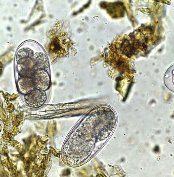
Infections acquired indoors are considered to
be rare and of minor importance. Non-migratory
strongylids are considered much less pathogenic
than the migratory strongylids like Strongylus spp.,
however a large number of Triodontophorus spp. (the
most common being T. serratus and T. brevicauda)
may damage the intestinal mucosa and result in
emaciation and diarrhoea because of their tendency
to feed all together like “worm herds”. More than 40
cyathostomin species are recognised in horses and
individual horses may be simultaneously infected
with several, often more than ten, cyathostomin
species.     Cyathostomins     can    cause    larval
cyathostominosis, a syndrome which is a result of the
synchronic restart of the development of numerous
inhibited/encysted L3 (Figure 3) and simultaneous
                                                          Figure 3: Large intestine of a horse with multiple encysted
migration of mucosa dwelling larval stages into the       small strongyle larvae in the mucosa
lumen with massive tissue destruction.

This disease is mostly seen in animals up to six years
of age and results in acute and persistent diarrhoea
(sometimes accompanied with colic, weight loss
or fever) and in a considerable number of cases a
fatal outcome. Normally, lumen-dwelling larval and
adult cyathostomins are considered to be of low
pathogenicity and most infected animals do not
appear to be clinically affected, even if fairly high
worm burdens are present. Nevertheless, some
studies have suggested a possible correlation
between cyathostomin infection and recurrent
diarrhoea and intermittent colic.                                                     L1

Diagnosis of a patent small strongyle infection is
                                                           Figure 4: Gastrointestinal strongyle eggs in horse faeces,
performed by faecal examination and identification         egg marked with L1 contains first stage larva
of the thin-shelled, ovoid, strongyle-type eggs which
measure approximately 80–100 µm in length (Figure 4).

Often larval/pre-adult and adult stages are found
in large numbers on the faeces of treated horses
(Figure 5).

Various methods can be employed allowing either the
qualitative or quantitative analysis of strongyle eggs.
There are no scientific data available concerning the
correlation between the number of strongyle eggs
per gram of faeces and the intestinal adult worm
burden in adult horses. One study, in which horses
younger than three years were examined, showed
                                                           Figure 5: Horse faeces with typically red-coloured
that low or even negative faecal egg counts can be         cyathostomin stages
found in horses with thousands of intestinal worms.

Overall, it may be assumed that the correlation between faecal strongyle egg counts and worm burden is
weak in all age groups. It is noteworthy that the eggs of small and large strongyles (e.g. Strongylus vulgaris)
are not reliably distinguishable based on morphological criteria. However, following in vitro culture, the third
stage larvae (L3) can be differentiated based on the number of their midgut/gut cells. This differentiation is
significant due to the considerably higher pathogenicity of large strongyles, which, owing to the widespread
use of effective anthelmintics, are now only considered to occur on a low percentage of farms. However,
recent data show that S. vulgaris is still present in the European horse population (see 4.b.).

                                                                                                                        9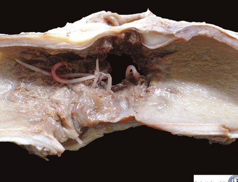
Horses first become infected with small strongyles as soon as they start grazing and start to shed strongyle
     eggs 6–14 weeks after infection. Accordingly, treatment and control measures should be applied to foals
     beginning at approximately two months of age. As a result of the widespread occurrence of anthelmintic
     resistance (AR), it is important to reduce the frequency of treatment to the minimum possible without risking
     the establishment of clinically relevant worm burdens. Under currently prevailing epidemiological conditions
     in most European countries, where the intensity of small strongyle infection is only low to moderate, effective
     three-monthly treatment of foals and yearlings can be considered appropriate. In adult horses it may be
     feasible to treat only twice yearly. In the absence of large (migrating) strongyles one annual treatment is
     sufficient, when faecal monitoring does not indicate further treatment and provided that strict quarantine
     procedures are being employed on the respective farm.

     Horses suffering from larval cyathostominosis should be treated palliatively i.e. reducing diarrhoea (for
     example using codeine phosphate), reducing mucosal inflammation and administering fluid therapy if
     needed. Irrespective of clinical status, all horses of the same group should receive anthelmintic treatment
     against the mucosal worm burden either using moxidectin (once orally 0.4 mg/kg bodyweight only in horses
     >4 months old) or fenbendazole (7.5 mg/kg bodyweight orally once daily over five days and only when the
     respective cyathostomin population is susceptible). It is recommended that such treatments against mucosal
     cyathostomin larvae are employed once a year for foals and young horses up to and including four years of
     age (e.g. at the end of the grazing season).

     With regard to AR, recent studies in France, Germany, Italy and the UK, showed that the small strongyle
     populations present on more than 80% of the farms studied had reduced susceptibility to the benzimidazole
     group of anthelmintics (BZs). In the case of pyrantel, this was only found on approximately 20–30% of farms.
     In contrast, the macrocyclic lactones (MLs) ivermectin and moxidectin were found to be fully effective with
     95–100% faecal egg count reduction at 14 days post treatment on nearly all of the farms tested. Nevertheless,
     a reduced egg reappearance period (ERP) post ML treatment has been reported occasionally and this is
     considered to be a sign of reduced efficacy. It is therefore advisable to regularly confirm/test the efficacy
     of any anthelmintic drug class used by, for example, conducting an annual faecal egg count reduction
     test (FECRT).

     4.b. Migratory strongylids (commonly named “large strongyles”)
     This group of parasitic worms occurring in the large intestine consists of migratory species of some
     strongylines (S. vulgaris, S. edentatus and S. equinus, Figure 6).

              Strongylus edentatus                          Strongylus equinus                       Strongylus vulgaris

     Figure 6: Anterior end of large strongyles depicting the buccal capsule, leaf crown and tooth-like structures at base of the
     buccal capsule

10
Clinically, these are the most important of the equine parasites with S. vulgaris considered a major threat
to equine health. Their larvae migrate extensively before developing to maturity in the large intestine:
in the anterior mesenteric and nearby arteries (S. vulgaris, Figure 7 and Figure 8), through the liver to
the subperitoneal connective tissues (S. edentatus) and to the liver and the pancreatic and renal region
(S. equinus). These larval migrations result in long prepatent periods, which are 6–7 months for S. vulgaris,
9 months for S. equinus and 11–12 months for S. edentatus. The damage caused by the migrating larvae leads to
severe pathological consequences and clinical signs which differ depending on the respective Strongylus species.
Figure 7: Strongylus vulgaris life cycle
Parasitic phase: Oral uptake of L3 with adult stages
grass, exsheathment in small intestine, egg
penetration into wall of large intestine
(other stages
and moulting to L4, migration on or not shown)
in intima of arteries of large intestine,
migration to cranial mesenteric artery egg
and moulting to pre-adult stage, with L1
migration to intestine and penetration
of intestinal wall to enter lumen where
development to adults is completed.
Free-living phase: Thin-shelled eggs L3 on
expelled with faeces, development grasses
to first stage larva (L1) within the egg, L1
moulting to second stage larva (L2)
and infective third stage larva (L3). L3
L2
Abbarant larvae
Aorta
Cranial
mesenteric L3
artery St5
St5
Figure 8: Development and migration of
Strongylus vulgaris larvae
Development: L3 penetrate the intestinal
L4 wall and moult to L4, migration of L4 into
the cranial mesenteric artery, moulting
to St5 from 90th day p.i., backwards
migration from arteries to the gut.
Modified after Deplazes et al., 2016,
Parasitology in Veterinary Medicine,
L4 Wageningen Academic Publishers, pp 269
11In the past, S. vulgaris, the “horse killer”, has
received most attention due to the clinical syndrome
of thromboembolic colic caused by larvae migrating
to the cranial mesenteric artery (Figure 9). Adult
strongyles feed on plugs of intestinal mucosa, the
resulting damage causing diarrhoea, weakness,
emaciation and sometimes anaemia.
The migrating larvae and the thromboses they cause
can lead to non-strangulating intestinal infarctions
most often seen in the large intestine. Depending
on the infection intensity, initial clinical signs of non-
strangulating intestinal infarctions may be mild,
often recurrent abdominal pain (colic), fever and
peritonitis. If the infarcted intestine is not recognised
Figure 9: Aorta at cranial mesenteric artery junction
and surgically resected, the intestine will necrotise showing several L4/pre-adult stages of Strongylus vulgaris
and rupture leading to the death of the horse.
It is noteworthy that sometimes even horses with severe intestinal necrosis caused by thrombosis, do not
show signs of serious pain. Peritonitis is therefore often the only sign advocating surgical intervention.
Detection of patent large strongyle infections is based on in vitro culture of the third stage larvae (L3) which
can be differentiated from those of other strongyles based on the number of midgut cells (see also 8.1.
Diagnosis of worm infections).
Previously, routine treatments at regular intervals have been recommended for all horses to minimise the level
of pasture contamination and thus reduce the risks associated with migrating S. vulgaris larvae. Due to years
of this intensive metaphylactic chemotherapy, infection with S. vulgaris has become uncommon. However,
during the recent past, a selective therapy approach has been increasingly recommended in an attempt to
reduce the development of anthelmintic resistance in the cyathostomins by reducing treatment intensity i.e. to
leave horses with low strongyle egg counts untreated. Consequently, specific diagnosis of patent S. vulgaris
infections is important.
For sustainable control of strongyle infections in horses, metaphylactic therapy programmes should therefore
be designed to avoid anthelmintic resistance (e.g. in cyathostomins and ascarids) and to simultaneously
minimise the potential for S. vulgaris transmission. To date there have been no convincing reports
of anthelmintic resistance in large strongyles. Biannual treatment of all horses with a drug effective against
S. vulgaris larvae (e.g. IVM or MOX) is likely to provide adequate control of this parasite.
124.c. Roundworms (Parascaris equorum and Parascaris univalens)
The equine roundworm species, Parascaris equorum and P. univalens, cannot be distinguished morphologically.
Recent findings indicate that P. univalens, and not P. equorum, is the species currently prevalent on most, if
not all, horse farms in Europe where equine roundworms are found. At present, there are no molecular tools
available for species differentiation and since both appear to have a similar pathogenesis and biology, we will
subsequently refer simply to Parascaris spp.
Infection with equine roundworms is mainly prevalent on stud farms and predominantly found in foals and
young horses. Recent cross-sectional studies in Europe provided prevalence rates of between 20% to over
80% in foals.
Measuring up to 50 cm in length at the adult stage, these worms which reside in the small intestine represent
one of the largest known parasitic nematode species. The females can shed hundreds of thousands of eggs
per day, thus contributing to considerable environmental contamination. The infective stage is the third stage
larva (L3) within the egg, which can survive in the environment for several years, even under harsh conditions
such as prolonged periods of frost. Consequently, both stables and pastures once contaminated will remain
a constant source of infection. Following ingestion of eggs, larvae are released and penetrate the small
intestinal wall to begin a somatic migration via the bloodstream through the liver, heart and lungs. There, the
larvae transfer to the respiratory system where they are transported with the mucosal flow to the larynx and,
after being swallowed, reach the small intestine approximately three weeks post infection. It then takes at
least another 7 weeks of maturation before the first shedding of eggs in the faeces (prepatent period 10–16
weeks, Figure 10).
B
C A
Figure 10: Parascaris equorum/
Parascaris univalens life cycle
A: Hatching of third stage larvae (L3) egg
in the stomach and small intestine,
penetration of intestinal veins.
B: Larvae reach liver via portal vein,
migration through liver tissue and
penetration of liver veins.
C: Larvae reach lung via vena cava and
right heart, penetration into lung alveoles
and migraton via trachea and pharynx to
egg + L3
small intestine (moulting to L4 and St5
prior to development into adults). egg + L1
13Often, no clinical signs are observed. Sometimes
during the somatic migration, clinical signs occur
mostly associated with pathological changes in the
lungs, whereas the migration through the liver does
not appear to cause clinical signs. In the lungs,
changes include haemorrhagic mucosal lesions
and heavy infections can result in coughing and
decreased weight gain in young stock and can
also lead to secondary bacterial or viral infections.
During the intestinal phase (Figure 11), Parascaris
spp. infected animals show reduced appetite and
a rough coat; intermittent colic and wasting may
also occur. Occasionally, heavy infections can result
in severe colic, obstruction of the small intestine,
perforation, invagination followed by peritonitis.
Under current epidemiological conditions in most
western European countries, the infection intensity is
low and the vast majority of cases in foals and young
horses are subclinical. Adult mares can occasionally
excrete eggs and so act as a source of infection for Figure 11: Small intestinal infection with Parascaris spp.
subsequent generations.
Diagnosis of Parascaris spp. infections relies on the direct detection of eggs (round, brownish, approximately
100 µm in length, thick-shelled) by faecal flotation and/or of pre-adult stages or adult worms in the faeces.
Coproscopic analysis is based upon the microscopic detection of the eggs either during a qualitative or
quantitative flotation protocol. As with ascarid infections in other host animals, it is impossible to reliably relate
the intensity of the intestinal worm burden to the level of egg shedding in the faeces and a positive faecal
examination should always be considered an indication for anthelmintic treatment. Due to the environmental
contamination and long survival times of Parascaris eggs, it has to be assumed that horses from the same age
group, sharing the same environment, which are currently not shedding eggs in the faeces, are also exposed
and probably infected and that the infection may be in the prepatent phase. All horses from the same age
group should be treated based upon a positive coproscopic analysis of any individual within the group. The
MLs are effective against larval stages in the lungs and intestines. Thus, the previous recommendation for 6–8
weekly treatments during the first year of the animal’s life is aimed at the prevention of contamination and the
consequent development of intestinal worm burdens. However, highly frequent treatment is considered to be
the main reason for the selection of ML-resistant Parascaris spp. populations.
Sustainable control approaches should include regular faecal monitoring (preferably individual samples).
Stable and pasture hygiene must accompany anthelmintic treatment, which should begin at two months
of age and repeated every three months during the first year of life, employing different drug classes. The
above-mentioned AR situation requires that each farm assesses the efficacy of the drug classes used, most
importantly the MLs, by performing a faecal egg count reduction test (FECRT) or at least a faecal examination
for Parascaris eggs, 14 days post treatment. The beneficial effects of pasture “cleaning” and the chemical
or physical disinfection of stables have been demonstrated in field surveys, and these have been associated
with significantly reduced Parascaris spp. prevalence. When using disinfectants it is important to use only
those which have been shown to be effective against worm eggs (i.e. containing cresol or peracetic acid,
see also chapter 5). Resistance to MLs has been widely reported for Parascaris spp. and more recently there
have been a few reports from North America and Australia suggesting that resistance to pyrantel and the BZs
may be emerging. On farms where there is confirmed resistance to the MLs, then BZs, pyrantel or piperazine
citrate (available in some EU countries only) can be used. However, the latter has to be given in comparatively
high doses and large volumes often requiring nasogastric intubation. Due to a potential risk of colic caused by
worm convolutes following the immediate killing/paralysis of neurotoxic drugs, MLs, pyrantel and piperazine
should not be used on foals with heavy infections.
144.d. Tapeworms (Anoplocephala perfoliata, Anoplocephala magna
and Paranoplocephala mamillana)
Two species of equine tapeworms are of significance in Europe: Anoplocephala perfoliata and A. magna.
Most cases of tapeworm infections in horses are caused by A. perfoliata which is endemic in many European
countries. Anoplocephala magna infections are rarely recognised, however there is evidence that this is
prevalent in Spain. Paranoplocephala mamillana has also been found occasionally, for example in Germany.
Infection with tapeworms occurs mainly during the second half of the grazing season and essentially only
on pasture by ingesting infected intermediate hosts which are oribatid mites or “box mites” (Figure 12).
The prepatent period is from six weeks to four months. Adult A. perfoliata (Figure 13) are 4–8 cm in length
and inhabit the caecum close to the ileocaecal junction while A. magna specimens (up to 80 cm in length)
occupy the small intestine. Higher A. perfoliata infections can be associated with clinical signs of colic in the
horse, due to bowel irritation, ileal impactions, intussusceptions and intestinal obstruction, which may lead to
recurrent episodes of spasmodic colic. The risk of gastrointestinal problems increases in horses with chronic
and heavy infections. Anoplocephala magna pathogenicity is limited to catarrhal inflammation and infections
generally pass unnoticed, with higher prevalence in young horses under 2 years old.
Figure 12: Anoplocephala perfoliata life cycle
Gravid proglotids filled with eggs are
expelled with the faeces (A), eggs (B)
are released and taken up by box mites E
as intermediate hosts within which the
infective cysticercoids develop (C),
following oral uptake of the infected
mite with grass (D), the cystercercoids are
released during digestion of the mite,
A proglotids
larvae attach to intestinal mucosa
and develop into adults (E).
adults B
D
mites egg
on grasses C
Figure 13: Head section of adult
Anoplocephala perfoliata
cysticercoids
Diagnosis of tapeworm infections in horses by faecal examination is limited in sensitivity as eggs are passed
intermittently and this is not associated with the number of worms present. To improve the detection of
Anoplocephala eggs in faeces, combined centrifugal sedimentation-flotation techniques have been developed
which process large faecal samples (15–50 g). To compensate for the limited sensitivity of the coproscopic
diagnosis, it is recommended that a group/farm diagnosis is performed and all animals treated if tapeworm
eggs are found in any of the examined samples. Commercial diagnostic assays, capable of detecting
A. perfoliata antibodies by either a serum ELISA (Diagnosteq, University of Liverpool, UK) or a saliva ELISA
(EquiSal, Austin Davis Biologics, Great Addington, UK) are available. Both tests can potentially generate
false-positive results in some horses due to the persistence of antibodies for up to four months e.g. in
previously-infected horses that have already been treated with anthelmintics. However, if allowances are
made for this, these tests can prove very useful, particularly for group/farm diagnosis using the serum test or
targeting of treatments to individuals using the saliva test.
15Treatment of tapeworms is based on the use of cestocidal anthelmintics and the drug of choice is praziquantel.
Praziquantel is often only available in combination with MLs (e.g. ivermectin or moxidectin). In a situation
where only drugs effective exclusively against nematodes are used, undiagnosed tapeworm infections can
persist for several years in groups of horses. Cestocidal drugs appear to have remained fully effective, but it
is difficult to evaluate the efficacy of tapeworm anthelmintics using current diagnostic methods, due to a lack
of sensitivity of the available tests.
Sustainable tapeworm control strategies should be related to regional climatic conditions and management
systems should be put into place to ascertain the significance of tapeworm infection at farm level. Routine
multiple treatments throughout the year, although justified for the control of cyathostomins, are not
recommended for the control of tapeworms, due to their different life cycle involving an intermediate host
and the marked seasonality of transmission. Generally, a single annual tapeworm treatment in late autumn or
winter will be sufficient to avoid significant infection, but in cases of heavy infection pressure, an additional
earlier treatment during the summer may be required. Regular (i.e. at least weekly) removal of faeces from
pasture may in the long term also reduce infection pressure.
4.e. Bot flies (Gasterophilus spp.)
Bot flies are arthropods of the genus Gasterophilus (Diptera: Oestridae). Gasterophilus intestinalis,
G. haemorrhoidalis, G. nasalis, G. inermis and G. pecorum are the most prevalent species in Europe.
Gasterophilus intestinalis, G. haemorrhoidalis and G. nasalis frequently infest grazing horses; G. inermis and
G. pecorum are found less often. Their larvae mainly cause gastrointestinal myiasis.
Adult flies resemble honey bees and females play the major role in the infection. In southern Europe they may
already be active in spring/early summer, while in temperate regions, egg laying takes place in late summer.
Females of most Gasterophilus species fly near horses and dart very rapidly close to the skin to attach an egg
to a hair (this fly activity produces a special buzzing noise which many horses find very disturbing). Females
die after laying small (1–2 mm), mostly operculate and yellowish eggs. The eggs can be seen fairly easily
with the naked eye, especially on animals with a dark hair coat. Regarding the egg localisation, G. intestinalis
places eggs on the hair of the forelimbs, shoulders and flanks while most other species deposit their eggs
on the head. G. pecorum is an exception as females lay eggs in the environment. Humans have occasionally
been infected showing conspicuous tracks in the cheeks, and even infection of the digestive tract.
The hatching of first stage larvae (L1) takes place after a mechanical stimulus (G. intestinalis and G. pecorum)
or spontaneously (G. nasalis). The L1 reach the oral cavity through oral uptake (licking or grazing for
G. intestinalis and G. pecorum respectively) or by larval migration. Second stage larvae (L2) are found in the
stomach and duodenum where they moult into third stage larvae (L3). The L3 measure 16–20 mm in length,
have a barrel-shaped form and bear two large mouth hooks. The segments have one or two rows of spines.
After several months, the L3 finally leave the host in the faeces and pupate in the soil, before the adult flies
emerge into the environment. The parasitic phase takes 8–10 months and the pupal phase 3–8 weeks. The
adults mostly emerge during June/July and are usually active until October or November, although their
activity may begin earlier and last longer in southern European regions.
Gasterophilus L2/L3 are found attached to the mucosa of the stomach (G. intestinalis), duodenum (G. nasalis,
G. haemorrhoidalis) or rectum (G. haemorrhoidalis, G. inermis), where they may cause focal, superficial
mucosal ulceration, cutting and piercing of tissues to facilitate feeding. When L1 are found in the oral
cavity, they migrate through the mucous membranes of the tongue, gums and palate, causing gingivitis
and pain which may affect food intake. Generally, the first clinical signs of gasterophilosis are characterised
by difficulties in swallowing due to the localisation of the larval stages in the throat. Remarkably, massive
infections with Gasterophilus spp. are not always associated with clinical signs and are thus considered much
less pathogenic than most nematode parasites. Nevertheless, gastric and intestinal ulcerations have been
associated with this infection, as well as chronic gastritis, gut obstructions, volvulus, rectal prolapses, rupture
of the gastrointestinal tract, peritonitis, anaemia and diarrhoea.
16The presence of Gasterophilus spp. can be confirmed in the summer/autumn by inspecting the horse’s
coat and finding yellowish eggs attached to the hair. Gastrointestinal examination through endoscopy may
allow the detection of Gasterophilus spp. larvae attached to the stomach and duodenum. ELISA based on
excretory/secretory antigens of G. intestinalis L2 for antibody detection and PCR techniques has been used
in Europe, however these tools are not yet considered routine laboratory techniques.
The larval stages of Gasterophilus are highly susceptible to MLs (particularly ivermectin) and will be eliminated
during regular deworming with these drugs. As fly activity ceases with the first frosts, an appropriate treatment
in late autumn, e.g. early November, should remove all larvae present within horses. The removal of the eggs
by hand with a special bot comb or bot knife or thoroughly washing the hair with warm water mixed with an
insecticide is recommended though it is usually not sufficient to effectively prevent gastrointestinal infection.
4.f. Threadworm (Strongyloides westeri)
The nematode Strongyloides westeri resides in the small intestine, mainly the duodenum. Patent infections
are largely found in young horses, i.e. foals up to six months old. Occasionally, older horses can harbour this
parasite and mares are an important source of infection for their foals. It is a unique parasite, since it only
develops female and no male parasitic stages. The very slender, small (maximum length 10 mm) parasitic
females reproduce through parthenogenesis. They shed small, thin-shelled, embryonated eggs (40–50 x 30–
40 µm) containing the first stage larvae (L1) which hatch in the environment. These can develop directly to
infective third stage larvae (L3) or give rise to free-living males and females that will reproduce and in turn
produce infective L3.
Infection may occur by ingestion of L3 in the mare’s milk (“lactogenic infection”) and this is the primary mode
of transmission of S. westeri to foals. Later on, transmission also occurs by ingestion of infective L3 from the
pasture or environment, or through percutaneous infection. When percutaneous infection occurs in immune
adult horses, S. westeri larvae do not become established in the alimentary tract and patent infections are rare.
Instead, they are distributed through various somatic tissues where they may remain viable for long periods,
probably years. In mares, the hormonal shifts during pregnancy and lactation probably stimulate these larvae
to resume migration to the mammary glands so that they are transmitted to the foal. After being ingested with
the milk, the larvae undergo a somatic migration starting with the penetration of the small intestinal wall. They
subsequently travel through the lungs, via the trachea and pharynx where they are swallowed finally reaching
the small intestine. Here they mature to adult female worms. The prepatent period can take some weeks but
can be as short as 5–8 days.
During massive percutaneous infection, local dermatitis can occur. The hair coat may become dull and animals
can be stressed by local skin irritation and itching, which is often a consequence of an allergic response to
re-infections. The major pathogenic effect of infection occurs in the intestine, where adult females embed in
the small intestinal mucosa and cause local enteritis, which can lead to diarrhoea. The role of S. westeri as the
cause of diarrhoea in young foals is unclear since there are reports of high faecal egg counts associated with
severe cases of diarrhoea while high Strongyloides egg shedding has also been found in animals showing no
clinical signs. Clinically affected foals may become anorexic and lethargic but in situations where regular worm
control is employed, it appears that most S. westeri infections are asymptomatic. It should be noted that there
are many cases of diarrhoea in foals aged 1–2 weeks which are not associated with S. westeri infection.
Diagnosis of S. westeri infection is performed by coproscopic detection of the typical eggs in faeces.
Treatment and control of S. westeri infections should involve both anthelmintics and basic hygiene measures.
Under the current epidemiological situation, the formerly often-employed, routine treatment of foals during
the first few weeks of life does not seem justified anymore due to the low prevalence and lack of evidence
for S. westeri associated disease in foals. On farms where S. westeri has previously been detected, regular
deworming of mares, before or shortly after parturition (i.e. 1–2 days) is thought to reduce the number of
larvae in milk and lower the incidence of diarrhoea in foals. For clinical cases, a number of drugs are available
including ivermectin or fenbendazole, the latter at a dosage of 50 mg/kg body weight (significantly higher than
the standard dosage of 7.5 mg/kg bodyweight). Pasture and stable hygiene, together with cleaning of the
mare’s udder should reduce the risk of environmental contamination and foal infection.
174.g. Pinworm (Oxyuris equi)
The equine pinworm Oxyuris equi (Figures 14a, 14b
and 15) has been reported as a common horse
parasite in Europe. Infections arise in stables and
may also occur on pasture, but usually only a few
horses appear to be clinically affected. Oxyuris equi
is rarely considered a major threat to equine health,
but heavy infections may result in fatigue, decreased
performance and loss of condition. Even massive
invasions of fourth stage larvae usually do not lead
to clinical signs, however, in single cases, they can
cause severe inflammation of the colonic mucosa
with non-specific intestinal signs.
Considerable numbers of O. equi eggs (up to tens
♀ ♂
to hundreds of thousands) are deposited by the
female worms on the skin of the perianal region.
The sticky fluid surrounding these eggs causes
an intense itching and an indication of O. equi Figure 14a: Oxyuris equi Figure 14b: Anterior end
infection is persistent anal pruritus and tail-rubbing (pinworm) adults of Oxyuris equi adult with
♂ 0.9–1.2 cm ♀ 2.5–15 cm, typical hourglass-shaped
which causes excoriations and bare patches around ♀ pointed rear end, oesophagus
the tail (Figures 16a and Figures 16b). bevelled anterior pole,
operculum, u-shaped larva
A C
B
egg
Figure 15: Oxyuris equi life cycle
egg + L3
Hatching of L3 in the small intestine (A),
histotrophic phase in the caecum and
egg + L1
colon (B), adults develop in the colon,
females emerge from the anus to lay
egg clusters on the perineum (C) egg + L2
18Figure 16a: Oxyuris equi infection with itching and dermatitis Figure 16b: Massive Oxyuris equi egg excretion with
of the tail root, tail rubbing, broken/matted hair (“rat tail”) cream-coloured, dried egg clusters
Diagnosis of pinworm infection is made by applying a transparent adhesive tape to the skin of the perianal
region, which is then removed and examined microscopically to identify characteristic oxyurid eggs, which
contain an embryo and are oval and flattened on one side, with an operculum at one end.
The perianal region of infected horses should be washed with hot water containing a mild disinfectant to
relieve the pruritus and to prevent the spread of pinworm eggs throughout the horse’s environment.
The MLs and BZs are effective against pinworms and their larval stages. Pyrantel has variable efficacy against
pinworms. Recent anecdotal reports on the reduced efficacy of MLs (ivermectin and moxidectin) against
O. equi must be viewed as potential resistance.
195. Measures against free-living/environmental stages
The control of parasite infections in horses currently relies mainly on the use of anthelmintic treatments
to eliminate intestinal worm burdens and thus reduce the contamination of the environment with eggs/
infective stages. However, as explained below, this strategy alone without any other measures designed to
prevent or minimise infection intensity is not sustainable, due to the development of anthelmintic resistance
in several parasite species. Consequently, stable and pasture hygiene are both important components of an
integrated worm control strategy and should therefore be employed. The infective stages of some equine
parasites have the potential to survive in the environment for months or years and it is important to consider
the following factors:
The eggs of important nematode species need, at appropriate temperatures, at least one week (strongyles)
or two weeks (Parascaris) to develop into infective stages. Therefore, the regular and frequent cleaning of
stables and the removal of faeces from pasture will reduce the risk of heavy infections. If possible, droppings
should be removed from the pasture on a daily basis. If this is impracticable, it should at least be done on
a twice weekly basis. Stables should also be cleaned on a daily basis, but when this is not possible e.g. in
deep litter management systems, stables should be thoroughly cleaned (mechanically and by steam) and
disinfected at least once a year using a disinfectant which has been shown to be effective against ascarid eggs
(e.g. as documented and listed by the committee for disinfection of the German Veterinary Society;
www.desinfektion-dvg.de/index.php?id=1793).
Using horse manure as a fertiliser will increase the risk of Parascaris spp. infection and thus should be
avoided. However, it has been shown that by effective windrow (long row) composting, Parascaris eggs will
be prevented from developing to the infective stage (or result in die-off), so that equine manure and soiled
bedding processed accordingly can be used for pasture fertilisation without increasing the infection risk.
All free-living stages of equine worms are vulnerable to low environmental humidity and therefore stables
should be kept dry.
To prevent importation of new parasite species and/or resistant parasite populations, each horse newly
introduced to a farm should be quarantined and treated upon arrival. Subsequently, the horse should only
be put out to pasture after a faecal examination conducted five days post therapy has confirmed that the
horse is not shedding worm eggs and that treatment was successful.
To date, measures aimed at the biological control of strongyle developmental stages in the environment
(i.e. L1, L2 and L3) are still in an early experimental phase and although promising it seems unclear if or
when these will become practical and available for routine use.
Agricultural practices such as deep ploughing of the paddock will help to reduce not only the presence
of infective nematode larval stages but also mites therefore also potentially reducing tapeworm infection,
provided no new contamination occurs.
206. General treatment strategies for foals, yearlings, adults and mares
(tabulated specific treatment recommendations in an annual context)
It needs to be noted that treatment-related factors, such as underdosing and frequent anthelmintic treatment,
are probably the most relevant reasons for the emergence of AR. Thus, to avoid selection for AR, treatment
should be administered as infrequently as possible without risking disease. This is managed by regular faecal
examinations including the differentiation of small and large strongyles, so that the infection status of the
individual animal or the respective age group is monitored throughout the year. Furthermore, thorough hygiene
and quarantine measures both in stables and on pasture are important to reduce the infection pressure and
accordingly the need for treatment.
To date, there are two alternative approaches for the control of small strongyles which are recommended
by experts working in the field of equine worm control. These are the ‘selective treatment’ and the
‘strategic treatment’ approaches. In the following section, both approaches will be briefly described and
discussed. Both strategies are considered to be effective in preventing clinical disease in adult horses when
employed according to these guidelines. Their specific potential to mitigate the development of anthelmintic
resistance will largely depend upon the resulting frequency of treatments per horse per year for each strategy.
Comparable data are not yet available but should be generated for the future analyses of both strategies.
Notwithstanding, it is essential that veterinarians and those responsible for equine health are aware of the
actual resistance status of the parasites occurring on their respective farms. On farms where resistance of a
certain drug class against a specific worm species has been identified (either by conducting post-treatment
efficacy check-ups or faecal egg count reduction tests), consideration is needed when deciding upon the
future use of this drug class. Generally, the respective drug class should not be used any more against the
respective worm species.
6.1. Selective treatment approach
Repeated small strongyle infections occur in all age groups of grazing horses; however, in the majority of adult
horses an immunological response leads to a suppression of small strongyle egg production. Several studies
provided evidence for consistency in strongyle egg shedding after acquisition of immunity in individual
horses. This phenomenon is the basis for selective treatment approaches in which only horses showing a
pattern of consistently high strongyle egg shedding, exceeding a certain threshold e.g. 200 eggs per gram
(EPG) of faeces, receive anthelmintic treatment. Practically, this approach involves a first year during which
faecal samples from each horse are examined at least four times. All horses with strongyle EPGs above the
threshold should be treated. If the responsible veterinarian regards the epidemiological situation as stable,
the frequency of diagnostic examination can be reduced to three in the following years (beginning, mid and
end of season, see Table 5).
The selective treatment approach is only recommended for adult horses and exclusively designed for the
control of small strongyles. It aims at increasing the proportion of small strongyle eggs/larvae on pasture
that were produced by worms that have not been exposed to anthelmintic treatment. This is known as a
refugium of susceptibility and it has been hypothesised that a large environmental parasite refugium prevents
or postpones the development of anthelmintic resistance. In various studies, including several from Europe,
the application of the selective treatment approach has been shown to significantly reduce the number of
anthelmintic treatments in horses. In these studies, horses did not develop clinical signs associated with
parasite infection.
21However, it is not completely certain that the intestinal worm burden of horses shedding only low numbers
of strongyle eggs is in fact negligible. As mentioned above, high treatment frequency is considered one of
the most relevant reasons for the emergence of AR. This is, however, much more of an issue in foals and
yearlings, where previous recommendations of 4–8 weekly treatments should now be avoided. To date, it
remains unproven whether the application of selective treatment in adult horses actually has a significant
impact on the selection for AR in horses or if the reduction of treatment frequency in foals and young horses
is more relevant. In this context, it is also worthwhile noting that it has been shown by a Danish study
that the highly pathogenic large strongyle species Strongylus vulgaris is more prevalent on farms which had
selectively treated their horses during recent years compared with those which had employed strategic whole
herd treatments. It should however be noted that the selective treatment approach employed in these farms
differed from that described herein, particularly concerning the monitoring of S. vulgaris presence and the
respective treatment decisions.
Strongylus vulgaris or other large strongyle species have not, or only very rarely, been found in recent
European studies using larval cultures and microscopic identification of L3. However, it has been reported in
several single cases and studies, often associated with severe clinical consequences, which demonstrates
that the parasite is nevertheless still present but at low levels. Consequently, monitoring the occurrence of
large strongyles using faecal larval cultures has to be an integrated component of the selective treatment
programme and this approach should not be recommended on farms where large strongyles have been shown
to occur. Before (re)integration of stables harbouring large strongyles into a selective control programme,
biannual anthelmintic treatments (i.e. late spring and autumn/winter) using drugs active against the adult
AND larval stages of the large strongyles (MLs and FBZ), should be given to all grazing horses for at least two
years. The large strongyle status of the farm should be documented by examination of pooled larval cultures
at least once per year. All other treatment decisions remain the responsibility of the veterinarian in discussion
with their horse-owning clients.
6.2. Strategic treatment approach
A horse’s age and usage can determine the appropriate worm treatment. Foals particularly but also young
horses need comprehensive protection with regular anthelmintic treatments, even on well-managed farms
with good stable and pasture hygiene. While in the past it was often recommended to treat foals frequently
(up to every 4–8 weeks during their first year), because of AR e.g. in ascarids and non-migrating strongylids,
this is no longer considered appropriate. Generally, the first treatment during the grazing season will be either
at turnout or 1–2 months post turnout, which is considered strategically more meaningful in order to obtain a
higher epidemiological impact on the production of strongyle larvae and thus pasture contamination.
The tabulated, age-group-specific schemes for treatment plans shown in tables 2–4 provide concrete guidance
as to which control measures (including monitoring of infection) should be employed at which time points
throughout the year. Using this approach, generally all animals of the same age group are treated.
One disadvantage of the strategic treatment approach is that a certain proportion of horses will be dewormed
even though they do not harbour any, or only very few, worms in their intestine. As mentioned above,
unfortunately these are not necessarily horses showing no worm eggs in their faeces. By reducing the use of
the same drug class to a maximum of twice a year, it is assumed that the selection of anthelmintic resistance
will be reduced. It is however, currently unclear whether two annual treatments select for resistance in equine
helminths and therefore some experts refrain from recommending this approach.
22Table 2: Age-specific scheme for a treatment plan1 of grazing foals.
Time point of treatment Indication Drug class2 Animals to be treated Remarks
Age approx. 4 weeks Strongyloides westeri BZ or ML All foals Monitoring3 by faecal
(e.g. April/May) examination, treatment
only if S. westeri found
on farm
Age 2 months Cyathostomins, BZ or PYR4 or ML5 All foals Monitoring3 at three
(e.g. May/June) Parascaris, large months of age by
strongyle larval stages faecal examination
Age 5 months Cyathostomins, BZ or PYR4, PZQ but All foals Monitoring3 by
(e.g. August/September) Parascaris, possibly only if tapeworms found faecal examination
tapeworms on farm
Age 8 months Cyathostomins, ML5, PZQ but only All foals Monitoring3 by
(November/December) Parascaris, possibly if tapeworms found faecal examination
Gasterophilus, on farm
tapeworms, large
strongyles
1
Treatment plans need to be adapted specifically to the farm and region.
2
Drug classes: benzimidazoles incl. pro-benzimidazoles (BZ), macrocyclic lactones (ML), the tetrahydropyrimidine pyrantel (PYR)
and the isochinoline praziquantel (PZQ).
3
Monitoring: These dates are suitable for the qualitative monitoring of the overall herd infection status. Individual animal testing
provides the most reliable data, however where this is not feasible, pooled sample testing (e.g. of up to five horses) can provide
qualitative information on the spectrum of parasites present. If monitoring provides positive results, a faecal egg count reduction
test could be performed to confirm drug efficacy. If performed quantitatively, the analysis of a pooled faecal sample may also
provide an estimate of the strongyle egg shedding intensity in the respective group.
4
BZ-resistance in cyathostomins is widespread and PYR-resistance also common therefore these drug classes should only be used
if efficacy has been confirmed on the farm using post-treatment coproscopic testing.
5
ML-resistance in Parascaris is widespread, particularly on stud farms therefore MLs should only be used if efficacy has been
confirmed on the farm using post-treatment coproscopic testing.
Table 3: Age-specific scheme for a treatment plan1 of grazing yearlings and young horses (up to and including four years old).
Time point of treatment Indication Drug class2 Animals to be treated Remarks
Age 11–12 months Cyathostomins, BZ or PYR4 All yearlings/young Monitoring3 by
(February/March) Parascaris horses however only faecal examination
if monitoring provided
evidence of infection
1–2 months post turn Cyathostomins, ML5 All yearlings/young Monitoring3 by
out (June/July) Parascaris, possibly horses faecal examination
large strongyles
4–5 months post turn Cyathostomins, BZ or PYR4 All yearlings/young Monitoring3 by
out (August/September) Parascaris, possibly horses faecal examination
tapeworms
At housing (November/ Cyathostomins, ML5, PZQ but only All yearlings/young Monitoring3 by
December) Parascaris, possibly if tapeworms found horses faecal examination
Gasterophilus, on farm
tapeworms, large
strongyles
1
Treatment plans need to be adapted specifically to the farm and region.
2
Drug classes: benzimidazoles incl. pro-benzimidazoles (BZ), macrocyclic lactones (ML), the tetrahydropyrimidine pyrantel (PYR)
and the isochinoline praziquantel (PZQ).
3
Monitoring: These dates are suitable for the qualitative monitoring of the overall herd infection status. Individual animal testing
provides the most reliable data, however where this is not feasible, pooled sample testing (e.g. of up to 5 horses) can provide
qualitative information on the spectrum of parasites present. If monitoring provides positive results, a faecal egg count reduction
test could be performed to confirm drug efficacy. If performed quantitatively, the analysis of a pooled faecal sample may also
provide an estimate of the strongyle egg shedding intensity in the respective group.
4
BZ-resistance in cyathostomins is widespread and PYR-resistance also common therefore these drug classes should only be used
if efficacy has been confirmed on the farm using post-treatment coproscopic testing.
5
ML-resistance in Parascaris is widespread, particularly on stud farms therefore MLs should only be used if efficacy has been
confirmed on the farm using post-treatment coproscopic testing.
23You can also read