BACK PAIN IN HORSES EPAXIAL MUSCULATURE - BY DR CATHERINE MCGOWAN, MS NARELLE STUBBS, PROF. PAUL HODGES AND PROF. LEO JEFFCOTT - AGRIFUTURES AUSTRALIA
←
→
Page content transcription
If your browser does not render page correctly, please read the page content below
Back Pain in Horses
                 Epaxial Musculature

By Dr Catherine McGowan, Ms Narelle Stubbs, Prof. Paul Hodges and Prof. Leo Jeffcott

                                  November 2007

                           RIRDC Publication No 07/118
                           RIRDC Project No UQ-111A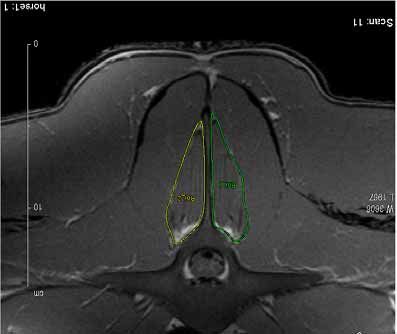
© 2007 Rural Industries Research and Development Corporation.
All rights reserved.

ISBN 1 74151 515 7
ISSN 1440-6845

Back Pain in Horses :Epaxial Musculatur
Publication No. 07/118
Project No. UQ-111A

The information contained in this publication is intended for general use to assist public knowledge and discussion
and to help improve the development of sustainable regions. You must not rely on any information contained in
this publication without taking specialist advice relevant to your particular circumstances.

While reasonable care has been taken in preparing this publication to ensure that information is true and correct,
the Commonwealth of Australia gives no assurance as to the accuracy of any information in this publication.

The Commonwealth of Australia, the Rural Industries Research and Development Corporation (RIRDC), the
authors or contributors expressly disclaim, to the maximum extent permitted by law, all responsibility and liability to
any person, arising directly or indirectly from any act or omission, or for any consequences of any such act or
omission, made in reliance on the contents of this publication, whether or not caused by any negligence on the
part of the Commonwealth of Australia, RIRDC, the authors or contributors.

The Commonwealth of Australia does not necessarily endorse the views in this publication.

This publication is copyright. Apart from any use as permitted under the Copyright Act 1968, all other rights are
reserved. However, wide dissemination is encouraged. Requests and inquiries concerning reproduction and rights
should be addressed to the RIRDC Publications Manager on phone 02 6271 4165.

Researcher Contact Details
Dr Catherine McGowan
c/o School of Animal Studies
The University of Queensland
Gatton 4343

Phone: 07 54601706
Fax: 0754601444
Email: c.mcgowan@uq.edu.au

From May 2007:
Department of Equine and Small Animal Medicine
Faculty of Veterinary Medicine
University of Helsinki
P.O. Box 57 (Viikintie 49)
00014 University of Helsinki
FINLAND
tel. +358 - 9 - 191 57367
fax. +358 - 9 - 191 57298
email: catherine.mcgowan@helsinki.fi

In submitting this report, the researcher has agreed to RIRDC publishing this material in its edited form.

RIRDC Contact Details
Rural Industries Research and Development Corporation
Level 2, 15 National Circuit
BARTON ACT 2600
PO Box 4776
KINGSTON ACT 2604

Phone:     02 6271 4100
Fax:       02 6272 5199
Email:     rirdc@rirdc.gov.au.
Web:       http://www.rirdc.gov.au

Published in November 2007
Printed by Union Offset

                                                          ii
Foreword
Back pain and diseases of the spine and pelvis are significant problems in all types of equine
performance causing poor performance, lost training days and wastage. As a result, back pain
represents a considerable economic and welfare issue for the equine performance industries.
Research to date has been led by the veterinary profession with a focus on pathoanatomical problems
underlying back pain. However, advances in human back pain have been led by physiotherapy
research. This has focussed on the neuromotor control model and the associated dysfunction especially
of the epaxial or deep back muscles that occurs as a result of back pain from different forms of
pathology.
Therefore, the overall aim was to increase the knowledge of back pain in horses by investigating this
multifacted problem by studying how pathology of the back itself or other parts of the musculoskeletal
system is reflected in the epaxial muscles. Also, the researcher aimed to determine the relationship
between atrophy or dysfunction of these muscles and pain and/or poor athletic performance. In order
to do this the project had three broad objectives: first, to describe the anatomy, biomechanics and
function of the equine epaxial muscles; second, to demonstrate that ultrasonography could reliably
measure the dimensions of the equine epaxial muscles; and third, to objectively measure the response
of equine epaxial muscles to back pain syndromes using ultrasonography.
The results of this project have shown that the anatomy and function of the equine epaxial muscles are
comparable to that of humans thereby justifying a similar approach to investigating back pain in
horses as in humans. Ultrasonography was found to be a repeatable and reliable tool for measurement
of the equine epaxial muscle size. When examined in clinical cases of equine back pathology, there
was a clear effect on the epaxial muscle size at the level of and close to areas of significant injury or
pathology. While ultrasonographic examinations were primarily focused on epaxial muscle size, it was
found that bony pathology was also detectable using this non-invasive tool.
In conclusion, use of a novel approach to equine back pain, borrowing from the human neuromotor
control model, has advanced the knowledge of the equine back, its function and the epaxial muscle
response to pathology of the spine and pelvis. Ultrasonography of the epaxial muscles is a valuable
non-invasive tool that will help detect back pain and associated pathology in horses. This information
will be valuable to veterinarians and physiotherapists managing back pain and poor performance
syndromes in athletic horses and will lead onto future work on the effect of physiotherapeutic
intervention on the recovery of epaxial muscle function following back pain.
This project was funded from industry revenue which is matched by funds provided by the Australian
Government.
This report, an addition to RIRDC’s diverse range of over 1600 research publications, forms part of
our Horses R&D program, which aims to aims to assist in protecting the Australian horse industry and
building and developing its future.
Most of our publications are available for viewing, downloading or purchasing online through our
website:
• downloads at www.rirdc.gov.au/fullreports/index.html
• purchases at www.rirdc.gov.au/eshop
Peter O’Brien
Managing Director
Rural Industries Research and Development Corporation
iiiAcknowledgments
The authors would like to acknowledge our collaborators, Professor David Hodgson, The University
of Sydney, Dr Chris Riggs, The Hong Kong Jockey Club and Dr Gary Cowin, The University of
Queensland for their valuable contributions to this work. We would also like to thank the staff and
management at the Hong Kong Jockey Club for allowing us to use their facilities and for being so
helpful in the study. We would also like to acknowledge the hard work Narelle Stubbs has contributed
to this project as part of her PhD studies.
Abbreviations
C cervical (vertebrae)
Ca caudal (vertebrae)
CSA Cross sectional area
CT Computed tomography
CV coefficient of variation
DJD degenerative joint disease
DSIL Dorsal sacroiliac ligament
DSP dorsal spinous process(es)
EIPH exercise induced pulmonary haemorrhage
L Lumbar (vertebrae)
LS Lumbosacral
m muscle
mm muscles
MRI Magnetic resonance imaging
S sacral (vertebrae)
SB Standardbred
SDFT superficial digital flexor tendon
SIJ sacroiliac joint
SID sacroiliac disease or dysfunction
SD standard deviation
T Thoracic (vertebrae)
TB Thoroughbred
TL Thoracolumbar
ivContents
Foreword ............................................................................................................................................... iii
Acknowledgments................................................................................................................................. iv
Abbreviations........................................................................................................................................ iv
List of Figures and Tables ................................................................................................................... vi
Executive Summary ............................................................................................................................ vii
Introduction ........................................................................................................................................... 9
Stability of the vertebral column and role of multifidus mm in man ................................................... 9
Back pain in humans and horses ....................................................................................................... 10
Anatomy of the equine epaxial musculature ..................................................................................... 11
Ultrasound Imaging of Multifidus ..................................................................................................... 11
Objectives ............................................................................................................................................. 13
Methodology ........................................................................................................................................ 14
Phase I: Functional anatomy of the caudal thoracolumbar and lumbosacral spine in the horse ....... 14
Phase II: Measurement of the equine epaxial muscles using ultrasonography ................................. 15
Phase III: Ultrasonography in equine back pain................................................................................ 16
Results .................................................................................................................................................. 18
Phase I Functional anatomy of the caudal thoracolumbar and lumbosacral spine in the horse ........ 18
Phase II: Measurement of the equine epaxial muscles using ultrasonography ................................. 22
Phase III: Ultrasonography in equine back pain................................................................................ 25
Discussion of Results ........................................................................................................................... 28
Functional anatomy of the caudal thoracolumbar and lumbosacral spine in the horse ..................... 28
Measurement of the equine epaxial muscles using ultrasonography ................................................ 29
Ultrasonography in equine back pain................................................................................................ 29
Implications.......................................................................................................................................... 31
Recommendations ............................................................................................................................... 31
References ............................................................................................................................................ 32
Appendices ........................................................................................................................................... 35
List of presentations .......................................................................................................................... 35
Publications: ...................................................................................................................................... 35
vList of Figures and Tables
Figure 1: MRI images of the equine 20
Figure 2: Anatomy of the equine epaxial muscles 21
Figure 3: Mean spinous process angles (°) of orientation relative to the vertebral body 22
Figure 4: Pooled SD for MRI CSA for 2 Standardbred horses 23
Figure 5: Multifidus mm CSA using ultrasonography 24
Figure 6. Difference between 2 operators (inter-operator reliability) for measurement of
multifidus mm. CSA (cm2) in 2 Australian Stock horses 25
Figure 7: Horse 33 showing marked asymmetry of upper hindlimb musculature with noticeable
atrophy on the right side 26
Table 1: Horse details, ultrasonography findings and post mortem findings in 22 racehorses 27
viExecutive Summary
Background
Back pain and diseases of the spine and pelvis are significant problems in all types of performance
horses, potentially causing poor performance, lost training days and wastage. As a result, back pain
represents a considerable economic and welfare issue for the equine performance industries.
Evaluation of back problems in performance horses is an important part of physiotherapy and
veterinary practice. Yet back pain syndromes are insidious and difficult to diagnose due to the
variability of presenting signs ranging from overt lameness or pain on palpation of the back to subtle
gait alterations or even behavioural changes. Complicating matters further, multiple problems often
coexist, particularly lameness and back pain
Research to date has been led by the veterinary profession with a focus on pathoanatomical problems
underlying back pain. However, advances in human back pain have been led by physiotherapy
research. Physiotherapy research has focussed on the neuromotor control model 1 and the associated
dysfunction, especially of the epaxial or deep back muscles that occurs as a result of back pain from
different forms of pathology.
Aim
The overall aim was to increase the knowledge of back pain in horses by using this novel approach.
Specifically; how pathology of the back itself or other parts of the musculoskeletal system is reflected
in the epaxial muscles and to determine the relationship between atrophy or dysfunction of these
muscles and pain and/or poor athletic performance.
Objectives
In order to do this we divided the research into three phases with the following objectives:
Phase I: To describe the anatomy, biomechanics and function of the equine epaxial muscles.
Phase II: To reliably measure the equine epaxial muscles using ultrasonography.
Phase III: To objectively measure the response of equine epaxial muscles to back pain
syndromes using ultrasonography.
Key findings
The results of this project have shown that the anatomy and function of the equine epaxial muscles are
comparable to that of humans. A difference, however, is the equine spinal anatomy and its variations,
especially in the lumbosacral region, with the variations occurring in about a third of Thoroughbred
horses, and but none of the Standardbreds. These variations could have a major effect on stability of
the lumbosacral joint and affect performance through altered mobility and or a predisposition to
pathology during an athletic career. Ultrasonography was found to be a repeatable and reliable tool for
measurement of the equine epaxial muscles and when examined in clinical cases of equine back
pathology, there was a clear reduction of the epaxial muscle size at the level of and close to areas of
significant injury or pathology. While ultrasonography was focused on epaxial muscle size, it was
found that bony pathology was also detectable using this non-invasive tool. Another finding of
particular interest was the high prevalence of serious back pathology in horses at the end of their
athletic careers, justifying the importance of new, and more sensitive methods to detect such
pathology.
1
For more information on the neuromotor control model in human or equine back pain please see link to paper
McGowan CM, Stubbs NC, Jull GA. Equine physiotherapy: a comparative view of the science underlying the
profession. Equine Vet J. 2007 Jan; 39(1):90-4.
viiConclusions and recommendations
In conclusion, use of a novel approach to equine back pain, using the human neuromotor control
model, has advanced knowledge of the equine back, its function and the epaxial muscle response to
back pathology. Ultrasonography of the epaxial muscles is a valuable non-invasive tool that will help
detect back pain and associated pathology in horses. This information will be valuable to veterinarians
and physiotherapists managing back pain and poor performance syndromes in athletic horses and will
lead onto future work on the effect of physiotherapeutic intervention on the recovery of epaxial muscle
function following back pain.
viiiIntroduction
Back pain and diseases of the spine are significant problems in equine sports and veterinary medicine
(Peham et al., 2001). The predominant feature identified is a substantial loss of performance (Jeffcott,
1980, 1999; Jeffcott et al., 1982; Denoix, 1998, 1999; Haussler, 1999a). As a result, evaluation of back
problems is an important part of physiotherapy and veterinary practice (Jeffcott, 1980; Jeffcott et al.,
1982; Denoix, 1998; Gellman, 1998). Yet, back pain syndromes are insidious and difficult to
diagnose. Presenting signs may vary from overt lameness or pain on palpation of the back to subtle
gait alterations or even behavioural changes. Diagnosis currently relies on a long process of
elimination due to the inherent difficulty in using diagnostic equipment in the equine vertebral column
(Jeffcott, 1980; Jeffcott et al., 1982; Denoix, 1998, 1999; Gellman, 1998).
In veterinary medicine, the focus on back pain has been on the underlying skeletal and ligamentous
pathology or a pathoanatomical model. Yet in humans, the focus encompasses neuromotor control and
the muscular system (Hodges, 2003; Lee, 2004). Numerous studies in humans and pigs have shown
that the deep epaxial muscles, especially the multifidus mm, play a key role in spinal function and
dysfunction The ability to assess the size and function of the multifidus muscles (mm) using
ultrasonography has been a valuable guide to assessment, management and prevention of recurrence of
back pain in man. Atrophy and dysfunction of multifidus mm occurs as a result of back pain and may
predispose to recurrence of back pain (Hides et al., 2001; Hodges, 2003). For this reason, assessment
and treatment based on this model have been developed for human back pain sufferers.
Stability of the vertebral column and role of multifidus mm in man
Dynamic spinal stability is known to be an important contributor to the pathogenesis of back pain in
man. Loss of control of a spinal segment’s stability allows more movement during activities, and is
described as the loss of control of, or increased size of the segement’s “neutral zone”. This loss of
neutral zone is associated with a predisposition to disc disease and spinal injury as well as allowing
more movement across joint surfaces predisposing to degenerative changes. (Panjabi, 1992). The
multifidus mm plays a key role in the stability of the lumbar spine in man (Moseley et al., 2002). It
provides segmental stabilisation of flexion torques during rotation, acting as bilateral stabiliser in
rotation (Bogduk and Twomey, 1987; Panjabi et al., 1989). Superficial fibres contribute to the control
of spinal orientation and deep fibres have an integral role in controlling inter-segmental and shear
forces. Fascicles act at right angles to spinous processes (2 vectors) exerting a rocking component into
extension where they “control flexion” i.e. multifidus mm acts as a posterior sagittal rotator. Pure
lumbar spinal rotation is an indirect motion due to the principal agonist muscles, the oblique
abdominal muscles, simultaneously flexing the lumbar spine. Hence the role of multifidus mm is to
balance unwanted flexion during rotation and stabilising axial rotation, maintaining the spinal
segment’s neutral zone (Bogduk and Twomey, 1987).
When compared to other muscles in close proximity to the last lumbar vertebrae in man (L4-L5),
multifidus mm contributed 2/3 of the increase in stiffness imparted by muscular action (Wilke et al.,
1995). In vivo studies in pigs confirmed multifidus mm as a major stabiliser of the lumbar spine
(Kaigle, 1995). What is also important is the recruitment of multifidus mm where deep fibres have
been shown to be preparatory in nature (switch on in anticipation of movement) similar to transverse
abdominus mm in the asymptomatic subject (Hodges and Richardson, 1996). This means that the
multifidus mm function is important in the protection of the production of abnormal rotation or shear
forces in the vertebral column.
In back pain patients, multifidus mm has been shown to be both reduced in size, segmentally (Hides
et al., 1994), and also have altered functional activation patterns. EMG studies showed that there was
decreased activation of multifidus mm at the initial period of axial rotational exertion - also at low
levels of exertion (Ng et al., 2002) – i.e. it loses the preparatory stabilisation or protective function
present in the normal patient and predisposes the patient with back pain to recurrence. Rantanen et al.
9(1993) found that subjects with back pain with multifidus mm pathology had a poor outcome post disc
surgery at five year follow up. Compared with the positive outcome group, on biopsy multifidus mm in
these patients was shown to have undergone pathological changes; “moth eaten appearance” possibly
in relation to type 1 fibres, decreased fibre diameter size of type 2 fibres, a more loose adipose
connective tissue, with denervation in the deep part of the multifidus mm.
It has been demonstrated that restoration of multifidus mm function and bulk is an important factor in
the prevention of recurrence of back pain in people with acute back injuries (Hides et al., 1996). The
recovery of multifidus mm has been shown not to be automatic in people, so that despite apparent
recovery or resolution of pain following an episode of acute back pain, the dysfunction of multifidus
mm persists (Hides et al., 1996). Specific physiotherapeutic intervention in people with multifidus mm
dysfunction following an episode of acute back pain reduced the rate of recurrence of injury to 30% in
physiotherapy intervention group compared with controls 84% (Hides et al., 2001).
We hypothesise that if multifidus mm in horses is shown to have the same role as in man, similar
physiotherapeutic strategies of strengthening and recruiting this muscle can be developed for the
treatment and rehabilitation of back pain in horses as have been in humans.
Back pain in humans and horses
Low back pain in the human general population is extremely common with a reported 60 – 80%
incidence of recurrence in the first year following the first episode of acute lumbar pain and 22 – 62%
in years the following four years (Liebensm, 1996). Back pain is also common and frequently
recurrent in horses ( Jeffcott 1980) with the literature reporting a variable prevalence of back pain in
horses from general veterinary practice (0.9%), Thoroughbred racehorse practice (2%), veterinary
school referrals (5%), mixed equine practice (dressage, showjumpers, eventing) (13%), spinal research
clinic (47%), to equine chiropractic clinic (94%) (Haussler, 1999a). Bailey et al. (1997) documented
that Sydney race horse trainers reported back problems as one of the most common injuries preventing
training and racing. Similarly in horses, of 190 horses with chronic back problems, 57% recovered
completely, 17% showed no improvement and 38% had a recurrence or continuation of signs of low
back pain (Jeffcott, 1979).
Equine back pain can result from a wide variety of different pathological processes. In a review of 443
cases of equine back pain, the major identified pathological lesions associated with thoracolumbar pain
were vertebral lesions (39%), soft tissue injuries (25%), sacroiliac strain (13%) and non-thoracolumbar
lesions (13%). Vertebral lesions were predominantly overriding dorsal spinous processes, while soft
tissue lesions were predominantly in the longissimus dorsi muscles and supraspinous ligament in the
caudal withers and cranial lumbar regions. Crowding and overriding dorsal spinous processes were
most common beneath the saddle at T12 to T17, and most prevalent in young adult to middle aged
horses used for jumping or dressage and in Thoroughbreds with short backs (Jeffcott, 1980).
Spinal muscular dysfunctions in horses with back pain are frequently secondary to underlying bone
pathology but may also be due to pathology of the muscles themselves, or of a generalised muscle
disorder (Valberg, 1999, Quiroz-Rothe et al., 2002). Spine and peripheral joint disease with pain can
cause reflex inhibition of motor neurones, resulting in weakness and atrophy of associated muscles
(Young, 1993). Local muscle damage attributed to a poorly fitting saddle, for example, can also cause
atrophy of the epaxial muscles (Gellman, 1998; Harman, 1999). In a survey of 443 cases referred with
thoraco-lumbar disorders, 23.37% had evidence of epaxial muscle pain (Jeffcott, 1980).
10Anatomy of the equine epaxial musculature
The longissimus dorsi m. is the most superficial of the epaxial muscles and therefore atrophy is most
visible. Signs of reduced epaxial muscle bulk are increased prominence of the vertebral spinous
processes and dipping away of the profile lateral to the spine (Harman, 1999). The multifidus mm are
deep and medial to the longissimus dorsi m. and not assessable by visual examination. Therefore it is
not possible to determine if wasting is due to the longissimus dorsi m. or the multifidus mm by eye
alone. In the horse, multifidus mm is described as originating from the transverse processes of the
thoracic, lumbar and sacral vertebrae and having insertions into the spinous processes of S1-2, L1-6
and T1-18, receiving innervation from the dorsal branches of the thoracic and lumbar nerves (Budras
et al., 2001).
Numerous skeletal muscles provide stability and flexibility in the spine (Valberg 1999). However, the
role of the multifidus and related muscles (especially sacrocaudalis dorsalis m) in the horse has been
unclear. The large epaxial muscles (m. longissimus dorsi, m. iliocostalis and m. middle gluteal) tend to
produce gross spinal extension and global spinal “stiffness” rather than dynamic” intersegmental
stability”, and the ventral muscles (iliopsoas minor/major and the abdominal complex) flex the spine.
It has been suggested that the deeper muscles such as multifidus mm may produce segmental
stabilization in the horse (Haussler, 1999b), with Nickel (1986) suggesting that the tail muscles may
contribute to spinal stability of the lumbar, sacral and caudal vertebrae.
In textbooks and the limited literature available there seems to be some discrepancy and lack of data as
to the exact anatomical arrangement and functional relationship and role of the sacrocaudalis dorsalis
m to multifidus mm and other epaxial muscles in the horse. Sacrocaudalis dorsalis m is described as
“extensions of the lumbar epaxial muscles predominantly the longissimus and multifidus muscles”
(Getty, 1975; Nickel et al., 1986; Evans, 1993) with sacrocaudalis dorsalis medialis m being an
extension of multifidus (Nickel et al., 1986; Evans, 1993). However, Getty (1975) suggests that the
action of the sacrocaudalis dorsalis m being elevation and lateral flexion of the tail (Getty, 1975) and
that sacrocaudalis dorsalis lateralis m is an extension of the multifidus muscle. Since the anatomical
arrangement is so vital to the biomechanical function of a muscle, clearly research on describing the
detailed anatomy of the multifidus mm and the sacrocaudalis dorsalis m are warranted.
Ultrasound Imaging of Multifidus
Monitoring muscle size is important in human physiotherapy practice as repeated measurements can
show changes that represent the effectiveness of the treatment implemented. Real-time ultrasound
imaging can be used to provide an immediate image of the structures beneath the probe and complete
cross-sections of muscle can be obtained (Stokes and Young, 1986). Ultrasound imaging can be used
to measure muscle cross-sectional area (CSA), therefore it may be able to provide a method of direct
assessment of epaxial muscle atrophy or hypertrophy and its response to therapy. Despite the quality
of more advanced techniques like magnetic resonance imaging (MRI) and computed tomography
(CT), no significant differences in the cross-sectional area of the multifidus mm were found when
compared to those obtained by ultrasound imaging (Hides et al., 1995).
Ultrasound imaging may therefore be a useful tool in the assessment of the efficacy of physiotherapy
on the muscular system (Hides et al., 1995). Intra-operator reliability has been demonstrated for the
use of ultrasound imaging to measure the anterior tibial m, quadriceps mm and multifidus mm in
humans (Hides et al., 1992; Martinson and Stokes, 1991; Stokes and Young, 1986). Ultrasound
imaging has further been used to examine the effect of lumbar back pain on the multifidus mm in
humans where there was significant reduction in cross-sectional area of multifidus mm on the
symptomatic side of the spine, showing a relationship between pain and muscle atrophy (Hides et al.,
1994). Despite the use of ultrasonography in the measurement of muscle size in humans, there are no
reports of its validation in horses.
11The clinical significance of being able to measure the size of the multifidus mm is to be able to
quantify atrophy present and thus the dysfunction that exists in the spine. This can be used both as a
diagnostic tool, and as an indicator of the risk of recurrence. If the presence of multifidus mm atrophy
can lead to degenerative effects in the human spine (Hides et al., 1996) then it could be speculated that
multifidus mm atrophy may allow the pre-cursor to degenerative changes. Further, it has been shown
that restoration of multifidus mm function and bulk is an important factor in the prevention of
recurrence of back pain in people with acute back injuries (Hides et al., 1996). Thus, the ability to
assess the size and function of multifidus mm in man has been a valuable guide to assessment,
management and prevention of recurrence of back pain.
The development of similar techniques in horses is warranted, and utilisation of the techniques will be
valuable in developing suitable assessment and management strategies in horses suffering from back
pain syndromes. The speculation that multifidus mm or epaxial muscle atrophy is present in both
primary and secondary back pain in the horse, as it is in man, provides a rationale for investigation of
ultrasonography for as an aid to diagnosis of equine back pain. Further, if atrophy is a consistent
finding, the response to therapy could be quantitated allowing rational evidence-based selection of
treatment techniques in horses with back pain.
Generalised secondary atrophy of the epaxial muscles, especially longissimus dorsi mm and middle
gluteal mm which are located on the dorsal aspect of the equine vertebral column has been reported in
horses with back pain (Jeffcott et al., 1982; Quiroz-Rothe et al., 2002) from which is can be suggested
that similar changes occur in equine epaxial muscles as has been found in humans. However, there has
been no research to support a neuromotor and muscular control theory of back pain in horses. We have
proposed that the human neuromotor control approach can be applied to the horse and utilisation of
ultrasonography in measuring epaxial muscle size, especially multifidus mm, will be valuable in
developing suitable assessment and management strategies in horses suffering from back pain
syndromes. Specifically; the aims of this project were to determine how pathology of the back itself or
other parts of the musculoskeletal system is reflected in the epaxial muscles and to determine the
relationship between atrophy or dysfunction of these muscles and pain and/or poor athletic
performance.
12Objectives
Our original stated objectives were as follows:
• to objectively measure the equine epaxial muscles
• to objectively measure the response of equine epaxial muscles to back pain syndromes
• to correlate clinical signs, ultrasonography, algometry and EMG analysis in affected horses
• to develop ultrasonography as a non-invasive tool for diagnosis of muscular dysfunction
associated with back pain in horses
• to develop ultrasonography as a non-invasive tool for monitoring response to therapy of back pain
in horses.
In order to address these objectives we divided the research into three methodological phases with the
following objectives:
• Phase I: To describe the anatomy, biomechanics and function of the equine epaxial muscles
Hypothesis: the equine multifidus and sacrocaudalis dorsalis mm have similar morphology,
biomechanics and function to man and the pig, acting as a dynamic stabiliser of spine and having
a key role in equine back pain
• Phase II: To reliably measure the equine epaxial muscles using ultrasonography
Hypothesis: That ultrasonography of the equine epaxial muscles (multifidus mm and longissimus
dorsi m) is a reliable method to determine muscle size, and variations with pathology
• Phase III: To objectively measure the response of equine epaxial muscles to back pain syndromes
using ultrasonography
Hypothesis: That clinical signs of back pain detected on clinical examination and diagnostic
imaging will be correlated with the underlying pathology detected at post mortem.
13Methodology
Studies were approved by the Institutional Animal Ethics Committee.
Phase I: Functional anatomy of the caudal thoracolumbar and
lumbosacral spine in the horse 2
This phase was originally planned to be the second phase, but on a review of the literature it became
apparent that the anatomy of the equine multifidus mm and its caudal extensions (especially
sacrocaudalis dorsalis m) in the horse were not well described and conflicting reports existed.
Ultrasonography relies on accurate anatomical descriptions of the area being imaged, so this phase of
the project was completed first. Further, following the initial anatomical dissections, it was noted that
there was considerable variation in the vertebral formula of each horse, leading us to expand this
section of the project.
Vertebral formula variation
120 horse cadavers were examined to identify variations in vertebral formula. There were 65
Thoroughbreds, 24 Standardbreds and 31 other breeds. A midline vertebral transection was performed
and the vertebral formulae (number of cervical, thoracic, lumbar and sacral vertebrae present) were
analysed and recorded for each horse. Observations were made of the site of divergence of the
lumbosacral spinous processes. The level of spinous process divergence and maximal dorsoventral
motion was identified by the presence of a bulk of muscle fibres between spinous processes (m.
interspinalis). The presence of transitional vertebrae, lumbar sacralisation and the number of
intervertebral discs were also noted.
Magnetic resonance imaging (MRI)
Images were made of the lumbosacral region in 3 cadavers (1 Thoroughbred, 2 Standardbred) to
identify gross anatomy and guide the detailed dissection and biomechanical analysis. Cadavers were
grossly dissected to provide a spinal block from T11 to caudal vertebra 3 with a cross sectional area of
40 cm2. Dorsally orientated spinal sections were imaged at 1 cm intervals in axial, coronal and sagittal
slices from T13 to caudal vertebra 3. MRI data were acquired on a Bruker AVANCE spectrometer
interfaced to an Oxford 2T whole body magnet. Spin echo images were acquired on the body coil with
the following limits: Field-of-view = 400 X 400 mm, slice thickness = 10 mm, slice separation = 0
mm, number of slices = 30, slice orientation = axial, TR=1000 ms, TE=17 ms, image matrix 512 X
512, spectral width = 1 kHz, number of averages = 2, total experiment time = 10 min. After acquisition
of a series of slices the bed was moved 260 mm and the next series acquired. Four series of images
were acquired.
Anatomical dissection
Detailed dissection of the vertebral column and deep epaxial muscles in the thoracolumbar and sacral
spine in 11cadavers (2 to 30 years) (6 Thoroughbred, 3 Standardbred, 2 others) was performed using
procedures adapted from Macintosh and Bogduk (1986 a, b). In 1 cadaver, cross-sectional slices T13,
T18, L3, L4, L5, L6, S1 and S3 with muscles kept intact were prepared using a band saw. These
sections were examined to identify the fascial divisions between multifidus, sacrocadualis caudalis
complex and dorsolateral epaxial musculature for comparison MRI.
The multifidus – sacrocadualis caudalis mm complex was isolated by careful resection of the large
superficial epaxial and hypaxial musculature and disarticulation of the sacroiliac joint to remove the
ilium. The distinct fascia covering over multifidus was removed so that overall appearance of the
intact muscle complex could be inspected. Two approaches were then used to determine the pattern,
orientation and attachments of the fascicles. In 5 horses, individual multifidus bundles of fascicles
2
Published as: Stubbs, N.C., Hodges, P.W., Jeffcott, L.B., Cowin, G., Hodgson D.R. and McGowan C.M.
Functional anatomy of the caudal thoracolumbar and lumbosacral spine in the horse. Equine Vet. J. Suppl. 36:
393-399. 2006.
14were detached from the T13 spinous process and lamina and the attachments were identified. Once the bundles had been resected the procedure was replicated for fascicles attaching to successive thoracic, lumbar and sacral vertebrae until all fibres of the muscle complex had been removed. In 6 horses the individual multifidus and sacrocadualis caudalis fascicles were identified by locating the cleavage planes with the caudal attachments. Fascicles were then detached, mobilised and traced to their cranial attachments. In both samples the left and right sides were dissected and recorded using digital video (Sony digital, 3CCD megapixel camera) and still photography (Olympus 3.2 megapixel camera). Biomechanics Spinous process orientation relative to the vertebral body was quantified in 6 cadavers (4 Thoroughbred, 2 Standardbred). Soft tissues other than multifidus and sacrocadualis caudalis mm were removed and the entire length of the right (n=3) or left (n=3) vertebral column. A band saw was used to transect the specimen sagittally from T9 to caudal vertebra 1 along the line of the medial lamina adjacent to the spinous processes. This procedure removed the facet joints, leaving the spinous processes intact with the longitudinally transected vertebral body in full view. Lumbosacral variations using the presence of the interspinalis mm and intervertebral discs were documented. To quantify the orientation of the spinous processes at T13, T18, L3, L4, L5, L6 and S1, the caudal aspect of the dorsal and ventral edges of the vertebral body were identified and marked with pins. A pin running perpendicular to this line was extended vertically (Figure. 1A, B). Second a steel pin, representing the spinous process orientation was aligned such that it bisected the mid-point between markers on the caudal and cranial aspects of the spinous process at points along the length of the spinous process from the lamina to the dorsal tip. Digital photographic images were taken in each region in a horizontal plane to the vertebral body. A metric ruler was placed on the dorsal aspect of the spinous process. Each spinous process angle was measured using image analysis software (Image J, version 1.32j, NIH, USA). Evaluation of the reliability of the spinous processes/vertebral body angle methodology was determined at 1 level (T13) in the 6 horses with 2 repeated measures on different days. Data Analysis Data are presented descriptively with the vertebral formula calculated and expressed as a percentage of the total number of horses and breed group. For the reliability analysis for the measurement of the angle of the spinous process, and Intraclass correlation coefficient (ICC [2,1]) and the standard error of measurement (SEM) were calculated. The mean and standard deviation were calculated in the 2 vertebral formulae groups for all levels and t-tests for independent samples were used to determine if there was a significant difference at any level between the 2 groups. Significance was set at P
Ultrasonographic multifidus mm CSA reliability
To determine the reliability of measure re-measure CSA from ultrasonography, 2 Australian Stock
horse geldings were imaged using real time ultrasonography using a Mysono-201 (Excelray, Australia)
ultrasound machine with a 4-7 MHz curved linear probe at a depth of 15cm with multifidus mm and its
fascial boarders in view at T13/14, T18/L1, L3/4 L5/6 and the Sacrocaudalis dorsalis m complex at
the 3rd Sacral vertebra. Cross-sectional area measurements were analysed blindly on 3 occasions using
specialized software (Image J) by the same examiner. The same operator repeated the ultrasonographic
examination at each site three times, with each image being analysed three times.

Data were analysed for intra- and inter-rater reliability at all levels,and left vs right side in the same
horse.

Effect of age, breed and conditioning on multifidus mm CSA
Multifidus mm CSA was measured by the same person at 4 sites of the back (T13, T18/L1, L5 and S3).
These were chosen based on the results of the reliability data. CSA was measured in 6 horses in 5
groups: 2 year old unbroken Australian Stock Horses, 2 year old Thoroughbreds in pre-race training,
mature Thoroughbreds in race training, mature Standardbreds in race training and aged horses. Images
were analysed using image J and CSA at the different levels compared. Measurement of multifidus mm
in Warmbloods CSA was attempted, but was not possible due to the large size of the muscle being
beyond the limits of the ultrasound probe being used.

Phase III: Ultrasonography in equine back pain
All racehorses presented for euthanasia at the Hong Kong Jockey Club and subjected to routine post
mortem procedures during the period of April – July 2006 were recruited. Jockey Club records and the
racing and training history were examined and a clinical examination of the horse performed.
Following routine post mortem procedures, the thoracolumbar spine and pelvis was dissected,
macerated and examined for the presence of bone pathology.

Clinical History
A thorough clinical history including clinical findings, performance history and results of lameness
examination and other diagnostic tests were obtained.

Ultrasonographic examination
Ultrasonographic examination was performed with the horses lightly sedated by the attending
veterinary surgeon, and standing in stocks using a VingMed system 5 ultrasound machine with
different probes ranging from 2.5 – 10 Mhz depending on the depth required. Images were taken and
stored using the software on the ultrasound machine for later analysis using Image J. As well as the 4
specific regions of the back, additional views were taken in these horses to determine the presence of
bony changes on the facet joints of the entire thoracolumbar spine, and images of the pelvis and dorsal
sacroiliac ligament were also obtained.

Clinical Examination
Clinical assessment comprising of a full gait assessment and observation of dynamic vertebral motion,
stability and dysfunctions was performed. A physical and manual examination was performed
including specific provocation tests. Where necessary this was performed in stocks to prevent lateral
or excessive movement. Digital video recording was taken of gait for subsequent analysis.
Photographs of conformation, muscle bulk and vertebral and pelvic symmetry were taken. All
information from the examination was recorded verbally on a voice recorder and later reviewed in
association with the images.

                                                     16
Post mortem examination
Gross soft tissue lesions; muscular, ligamentous and fascia were noted and photographed. The
thoracolumbar vertebral column and sacropelvic region were assessed for the following abnormalities:
• morphology of the spinal column; number of vertebrae, sacralisation of lumbar vertebrae, presence
and locations(s) of interspinalis mm, presence and location(s) of lumbar dorsal spinous process
bursa
• interference or impingement of dorsal spinous processes / overriding
• interference or impingement of transverse processes / overriding
• evidence of gross bone remodelling/ gross degenerative changes of the facet joints (zygophyseal
joints); gross enlargement of the dorsal facets
• evidence of gross bone remodelling/ gross degenerative changes of the thoracolumbar and
lumbosacral articulations and intervertebral discs (degenerative intervertebral disc disease)
• evidence of gross bone remodelling – gross degenerative changes of the lumbar inter-transverse
joints
• evidence of acute or chronic sacroiliac joint injury or luxation; chronic laxity or signs of
subluxations
• presence (signs of) of spondylosis or ankylosis
• osseous lesions; fractures
• ligamentous lesions
• fascial lesions
• muscular lesions
• Multifidus mm appearance; adipose tissue within the muscle.
A steel rod was passed down the length of the spinal canal and fixed at both ends to ensure vertebrae
stayed in correct alignment. The spine was then boiled to remove all remaining soft tissues and stored
until examination. Spines were subsequently examined for evidence of gross bone pathology.
The thoracolumbar vertebral column and sacropelvic region were divided into 5 regions and scored
using the scale of 0 = no changes, 1 = mild changes, 2 = moderate changes and 3 = severe changes.
The regions were:
• vertebral bodies and end plates
• facet joints and articular processes
• Lumbosacral complex: Lumbosacral joint, intertransverse joints
• sacroiliac complex; sacral and ilial surfaces photographed and graded as per Jeffcott et al.
(1985); CSA (Image J) and degenerative joint disease
• over riding thoracolumbar dorsal spinous process and lumbar transverse processes.
Each region was assessed and scored for following abnormalities:
• fractures
• spondylosis
• evidence of lysis of and/or periosteal new bone formation on the dorsal spinous processes
• evidence of lysis of and/or periosteal new bone formation on the transverse processes
• periarticular new bone formation associated with the dorsal facet joints
• lysis of bone associated with the dorsal facet joints
• any other lesions.
Active new bone formation was considered a major finding and its presence required before a lesion
could be graded as severe.
The identity of all macerated vertebral samples was coded and the bone pathology recorded by the
investigators blinded to the identity of the horse.
17Results
Phase I Functional anatomy of the caudal thoracolumbar and
lumbosacral spine in the horse
Vertebral formula variation
The conventional cervical and thoracic vertebral formula was found in all horses examined. However,
the expected L6, S5 formula with L6-S1 as the maximal dorsoventral motion, was only found in 67 %
(80/120) horses. By breed, the expected lumbosacral vertebral formula was found in; 60% of
Thoroughbreds (39/65), 100% Standardbreds (24/24) and 55% others (17/31).
Lumbosacral variations were found in 33% of horses (40/120). The variations that occurred included:
• Five lumbar vertebrae instead of six, but the normal 5 sacral formula, with maximal dorsoventral
motion L5-S1. This occurred in 8% (5/65) Thoroughbreds, 0% Standardbreds and 16% (5/31)
others.
• The conventional L6-S5 formula but a variation of the lumbosacral region with maximal
dorsoventral motion at the L5-L6 intervertebral segment. This occurred in 32% (21/65)
Thoroughbreds, 0% Standardbreds, 29% (9/31) others. This was apparent as a spinous
process/vertebral orientation divergence of L5 cranially and L6 caudally, and interspinalis mm
between L5 and L6 (Figure 1).
This variation had two subsets:
1. L5-L6 divergence with m. interspinalis present between L5 and L6 only, and various
stages of L6-S1 intervertebral disc sacralisation characteristically evident. This occurred in
24% (16/120) Thoroughbreds, 0% Standardbreds and 23% (7/120) others (Figure 2)
2. L5-L6 divergence with a more normalised lumbosacral intervertebral disc, with L6 acting
as a transitional vertebra. The striking feature in this sub-population was the presence of a
less developed interspinalis m between L6-S1 as well as between L5-L6. This occurred in
8% (5/120) Thoroughbreds, 0% Standardbreds and 6% (2/120) others.
18MRI
MRI images showed the detail of the muscles and the cross sectional arrangement of the multifidus
mm. Of note was the presence of adipose tissue between the spinous processes, lamina and lateral
sacrum and the multifidus mm -sacrocaudalis dorsalis m, and in close proximity to the facet and
sacroiliac joint. On MRI and gross inspection the intact multifidus - sacrocaudalis dorsalis m complex
appeared homogenous beneath its delineating fascial encasement, comparable to the human lumbar
multifidus (Macintosh and Bogduk 1986 a, b) (Figure 1).
Longissimus dorsi mm
Multifidus mm
Multifidus mm
Lumbar vertebrae 5
Thoracic Vertebra 13
Figure 1a T13 Figure 1b L5
tuber sacrale
longissimus Multifidus mm and
& SCDL Sacrocaudalis mm complex
wing
of
ilium
Multifidus mm
sacroiliac joint
Sacral vertebra 3
Sacrum
Figure 1c S1 Figure 1d S3
Figure 1: MRI images of the equine back at T13 (1a), L5 (1b), S1 (1c) and S3 (1d). Note muscle is
grey, fascia is black and adipose tissue is white. In 1b the multifidus has been traced.
19Anatomical dissection
In the TL/LS regions there were 5 distinct segmental bands of multifidus mm fascicles (cleavage
plane). Each band extended caudolaterally from midline and emanated from 1 spinous process and
lamina (Figure 2). The fascicles were multipennate with a fleshy body and tendinous portion both
running the length of the muscle. Fascicles were confluent with one another cranially arising from the
tip of the spinous process to the vertebral lamina, but distinct with independent attachments caudally.
The most dorsal fascicle of multifidus mm overlaid the others and crossed 2-4 intervertebral discs,
arising from the caudal edge and lateral surface of the spinous process. A fleshy portion crossed 4
intervertebral discs. The remaining fascicles crossed 4, 3 and 2 intervertebral discs, from the tendinous
insertion alone on the dorsocaudal aspect of the spinous process to the lamina. The deepest and
shortest fascicle only crossed 1 intervertebral disc and arose from the vertebral lamina (Figure 2).
Multifidus mm fascicles m. Sacrocaudalis dorsalis
lateralis
m. Sacrocaudalis dorsalis medialis
T13
T18
L6
S3
L3
L5
m. Interspinalis
Figure 2: Anatomy of the equine epaxial muscles. The multifidus mm can be seen as 5 overlapping
fascicles at each vertebral level (only T13, T18 and L6 are shown for clarity). The multifidus mm
increased in size caudally, with the largest bulk of muscle evident at the lumbosacral region, where
most motion occurs. In the lumbosacral region the sacrocaudalis dorsalis lateralis m is a caudal
extension of the multifidus mm. The spine depicted in this figure represents 30% of the Thoroughbreds
examined with the divergence of the lumbar vertebrae occurring at L5-L6, instead of the expected L6-
S1. Note the presence of interspinalis m between the diverging vertebrae (L5 - L6).
Fascicles of sacrocaudalis dorsalis lateralis m appeared to have the same morphology as the
multifidus mm, replicating and replacing the most lateral of the multifidus fascicles commencing at L4,
L5, and L6 depending on LS variations. The tendinous portion of the muscle bundle originated from
the dorsal aspect of L4, L5 or L6, blending with the TL fascia and the supraspinous ligament in this
region. In 7 cases a bursa in this tendinous portion over L5 or L6 dorsal spinous process was noted.
Deeper fibres of sacrocaudalis dorsalis lateralis m also followed the same pattern as multifidus mm
attaching to the lateral border of L5 or L6 and the lateral border and lamina of the sacrum. Fascicles of
sacrocaudalis dorsalis lateralis m continued beyond the sacrum following similar morphology along
the caudal vertebrae. Sacrocaudalis dorsalis m medialis attached from S3 in a similar pattern to the
multifidus fascicles elsewhere, attaching to the lateral border of the sacrum and extending caudally to
the caudal vertebrae. On direct visual inspection the cross-sectional area of the multifidus mm and
20sacrocaudalis dorsalis lateralis m bundles were much larger at the LS junction. This gradual increase in size continued caudally with sacrocaudalis dorsalis medialis m from S3 caudally. The interspinalis muscle was present in the region of maximal dorsoventral motion i.e. the level of divergence of the spinous process and maximal dorsoventral motion, but not elsewhere in the thoracolumbar spine. Adipose tissue of variable size (individual to the horse and vertebral level) separated the multifidus, sacrocaudalis dorsalis lateralis m and sacrocaudalis dorsalis medialis m mm from the bone and the interspinalis mm (Figure 2). Biomechanics The reliability for measurement of the angle of the spinous processes was very good with an ICC [2,1] of 0.99. The standard error of the measurement (SEM) was 0.41° and the smallest detectable difference was 1.1°. The mechanical function (moment arm and force vector) of the multifidus mm fascicles were affected by the orientation of the spinous process relative to the vertebral body. Based on variations in vertebral formula and the location of m. interspinalis, horses were allocated into; L6-S1 (n=3) formula or L5-L6 formula (n=3) groups. This variation in level of the divergence of the spinous processes was associated with significant changes in the angle of the spinous process of L6 relative to the vertebral body and varied significantly between the two groups (p
Phase II: Measurement of the equine epaxial muscles using
ultrasonography
MRI multifidus mm CSA reliability
Median coefficient of variation (CV) for all 56 individual readings was 1.7%. All readings except one
(at T14 in horse 1) were below 5% CV showing good intra-rater reliability of measurement of
multifidus CSA using MRI. The Standard Deviations (SD) were calculated for all readings and
graphed (Figure 4). The pooled SD for all readings was calculated to be 0.22 cm2 to derive a 95%
confidence interval of ± 0.43cm. There was consistency of measurement along the spine with no
specific region or CSA size shown to create more measurement error than any other region. The
results confirm that MRI has excellent intra-rater reliability and therefore may be used as a standard
for further comparative investigation of non invasive measurement methods such as real time
ultrasound imaging to measure multifidus mm in the normal/abnormal horse with back pain (see also
Figure 1).

                     Pooled Standard Deviation by Veterbral region

     0.4

    0.35

     0.3

    0.25

                                                                      Subject 1
  SD 0.2
                                                                      Subject 2

    0.15

     0.1

    0.05

       0
               T14              T18                   L6         S2
                                      Spinal region

Figure 4: pooled SD for MRI CSA for 2 Standardbred horses showing a very low SD (0.22 cm2)
indicating the excellent reliability of measurement of muscle CSA using this technique.

Ultrasonographic multifidus mm CSA reliability
Ultrasonography of epaxial musculature along the vertebral column from T13 to S5 reveals striking
differences in the shape and size of multifidus mm, with multifidus largest in the lumbosacral region
(Figure 5). This fits with the biomechanical function of the lumbosacral region having the most
dorsoventral motion and therefore requiring the greatest amount of stability (see Phase I). The “long
view”, over the intervertebral space at all levels was most repeatable other than at T13 where there was
no difference. The greater variability of the facet or “short” view may be due to variability of facet size
and/or shape (see Phase III). The longissimus dorsi m. cross sectional area was able to be measured in
the lumbosacral region (L5) where it narrows down sufficiently just cranial to the tuber sacrale to
enable a cross sectional area to be obtained.

                                                           22
Figure 5a T13
Figure 5b T18/L1
Multifidus Longissimus
mm dorsi m
Figure 5 c L5
23Biceps femoris
Multifidus mm
SCDL
SCDM
Figure 5d S3
Figure 5: Multifidus mm CSA using ultrasonography at levels T13 (5a), T18/L1 (5b), L5 (5c) and S3
(5d).
The reliability for measurement of the multifidus mm CSA was good with an ICC [2,1] of 0.83. The
standard error of the measurement (SEM) was 0.78 cm with the smallest detectable difference of
< 1.5cm occurring at all levels. The relative variance components of measurement of CSA were 56%
inter-operator variance, 11% intra-operator and 33% image variance i.e. the variance was minimised
by having just one operator and taking several images of each site. There was significant effect of
horse but not of side of the image indicating that ‘normal’ horses have symmetrical epaxial muscles.
Compared to the MRI measurement of CSA repeatability, the pooled SD was 0.4 cm2, thus almost
double, but still within what was considered acceptable limits.
14.00
Multifidus mm CSA (cm2)
12.00
10.00
8.00
Scanner C
Scanner N
6.00
4.00
2.00
0.00
L1 L3 L5 S3 T13
Vertebral Level
Figure 6. Difference between 2 operators (inter-operator reliability) for measurement of multifidus mm.
CSA (cm2) in 2 Australian Stock horses.
24You can also read