Cellulose Binding Domains of a Phytophthora Cell Wall Protein Are Novel Pathogen-Associated Molecular Patterns W
←
→
Page content transcription
If your browser does not render page correctly, please read the page content below
This article is published in The Plant Cell Online, The Plant Cell Preview Section, which publishes manuscripts accepted for publication after they
have been edited and the authors have corrected proofs, but before the final, complete issue is published online. Early posting of articles reduces
normal time to publication by several weeks.

Cellulose Binding Domains of a Phytophthora Cell Wall Protein
Are Novel Pathogen-Associated Molecular Patterns                                                                           W

Elodie Gaulin,a,1 Nani Dramé,a Claude Lafitte,a Trudy Torto-Alalibo,b Yves Martinez,c Carine Ameline-Torregrosa,a
Moustafa Khatib,a Honoré Mazarguil,d François Villalba-Mateos,a,2 Sophien Kamoun,b Christian Mazars,a
Bernard Dumas,a Arnaud Bottin,a Marie-Thérèse Esquerré-Tugayé,a and Martina Rickauera,3
a Unité Mixte de Recherche 5546, Centre National de la Recherche Scientifique–Université Paul Sabatier-Toulouse III,

Pôle de Biotechnologie Végétale, 31326 Castanet-Tolosan, France
b Ohio State University/Ohio Agricultural Research and Development Center, Department of Plant Pathology, Wooster,

Ohio 44691
c Institut Fédératif de Recherche 40, Pôle de Biotechnologie Végétale, BP42617, 31326 Castanet-Tolosan, France
d Unité Mixte de Recherche–Centre National de la Recherche Scientifique 5089, Institut de Pharmacologie

et de Biologie Structurale, 31077 Toulouse Cedex 04, France

The cellulose binding elicitor lectin (CBEL) from Phytophthora parasitica nicotianae contains two cellulose binding domains
(CBDs) belonging to the Carbohydrate Binding Module1 family, which is found almost exclusively in fungi. The mechanism by
which CBEL is perceived by the host plant remains unknown. The role of CBDs in eliciting activity was investigated using
modified versions of the protein produced in Escherichia coli or synthesized in planta through the potato virus X expression
system. Recombinant CBEL produced by E. coli elicited necrotic lesions and defense gene expression when injected into
tobacco (Nicotiana tabacum) leaves. CBEL production in planta induced necrosis. Site-directed mutagenesis on aromatic
amino acid residues located within the CBDs as well as leaf infiltration assays using mutated and truncated recombinant
proteins confirmed the importance of intact CBDs to induce defense responses. Tobacco and Arabidopsis thaliana leaf
infiltration assays using synthetic peptides showed that the CBDs of CBEL are essential and sufficient to stimulate defense
responses. Moreover, CBEL elicits a transient variation of cytosolic calcium levels in tobacco cells but not in protoplasts.
These results define CBDs as a novel class of molecular patterns in oomycetes that are targeted by the innate immune system
of plants and might act through interaction with the cell wall.

INTRODUCTION                                                                 bacteria, and viruses. Among eukaryotic plant pathogens, the
                                                                             genus Phytophthora contains >60 species that are pathogenic
During their whole life, plants are exposed to pathogenic micro-             on a wide array of plants, causing economically important
organisms in their environment. Similar to animals, they have                diseases worldwide. Many elicitors of various structures have
developed various defense mechanisms to avoid disease and                    been isolated from Phytophthora species, among them a cell
death. Besides being induced by contact with pathogenic mi-                  wall–derived heptaglucan (Sharp et al., 1984), extracellular (glyco)-
croorganisms, active defense reactions can also be triggered by              proteins such as a transglutaminase (Brunner et al., 2002), eli-
treatment with microbial compounds called elicitors, which may               citins (Ricci et al., 1989; Kamoun et al., 1997), GP32 (Bailleuil et al.,
be characteristic of a whole group of organisms or limited to                1996), and cellulose binding elicitor lectin (CBEL) (Villalba-
specific strains of a microbial species (Bonas and Lahaye, 2002;             Mateos et al., 1997).
Montesano et al., 2003; Jones and Takemoto, 2004). Such                         In recent years, remarkable similarities between defense
compounds have been characterized from fungi, oomycetes,                     mechanisms triggered by elicitors in plants and what is known
                                                                             as the innate immune response in animals have been found
1 To whom correspondence should be addressed. E-mail gaulin@scsv.            (Gomez-Gomez and Boller, 2000; Parker, 2003; Nürnberger et al.,
ups-tlse.fr; fax 33-562-193-502.                                             2004; Zipfel and Felix, 2005). A model has emerged in which
2 Current address: Bayer CropScience, 69009 Lyon, France.
                                                                             discrimination from self is achieved through receptors that
3 Current address: Laboratoire Biotechnologie et Amélioration des
                                                                             recognize pathogen-associated molecular patterns (PAMPs)
Plantes/Ecole Nationale Supérieure d’Agronomie Toulouse, Pôle de
                                                                             (Janeway and Medzhitov, 2002). Such patterns correspond to
Biotechnologie Végétale, 18 Chemin de Borde-Rouge, BP 32607, 31326
Castanet-Tolosan, France.                                                    motifs or domains with conserved structural traits found in widely
The author responsible for distribution of materials integral to the         occurring compounds of microbes but not present in their hosts
findings presented in this article in accordance with the policy described   and essential for microbial fitness. High-affinity binding sites
in the Instructions for Authors (www.plantcell.org) is: Elodie Gaulin        in plants have been described for several general elicitors of
(gaulin@scsv.ups-tlse.fr).
W
  Online version contains Web-only data.                                     bacterial, fungal, and oomycete origin, such as flagellin, chitin
Article, publication date, and citation information can be found at          fragments, a b-heptaglucoside, and cryptogein (Bourque et al.,
www.plantcell.org/cgi/doi/10.1105/tpc.105.038687.                            1999; Gomez-Gomez and Boller, 2000; Bradley Day et al., 2001),

The Plant Cell Preview, www.aspb.org ª 2006 American Society of Plant Biologists                                                              1 of 12
2 of 12 The Plant Cell
and a few of them have been cloned. Four peptides were recently In the first approach, CBEL was produced in E. coli using
designated as PAMPs in phytopathogenic microorganisms, the pFLAG-ATS vector (pATS:CBEL), which carries the OmpA
similar to those involved in the innate immune response in secretion sequence, directing the recombinant protein to the
mammals and insects. These include a stretch of 22 amino acids periplasmic space. Upon isopropylthio-b-galactoside induction
represented by the peptide flg22 from a conserved domain in of BL21 cells containing pATS:CBEL, the corresponding protein
bacterial flagellin (Felix et al., 1999), the Pep-13 domain from the was expressed and accumulated as insoluble inclusion bodies.
cell wall elicitor GP42 of Phytophthora sojae (Brunner et al., After solubilization, efficient refolding of the denatured protein
2002), the RNA binding motif RNP-1 of bacterial cold-shock was achieved by adapting a protocol described by Berdichevsky
proteins (Felix and Boller, 2003), and the N terminus of bacterial et al. (1999). After dialysis against distilled water, the recombi-
elongation factor Tu (Kunze et al., 2004). nant protein (CBELrec) appeared as a single band on SDS-PAGE
We reported previously on the characterization and cloning of after Coomassie blue staining (data not shown), with final
CBEL, a cell wall glycoprotein from Phytophthora parasitica var amounts of 4 mg of protein per liter of cell culture. The elicitor
nicotianae (Ppn), the causal agent of the black shank disease of activity of CBELrec was assessed by infiltrating a 200 nM protein
tobacco (Nicotiana tabacum) (Villalba-Mateos et al., 1997). This solution into the mesophyll of tobacco leaves, with two leaves on
glycoprotein is widespread in the genus Phytophthora (Khatib three different plants for each assay. The infiltrated area turned
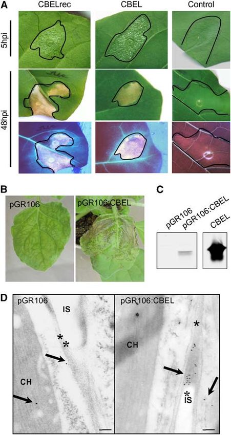
et al., 2004) and is present during the growth of Ppn in vitro and in slightly bright on the abaxial face at 4 to 5 h after infiltration,
planta (Séjalon-Delmas et al., 1997). CBEL is a potent elicitor in started to desiccate at 9 to 12 h after infiltration, and was fully
the Ppn host plant tobacco, in which it induces local hypersen- desiccated by 24 h after infiltration. The necrosis turned brown
sitive response (HR)–like lesions, defense responses, and pro- thereafter and remained strictly limited to the infiltrated area
tection against subsequent infection with the oomycete. It is also (Figure 1A). UV light examination revealed that both the necrotic
active in various nonhost plants, among them Arabidopsis area and the surrounding tissues displayed a blue autofluores-
thaliana (Khatib et al., 2004). Using Arabidopsis mutants affected cence indicative of the accumulation of defense-related aro-
in the signaling pathways that involve salicylic acid, jasmonic matic compounds. These symptoms and their time course are
acid, or ethylene, it was shown that the three pathways are similar to those observed previously with native CBEL at the
triggered by the elicitor and that its necrosis-inducing activity same protein concentration (Villalba-Mateos et al., 1997), indi-
depends on ethylene and jasmonic acid (Khatib et al., 2004). The cating that the recombinant form of CBEL produced in E. coli is
protein moiety of CBEL is composed of two direct repeats of as active as the native protein and that the glycan part is not
Cys-rich domains, connected by a linker. Each repeat contains a required for its elicitor activity.
motif that closely resembles the fungal type I cellulose binding The second approach consisted of producing CBEL in planta
domain (CBD) consensus pattern found in cellulases from var- by means of a viral expression vector derived from PVX. This
ious fungi (Gilkes et al., 1991). CBEL is able to bind to crystalline virus infects Nicotiana benthamiana, and the elicitor activity of
cellulose and tobacco cell walls in vitro in a dose-dependent the heterologous proteins produced is visible either as a local
manner, but in contrast with cellulases, it does not possess any HR-like necrosis or as systemically spreading necrotic lesions.
detectable enzyme activity on various polysaccharides (Villalba- The entire CBEL coding sequence, including its own secretion
Mateos et al., 1997). Phenotypic characterization of Ppn strains signal, was cloned into the binary PVX vector pGR106 (Lu et al.,
suppressed in CBEL expression revealed that this glycoprotein is 2003), resulting in plasmid pGR106:CBEL.
involved in organized polysaccharide deposition in the cell wall This plasmid was introduced into Agrobacterium tumefaciens
and in adhesion of the mycelium to cellulosic substrates (Gaulin to allow the delivery of PVX into N. benthamiana plants via
et al., 2002). Thus, the role of CBEL in the biology of Ppn and its inoculation with the bacteria. Control plants inoculated with
occurrence among various species of Phytophthora, as well bacteria containing the empty vector pGR106 (PVX) developed
as the wide range of plants responding to the elicitor, support the chlorotic mosaic symptoms and leaf curling characteristic
the view that CBEL might contain a PAMP of eukaryotic plant of PVX infection in systemically infected leaves at 11 d after
pathogens. According to this concept, it is assumed that con- inoculation (Figure 1B). Inoculation with bacteria carrying pGR106:
served structural motifs of the molecule account for its activity. CBEL first induced systemic PVX mosaic symptoms in the upper
In this study, we investigated the role of the CBDs of CBEL in noninoculated leaves, followed rapidly by localized necrotic
elicitor activity. It was evaluated both by infiltrating tobacco leaves lesions that became confluent soon afterward (Figure 1B). Pro-
with recombinant forms of the protein produced in Escherichia coli tein gel blot analysis with polyclonal antibodies against CBEL
and by producing the protein in planta through the potato virus X showed that leaves developing necrosis contained substantial
(PVX) expression system (Jones et al., 1999). Deletion and muta- amounts of CBEL (Figure 1C).
tional analysis, along with the use of synthetic peptides, allowed Immunocytochemistry was used to localize CBEL in leaf
us to assign the role of PAMP to the CBDs of CBEL. tissues developing small necroses. Immunogold labeling using
a purified CBEL antibody showed that the protein deposition
RESULTS occurred in the cell wall and junctions of pGR106:CBEL-infected
parenchyma cells, whereas only a few gold particles were
Elicitor Activity of CBEL Produced in E. coli or in Planta observed in the control samples (Figure 1D). A faint background
labeling of chloroplasts and cytoplasm could be observed in both
To study the relationship between the structure of CBEL and its elic- pGR106:CBEL-infected samples and the pGR106-infected con-
itor activity, we set up its expression in two heterologous systems. trols. Together, these results show that CBEL retains its elicitor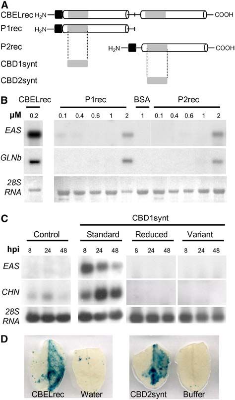
Phytophthora CBDs and Plant Immunity              3 of 12

                                                                              activity when expressed in plant cells and that its endogenous
                                                                              oomycete signal peptide is functional in directing the protein to
                                                                              the extracellular space. They also suggest that CBEL binds to the
                                                                              plant cell wall in situ, as no labeling was observed in the
                                                                              intercellular spaces.

                                                                              Aromatic Amino Acids in the CBD of CBEL Are Important
                                                                              for Elicitor Activity

                                                                              Studies Using CBEL Produced in Planta

                                                                              Because the PVX-based expression system does not require
                                                                              purification of the proteins under study, it is very convenient for
                                                                              screening a large array of modified proteins. This approach was
                                                                              first used to search for structural motifs involved in the elicitor
                                                                              activity of CBEL.
                                                                                 The occurrence of two CBDs homologous with those found in
                                                                              fungal cellulases is one of the main characteristics of CBEL.
                                                                              Structure–function relationship studies on the CBD of cellobio-
                                                                              hydrolase I, CBDCBHI (also named CBDCel7A), from the filamen-
                                                                              tous fungus Trichoderma reesei have demonstrated an essential
                                                                              role of the aromatic amino acids Y5, Y31, and Y32 for its binding
                                                                              to cellulose (Linder et al., 1995b). Based on sequence alignment
                                                                              of the two CBDs (CBD1 and CBD2) of CBEL with the T. reesei
                                                                              CBD (Figure 2A), the amino acids Y51 and Y52 in CBD1 and F187
                                                                              and Y188 in CBD2 were predicted to be surface-exposed and
                                                                              involved in the cellulose binding of CBEL. Hence, these residues
                                                                              were replaced individually or in pairs by Ala through PCR-based
                                                                              site-directed mutagenesis. The mutant fragments were cloned
                                                                              into pGR106, resulting in constructs whose expression products
                                                                              were named Y51A, Y52A, Y51A_Y52A, Y188A, F187A_Y188A,
                                                                              and Y52A_Y188A (Figure 2B). The various plasmids were then
                                                                              introduced into A. tumefaciens for N. benthamiana inoculation.
                                                                              Three independent experiments were performed on 4-week-old
                                                                              plants; in each experiment, three plants were inoculated with
                                                                              each PVX construct, and symptoms were scored from 9 to 15 d
                                                                              after inoculation. Representative phenotypes of the five classes
                                                                              of the symptom scale are shown at the top of Figure 2C. Whereas
                                                                              control plants inoculated with pGR106 displayed systemic mo-
                                                                              saic symptoms after 9 d, 90% of N. benthamiana leaves inoc-
                                                                              ulated with pGR106:CBEL exhibited local or spreading necrosis
                                                                              at this time. The lesions spread rapidly, affecting 100% of the
Figure 1. Elicitor Activity of CBEL Produced in E. coli or in Planta.         plants after 11 d and causing plant death after 15 d. Lesions were
(A) Symptoms observed on tobacco leaves after infiltration of recombi-        less extended and developed less rapidly with all mutant forms,
nant CBEL produced in E. coli. Leaves of 2-month-old tobacco plants           notably those involving residue Y188; they appeared as localized
were infiltrated with ;100 mL of a 200 nM solution of purified CBELrec,       spots between 11 and 13 d after inoculation and only rarely did
native CBEL, or BSA as a control. Symptoms were observed at 5 and 48          they spread throughout the whole plant. No additive effect of
h after inoculation (hpi) under white light (top) or UV light (bottom).       these mutations was observed. Hence, this symptom scoring
(B) Systemic symptoms observed on N. benthamiana plants after inoc-
                                                                              shows that the necrosis-inducing activity of CBEL is significantly
ulation with A. tumefaciens strains carrying the pGR106 or pGR106:
                                                                              reduced by point mutations of aromatic residues potentially
CBEL vector. Photographs were taken at 11 d after inoculation.
(C) Protein gel blot analysis of CBEL production in planta. Leaf samples      involved in cellulose binding.
were collected at 12 d after inoculation, and total proteins were extracted      To determine whether the point mutations affected the ability
and subjected to protein gel blot analysis using an anti-CBEL polyclonal      of CBEL to bind to plant cell walls, proteins were extracted from
antiserum. The quantity of total protein corresponds to 3 cm2 of leaf area.   systemically infected leaves at 14 d after inoculation, and the
Purified native CBEL (20 ng) was used as a control.
(D) Immunogold labeling of leaves at 11 d after inoculation with pGR106
(left) or pGR106:CBEL (right). Labeling was achieved with a purified          tographs were taken at 325,000 magnification. Arrows show gold
polyclonal antiserum against CBEL and gold-conjugated goat antiserum          particles located in the cell wall; asterisks indicate the plant cell wall.
to rabbit IgG. Sections were contrasted with uranyl acetate, and pho-         CH, chloroplast; IS, intercellular space.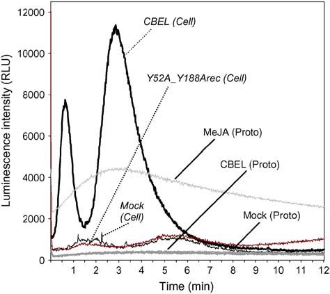
4 of 12    The Plant Cell

Figure 2. Site-Directed Mutagenesis Analyses of CBEL Using the PVX Expression System.
(A) Alignment of CBD1 and CBD2 of CBEL and the cellobiohydrolase I CBD from T. reesei. The deduced amino acid sequences of CBDs from CBEL
(CBD1, CBD2) and cellobiohydrolase I CBD from T. reesei (CBDCBHI) were aligned according to the ClustalW and BoxShade sequence analysis software
Phytophthora CBDs and Plant Immunity            5 of 12

                                                                                inoculated with the various deleted constructs developed only
                                                                                systemic mosaic symptoms typical of PVX infection, comparable
                                                                                to control plants inoculated with the empty vector. Protein gel
                                                                                blot analyses were performed at 12 d after inoculation to check
                                                                                the presence of the truncated CBEL proteins (Figure 4B).
                                                                                Whereas a major protein band was revealed in leaves system-
                                                                                ically infected with pGR106:CBEL, the protein band was very
                                                                                faint or undetectable in all cases of truncated CBEL versions.
                                                                                This finding indicates that the failure of CBEL deletion mutants to
                                                                                elicit necrosis was probably the result of insufficient accumula-
                                                                                tion of the proteins in the leaves.
                                                                                   In summary, PVX results demonstrate that necrosis on N.
                                                                                benthamiana plants is mediated by amino acids implicated in the
                                                                                interaction of CBEL with plant cell walls.

                                                                                Studies Using Recombinant CBEL
Figure 3. Affinity of Wild-Type CBEL and Double Mutant Protein to the
Plant Cell Wall.                                                                Three proteins with mutated CBDs (Y52Arec, Y188Arec, and
Insoluble (I) and soluble (S) proteins were extracted from PVX system-          Y52A_Y188Arec), previously tested using the PVX system, were
ically infected leaves at 14 d after inoculation and subjected to protein gel   also produced and purified to homogeneity from E. coli inclusion
blot analysis using an anti-CBEL polyclonal antiserum. The specificity of       bodies (Figure 5A). They were infiltrated into the mesophyll of
labeling is shown with extracts from plants inoculated with bacteria            tobacco leaves, and symptoms were monitored from 24 h to 5 d
carrying the empty vector pGR106.                                               after inoculation. At a concentration of 0.2 mM, equivalent to the
                                                                                concentration at which unmodified CBEL (CBELrec or CBEL)
                                                                                induced necrosis, none of them induced macroscopic symp-
soluble and insoluble fractions were analyzed separately. As                    toms; only at much higher concentrations (1.2 to 2.4 mM) was a
shown in Figure 3, the wild-type CBEL was recovered only in the                 faint fluorescence observed. The infiltrated areas (six areas per
insoluble fraction, indicating that it was associated with cell wall            assay) were collected at 8 and 24 h after infiltration, and total
components, whereas the double mutant version Y52A_Y188A                        RNA was extracted for RNA gel blot analyses (Figure 5B).
was recovered in both the soluble and insoluble fractions.                      CBELrec at 0.2 mM induced the accumulation of defense gene
Substitution of Y52 or of Y188 alone by Ala did not result in a                 transcripts such as 5-epi-aristolochene synthase, also called
significant reduction of this cell wall binding capacity (data not              sesquiterpene cyclase (EAS) and basic glucanase as efficiently
shown). Together, these results show that a single mutation on                  as the native glycoprotein (Villalba-Mateos et al., 1997). By
the aromatic amino acids of either CBD is sufficient to strongly                contrast, the Y52Arec, Y188Arec, and Y52A_Y188rec mutated
decrease the elicitor activity of CBEL, whereas binding to the                  forms of CBEL failed to induce defense gene expression at this
plant cell wall is affected only when both CBDs are mutated at                  concentration. Even at higher concentrations up to 1.2 mM, the
functionally equivalent positions.                                              mutated proteins were inactive except in rare cases in which a
   The fact that CBD double mutant versions of CBEL retained                    slight induction was observed. These results confirm the data
residual necrosis-inducing activity suggested that other motifs of              obtained by PVX expression showing that mutations either in Y52
CBEL might also be involved in elicitation. To determine the                    or Y188 impair CBEL recognition by the plant cell.
minimal length required for CBEL activity, constructs progres-                     To test whether such mutations would affect the CBEL cellu-
sively shortened by C-terminal deletions were tested using the                  lose binding affinity, the purified Y52Arec, Y188Arec, and Y52A_
PVX system on N. benthamiana plants (Figure 4A). All plants                     Y188Arec were assayed for their interaction with crystalline

Figure 2. (continued).
programs. Single-letter codes are used for amino acid residues, black or gray boxes indicate identical or similar residues, and dashes indicate gaps
introduced to allow optimal alignment of the sequence. Asterisks mark the positions of the aromatic amino acids of the CBD from T. reesei implicated in
cellulose binding affinity.
(B) Scheme indicating the positions of mutations introduced into the CBEL sequence. Y51A, Y52A, and Y188A correspond to a single amino acid
exchange of Tyr-51, Tyr-52, and Tyr-188 with Ala, respectively. Y51A_Y52A, F187A_Y188A, and Y52A-Y118A are double point-mutated versions of
CBEL.
(C) Comparative analysis of necrosis-inducing activity of the mutated versions of CBEL. At top is the symptom scale used in the experiments,
corresponding to the following phenotypes: no symptoms (I), typical mosaic disease symptoms (II), localized necrosis (III), confluent or spreading
necrosis (IV), and death of the plant (V). Observations were conducted every 2 d from 9 to 15 d after inoculation (DPI). Control plants were inoculated
with A. tumefaciens harboring the empty PVX expression vector (pGR106). Test plants were inoculated with agroinfected PVX strain containing a wild-
type (pGR106:CBEL), point-mutated (pGR106:Y51A, pGR106:Y52A, pGR106:Y188A), and double mutated (pGR106:Y51A_Y52A, pGR106:F187A_Y188A,
pGR106:Y52A_Y188A) DNA construct. Each box symbolizes one inoculated plant.
6 of 12 The Plant Cell
To screen smaller sections of P1rec and P2rec, peptides
corresponding to the CBD1 and CBD2 domains of CBEL
(CBD1synt and CBD2synt, respectively) were chemically syn-
thesized (Figure 6A). The elicitor activity of CBD1synt was
analyzed by RNA gel blot upon infiltration of a 5 mM solution
into tobacco leaves. As shown in Figure 6C, the peptide induced
the expression of two defense genes coding EAS and chitinase,
with maxima at 8 and 24 h after infiltration, respectively. The
elicitor activity of CBD2synt was assessed by infiltration into
leaves of transgenic Arabidopsis plants expressing the uidA
Figure 4. Expression of CBEL Truncated Proteins in Plant Cells via PVX. gene under the control of a PR1 promoter. GUS activity staining
(A) Schemes of the protein constructs. The two direct Cys-rich repeats
was detected 48 h after infiltration with CBELrec (0.2 mM) or
are represented as white boxes and separated by a line corresponding to CBD2synt (5 mM) with a similar intensity, as shown in Figure 6D.
the Thr/Pro linker containing a central Met. Gray boxes symbolize the These findings demonstrate that the CBDs are sufficient to
CBD subdomains.
(B) Protein gel blot analysis of CBEL accumulation in N. benthamiana
systemically infected leaves. Protein extracts were obtained from inoc-
ulated plants at 14 d after inoculation and analyzed by use of an anti-
CBEL polyclonal antiserum. ND, not determined.
cellulose in vitro. Proteins were incubated for 1 h at room tem-
perature in a 2% cellulose suspension according to Villalba-
Mateos et al. (1997). Subsequent centrifugation resulted in the
cellulose-bound (pellet) and unbound (supernatant) protein frac-
tions. Their analysis by SDS-PAGE revealed that part of the dou-
ble mutated protein (Y52A_Y188Arec) was in the supernatant,
whereas unmodified CBELrec was detected only in the pellet.
The two mutant proteins containing a single Ala exchange
(Y52Arec and Y188Arec) were detected preferentially in the
cellulose pellet (Figure 5C). Hence, the simultaneous mutation of
the two CBDs slightly decreased the affinity of CBEL for cellu-
lose, whereas mutation of only one CBD had no effect on binding.
Together, the results observed with recombinant CBEL con-
firm those obtained with PVX expression (i.e., that amino acids
putatively involved in cellulose binding are important for elicitor
activity).
CBDs of CBEL Mediate Defense Responses in Plants
To more precisely define the CBEL domain involved in elicitor Figure 5. Elicitor and Cellulose Binding Activity of CBEL Mutant Re-
activity, the two moieties of CBEL containing their respective CBD combinant Proteins.
(CBD1 or CBD2) were produced in E. coli (Figure 6A). Considering
(A) Purified CBEL mutant proteins. Y52Arec (lane 1), Y188Arec (lane 2),
the protein as a repeated structure organized around the central
and Y52A_Y118Arec (lane 3) were expressed in E. coli and purified from
Met residue, constructs for the production of either the N-terminal inclusion bodies as described in Methods. Proteins from purified extract
half (P1rec) or the C-terminal half (P2rec) of CBEL were cloned into (5 mg) were subjected to SDS-PAGE analysis and revealed by Coomas-
the pFLAG:ATS vector and the proteins were purified from inclu- sie blue staining.
sion bodies as described above. After infiltration of P1rec and (B) Defense gene induction by CBEL mutant proteins in tobacco leaves.
P2rec into tobacco mesophyll, no macroscopic symptom could Leaf infiltrations were performed with 0.6 or 1.2 mM solutions of each
be observed on leaves, in contrast with the areas infiltrated with protein (Y52Arec [lane 1], Y188Arec [lane 2], Y52A_Y188Arec [lane 3],
CBELrec. However, under UV light examination, a faint blue 0.2 mM CBELrec [lane 4], or 1 mM BSA as a control). Total RNA was isolated
autofluorescence, which is indicative of the accumulation of from tobacco leaves at 8 and 24 h after treatment and analyzed for the
expression of defense genes encoding sesquiterpene cyclase (EAS) and
aromatic defense-like compounds, was detected after infiltration
basic glucanase (GLNb). Equal loading of the gel was checked on the
with either polypeptide. Dose–response experiments, in which the
membrane by visualization of rRNAs (28S RNA) under UV light (l ¼ 254 nm).
infiltrated P1rec and P2rec concentrations ranged from 0.1 to 2 (C) Cellulose binding assays. Recombinant mutant proteins (lane 1,
mM, showed that EAS and GLN gene expression was induced at a Y52Arec; lane 2, Y188Arec; lane 3, Y52A_Y188Arec [124 ng]) and CBELrec
concentration of 2 mM (Figure 6B). Thus, its appears that P1rec (lane 4 [140 ng]) were incubated with 400 ng of cellulose. Proteins in the
and P2rec each contains functional domains recognized by the supernatant (SN) or solubilized from the cellulose pellet (P) were analyzed
plant cell to elicit defense responses. by SDS-PAGE, and the proteins were visualized by silver staining.Phytophthora CBDs and Plant Immunity 7 of 12
activate plant defense reactions. The fact that CBD concentra-
tions greater than CBEL concentrations are required for similar
elicitor effects correlates with the fact that only a small proportion
of the synthesized CBDs (8 of 12 The Plant Cell
the fact that only one of the three surface-exposed aromatic re-
sidues was replaced in each CBD. It might also be attributable to
a synergistic interplay between the two CBDs for binding to cel-
lulose, which would result in stabilization of the CBEL–cellulose
complex, as already described in the case of a genetically
engineered double CBD of cellobiohydrolases from T. reesei
(Linder et al., 1996). So, although they confirm that the aromatic
amino acids indeed contribute to the cellulose binding and
elicitor activity of CBEL, our results show that the two activities
are not strictly correlated.
Synthetic peptides corresponding to the surface exposed
CBDs of CBEL were used to check whether these domains are
sufficient to act as elicitors in plants (i.e., tobacco and Arabi-
dopsis). It was found that the synthetic CBD peptides did not
induce necrosis but were still able to elicit defense. However,
they generally had to be infiltrated at higher concentrations
(5 versus 0.2 mM) to induce comparable defense gene expression
(Figure 6C; see Supplemental Figure 2 online). Such differences
could be attributable to several causes, including susceptibility
to plant proteases or peptide stability. An example of peptide
Figure 7. Changes in Luminescence Intensity in Aequorin-Transformed
degradation is illustrated by flagellin-derived peptides that are
Cells and Protoplasts upon CBEL Treatment.
not stable in suspension-cultured cells; thus, elicitor activity
Tobacco cells or protoplasts were treated with 1.5 mM CBEL [CBEL rapidly disappeared, except after heat treatment of the medium
(Cell), CBEL (Proto)], 1.5 mM Y52A_Y188Arec [Y52A_Y188Arec (Cell)], (Meindl et al., 2000). Correct folding of the synthetic peptides is
5 mM methyl jasmonate [MeJA (Proto)], or water [Mock (Cell), Mock
also important. Indeed, CBD1synt and CBD2synt are long syn-
(Proto)], and luminescence counts were recorded continuously at 1-s
thetic peptides (36 and 35 amino acids) containing four Cys
intervals for 12 min. Data correspond to one representative experiment
out of three. RLU, relative light units.
residues involved in two disulfide bonds. Because the disulfide
bonds of CBD are required for proper folding and cellulose
binding activity (Gilkes et al., 1991), we investigated the possi-
bility that the difference in specific activity between CBEL and the
To investigate the importance of CBDs in the elicitor activity of peptides might reflect misfolding of part of the synthetic CBDs.
CBEL, site-directed mutagenesis was performed. Because the Upon incubation of the synthetic peptides with cellulose, it
CBDs of CBEL contain aromatic residues at positions that are appeared that only a fraction was able to bind crystalline cellu-
highly conserved in all fungal CBDs and are known to be lose (see Supplemental Figure 1 online), suggesting that a sub-
important in the cellulose binding affinity of these domains stantial amount of the solution was not folded correctly, thereby
(Linder et al., 1995b), their contribution was evaluated by Ala explaining why a greater amount of CBDs was required to elicit
replacement. Single mutations on either CBD1 or CBD2 (Y51A, defense responses. In addition, synthetic CBD conformation
Y52A, or Y188A) strongly reduced CBEL necrosis-inducing may differ from their native conformation in the context of the
activity in planta. Double mutations (Y51A_Y52A, F187A_Y188A, intact CBEL protein. The same situation was described for the
or Y52A_Y188A) did not further decrease the activity of the synthetic pentapeptide TKLGE from the ethylene-inducing xyla-
elicitor. Infiltration assays using recombinant modified proteins nase from Trichoderma viride, which was unable to elicit a
fully confirmed that the integrity of each CBD must be preserved hypersensitive response in tobacco but was shown to be es-
for full defense induction (i.e., necrosis and defense gene ex- sential for the elicitor activity of the protein (Rotblat et al., 2002).
pression). Thus, mutations within the CBDs revealed the impor- The possibilities that some regions outside of the two CBDs also
tance of conserved amino acids in these carbohydrate binding play a role, and that the two CBDs should be physically linked to
modules for elicitor activity. Interestingly, the data also show that achieve optimal perception by the plant cell, also should be
the inductions of HR-like necrosis and defense gene expression considered. This latter hypothesis is supported by the fact that
are not correlated, because neither P1rec nor P2rec (corre- P1rec and P2rec (corresponding to half of the native CBEL)
sponding to half of the native CBEL) induced necroses in infil- displayed weaker elicitor activities than CBEL, even when they
trated leaves, although they stimulated defense gene expression. were infiltrated simultaneously into plant tissues. Despite these
This finding suggests that the two halves of CBEL have to be limitations, CBDs are sufficient for the activation of a biological
physically linked for the induction of necrosis. This is consistent response.
with the previous finding that the inductions of defense gene The family 1 carbohydrate binding modules (Pfam; PF00734)
expression and of necrosis by CBEL are regulated by different represented in CBEL by two CBDs are known to occur as
pathways in Arabidopsis (Khatib et al., 2004). structurally independent, well-defined, and very compact stable
It was noticed that the simultaneous substitution of Y52 and domains in most fungal glycanases (Linder et al., 1995a, 1995b;
Y188 by Ala only slightly reduced the binding affinity of CBEL to Bray et al., 1996). Importantly, CBM_1 is found in most fungi and
plant cell walls and crystalline cellulose. This might result from has not been detected in higher plants (http://afmb.cnrs-mrs.fr/Phytophthora CBDs and Plant Immunity 9 of 12
CAZY/CBM_1.html). In silico analysis of the occurrence of this Altogether, the data obtained in this work on CBEL, a general
module in sequenced annotated genomes of filamentous fungi elicitor of original structure, showed that CBDs of the family
showed that the genome of Neurospora crassa contains two to 1 carbohydrate binding module are sufficient and necessary for
five times more CBDs than the genomes of the plant pathogens the induction of plant defense gene expression. They add to
Magnaporthe grisea and Fusarium graminearum (see Supple- already compelling evidence that the cell wall has a key position
mental Figure 3 online). The low occurrence of CBDs in fungal in the molecular dialogue between plants and microorganisms.
pathogens might reflect an adaptive mechanism that minimizes Future work will aim to discover the precise mechanism by which
the perception and induction of basal plant defense responses. CBDs are perceived by the plant cell.
The characteristic features of the CBDs from CBEL are in
agreement with the definition of PAMPs, used when referring METHODS
to microbial components that elicit innate immune responses.
PAMPs are molecular patterns present in molecules that are Plant Material
unique to microbes and important for microbial fitness (Nürnberger
Tobacco (Nicotiana tabacum) plants were grown on vermiculite in a
et al., 2004). To date, CBEL is the only PAMP-containing mol- growth chamber at 75% hygrometry, with a photoperiod of 12 h of light at
ecule from phytopathogenic microorganisms for which a func- 258C and 12 h of dark at 228C. Plant leaves were infiltrated 7 weeks after
tional study in the corresponding microorganism was performed, seed germination. Nicotiana benthamiana plants were grown under the
by generating CBEL-silenced Phytophthora mutants (Gaulin same conditions in pots containing a 1:2 sand:soil mix. Plants were
et al., 2002). The phenotype of the mutants clearly showed that challenged with recombinant PVX constructs at ;4 to 6 weeks after seed
CBEL is involved in exogenous cellulose perception and Phy- germination.
tophthora cell wall organization. In this context, CBDs belonging
to the CBM_1 family can be considered as PAMPs. BY-2 Cell Suspension Culture and Protoplast Isolation
The mechanism of CBD perception in plants remains to be Aequorin-transformed BY-2 tobacco cells were grown as described by
elucidated. It has been postulated that plants have evolved to Pauly et al. (2001). For protoplast isolation, aequorin-transformed BY-2
recognize PAMPs, probably through the presence of pattern- tobacco cells were resuspended into 25 mM Tris-MES buffer, pH 5.5, and
recognition receptors (Ausubel, 2005), as demonstrated for 0.6 M mannitol supplemented with 1 mg/mL Pectolyase Y23 (MP Bio-
flg22 and Pep-13 (Nürnberger et al., 1995; Bauer et al., 2001; medicals), 10 mg/mL Cellulase RS (Onozuka), 2 mg/mL Driselase (Sigma-
Chinchilla et al., 2006). Because the aromatic amino acids Aldrich), and 10 mg/mL BSA for protoplast isolation. One gram of BY-2
involved in cellulose binding of CBDs are surface-exposed cells per 10 mL of enzyme cocktail was used for each experiment. The
enzymatic digestion of the cell walls was performed for 2 h with mild
residues, they might easily interact with a putative receptor at
agitation at 378C. Protoplasts were then successively centrifuged (5 min,
the plasma membrane. However, one might also consider the
48C, 160g) and washed using 15 mL of 25 mM Tris-MES buffer, pH 5.5.
possibility that the binding of CBEL to cellulose in plant cell walls The pellet was resuspended in 2 mL of 20% Ficoll PM-400 (Sigma-
is involved in its perception. Indeed, CBEL triggered a bimodal Aldrich) and then overlaid with 2 mL of 10% Ficoll and the same volume of
increase in [Ca2þ]cyt when administered to aequorin-transformed 25 mM Tris-MES buffer, pH 5.5. The resulting step gradient was centri-
BY-2 tobacco cells and not to tobacco protoplasts. Induction of fuged at 160g for 30 min at 48C. Intact protoplasts were collected from the
biphasic increases of [Ca2þ]cyt have been reported for tobacco 10/20% Ficoll interface, washed once with 25 mM Tris-MES buffer, pH
cells treated with cryptogein and oligouronide elicitors (Lecourieux 5.5, and diluted (5 3 106 protoplasts/mL).
et al., 2002) and Pep-13–treated parsley (Petroselinum crispum)
cells (Blume et al., 2000). In these studies, the induced calcium Heterologous Expression of CBEL and Its Structural Derivatives
fluxes were shown to be tightly correlated to elicitor perception in Escherichia coli
and were required for defense activation. Thus, the induction of The CBEL coding region was amplified by PCR from cDNA and cloned
calcium fluxes by an elicitor can be considered as a defense- into pATS (Sigma-Aldrich) between 59 HindIII and 39 EcoRI sites.
related response, and in the case of CBEL, this response re- pATS:CBEL was subjected to site-directed mutagenesis using the
quires the presence of the cell wall. A close link between cell wall GenEditor in vitro site-directed mutagenesis system (Promega) accord-
components and plant defense is indicated by several published ing to the recommendations of the supplier. CBEL and mutants were
data (reviewed in Vorwerk et al., 2004). Characterization of expressed in E. coli BL21. Expression was induced by the addition of
500 mM isopropylthio-b-galactoside (4 h, 288C). Recovery and solubili-
several Arabidopsis cell wall mutants carrying a mutation in the
zation of inclusion bodies were performed as described by Berdichevsky
cellulose synthase gene CeSA3 (i.e., cev1 and eli1 mutants)
et al. (1999). The urea-solubilized inclusion bodies were dialyzed against
further supports the notion that the cell wall can signal stress 10 L of a sodium acetate buffer (100 mM, pH 5.2) at 48C and subjected to
responses in plant–pathogen interactions (Ellis and Turner, 2001; CM-Sepharose chromatography using the same buffer. The column was
Ellis et al., 2002, Cano-Delgado et al., 2003). Thus, it is likely that eluted with a NaCl gradient from 0 to 1 M. The eluted proteins were com-
the interaction of a CBD with plant cellulose microfibrils in planta bined and neutralized by dialysis against 100 volumes of water at 48C for
results in a local nonenzymatic disruption of plant cellulose 16 h. Purified proteins were stored at 48C, and concentration was esti-
microfibrils, as has been shown for the CBDs found in cellulases mated with the Bio-Rad protein assay kit using BSA as a standard.
and expansin (Din et al., 1991; Shpigel et al., 1998; Levy et al.,
2002). Such an alteration could be sufficient to trigger defense Construction of Recombinant Agrobacterium tumefaciens
Binary PVX Vectors
gene expression, whereas the simultaneous interaction of the two
CBDs in CBEL with microfibrils could have an additional effect on The construct pGR106:CBEL was based on the entire CBEL coding
cell wall integrity and lead to necrosis. sequence, which was amplified by PCR using CBEL cDNA as a template.10 of 12 The Plant Cell
The ClaI-digested PCR fragment was ligated into pGR106 (Lu et al., 2003) QCIQPPA-39; and CBD1synt variant, 59-SFGNSGSDAAGVSSSQSTQY-
(kindly provided by D.C. Baulcombe). pGR106:Y51A, pGR106:Y52A, SQPWNANYYQSLDLPA-39) were synthesized by automated solid-phase
pGR106:Y51A_Y52A, pGR106:Y188A, pGR106:F187A_Y188A, and pGR106: synthesis using 9-fluorenylmethoxycarbonyl amino acids chemistry
Y52A_Y118A vectors were based on pATS:Y51A, pATS:Y52A, pATS: (Fmoc) and purified by reverse-phase HPLC (Bonnard et al., 2002). A
Y51A_Y52A, pATS:Y188A, pATS:F187A_Y188A, and pATS:Y52A_Y188A major peak was collected and lyophilized, and the integrity of each
vectors, respectively. In the first step, gene-specific primers comple- purified peptide was checked by mass spectrometry.
mentary to the 59 and 39 ends of the CBEL native signal peptide, including
restriction site overhangs for cloning into pUC19 and pGR106, were PVX Expression Assay
designed: PS-F, 59-GAGCTCATCGATCCATTGCTATTCGCATTACCG-
TAGTCATCGCTGG-39; and PS-R, TCTAGAGCGGCCGCGGTACCCTC- Agrobacteria containing the various PVX constructs were allowed to grow
GAGGCCGGCAGCATCAGAGCCACAGTTGC-39. for 2 d at 288C on Luria-Bertani agar plates supplemented with tetracy-
After ligation into SacI and XbaI pUC19 restriction sites, vectors were cline and kanamycin. The colonies were toothpick-inoculated by a
digested by NaeI and KpnI. Digested fragments were ligated into perforation at each side of the central vein of one lower leaf of 4-week-
pUC19:SP, resulting in a translational fusion of the CBEL signal peptide old N. benthamiana plants. Culture of the inoculated plants continued
with the mutated CBEL open reading frame. In the third step, recombinant under the conditions described above, and symptoms were scored daily
plasmids were digested by ClaI and the purified inserts were ligated into on a scale ranging from mosaic chlorosis to death of the plant.
pGR106. Shortened C-terminal CBEL deletion vectors were obtained by
PCR using pGR106:CBEL vector as a template. A ClaI site was intro- Aequorin Luminescence Measurements
duced at the 59 end of both forward and reverse primers: CBEL-F,
59-GGGAAATCGATCCATGGCTATTCGCATTACCGTAGTCTTCGC-39; At the end of the exponential phase (12 d), tobacco cells expressing
PpD580-R, 59-GGGAAATCGATTTATGGCGGCTGGATGCACTGGTAA-39; cytosolic apoaequorin were washed with an incubation buffer (175 mM
PpD460-R, 59-GGGAAATCGATTTAGGTTGGCGATGATGTCGTCGGC- mannitol, 0.5 mM CaCl2, 0.5 mM K2SO4, and 2 mM MES adjusted to pH
GTTGGC-39; PpD400-R, 59-GGGAAATCGATTTACGTCAAGATGCCCG- 5.8). The cells were then diluted with this buffer to obtain a packed cell
AGACTGC-39; PpD260-R, 59-GGGAAATCGATTTAGCGCGTGCAGCAT- volume of 20% (v/v). Washed cells were supplemented with 2.5 mM native
TCTCCGGGCTG-39; PpD170-R, 59-GGGAAATCGATTTAATCGAGGCA- coelenterazine (Molecular Probes) to perform in vivo reconstitution of
CTGGTAGTAGTTGGC-39. aequorin and incubated overnight in the dark (150 rpm, 248C). Purified
The ClaI-digested PCR fragments were ligated into pGR106 that was protoplasts were incubated overnight at 48C with 2.5 mM native coelen-
linearized previously with the same enzyme. The binary expression con- terazine. Bioluminescence measurements were made using a digital
structs were introduced into A. tumefaciens strain GV3101 by electro- luminometer (Lumat LB9507; Berthold) as described by Pauly et al.
poration, and transformed bacteria were selected on Luria-Bertani agar (2001). Briefly, 100 mL of culture cells/protoplasts were transferred
plates supplemented with tetracycline and kanamycin. carefully to a luminometer glass tube, and the luminescence counts
were recorded continuously at 1-s intervals. Results are expressed as
relative light units per second.
RNA Gel Blot Analysis
Total RNA was extracted from leaves by use of the Extract-all reagent Protein Extraction and Analysis
(Eurobio). RNA gel blot analysis was performed with 15 mg of total RNA Fourteen days after inoculation, samples were taken from systemically
as described previously (Rickauer et al., 1997). The probes used in this infected N. benthamiana leaves (60 mg), immediately frozen in liquid
study were pBS-TEAS for EAS (Facchini and Chappell, 1992), pNT517 nitrogen, and ground in a mortar with a pestle. Extraction was achieved in
for glucanase (Godiard et al., 1990), and pCHN50 for chitinase (Shinshi 50 mM Tris-HCl buffer, pH 6.8. After centrifugation of the suspension
et al., 1987), all kindly provided by the authors cited. The corresponding (5 min, 13,000 rpm, room temperature), the supernatant was recovered
inserts were labeled with [a-32P]dCTP by random priming using the and the pellet was washed two times with fresh extraction buffer. Protein
RadPrime DNA labeling system (Life Technologies). After hybridization gel blot analyses were performed as described by Séjalon-Delmas et al.
and washing, the membranes were exposed to Hyperfilm MP films (1997). Molecular mass standards were purchased from Pharmacia, and
(Amersham) at 808C for 24 h. Each experiment, including sample silver nitrate staining was performed as described by Oakley et al. (1980).
preparation and RNA isolation, was performed three times indepen-
dently.
Electron Microscopy and Immunogold Labeling
Recombinant Protein Infiltration Assays, Binding Studies, Small pieces were cut from systemically infected N. benthamiana leaves
and GUS Activity 11 d after inoculation and embedded in LR White resin as described
previously (Gaulin et al., 2002). In the case of pGR106:CBEL-inoculated
Recombinant proteins were infiltrated into the mesophyll of fully ex- plants, only leaves with beginning point necrosis were selected to study
panded leaves of 2-month-old plants with a syringe without a needle. Two undamaged tissue. Ultrathin sections (90 nm thickness) were cut with a
leaves per plant from three individual plants were infiltrated, and the diamond knife on an UltraCut E ultramicrotome (Reichert-Leica) and
corresponding area was delineated with a marker pen. The tissues cor- collected on gold grids. Immunogold labeling experiments were per-
responding to the different infiltrated areas (six per sample) were har- formed as described by Boudart et al. (2003) using polyclonal antibodies
vested, frozen in liquid nitrogen, and stored at 808C until use. directed against the protein part of CBEL (dilution, 1:50) and goat anti-
In vitro cellulose binding studies using recombinant proteins and GUS rabbit antibodies coupled to colloidal gold (Sigma-Aldrich) at a dilution of
enzyme assays were performed as described by Villalba-Mateos et al. 1:400. After contrasting with 5% uranyl acetate, the sections were
(1997) and Jefferson et al. (1987), respectively. observed on an H600 electron microscope (Hitachi) at 75 kV.
Peptide Synthesis Accession Number
Peptides (CBD1synt, 59-SFGNCGSDAAGVSCCQSTQYCQPWNANYYQ- Sequence data for CBEL can be found in the GenBank/EMBL data
CLDLPA-39; CBD2synt, 59-PYGSCGSSNGATCCPSGYYCQPWNDSFY- libraries under accession number X97205.Phytophthora CBDs and Plant Immunity 11 of 12
Supplemental Data Bradley Day, R., Okada, M., Ito, Y., Tsukada, K., Zaghouani, H.,
Shibuya, M., and Stacey, G. (2001). Binding site for chitin oligosac-
The following materials are available in the online version of this article.
charides in the soybean plasma membrane. Plant Physiol. 126, 1162–
Supplemental Figure 1. HPLC Elution Profile of a C4 Column of 1173.
CBD1synth before and after Incubation with Crystalline Cellulose, and Bray, M.R., Johnson, P.E., Gilkes, N.R., McIntosh, L.P., Kilburn,
Corresponding Histograms Showing the Proportions of the Two D.G., and Warren, R.A. (1996). Probing the role of tryptophan
Peaks. residues in a cellulose-binding domain by chemical modification.
Supplemental Figure 2. Dose–Response Experiments Using CBEL Protein Sci. 5, 2311–2318.
and Its Recombinant and Synthetic Derivatives. Brunner, F., Rosahl, S., Lee, J., Rudd, J.J., Geiler, C., Kauppinen, S.,
Rasmussen, G., Scheel, D., and Nürnberger, T. (2002). Pep-13, a
Supplemental Figure 3. CBM_1 Modules Repartition among Se-
plant defense-inducing pathogen-associated pattern from Phytoph-
quenced Plants, Filamentous Fungi, and Oomycetes.
thora transglutaminases. EMBO J. 21, 6681–6688.
ACKNOWLEDGMENTS Cano-Delgado, A., Penfield, S., Smith, C., Catley, M., and Bevan, M.
(2003). Reduced cellulose synthesis invokes lignification and defense
We thank D.C. Baulcombe (Sainsbury Laboratory) for kindly providing responses in Arabidopsis thaliana. Plant J. 34, 351–362.
pGR106, Y. Marco (Institut National de la Recherche Agronomique) for Chinchilla, D., Bauer, Z., Regenass, M., Boller, T., and Felix, G.
pNT517, J. Chappell (University of Lexington) for pBS-TEAS, F. Meins, (2006). The Arabidopsis receptor kinase FLS2 binds flg22 and de-
Jr. (Friedrich Miescher Institute) for pCHN50, and A. Shapiro and B. termines the specificity of flagellin perception. Plant Cell 18,
Staskawicz (University of California) for the PR1-GUS line. We thank G. 465–476.
Borderies and M. Rossignol (Unité Mixte de Recherche 5546) for Din, N., Gilkes, N.R., Tekant, B., Miller, R.C., Jr., Warren, A.J., and
proteins and synthetic peptide analyses. We are grateful to A. Jauneau Kilburn, D.G. (1991). Non hydrolytic disruption of cellulose fibres by
(Institut Fédératif de Recherche 40) and R. O’Connell (Max Planck the binding domain of bacterial cellulase. Biotechnology 9, 1096–
Institute für Züchtungsforschung) for helpful discussions and to P. Rech 1099.
(Université Paris VI) for constructive suggestions. E.G. and F.V.-M. Ellis, C., Karafyllidis, I., Wasternack, C., and Turner, J.G. (2002). The
received a grant from the French Ministry of Education and Research. Arabidopsis mutant cev1 links cell wall signaling to jasmonate and
ethylene responses. Plant Cell 14, 1557–1566.
Ellis, C., and Turner, J.G. (2001). The Arabidopsis mutant cev1 has
Received October 12, 2005; revised April 21, 2006; accepted May 11, constitutively active jasmonate and ethylene signal pathways and
2006; published June 9, 2006. enhanced resistance to pathogens. Plant Cell 13, 1025–1033.
Facchini, P.J., and Chappell, J. (1992). Gene family for an elicitor-
induced sesquiterpene cyclase in tobacco. Proc. Natl. Acad. Sci. USA
REFERENCES
89, 11088–11092.
Felix, G., and Boller, T. (2003). Molecular sensing of bacteria in plants.
Ausubel, F.M. (2005). Are innate immune signalling pathways in plants
and animals conserved? Nat. Immunol. 6, 973–979. The highly conserved RNA-binding motif RNP-1 of bacterial cold
Bailleuil, F., Fritig, B., and Kauffman, S. (1996). Occurrence among shock proteins is recognized as an elicitor signal in tobacco. J. Biol.
Phytophthora species of a glycoprotein eliciting a hypersensitive Chem. 278, 6201–6208.
response in tobacco and its relationships with elicitins. Mol. Plant Felix, G., Duran, J.D., Volko, S., and Boller, T. (1999). Plants have a
Microbe Interact. 9, 214–216. sensitive perception system for the most conserved domain of bac-
Bauer, Z., Gomez-Gomez, L., Boller, T., and Felix, G. (2001). Sensi- terial flagellin. Plant J. 18, 265–276.
tivity of different ecotypes and mutants of Arabidopsis thaliana toward Gaulin, E., Jauneau, A., Villalba, F., Rickauer, M., Esquerré-Tugayé,
the bacterial elicitor flagellin correlates with the presence of receptor- M.T., and Bottin, A. (2002). The CBEL glycoprotein of Phytophthora
binding sites. J. Biol. Chem. 49, 45669–45676. parasitica var nicotianae is involved in cell wall deposition and
Berdichevsky, Y., Lamed, R., Frenkel, D., Gophna, U., Bayer, E.A., adhesion to cellulosic substrates. J. Cell Sci. 115, 4565–4575.
Yaron, S., Shoham, Y., and Benhar, I. (1999). Matrix-assisted refold- Gilkes, N.R., Henrissat, B., Kilburn, D.G., Miller, R.C., Jr., and Warren,
ing of single-chain Fv- cellulose binding domain fusion proteins. R.A. (1991). Domains in microbial beta-1,4-glycanases: Sequence
Protein Expr. Purif. 17, 249–259. conservation, function, and enzyme families. Microbiol. Rev. 55,
Blume, B., Nürnberger, T., Nass, N., and Scheel, D. (2000). Receptor- 303–315.
mediated increase in cytoplasmic free calcium required for activation Godiard, L., Ragueh, F., Froissard, D., Leguay, J., Grosset, J.,
of pathogen defense in parsley. Plant Cell 8, 1425–1440. Chartier, Y., Meyer, Y., and Marco, Y. (1990). Analysis of synthesis
Bonas, U., and Lahaye, T. (2002). Plant disease resistance triggered by of several pathogenesis related proteins in tobacco leaves infiltrated
pathogen-derived molecules: Refined models of specific recognition. with water and with compatible and incompatible isolates of Pseu-
Curr. Opin. Microbiol. 5, 44–50. domonas solanacearum. Mol. Plant Microbe Interact. 3, 207–213.
Bonnard, E., Mazarguil, H., and Zajac, J.M. (2002). Peptide nucleic Gomez-Gomez, L., and Boller, T. (2000). FLS2: An LRR receptor-like
acids targeted to the mouse proNPFFA reveal an endogenous opioid kinase involved in the perception of the bacterial elicitor flagellin in
tonus. Peptides 23, 1107–1113. Arabidopsis. Mol. Cell 5, 1003–1011.
Boudart, G., Charpentier, M., Lafitte, C., Martinez, Y., Jauneau, A., Janeway, C.A., Jr., and Medzhitov, R. (2002). Innate immune recog-
Gaulin, E., Esquerré-Tugayé, M.T., and Dumas, B. (2003). Elicitor nition. Annu. Rev. Immunol. 20, 197–216.
activity of a fungal endopolygalacturonase in Nicotiana tabacum Jefferson, R.A., Kavanagh, T.A., and Bevan, M.W. (1987). GUS
requires a functional catalytic site and cell wall localization. Plant fusions: Beta-glucuronidase as a sensitive and versatile gene fusion
Physiol. 131, 93–101. marker in higher plants. EMBO J. 6, 3901–3907.
Bourque, S., Binet, M.N., Ponchet, M., Pugin, A., and Lebrun-Garcia, Jones, D.A., and Takemoto, D. (2004). Plant innate immunity—Direct
A. (1999). Characterization of the cryptogein binding sites on plant and indirect recognition of general and specific pathogen-associated
plasma membranes. J. Biol. Chem. 274, 34699–34705. molecules. Curr. Opin. Immunol. 16, 48–62.12 of 12 The Plant Cell Jones, L., Hamilton, A.J., Voinnet, O., Thomas, C.L., Maule, A.J., elicitor to its receptor in parsley membranes. Proc. Natl. Acad. Sci. and Baulcombe, D.C. (1999). RNA–DNA interactions and DNA USA 6, 2338–2342. methylation in posttranscriptional gene silencing. Plant Cell 11, Oakley, B.R., Kirsch, D.R., and Morris, N.R. (1980). A simplified 2291–2301. ultrasensitive silver stain for detecting proteins in polyacrylamide gels. Kamoun, S., Lindqvist, H., and Govers, F. (1997). A novel class of Anal. Biochem. 105, 361–363. elicitin-like genes from Phytophthora infestans. Mol. Plant Microbe Parker, J.E. (2003). Plant recognition of microbial patterns. Trends Plant Interact. 10, 1028–1030. Sci. 8, 245–247. Khatib, M., Lafitte, C., Esquerré-Tugayé, M.-T., Bottin, A., and Pauly, N., Knight, M.R., Thuleau, P., Graziana, A., Muto, S., Ranjeva, Rickauer, M. (2004). The CBEL elicitor of Phytophthora parasitica R., and Mazars, C. (2001). The nucleus together with the cytosol var. nicotianae activates defence in Arabidopsis thaliana via three generates patterns of specific cellular calcium signatures in tobacco different signalling pathways. New Phytol. 162, 501–510. suspension culture cells. Cell Calcium 30, 413–421. Kunze, G., Zipfel, C., Robatzek, S., Niehaus, K., Boller, T., and Felix Ricci, P., Bonnet, P., Huet, J.C., Sallantin, M., Beauvais-Cante, F., G. (2004). The N terminus of bacterial elongation factor TU elicits Bruneteau, M., Billard, V., Michel, G., and Pernollet, J.C. (1989). innate immunity in Arabidopsis plants. Plant Cell 16, 3496–3507. Structure and activity of proteins from pathogenic fungi Phytophthora Lecourieux, D., Mazars, C., Pauly, N., Ranjeva, R., and Pugin, A. eliciting necrosis and acquired resistance in tobacco. Gene 183, (2002). Analysis and effects of cytosolic free calcium increases in 555–563. response to elicitors in Nicotiana plumbaginifolia cells. Plant Cell 10, Rickauer, M., Brodschelm, W., Bottin, A., Véronési, C., Grimal, H., 2627–2641. and Esquerré-Tugayé, M.T. (1997). The jasmonate pathway is in- Levy, I., Shani, Z., and Shoseyov, O. (2002). Modification of poly- volved differentially in regulation of different defence responses in saccharides and plant cell wall by endo-1,4-beta-glucanase and tobacco cells. Planta 202, 155–162. cellulose-binding domains. Biomol. Eng. 19, 17–30. Rotblat, B., Enshell-Seijffers, D., Gershoni, J.M., Schuster, S., and Linder, M., Lindeberg, G., Reinikainen, T., Teeri, T.T., and Pettersson, Avni, A. (2002). Identification of an essential component of the G. (1995a). The difference in affinity between two fungal cellulose- elicitation active site of the EIX protein elicitor. Plant J. 32, 1049– binding domains is dominated by a single amino acid substitution. 1055. FEBS Lett. 372, 96–99. Séjalon-Delmas, N., Villalba Mateos, F., Bottin, A., Rickauer, M., Linder, M., Mattinen, M.L., Kontteli, M., Lindeberg, G., Stahlberg, J., Dargent, R., and Esquerré-Tugayé, M.T. (1997). Purification, elicitor Drakenberg, T., Reinikainen, T., Pettersson, G., and Annila, A. activity, and cell wall localization of a glycoprotein from Phytophthora (1995b). Identification of functionally important amino acids in the parasitica var. nicotianae, a fungal pathogen of tobacco. Phytopathology cellulose-binding domain of Trichoderma reesei cellobiohydrolase I. 87, 899–909. Protein Sci. 4, 1056–1064. Sharp, J.K., Valent, B., and Albersheim, P. (1984). Purification and Linder, M., Salovuori, I., Ruohonen, L., and Teeri, T.T. (1996). partial characterization of a beta-glucan fragment that elicits phyto- Characterization of a double cellulose-binding domain. Synergistic alexin accumulation in soybean. J. Biol. Chem. 259, 11312–11320. high affinity binding to crystalline cellulose. J. Biol. Chem. 271, 21268– Shinshi, H., Mohnen, D., and Meins, F., Jr. (1987). Regulation of a 21272. plant pathogenesis-related enzyme: Inhibition of chitinase and chiti- Lu, R., Malcuit, I., Moffett, P., Ruiz, M., Peart, J.R., Wu, A.J., Rathjen, nase mRNA accumulation in cultured tobacco tissues by auxin and J.P., Bendahmane, A., Day, L., and Baulcombe, D.C. (2003). High cytokinin. Proc. Natl. Acad. Sci. USA 66, 773–779. throughput virus-induced gene silencing implicates heat shock pro- Shpigel, E., Roiz, L., Goren, R., and Shoseyov, O. (1998). Bacterial tein 90 in plant disease resistance. EMBO J. 22, 5690–5699. cellulose-binding domain modulates in vitro elongation of different Meindl, T., Boller, T., and Felix, G. (2000). The bacterial elicitor flagellin plant cells. Plant Physiol. 117, 1185–1194. activates its receptor in tomato cells according to the address- Villalba-Mateos, F., Rickauer, M., and Esquerré-Tugayé, M.T. (1997). message concept. Plant Cell 12, 1783–1794. Cloning and characterization of a cDNA encoding an elicitor of Montesano, M., Brader, G., and Palva, E.T. (2003). Pathogen derived Phytophthora parasitica var. nicotianae that shows cellulose-binding elicitors: Searching for receptors in plants. Mol. Plant Pathol. 4, 73–79. and lectin-like activities. Mol. Plant Microbe Interact. 10, 1045–1053. Nürnberger, T., Brunner, F., Kemmerling, B., and Piater, L. (2004). Vorwerk, S., Somerville, S., and Somerville, C. (2004). The role of Innate immunity in plants and animals: Striking similarities and obvi- plant cell wall polysaccharide composition in disease resistance. ous differences. Immunol. Rev. 198, 249–266. Trends Plant Sci. 9, 203–209. Nürnberger, T., Nennstiel, D., Hahlbrock, K., and Scheel, D. (1995). Zipfel, C., and Felix, G. (2005). Plants and animals: A different taste for Covalent cross-linking of the Phytophthora megasperma oligopeptide microbes? Curr. Opin. Plant Biol. 8, 353–360.
You can also read