Siglec-7 Mediates Immunomodulation by Colorectal Cancer-Associated Fusobacterium nucleatum ssp. animalis
←
→
Page content transcription
If your browser does not render page correctly, please read the page content below
ORIGINAL RESEARCH
                                                                                                                                                published: 01 October 2021
                                                                                                                                          doi: 10.3389/fimmu.2021.744184

                                          Siglec-7 Mediates
                                          Immunomodulation by Colorectal
                                          Cancer-Associated Fusobacterium
                                          nucleatum ssp. animalis
                                          Dimitra Lamprinaki 1, Pilar Garcia-Vello 2, Roberta Marchetti 2, Charlotte Hellmich 3,
                                          Kelli A. McCord 4, Kristian M. Bowles 3,5, Matthew S. Macauley 4, Alba Silipo 2,
                                          Cristina De Castro 6, Paul R. Crocker 7 and Nathalie Juge 1*
                                          1 Quadram Institute Bioscience, Norwich Research Park, Norwich, United Kingdom, 2 Department of Chemical Sciences,

                                          University of Naples Federico II, Naples, Italy, 3 Norfolk and Norwich University Hospitals, NHS Foundation Trust,
                                          Norwich, United Kingdom, 4 Departments of Chemistry, and Medical Microbiology and Immunology, University of Alberta,
                                          Edmonton, AB, Canada, 5 Norwich Medical School, University of East Anglia, Norwich, United Kingdom, 6 Department of
                                          Agricultural Sciences, University of Naples Federico II, Portici, Italy, 7 Division of Cell Signalling and Immunology, School of
                        Edited by:
                                          Life Sciences, University of Dundee, Dundee, United Kingdom
                   Takashi Angata,
            Academia Sinica, Taiwan

                      Reviewed by:        Fusobacterium nucleatum is involved in the development of colorectal cancer (CRC)
                  Yoshiki Yamaguchi,
                                          through innate immune cell modulation. However, the receptors of the interaction
 Tohoku Medical and Pharmaceutical
                    University, Japan     between F. nucleatum ssp. and immune cells remain largely undetermined. Here, we
               Stephan von Gunten,        showed that F. nucleatum ssp. animalis interacts with Siglecs (sialic acid–binding
      University of Bern, Switzerland
                                          immunoglobulin-like lectins) expressed on innate immune cells with highest binding to
                 *Correspondence:
                      Nathalie Juge
                                          Siglec-7. Binding to Siglec-7 was also observed using F. nucleatum-derived outer
       Nathalie.Juge@quadram.ac.uk        membrane vesicles (OMVs) and lipopolysaccharide (LPS). F. nucleatum and its derived
                                          OMVs or LPS induced a pro-inflammatory profile in human monocyte-derived dendritic
                  Specialty section:
        This article was submitted to
                                          cells (moDCs) and a tumour associated profile in human monocyte-derived macrophages
              Microbial Immunology,       (moMfs). Siglec-7 silencing in moDCs or CRISPR-cas9 Siglec-7-depletion of U-937
              a section of the journal    macrophage cells altered F. nucleatum induced cytokine but not marker expression. The
             Frontiers in Immunology
                                          molecular interaction between Siglec-7 and the LPS O-antigen purified from F. nucleatum
           Received: 19 July 2021
     Accepted: 15 September 2021          ssp. animalis was further characterised by saturation transfer difference (STD) NMR
       Published: 01 October 2021         spectroscopy, revealing novel ligands for Siglec-7. Together, these data support a new
                            Citation:     role for Siglec-7 in mediating immune modulation by F. nucleatum strains and their OMVs
        Lamprinaki D, Garcia-Vello P,
Marchetti R, Hellmich C, McCord KA,
                                          through recognition of LPS on the bacterial cell surface. This opens a new dimension in
 Bowles KM, Macauley MS, Silipo A,        our understanding of how F. nucleatum promotes CRC progression through the
De Castro C, Crocker PR and Juge N        generation of a pro-inflammatory environment and provides a molecular lead for the
            (2021) Siglec-7 Mediates
    Immunomodulation by Colorectal        development of novel cancer therapeutic approaches targeting F. nucleatum-Siglec-
   Cancer-Associated Fusobacterium        7 interaction.
             nucleatum ssp. animalis.
         Front. Immunol. 12:744184.       Keywords: Fusobacterium nucleatum, colorectal cancer, Siglec-7, outer membrane vesicle, innate
   doi: 10.3389/fimmu.2021.744184         immunity, lipopolysaccharide

Frontiers in Immunology | www.frontiersin.org                                       1                                         October 2021 | Volume 12 | Article 744184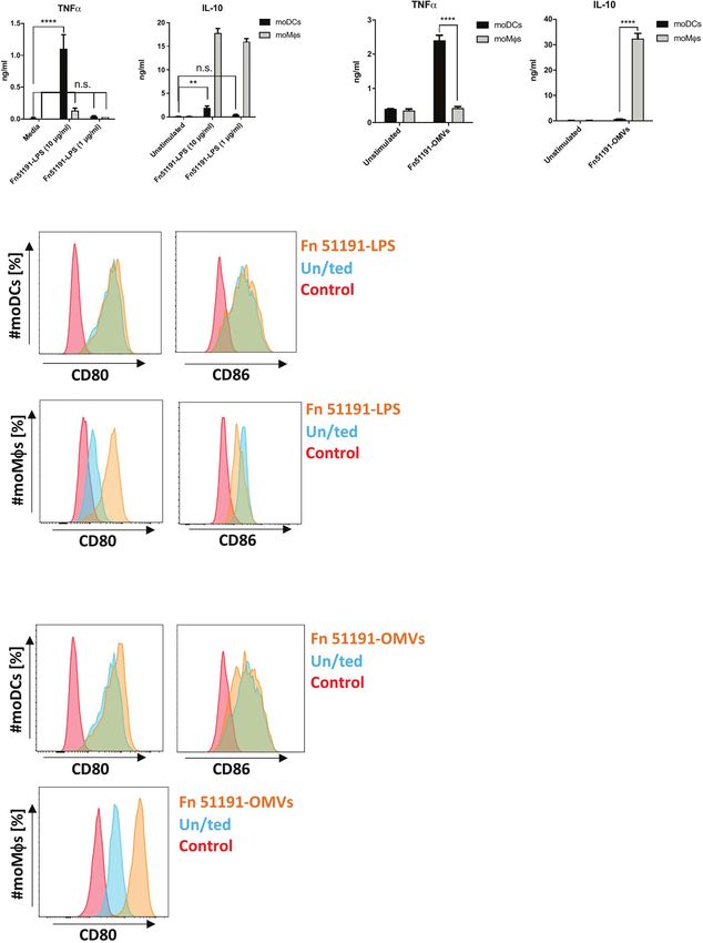
Lamprinaki et al.                                                                                         Siglec-7-Fusobacterium nucleatum Interaction

INTRODUCTION                                                                transmembrane proteins, which possess an extracellular portion
                                                                            characterized by a V-set immunoglobulin-like domain,
Colorectal cancer (CRC) is one of the most frequently diagnosed             containing the carbohydrate recognition domain (CRD), and
malignancies worldwide, accounting for 10% of all cancers and for           one or more C2-set immunoglobulin-like domains. The
approximately 20% of all cancer-related deaths in developed                 majority of Siglecs possess immunoreceptor tyrosine-based
countries (1). While CRC incidence commonly appears in ages                 inhibitory motifs (ITIMs) in their intracellular domain (26).
over 50 years old, recent years have shown an increased incidence in        Siglec ligands can be presented on the cell on which the Siglec is
younger adults which may be associated to lifestyle factors (2, 3).         expressed (cis ligands), or on glycans in the extracellular matrix
    Tumours that arise at epithelial barrier surfaces of the body           of other cells (trans ligands) (28). Although the CRDs of most
harbour extensive microbiota, and the importance of these                   Siglecs have some specificities towards certain sialylated
microbes in CRC is now widely acknowledged. The enrichment of               structures, several Siglecs have a broad and overlapping ligand
Fusobacterium spp. in CRC tissues, as revealed by whole genome              specificity (28). These glyco-immune checkpoints have been
sequencing, showed that the most abundant species is Fusobacterium          proposed as new targets for cancer immunotherapy (31–33).
nucleatum (4–6). Patients with F. nucleatum associated carcinoma            The working hypothesis on the role played by Siglecs in cancer
have a shorter survival period (7). In addition, F. nucleatum appears       is that immune cells expressing Siglecs are inhibited upon
to contribute to the chemoresistance of CRC (8–10). Among F.                binding to their ligands on cancer cells. Indeed, enzymatic
nucleatum subspecies, F. nucleatum ssp. animalis is most                    removal of sialic acids from cancer cell surfaces was shown to
predominant in CRC specimens (11).                                          enhance immune cell-mediated clearance of those cells through
    F. nucleatum potentiates intestinal tumorigenesis mainly by             loss of Siglec-7 and Siglec-9 binding in cis (34) although the
recruitment of tumour infiltrating immune cells, in particular               range of physiological ligands of Siglec-7 and Siglec-9 remain to
myeloid-derived immune cells such as tumour associated                      be identified (35). Recently, a genome-wide CRISPR screens
macrophages (TAMs), myeloid-derived suppressor cells                        revealed the glycoprotein CD43 expressed on leukemia cells as a
(MDSCs), dendritic cells (DCs), tumour associated neutrophils               highly specific ligand for Siglec-7 and blocking the interaction
(TANs) (11–13), and inhibits human T-cell response (12, 14, 15),            relieved Siglec-7-mediated inhibition of immune killing activity
leading to colorectal neoplasia progression. A high abundance of            (33). In addition, the tumour immune-suppressive effect of Siglec-7
F. nucleatum in CRC tissues is associated with increased nuclear            was recently demonstrated in vivo (36), further supporting the
factor kappa B (NFkB) activation and induction of a pro-                    proposed role of Siglec-7 as an immune checkpoint receptor.
inflammatory profile (12). Well-characterised virulence factors of                Several clinically relevant pathogens have evolved mechanisms
F. nucleatum such as membrane proteins FadA or Fap2 are involved            of molecular mimicry by displaying sialylated structures on their
in the binding of F. nucleatum to colon cancer cells inducing               surface to overcome the cis interactions of Siglecs on the surface of
oncogenic response (16, 17). In the epithelium, F. nucleatum                immune cells. For example, Campylobacter jejuni strains can
induces the expression of cell signalling proteins (cytokines), such        interact with Siglec-7 and sialoadhesin (Siglec-1) via their
as tumour necrosis factor (TNF)-a and interleukin (IL)-8 in                 lipooligosaccharides (37) and to Siglec-10 via a sialic acid-like
addition to epithelial-mesenchymal transition (18). Recently, the           molecule, pseudaminic acid present in the flagella (38), while
structures of O-chain polysaccharides (O-antigens) of the LPS from          Siglec-7 showed sialic acid-independent binding to b-protein
F. nucleatum strains ATCC 23726 (ssp. nucleatum) (19), ATCC                 expressed on Group B Streptococcus surface (39). Additionally,
25586 (ssp. nucleatum) (20), ATCC 10953 (ssp. polymorphum)                  human Siglecs have evolved to recognise non-Neu5Ac ligands
(21), ATCC 12230 (22), MJR 7757 B (23) and ATCC 51191 (ssp.                 present on external stimuli such as microbes (40). In this work we
animalis) (24) have been elucidated, showing strain-specific                 hypothesised that Siglecs may be involved in the recognition of
differences in the trisaccharide repeat unit containing either sialic       F. nucleatum strains by immune cells, contributing to the
acid/N-acetylneuraminic acid (Neu5Ac) (21), fusaminic acid (20)             tumorigenesis of these strains in CRC.
or monosaccharides other than nonulosonic acid residues (24).
However, the mechanisms underpinning the interaction of
F. nucleatum with immune cells remain undefined.                             MATERIALS AND METHODS
    Immune cells express a large variety of glycan-binding
receptors or lectins, which sense and respond to changes in                 Materials
the glycan signature of their environment leading to the                    All reagents were purchased from Sigma unless otherwise stated.
activation or inhibition of immune processes (25). Siglecs                  Recombinant Siglecs, human Siglec-3, Siglec-5, Siglec-7, Siglec-9 and
(sialic acid–binding immunoglobulin-like lectins) are a large               -10 and CHO-expressing Siglec-7-Fc (CHO-Siglec-7-Fc) cell line
family of lectins found on innate immune cells and tumour-                  were a kind gift from Prof. Paul Crocker (University of Dundee).
infiltrating T cells, which inhibit immune activation after                  Recombinant Siglec-7-Fc was also obtained commercially
sensing sialic acid-containing glycans (26, 27). Individual                 (R&D Systems).
family members exhibit preferences for sialosides of various
linkages to underlying glycan motifs, but many of the                       Bacteria Growth, Preparation
physiological ligands, glycoproteins or glycolipids, they                   and Quantification
interact with remain largely unknown (28, 29). The expression               F. nucleatum ssp. animalis ATCC 51191 isolated from clinical
of Siglecs on immune cells is cell type dependent (30). Siglecs are         samples was obtained from ATCC in partnership with LGC

Frontiers in Immunology | www.frontiersin.org                           2                                 October 2021 | Volume 12 | Article 744184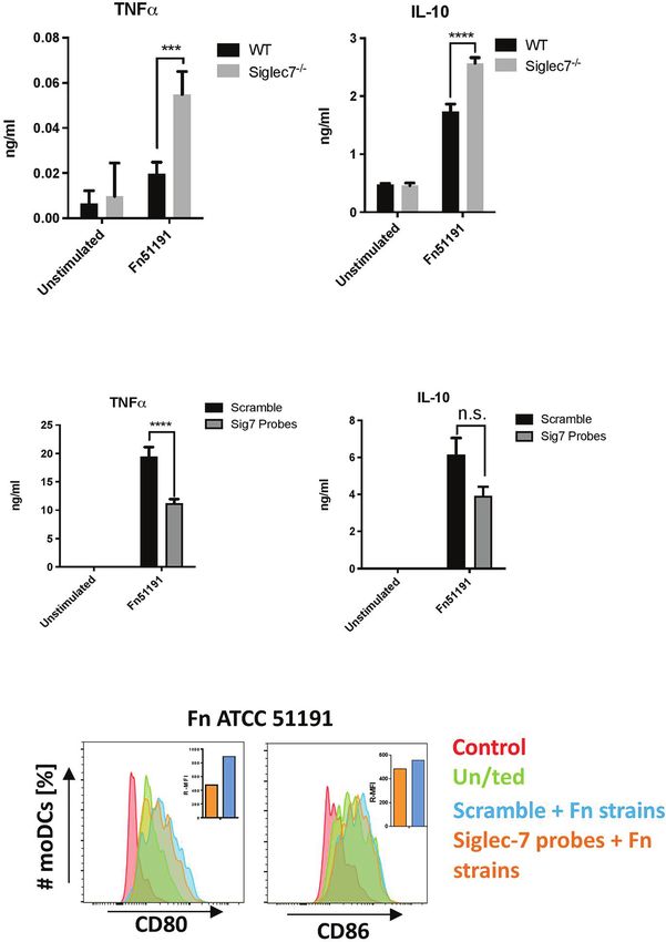
Lamprinaki et al.                                                                                          Siglec-7-Fusobacterium nucleatum Interaction

standards ltd. F. nucleatum was cultured in tryptic soy broth                Gel (BIO-RAD). The OMV-containing fractions were diluted with
media (Becton Dickinson) supplemented with 5 mg/ml hemin                     sterile DPBS and ultracentrifuged at 200,500 x g for 2 h at 4°C using
(Sigma) and 1 mg/ml menadione (Sigma). For binding and                       a Type 45 Ti rotor (Beckman Coulter). OMVs were resuspended in
human cell co-culture experiments, bacteria were centrifuged                 sterile DPBS and then filtered using a 0.22 mm membrane.
at 15,000 x g for 5 min, and the cells were fixed with 4%                         Purified OMVs were quantified and measured for their particle
paraformaldehyde (PFA) (Electron Microscopy Sciences/CN                      size using a NanoSight LM12 (Malvern Panalytical). Briefly, the
Technical Services ltd) for 45 min at room temperature (RT),                 samples were diluted 100 times in 1 ml DPBS and loaded onto the
in the dark, followed by two washes in Dulbecco’s Phosphate-                 instrument’s chamber by a syringe and the sample were slowly
buffered Saline (DPBS) (Lonza).                                              released. The considered particle size of each OMV sample were the
    For bacteria de-sialylation, 107 cells were treated with 20 U of         mean of triplicates. Instrument settings used: camera shutter 1035,
sialidase a2-3,6,8,9 neuraminidase A (NEB) in 1X GlycoBuffer I (NEB)         camera gain 680, capture duration 60 sec.
or control treated in 1X GlycoBuffer I alone, overnight (o/n) at 37°C.
    Bacteria were quantified by spectroscopy with OD600nm of 1                Semi-Quantitative Analysis of LPS in
corresponding to 109 cells/ml or by imaging flow cytometry                    OMVs by Gas Chromatography-Mass
(Amnis ImageStreamx Mk II) (as described below).                             Spectrometry (GC-MS)
                                                                             The content of LPS in F. nucleatum-derived OMVs was evaluated
LPS Extraction                                                               by analysing the fatty acids content. Based on the chemical
F. nucleatum ATCC 51191 bacterial cells were harvested by                    structure of the lipid A component of F. nucleatum LPS in the
centrifugation, lyophilised, and extracted by the hot phenol/water           bacteria (44), C14:0 (or myristic acid) was considered as the
method, as previously described (41). Each phase was dialysed                reporter group for LPS, while C16:0 (palmitic acid) and C18:0
against distilled water to remove the phenol, freeze-dried, and              (stearic acid) were considered the reporters for the phospholipids.
analysed by 12% sodium dodecyl sulphate polyacrylamide gel                       F. nucleatum-derived OMVs (1 mg) were treated with HCl/
electrophoresis (SDS-PAGE). After the water/phenol extraction,               MeOH (1 ml, 1.25 M, 80°C, 16 h) and lipids, derivatised as
F. nucleatum LPS extracted from was detected in the water phase by           methylesters, were extracted with hexane (41). This analysis
silver nitrate staining (42). The phases containing LPS were further         estimated the amount of each fatty acid (C14:0, C16:0 and
purified by enzymatic digestion (DNAse, RNAse and proteinase K)               C18:0) by correcting the areas of the corresponding peaks with
as previously described (41), followed by centrifugation at 6,000            a response factor, made by using an array of standard solutions
rpm for 30 min at 4°C and ultracentrifugation at 30,000 rpm for 4 h          and by setting C16:0 as internal standard. Areas were correlated
at 4°C. To separate the O-antigen (OPS) and lipid A domains, LPS             by a linear regression. The methanol layer after extraction with
were mild acid hydrolysed by acetic acid 1% (100°C, 2-3 h). The OPS          hexane, was used to countercheck the data from lipid analysis, by
domain of the strains containing ulosonic residues was further               verifying the presence of 3-deoxy-2-keto-D-manno-octulosonic
partial depolymerised. The solution was centrifuged and the                  acid and L-glycero-D-manno-heptose, both markers of the LPS
supernatants were freeze-dried and further purified by gel                    molecules. Identification of the fatty acids or the monosaccharide
filtration chromatography.                                                    constituents, was performed by comparing the retention time and
                                                                             the fragmentation pattern of each peak to a relevant standard.
F. nucleatum OMV Purification                                                     All chemical derivatives were analysed by using a Gas
and Characterisation                                                         Chromatography-Mass Spectrometry (GC-MS) Agilent
F. nucleatum derived OMVs were collected from the cell culture               Technologies 7820A (Santa Clara, CA, USA) equipped with a
supernatant, as described previously by Liu et al. (43) with some            mass selective detector 5977B and a HP-5ms capillary column
modifications. Briefly, F. nucleatum ssp. cells were cultured until            Agilent, Italy (30 m x 0.25 mm i.d., 0.25 mm as film thickness,
reaching OD600nm of 0.7-1.2. Cells were centrifuged at 8,500 x g             flow rate 1 ml/min, He as carrier gas). Electron impact mass
for 15 min at 4°C. The supernatant was collected, and vacuum                 spectra were recorded with ionisation energy of 70 eV and an
filtered using 0.22 mm membrane. The filtered supernatant was                  ionising current of 0.2 mA. The temperature program used was:
concentrated by spin-filtration using 100,000 molecular weight                150°C for 5 min, 150 up to 300°C at 10°C/min, 300°C for 12 min.
cut-off filter unit (Sartorius). OMVs were recovered from the
filter using sterile DPBS and further purified by density gradient             Expression and Purification of
ultra-centrifugation. For the gradient, Optiprep media (60% w/v,             Recombinant Siglec-Fc Proteins
Sigma) was diluted in 0.85% w/v NaCl and 10 mM tricine-NaOH                  CHO-Siglec-7-Fc cells were cultured in Glasgow Modified
pH 7.4 solution to make 35%, 30%, 25% and 20% density                        Essential Medium (GMEM) without L-glutamine media
solutions. The OMVs were mixed with 40% Optiprep solution                    (Sigma) supplemented with 10% fetal bovine serum (FBS)
and placed at the bottom of a 13.2 ml Ultra-clear tube (Beckman              (Thermo Scientific Gibco), 100 U/ml penicillin and 100 mg/ml
Coulter) and Optiprep (2 ml) was added subsequently by                       streptomycin (Lonza) and 50X GS supplements (Sigma).
density-decreasing order. The preparation was ultracentrifuged                   Adherent CHO-Siglec-7-Fc cells (80-90% confluence) were
at 135,000 x g for 16 h at 4°C with minimum acceleration and                 washed twice with Dulbecco’s phosphate-buffered saline (DPBS)
deceleration using a SW41 Ti rotor (Beckman Coulter). From the               (Thermo Scientific Gibco) and protein expression was induced
top to the bottom, 1 ml fractions were collected and analysed by             by culturing the cells with GMEM without L-glutamine media
SDS-PAGE in 4–15% Mini-PROTEAN® TGX™ Precast Protein                         (Sigma) supplemented with 200X FetalClone II (Thermo Fisher

Frontiers in Immunology | www.frontiersin.org                            3                                 October 2021 | Volume 12 | Article 744184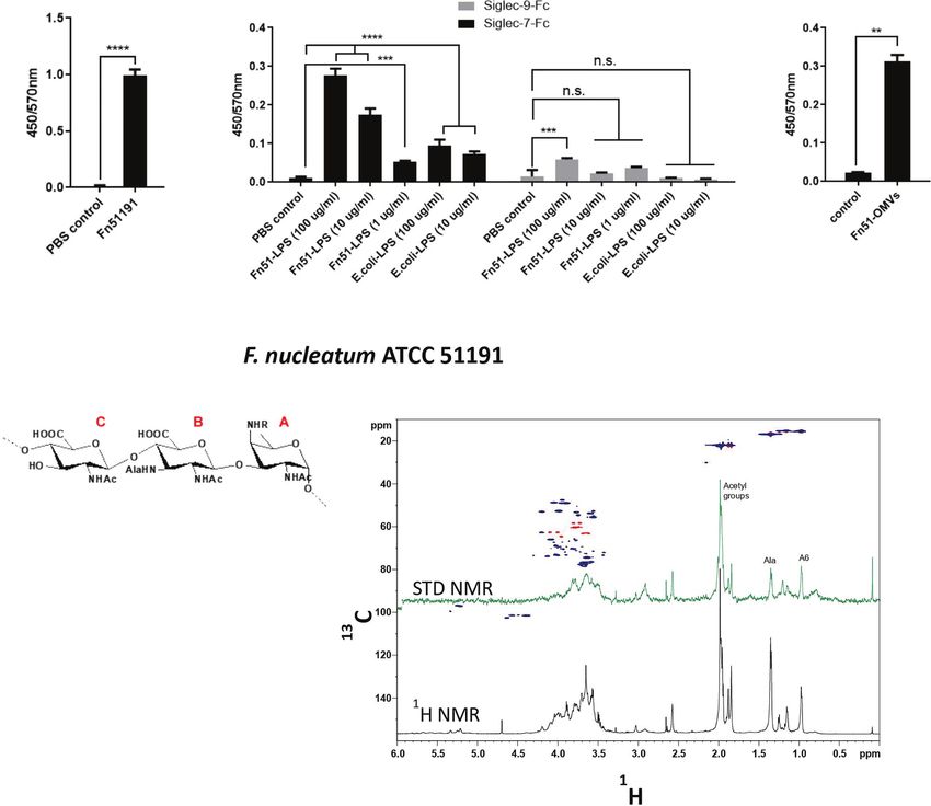
Lamprinaki et al.                                                                                      Siglec-7-Fusobacterium nucleatum Interaction

Scientific), 100 U/ml penicillin and 100 mg/ml streptomycin                ratios varying from 1: 20 to 1: 80 with 15 µM of Siglec-7-Fc
(Lonza), 50X GS supplements (Sigma) and 100 mg/ml MSX                     protein. STD NMR experiments were acquired at 298 K with 32 k
(Sigma). After 4 days, the supernatant was collected for Siglec-          data points and zero-filled up to 64 k data points prior to
7-Fc purification.                                                         processing. The Siglec-7-Fc resonances were saturated applying
   Siglec-7-Fc purification was carried out using gravity-flow              40 Gauss pulses with a length of 50 ms, setting the on-resonance
column (BIO-RAD) packed with protein A-Sepharose (Sigma)                  pulse at aromatic region (7.5/6-5 ppm) and the off-resonance
washed with DPBS. The harvested CHO supernatant was added                 pulse frequency at 100 ppm. Under these experimental
to the column and the column washed with DPBS. To elute                   conditions, very low residual signals were observed in some of
Siglec-7-Fc, a solution of 0.1 M glycine, pH 3 was added to the           the STD NMR spectra for the ligands in the free state which were
column and fractions (0.5 ml) were collected in 1 M Tris, pH 8            taken into account during data processing. To suppress the water
(for neutralisation). The protein concentration in fractions was          signal, an excitation sculpting with gradient pulses (esgp) was
quantified by Nanodrop (Thermo Fisher Scientific).                          applied and to reduce the NMR signals of Siglec-7-Fc, a spin-lock
                                                                          filter (20 ms) was used.
Binding Assays
For the flow cytometry binding assays between F. nucleatum ssp.            Culture of Human Primary Immune Cells
animalis ATCC 51191 and recombinant Siglecs (Siglec-3, Siglec-            and U-937 Monocytic Cell Line
5, Siglec-7, Siglec-9 and -10), bacteria (107 cells) were incubated       Human peripheral blood was obtained from haemochromatosis
with the pre-complex of recombinant Siglec-Fc (4 mg/ml) and               patients undergoing a therapeutic venesection at the Norfolk and
mouse a-Fc-PE Ab (1 mg/ml) (R&D Systems) in DPBS for 1 h at               Norwich University Hospital (Norwich, UK). Blood collection in
37°C. Following centrifugation at 14,000 x g for 4 min, bacterial         this study was approved by the Faculty of Medicine and Health
cells were washed with DPBS and analysed by Fortessa (BD                  Sciences Research Ethics Committee REC reference number
Biosciences). For the inhibition assays, Siglec-7-Fc and a-Fc-            2013/2014 -14HT (University of East Anglia).
phycoerythrin (PE) Ab pre-complex was first incubated with                     For monocyte-derived dendritic cell (moDC) and macrophage
disialoganglioside with three glycosyl groups GD3 (Sigma) at 50           (moMf) generation, peripheral blood mononuclear cells (PBMCs)
mg/ml for 30 min at 4°C. For the flow cytometry binding assays             were isolated from whole blood following centrifugation using
between F. nucleatum ssp. and human cells, U-937 (WT or                   Ficoll-Paque gradient media (GE Healthcare). Monocytes
Siglec-7-/-) cells were first stained with 10,000X cell trace violet       (CD14+ cells) were isolated from PBMCs using CD14 positive
(CTV) (Thermo Fisher Scientific) for 15 min at RT and F.                   selection microbeads (StemCell technologies) according to the
nucleatum ssp. were stained with 10 mg/ml of fluorescein                   manufacturer’s instructions. Freshly isolated CD14+ monocytes
isothiocyanate (FITC) (Sigma). Following two washes with DPBS,            (106 cells/ml) were cultured in Mercedes medium (RPMI 1640
U-937-CTV (5 x 10⁵ cells) were incubated with F. nucleatum-FITC           medium (Lonza) supplemented with 25 mM HEPES, 10% FBS
(5 x 10⁶ cells) for 1 h at 4°C. Cells were washed with FACS buffer        (Thermo Scientific Gibco), 55 mM 2-mercaptoethanol, 100 U/ml
(HBSS containing 0.01% bovine serum albumin (BSA), and 2 mM               penicillin and 100 mg/ml streptomycin (Lonza), 2 mM glutamine
EDTA), centrifuged at 510 x g for 3 min and analysed using Fortessa       (Lonza), 1 mM non-essential amino acids (Lonza) and 1 mM
(Threshold of FSC parameter set to 1000). Flow cytometry data             sodium pyruvate (Lonza), were incubated with granulocyte-
were processed in FlowJo (TreeStar) software.                             macrophage colony-stimulating factor (GM-CSF) and IL-4
    For the ELISA-based binding assays, bacteria (107 cells) or           (PeproTech) (25 ng/ml) for differentiation of monocytes to
bacteria-derivatives (10 mg/ml LPS or 108 OMV particles) in               moDCs or with macrophage colony-stimulating factor (M-CSF)
DPBS solution were coated in a 96-well plate, o/n at 4°C.                 (PeproTech) (25 ng/ml) for differentiation of monocytes to moMfs.
Following a washing step with 0.05% tween in PBS (washing                 The cells were incubated for 7 days at 37°C, with addition of the
buffer) the plate was incubated with 1% BSA for 1 h at RT.                above cytokines on day 3, as previously described (45).
Followed by 3 times washing the plates were incubated with pre-               For U-937 differentiation, U-937 (5 x 10⁵ cells/ml) cultured in
complexed Siglec-Fc and a-Fc-HRP for 2 h at RT. Briefly, Siglec-           RPMI 1640 medium (Lonza) supplemented with 25 mM HEPES,
Fc protein (4 mg/ml) was incubated with 50,000X a-human-Fc-               10% FBS (Thermo Scientific Gibco), 55 mM 2-mercaptoethanol,
HRP (Abcam) for 1 h at RT. Following 3 washes with                        100 U/ml penicillin and 100 mg/ml streptomycin (Lonza), 2 mM
washing buffer, the plate was incubated with 3,3′,5,5′-                   glutamine (Lonza) and 1 mM sodium pyruvate (Lonza) were
tetramethylbenzidine (TMB) (Biolegend) until colour                       differentiated with 100 ng/ml phorbol 12-myristate 13-acetate
development. Colour development was stopped by the addition               (PMA) (Sigma) for 28 h at 37°C, as previously described (46).
of 2 N H2SO4 and the absorbance was measured at 450 nm with               Adherent cells were detached by PBS-EDTA (Lonza) and
reference at 570 nm. Data were analysed in GraphPad Prism 6.              scraping and collected for functional assays.

STD NMR Analysis                                                          Generation of Siglec-7-/- U-937 Cells by
Spectra were acquired on a Bruker 600 MHz AVANCE NEO                      CRISPR-Cas9
equipped with a cryo probe and analysed using the TOPSPIN                 CRISPR RNA (crRNA) was designed to target human Siglec-7
4.1.0 software. The partial depolymerised OPS derived from                (CATGCCCTCTTGCACGGTCA, IDT) in U-937 cells (ATCC®
F. nucleatum ssp. LPS were prepared in deuterated PBS buffer              CRL-1593.2™). guide RNA (gRNA) (1 mM crRNA, 1 mM
(20 mM PBS, NaCl 150mM, pH= 7.4), using protein-ligand                    ATTO-550 labeled tracrRNA (IDT)) was boiled at 95°C for

Frontiers in Immunology | www.frontiersin.org                         4                                October 2021 | Volume 12 | Article 744184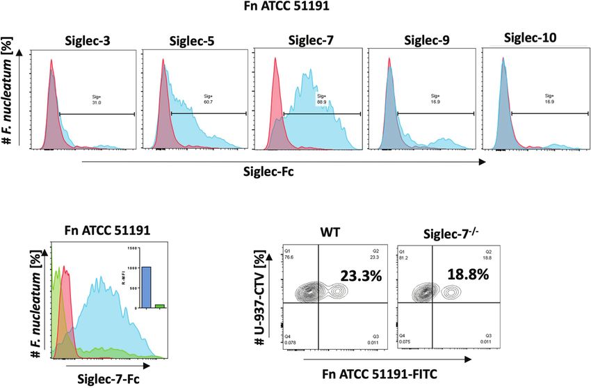
Lamprinaki et al.                                                                                            Siglec-7-Fusobacterium nucleatum Interaction

5 min. A solution of 20 pmol gRNA, 20 pmol Cas9 nuclease (IDT),                FACS buffer and analysed by ImageStreamx Mk II (Amnis).
8 ml Cas9 PLUS reagent (IDT), 16 ml CRISPRMAX reagent                          Using the INSPIRE software, a total of 5,000 FITC stained cells
(Thermo Fisher) in 600 µl of Opti-MEM medium (Gibco) was                       were collected. The percentage of internalised bacteria were
prepared. 750,000 U-937 cells were washed with Opti-MEM                        determined using the internalisation wizard with erode mask
medium (Gibco) and centrifuged at 300 x g for 5 min. The cell                  function at 7 number of pixels.
pellet was resuspended in the prepared solution and incubated at
37°C, 5% CO2. After a 24 h incubation, cells were centrifuged at               Statistical Analyses
300 x g for 5 min, then resuspended in 400 ml flow buffer (HBSS,                One-way ANOVA followed by Tukey’s test were used for
1% FBS, 500 µM EDTA). The top 5% ATTO-550 positive cells                       multiple comparisons, t-test or two-way ANOVA were used
were sorted on a BD FACSMelody™ Cell Sorter into four 96-well                  for two-group comparisons, on Prism software (GraphPad). P <
flat-bottom plates containing media at one cell per well.                       0.05 was considered as statistically significant. *p < 0.05, **p <
Approximately 2 weeks later, colonies were screened for Siglec-7               0.01, ***p < 0.001, ****p < 0.0001, n.s., not statistically difference.
expression by flow cytometry using PE-conjugated Siglec-7 at
1:100 dilution (BioLegend) and Siglec-7-/- clones were collected.

Siglec-7 RNA Silencing of Primary                                              RESULTS
Immune Cells
moDCs were transfected with two pre-designed small interfering                 F. nucleatum ssp. animalis Binding to
RNA (siRNA) Silencer Select SIGLEC7 probes (ID# s25729 and                     Siglecs Revealed Specific Binding
s25730) or with the scramble siRNA (Invitrogen) with reverse                   to Siglec-7
transfection, as described previously (47). Briefly, 3 × 10⁵ moDC               The binding of F. nucleatum ATCC 51191 was first tested against a
or moMf cells were incubated with the complex of two probes to                 range of human recombinant CD33-related Siglec-Fc proteins
a final 200 nM concentration or with the negative control                       including Siglec-3, Siglec-5, Siglec-7, Siglec-9 and -10 by flow
(scramble) and 1% HiPerFect transfectant (Qiagen) in warm                      cytometry. A clear shift in fluorescence was observed in the
RPMI 1640 (non-supplemented) media in a 24-well plate for                      presence of Siglec-7 with approx. 90% of F. nucleatum population
three days.                                                                    bound to Siglec-7, while 60% and 30% of F. nucleatum population
                                                                               bound to Siglec-5 and Siglec-3, respectively, and only 17% of the
Cytokine and Cell Surface Marker Analysis                                      population bound to Siglec-9 and -10 (Figure 1A). To determine if
moDCs or moMfs or U-937-PMA (105 cells) were cultured in                       the binding to Siglec-7 was glycan-mediated, inhibition binding
96-well plates in the Mercedes medium as described above and                   assays were carried out in the presence of ganglioside GD3, a known
stimulated with PFA-fixed F. nucleatum ssp. at multiplicity of                  Siglec-7 ligand (48). A significant decrease in Siglec-7 binding to F.
infection (MOI) of 50 or 5, F. nucleatum ssp.-derived LPS at 10 or 1           nucleatum ATCC 51191 was observed in the presence of GD3,
mg/ml, the E. coli O111:B4 control at 1 mg/ml, or OMVs at 5 x 10⁷              showing an approx. 92% reduction of the bacterial cell population
particles/ml for 18 h at 37°C. The cells were centrifuged at 510 x g for       bound to Siglec-7 (Figure 1B). This result suggests that Siglec-7 V-
3 min and the supernatant and pellet collected for analysis.                   set domain is implicated in the binding between Siglec-7 and F.
   For cytokine analysis, human TNFa, IL-10, IL-8 production                   nucleatum ssp. To investigate whether the binding of F. nucleatum
in the supernatant was quantified by ELISA (BioLegend)                          ATCC 51191 to Siglec-7 was mediated by sialic acid exposed on the
according to the manufacturer’s instructions.                                  bacterial cell surface, the bacterial cells were treated with
   For cell surface marker analysis, moDC or moMf pellets were first            neuraminidase A, a sialidase with broad specificity to (a2-3,6,8,9)
incubated with human Fc block (BioLegend) and then incubated with              sialylated linkages, cleaving linear and branched non-reducing
antibodies for 30 min at 4°C as follows: programmed death-ligand 1             terminal sialic acid residues from glycoconjugates. The sialidase
(PD-L1)-PE at 1:50 dilution or CD80-PE at 1:100 dilution, CD86-                pre-treatment only led to a small reduction in the binding of F.
Alexa488 at 1:200 dilution, isotype controls mouse IgG1-PE,k at                nucleatum to Siglec-7 as shown by flow cytometry (Figure S1), in
1:100 (BioLegend) at 1:25 dilution (BioLegend) and with propidium              agreement with the absence of sialic acid in F. nucleatum ATCC
iodide (PI) or 4′6-diamidino-2-phenylindole (DAPI) at 0.1 or 1 mg/             51191 LPS (24). Next, we conducted binding assays between the
ml for dead cell staining, respectively. The cells were then washed with       monocytic cell line U-937 (wild type (WT) or Siglec-7-/-) and F.
DPBS supplemented with 1% BSA (FACS buffer) analysed by flow                    nucleatum ATCC 51191 by flow cytometry. Our results showed a
cytometry using Fortessa.                                                      reduction of F. nucleatum associated with U-937-Siglec-7-/- cells as
                                                                               compared to WT cells (Figure 1C), supporting an interaction
Imaging Flow Cytometry                                                         between F. nucleatum ssp. and Siglec-7 when expressed on the
For counting bacteria, 10,000 events were collected and                        cell surface although other receptors may be involved in the
processed using the IDEAS (Amnis) software. Bacteria density                   interaction between F. nucleatum ATCC 51191 and U-937 cells.
‘‘objects/ml’’ were selected in the bright field channel (M04) of
the Aspect Ratio_M04 versus Area_M04 dot plot.                                 F. nucleatum Derived LPS or OMVs Bind
    For internalisation assays, human cells (monocyte-derived or               to Siglec-7
U-937-PMA) at 5 × 10⁶ cells/ml were incubated with 5 × 10⁷                     To determine the role of LPS in the binding of F. nucleatum
FITC-stained F. nucleatum ssp. for 4 h. Cells were washed with                 ATCC 51191 to Siglec-7, LPS was extracted from F. nucleatum

Frontiers in Immunology | www.frontiersin.org                              5                                  October 2021 | Volume 12 | Article 744184
Lamprinaki et al. Siglec-7-Fusobacterium nucleatum Interaction
A
B C
FIGURE 1 | Binding of F. nucleatum ATCC 51191 to Siglecs using flow cytometry. (A) Binding of F. nucleatum to recombinant Siglec-Fc proteins (in blue).
(B) Binding of F. nucleatum to Siglec-7 in the presence of GD3 inhibitor (in green) or untreated cells (in blue). (C) Binding of F. nucleatum to WT or Siglec-7-/-
U-937 cells. Bacteria incubated with a-Fc-PE Ab only was used as a control (in red). Fn, F. nucleatum.
ATCC 51191 by the hot phenol/water method (41) and further and GlcNAc3NAlaA) and fucosamine (FucNAc4N) residues.
purified by enzymatic digestion (41). The SDS-PAGE of the Therefore, STD NMR analysis confirmed binding of Siglec-7 to
extracted F. nucleatum-derived LPS showed the typical LPS F. nucleatum ATCC 51191 OPS, even though it lacks nonulosonic
ladder-like pattern and lower average molecular weight acid residues (Figure 2D).
distribution when compared to E. coli O127:B8 (Figure S2). Next, we purified outer-membrane vesicles (OMVs)
Using an ELISA-type assay, we showed that the whole F. produced by F. nucleatum ATCC 51191 by density gradient
nucleatum cells and the derived LPS bound to Siglec-7-Fc ultracentrifugation, resulting in pure and spherical particles with
(Figures 2A, B) while no binding of F. nucleatum-derived LPS a diameter range from 30 to 250 nm (Figure S3A). We showed
was observed against Siglec-9-Fc used as a control (Figure 2B). To by GC-MS that LPS is a main constituent (approximately in 60-
map the relevant positions of F. nucleatum LPS involved in the 70% mol/mol) of F. nucleatum-derived OMVs (Figure S3B). We
interaction with Siglec-7 and gain a first evaluation of the ligand then tested the ability of F. nucleatum-derived OMVs to bind to
epitopes, the partially depolymerised O-antigen chain (OPS) Siglec-7 (Figure 2C). F. nucleatum-derived OMVs from ATCC
isolated from F. nucleatum ATCC 51191 was analysed by STD 51191 bound to Siglec-7 at levels comparable to LPS under the
NMR (49) (Figure 2D). Interestingly, STD enhancements, together conditions tested (Figure 2C).
with changes in the relative intensity of STD signals with respect to Together these data identified F. nucleatum LPS present on
the reference spectrum, were detected, clearly indicating that F. whole cells or OMVs as a new ligand to Siglec-7-Fc.
nucleatum ATCC 51191 OPS structure was recognised by and
interacted with Siglec-7-Fc. Despite the significant overlapping of F. nucleatum Modulates Immune
ligand resonances which impaired a detailed analysis of the protons Response in a Cell Subset Specific
involved in the recognition and binding process, the fingerprint of Manner
STD NMR spectrum allowed to identify the ligand regions in close To investigate the impact of F. nucleatum ssp. on the host immune
contact with Siglec-7-Fc. F. nucleatum ATCC 51191 OPS contains a response, myeloid cells, moDCs and moMfs, were generated from
linear trisaccharide made up of glucosaminuronic (GlcNAcA and human blood, and stimulated with F. nucleatum ATCC 51191 or
GlcNAc3NAlaA) and fucosamine (FucNAc4N) residues, [!4)‐b‐ with F. nucleatum ATCC 51191-derived LPS and OMVs.
D‐GlcpNAcA‐(1!4)‐b‐D‐GlcpNAc3NAlaA‐(1!3)‐a‐D‐ F. nucleatum bacterial cells were shown to associate with the
FucpNAc4NR‐(1!], with the N‐4 of the fucosamine partly cell surface of moDCs or moMfs as determined by imaging flow
acetylated (60 %). The analysis of signals in isolated regions of the cytometry (Figure S4). Stimulation of moDCs with F. nucleatum
spectrum, i.e., in the range between 0.8 – 1.5 ppm, demonstrated the at MOI 5 resulted in a marked increase in cytokine production of
contribution to the interaction from glucosaminuronic (GlcNAcA TNFa, IL-8 (p < 0.0001) (Figure 3A) and an induction of CD86
Frontiers in Immunology | www.frontiersin.org 6 October 2021 | Volume 12 | Article 744184Lamprinaki et al. Siglec-7-Fusobacterium nucleatum Interaction
A B C
D
FIGURE 2 | Binding of F. nucleatum ATCC 51191 cells, LPS or OMVs to Siglec-7-Fc. Immobilised F. nucleatum 51191 cells were tested for binding to (A) Siglec-7-
Fc and the extracted LPS to (B) Siglec-7-Fc or Siglec-9-Fc by ELISA. (C) Immobilised OMVs extracted from F. nucleatum 51191 were tested for binding to Siglec-7-
Fc by ELISA. PBS was used as control. Data shown are the mean of duplicates ± SD derived from one representative experiment reproduced in three independent
experiments. Fn, F. nucleatum. (D) STD NMR analysis of the binding between Siglec-7 and partially depolymerised OPS from F. nucleatum ATCC 51191. The panel
shows the superimposition of the reference 1H NMR spectrum (in black) and STD NMR spectrum (in green), the 1H-13C HSQC spectrum (blue/red) and the chemical
structure of F. nucleatum OPS repeating units. Statistical analyses were performed by t-test (for panels 2Aand 2C) or one-way ANOVA followed by Tukey’s test.
P < 0.05 was considered as statistically significant. **p < 0.01, ***p < 0.001, ****p < 0.0001, n.s., not statistically difference.
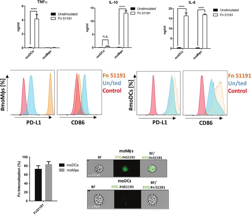
and PD-L1 as compared to the unstimulated control moDCs showing approx. 10% less internalisation as compared to
(Figure 3B). A different profile was observed with moMfs, moMfs (Figure 3C).
where stimulation with F. nucleatum led to a significant Next, we stimulated moDCs or moMfs with F. nucleatum
induction of IL-10 and IL-8 production (p < 0.0001), low levels ATCC 51191-derived LPS or OMVs. In moDCs, treatment with
(n.s.) of TNFa production (Figure 3A) and to the induction of OMVs or with LPS at 10 mg/ml but not 1 mg/ml induced TNFa
PD-L1 and downregulation of CD86 cell surface markers as production (Figures 4A, B). In moMfs, stimulation OMVs
compared to the unstimulated control (Figure 3B). The results or LPS (at both 10 or 1 mg/ml) showed significant induction of
were dose-dependent, with a marked increase in cytokine IL-10 at levels comparable to the whole bacteria (Figures 4A, B).
production when cells were stimulated at MOI 50 as compared When moMfs or moDCs were treated with LPS or OMVs, there
to MOI 5 (Figure S5A). This acquired moMf phenotype was was an upregulation of the CD80 cell surface marker expression
also observed using the macrophage like cell line U-937 after as compared to the unstimulated control, as showed with the
F. nucleatum ATCC 51191 stimulation, leading to high IL-10 and whole bacteria. In moDCs, LPS stimulation led to an induction of
low TNFa levels (Figure S5B). Consistent with these results, we CD86 expression (Figure 4C), as also observed with the whole
showed, using imaging flow cytometry, that both moDCs and bacteria, while stimulation with OMVs showed a reduction of
moMfs were able to internalise F. nucleatum (Figure 3C), with CD86 expression compared to the unstimulated control
Frontiers in Immunology | www.frontiersin.org 7 October 2021 | Volume 12 | Article 744184Lamprinaki et al. Siglec-7-Fusobacterium nucleatum Interaction
A
B
C
FIGURE 3 | Effect of F. nucleatum ATCC 51191 on human myeloid cells. Analysis of (A) cytokine and (B) cell surface marker expression in moDCs or moMfs by
flow cytometry. Human cells were stimulated with F. nucleatum ATCC 51191 (in orange). Unstained cells (in red) and unstimulated (un/ted) cells (in blue) were used
as controls. (C) Internalisation of F. nucleatum ATCC 51191 into moDCs or moMfs. Images were taken with a 40X objective. For the cytokine quantification, data
shown are the mean of triplicates ± SD derived from one representative experiment reproduced in three independent experiments. Statistical analyses were
performed by one-way ANOVA followed by Tukey’s test. P < 0.05 was considered as statistically significant. ****p < 0.0001, n.s., not statistically difference.
(Figure 4D). Stimulation of moMfs with LPS (at 10 or 1 mg/ml) ssp.-stimulated U-937-Siglec-7 -/- as compared to WT
showed a reduction of CD86 expression (Figure 4C), as also cells (Figure 5A).
observed with the whole bacteria. Next, we carried out silencing of Siglec-7 in primary moDCs
Overall, our results suggest that moDCs stimulated with and moMfs by siRNA. Using flow cytometry, we confirmed that
F. nucleatum ATCC 51191 and derived components (OMVs Siglec-7 was expressed on the cell surface of moDCs and moMfs
and LPS) show a pro-inflammatory profile while F. nucleatum- (Figure S7A), and that expression could be reduced by up to 40%
treated moMfs acquire a M2-phenotype which is associated with in moDCs as compared to the scramble control (Figure S7B)
tumour progression (50). while no significant reduction in expression could be achieved in
moMfs. We then analysed the cytokine profile and expression of
Siglec-7 Is Involved in F. nucleatum- cell surface markers following stimulation of silenced or
Mediated Immune Response scramble control moDCs with F. nucleatum ATCC 51191 at
To obtain direct evidence of the contribution of Siglec-7 in MOI 5 (Figure 5B). We showed that stimulation of Siglec-7
F. nucleatum ATCC 51191 interaction with myeloid human silenced moDCs with F. nucleatum produced statistically
cells, we used CRISPR-Cas9 editing to generate Siglec-7 significant (p < 0.05) lower TNFa levels as compared to
deficient U-937 cells and assayed the effect of F. nucleatum scramble moDCs (Figure 5B). No differences in cell surface
stimulation on the immune response of differentiated U-937 WT marker expression were observed between F. nucleatum-
or Siglec-7-deficient (Siglec-7-/-) cell lines (Figure 5). The stimulated Siglec-7 silenced or scramble moDCs (Figure 5C).
expression of Siglec-7 in these cell lines was confirmed by flow Together these data suggest that Siglec-7 is involved in the
cytometry (Figure S6). A significant increase (p < 0.01) in TNFa induction of a pro-inflammatory response in moDCs by F.
and IL-10 cytokine production was observed in F. nucleatum nucleatum ATCC 51191.
Frontiers in Immunology | www.frontiersin.org 8 October 2021 | Volume 12 | Article 744184Lamprinaki et al. Siglec-7-Fusobacterium nucleatum Interaction
A B
C
D
FIGURE 4 | Effect of F. nucleatum ATCC 51191-derived LPS or OMVs on human myeloid cells. Analysis of (A) F. nucleatum LPS or (B) F. nucleatum OMVs on
cytokine production and (C) F. nucleatum LPS or (D) F. nucleatum OMVs on cell surface marker expression. Unstimulated (un/ted) cells (in blue) and unstained
cells (in red) were used as controls. Data shown are the mean of triplicates ± SD derived from one representative experiment reproduced in three independent
experiments. Statistical analyses were performed by one-way ANOVA followed by Tukey’s test. P < 0.05 was considered as statistically significant. **p < 0.01,
****p < 0.0001, n.s., not statistically difference.
DISCUSSION invasion (51), enabling it to reside intracellularly in tumour cells
(52), and, once there, potentially influencing tumorigenesis.
F. nucleatum is the most abundant bacterial species in the Adhesion and invasion of F. nucleatum to epithelial cells are
colorectal tumour microenvironment with F. nucleatum ssp. mediated by the Fap2 lectin and FadA adhesin expressed on the
animalis ATCC 51191 being enriched in CRC tissues (11). F. surface of F. nucleatum. The Fap2 lectin interacts with Gal-
nucleatum encodes an array of genes related to adhesion and GalNAc glycans which are overexpressed on tumour cells (53),
Frontiers in Immunology | www.frontiersin.org 9 October 2021 | Volume 12 | Article 744184Lamprinaki et al. Siglec-7-Fusobacterium nucleatum Interaction
A
B
C
FIGURE 5 | Effect of Siglec-7 on F. nucleatum ATCC 51191 interaction with human immune cells. (A) Cytokine production of U-937-PMA (WT or Siglec-7-/-)
stimulated with F. nucleatum ATCC 51191. Bars represent the median values from 3 technical replicates. (B) Cytokine production and (C) cell surface marker
expression of Siglec-7 silenced moDCs (in orange) or scramble control cells (in blue) stimulated with F. nucleatum. Unstimulated (un/ted) cells (in green) and
unstained cells (in red) were used as controls. For the cytokine and internalisation analyses, data shown are the mean of triplicates ± SD and duplicated ± SD,
respectively, derived from one representative experiment reproduced in three independent experiments. Statistical analyses were performed by two-way ANOVA
followed by Tukey’s test. P < 0.05 was considered as statistically significant. ***p < 0.001, ****p < 0.0001, n.s., not statistically difference.
while the FadA adhesin, recognises and binds host surface dendritic cells constitutively express Siglec-7, and the colonic
components, such as vascular endothelial cadherin (54) and lamina propria monocytes and macrophages represent the major
epithelial cadherin (16). F. nucleatum FadA expression was Siglec-7 positive populations (55). Extracellularly, Siglec-7 has a
also shown to be upregulated in CRC tissue (16). Interaction of sialic acid-binding V-set domain which we demonstrated was
FadA with E-cadherin triggers the expression of oncogenes, such implicated in the binding to F. nucleatum. Siglec-7 has been
as c-MYC and inflammatory genes, through the b-catenin shown to bind to the sialylated ganglioside GD3 (56), and N-
cascade and the upregulation of annenix A1 (16, 17). However, linked disialyl Lewisa in the normal colonic epithelium (55). At
the receptors involved in the interaction between F. nucleatum the molecular level, Siglec-7 has been reported to bind to
ssp. and immune cells remain largely unknown. terminal sialic acid moieties with diverse underlying glycan
Here, we showed that F. nucleatum ssp. animalis ATCC structures. We recently uncovered the LPS structure of F.
51191 interacts with Siglec-7 expressed by immune cells and nucleatum ATCC 51191, revealing a novel sugar repeating unit
that binding is LPS-mediated. Human NK cells, macrophages, in the O-antigen structure [!4)-b-D-GlcpNAcA-(1!4)-b-D-
Frontiers in Immunology | www.frontiersin.org 10 October 2021 | Volume 12 | Article 744184Lamprinaki et al. Siglec-7-Fusobacterium nucleatum Interaction
GlcpNAc3NAlaA-(1!3)-a-D-FucpNAc4NR-(1!], (R= differences in cell immune response could be attributed to the mode
Acetylated 60%), and a bis-phosphorylated hexa-acylated lipid of recognition, the nature of the interactions (cell-cell or cell-
A moiety (24). It therefore likely that the LPS glycans other than microbe), or the heterogeneity in pattern recognition receptor
sialic acid moieties may contribute to the binding of F. nucleatum (PRR) expression in the different cell subsets used in the in vitro
ATCC 51191 to Siglec-7, consistent with the results of the studies. Indeed, other PRRs may act synergistically with Siglec-7 to
sialidase treatment. This was further confirmed by STD NMR, contribute to a distinct immune response. For example, TLR-4, a
showing that the OPS extracted from F. nucleatum ATCC 51191 toll-like receptor with an intracellular activation motif has been
was recognised by Siglec-7, revealing new ligand epitopes not shown to establish a direct interaction with Siglecs including Siglec-
restricted to nonulosonic acids (neuraminic acid and fusaminic 7 (64). Therefore, since TLR-4 is expressed in U-937 (65) and
acid). The discovery that F. nucleatum LPS is a ligand for moDCs (66), our findings could be the result of a synergetic effect
immune checkpoint receptors like Siglec-7 opens up new between Siglec-7 and TLR-4. This interaction could also contribute
blockade strategies and studies are in progress to gain further to the capacity of F. nucleatum ssp. to promote chemoresistance of
structural insights into the broad ligand specificity of Siglec-7 CRC by inhibition of cancer cell apoptosis (8–10). A recent in vivo
towards the bacterial glycan structures revealed in this work. study using humanized immunocompetent mice, showed that
We showed that F. nucleatum ATCC 51191 induced a pro- Siglec-7 and -9 could be potential targets to enhance anti-tumour
inflammatory profile in moDCs and a tumour associated profile in immunity (36). In the future, it will be interesting to study the effect
macrophages (moMfs and U-937 cells) and that Siglec-7 contributed of F. nucleatum-Siglec-7 interaction in vivo, using humanised
to these cell-specific responses using Siglec-7 RNA-silenced moDCs immunocompetent murine model, as Siglec-E, the closest murine
and Siglec-7 deficient U-937 cells. In macrophages, F. nucleatum homolog of Siglec-7, does not recognise F. nucleatum ssp. (data not
ATCC 51191 induced the expression of IL-10, IL-8 cytokines and shown), consistent with the lack of direct homology between
PD-L1 marker and a downregulation of CD86 cell surface marker murine and human Siglecs (67).
expression, characteristic of macrophage type 2 (M2) polarisation It was recently reported that Siglec−7 is expressed in
(12, 13). These results are in agreement with previous studies showing macrophages in CRC tissue from patients and that high levels of
an infiltration of M2-macrophages in F. nucleatum ssp. positive Siglec−7 expression in tumour tissues are associated with shorter
clinical CRC specimens (57) and a M2 acquired phenotype in overall survival in patients treated with immunotherapy for
macrophage-like cell lines stimulated with F. nucleatum ATCC metastatic CRC (68). Mirroring this, an independent human
10953 (13), and F. nucleatum ATCC 25586 (58). A recent study study reported that patients with high relative abundance of F.
showed that Siglec-7 and -9 induce the polarisation of monocytes into nucleatum in tumour tissues compared to matched control tissues
a tumour-associated macrophage (TAM) phenotype and the have a higher incidence of regional lymph node metastases (69).
induction of tumour-associated cell surface markers such as PD-L1 These studies support the translation of our findings to humans.
(59). The moDC response to F. nucleatum ATCC 51191 suggests that The interaction of F. nucleatum ssp. with Siglec-7, leading to a pro-
Siglec-7 contributes to the pro-inflammatory response. These cell- inflammatory microenvironment, provides a mechanism
specific phenotypes could be recapitulated using F. nucleatum underpinning these associations in patients and initial evidence
derived OMVs or LPS, implicating LPS as a ligand of the that blocking this interaction may be a potential strategy to alleviate
interaction with Siglec-7. The interaction of F. nucleatum derived the progression of F. nucleatum associated CRC.
LPS has been reported with TLR-4 leading to polarisation of In summary, our results reporting LPS-mediated interaction of
macrophages, a process that is associated with tumour cell F. nucleatum and derived OMVs with Siglec-7, add a new
proliferation and metastasis (13). Our findings that LPS and OMVs dimension in our understanding of the role of Siglecs in cancer
influence innate immune cell responses is supported by recent studies progression. Given the role of F. nucleatum in influencing CRC
showing that F. nucleatum OMVs can trigger inflammation of tumorigenesis and response to cancer treatment, there is significant
human intestinal epithelial cells (IECs) (60), by promoting NF-kB interest in developing strategies that target F. nucleatum, preferably
activation in a TLR-2-dependent manner (61). in the tumour tissue. However, antimicrobial strategies are limited
Siglec-7 can bind to a range of human cell types (such as due to concerns about antibiotic resistance and the mutualistic role
basophils, eosinophils, NK cells and splenocytes), illustrating its of F. nucleatum in the oral cavity and other mucosal sites of humans
role of ‘self’ recognition (62). The response we observed upon (70). Targeted glycan interventions to displace Siglec-7-F.
interaction with F. nucleatum differs from the canonical inhibition nucleatum interactions could prove an effective way of improving
of immune activity observed between immune cells and cancer cells current approaches for the treatment of cancer by targeting F.
(25) but is consistent with in vivo work using ApcMin/+ model nucleatum in the tumour environment and without compromising
reporting that F. nucleatum induced expression of the genes the rest of the gut microbiome or inducing antimicrobial resistance.
encoding several pro-inflammatory cytokines, including TNFa,
IL-6, IL-8 and IL-1b (12) which mirrors human RNA-seq data from
patients bearing high F. nucleatum loads in their colorectal tumours DATA AVAILABILITY STATEMENT
(12). To our knowledge, only one study, using Siglec-7 silencing
approach in monocytes, also showed association of Siglec-7 with The original contributions presented in the study are included in
pro-inflammatory cytokine production, upon interaction with the article/Supplementary Material. Further inquiries can be
yeast particles in a sialic acid-independent manner (63). These directed to the corresponding author.
Frontiers in Immunology | www.frontiersin.org 11 October 2021 | Volume 12 | Article 744184Lamprinaki et al. Siglec-7-Fusobacterium nucleatum Interaction
ETHICS STATEMENT and its constituent project BBS/E/F/000PR10353 (Theme 1,
Determinants of microbe-host responses in the gut across life).
Blood collection in this study was approved by the Faculty of DL was supported by the BBSRC Norwich Research Park
Medicine and Health Sciences Research Ethics Committee REC Biosciences Doctoral Training Partnership grant number BB/
reference number 2013/2014 -14HT (University of East Anglia). M011216/1. PGV acknowledges financial support from the
European Commission via the International Training Network
Train2Target (721484). AS was supported by PRIN 2017
AUTHOR CONTRIBUTIONS (2017XZ2ZBK, 2019-2022), RM was supported by the European
Research Council (ERC) under the European Union’s Horizon
Conceptualization, DL and NJ. Experimental work, DL, PG-V, 2020 research and innovation programme, grant agreement
KM, and RM. Resources and materials, NJ, PC, MM, CH, and number 851356. Work in the PRC lab was supported by
KB. Writing—original draft preparation, NJ and DL. Review and Wellcome Trust grant 103744/Z/14/Z. MM acknowledges
editing, DL, PC, CC, and NJ. Supervision, NJ, AS, MM, CC, and funding from GlycoNet, NSERC, and a Canada Tier II research
KB. Funding acquisition, NJ. All authors contributed to the chair in Chemical Glycoimmunology. KM acknowledges support
article and approved the submitted version. through an Alberta Graduate Excellence Scholarship.
ACKNOWLEDGMENTS
SUPPLEMENTARY MATERIAL
The authors gratefully acknowledge the support of the
Biotechnology and Biological Sciences Research Council The Supplementary Material for this article can be found online
(BBSRC); this research was funded by the BBSRC Institute at: https://www.frontiersin.org/articles/10.3389/fimmu.2021.
Strategic Programme Gut Microbes and Health BB/R012490/1 744184/full#supplementary-material
REFERENCES 12. Kostic AD, Chun E, Robertson L, Glickman JN, Gallini CA, Michaud M, et al.
Fusobacterium Nucleatum Potentiates Intestinal Tumorigenesis and
1. The Global Cancer Observatory G. Source: Globocan 2018. World Heal Organ Modulates the Tumor-Immune Microenvironment. Cell Host Microbe
(2019) 876:2018–9. (2013) 14:207–15. doi: 10.1016/j.chom.2013.07.007
2. Loomans-Kropp HA, Umar A. Increasing Incidence of Colorectal Cancer in 13. Chen T, Li Q, Wu J, Wu Y, Peng W, Li H, et al. Fusobacterium Nucleatum
Young Adults. J Cancer Epidemiol (2019) 2019:1–9. doi: 10.1155/2019/ Promotes M2 Polarization of Macrophages in the Microenvironment of
9841295 Colorectal Tumours via a TLR4-Dependent Mechanism. Cancer Immunol
3. Keum NN, Giovannucci E. Global Burden of Colorectal Cancer: Emerging Immunother (2018) 67:1635–46. doi: 10.1007/s00262-018-2233-x
Trends, Risk Factors and Prevention Strategies. Nat Rev Gastroenterol 14. Mima K, Sukawa Y, Nishihara R, Qian ZR, Yamauchi M, Inamura K, et al.
Hepatol (2019) 16:713–32. doi: 10.1038/s41575-019-0189-8 Fusobacterium Nucleatum and T Cells in Colorectal Carcinoma. JAMA Oncol
4. Kostic AD, Gevers D, Pedamallu CS, Michaud M, Duke F, Earl AM, et al. (2015) 1:653–61. doi: 10.1001/jamaoncol.2015.1377
Genomic Analysis Identifies Association of Fusobacterium With Colorectal 15. Yu X, Harden K, Gonzalez LC, Francesco M, Chiang E, Irving B, et al. The
Carcinoma. Genome Res (2012) 22:292–8. doi: 10.1101/gr.126573.111 Surface Protein TIGIT Suppresses T Cell Activation by Promoting the
5. Gao R, Kong C, Huang L, Li H, Qu X, Liu Z, et al. Mucosa-Associated Generation of Mature Immunoregulatory Dendritic Cells. Nat Immunol
Microbiota Signature in Colorectal Cancer. Eur J Clin Microbiol Infect Dis (2009) 10:48–57. doi: 10.1038/ni.1674
(2017) 36:2073–83. doi: 10.1007/s10096-017-3026-4 16. Rubinstein MR, Wang X, Liu W, Hao Y, Cai G, Han YW. Fusobacterium
6. Dai Z, Coker OO, Nakatsu G, Wu WKK, Zhao L, Chen Z, et al. Multi-Cohort Nucleatum Promotes Colorectal Carcinogenesis by Modulating E-Cadherin/
Analysis of Colorectal Cancer Metagenome Identified Altered Bacteria Across b-Catenin Signaling via Its FadA Adhesin. Cell Host Microbe (2013) 14:195–
Populations and Universal Bacterial Markers. Microbiome (2018) 6:70. 206. doi: 10.1016/j.chom.2013.07.012
doi: 10.1186/s40168-018-0451-2 17. Rubinstein MR, Baik JE, Lagana SM, Han RP, Raab WJ, Sahoo D, et al.
7. Kunzmann AT, Proença MA, Jordao HW, Jiraskova K, Schneiderova M, Levy Fusobacterium Nucleatum Promotes Colorectal Cancer by Inducing Wnt/b-
M, et al. Fusobacterium Nucleatum Tumor DNA Levels are Associated With Catenin Modulator Annexin A1. EMBO Rep (2019) 8:e47638. doi: 10.15252/
Survival in Colorectal Cancer Patients. Eur J Clin Microbiol Infect Dis (2019) embr.201847638
38:1891–9. doi: 10.1007/s10096-019-03649-1 18. Yan X, Liu L, Li H, Qin H, Sun Z. Clinical Significance of Fusobacterium
8. Geller LT, Barzily-rokni M, Danino T, Jonas OH, Shental N, Nejman D, et al. Nucleatum, Epithelial–Mesenchymal Transition, and Cancer Stem Cell
Potential Role of Intratumor Bacteria in Mediating Tumor Resistance to the Markers in Stage III/IV Colorectal Cancer Patients. Onco Targets Ther
Chemotherapeutic Drug Gemcitabine. Science (2017) 1160:1156–60. doi: (2017) 10:5031–46. doi: 10.2147/OTT.S145949
10.1126/science.aah5043 19. Vinogradov E, St Michael F, Cox AD. Structure of the LPS O-Chain From
9. Ramos A, Hemann MT. Drugs, Bugs, and Cancer: Fusobacterium Nucleatum Fusobacterium Nucleatum Strain ATCC 23726 Containing a Novel 5,7-
Promotes Chemoresistance in Colorectal Cancer. Cell (2017) 170:411–3. Diamino-3,5,7,9-Tetradeoxy-L-Gluco-non-2-Ulosonic Acid Presumably
doi: 10.1016/j.cell.2017.07.018 Having the D-Glycero-L-Gluco Configuration. Carbohydr Res (2018)
10. Yu TC, Guo F, Yu Y, Sun T, Ma D, Han J, et al. Fusobacterium Nucleatum 468:69–72. doi: 10.1016/j.carres.2018.08.011
Promotes Chemoresistance to Colorectal Cancer by Modulating Autophagy. 20. Vinogradov E, Michael FS, Cox AD. The Structure of the LPS O-Chain of
Cell (2017) 170:548–63.e16. doi: 10.1016/j.cell.2017.07.008 Fusobacterium Nucleatum Strain 25586 Containing Two Novel Monosaccharides ,
11. Ye X, Wang R, Bhattacharya R, Boulbes D, Fan F, Xia L, et al. Fusobacterium 2-Acetamido-2 , 6- non-2-Ulosonic Acid. Carbohydr Res (2017) 440–441:10–5.
Nucleatum Subspecies Animalis Influences Pro-Inflammatory Cytokine Expression doi: 10.1016/j.carres.2017.01.002
and Monocyte Activation in Human Colorectal Tumors. Cancer Prev Res (2017) 10: 21. Vinogradov E, St. Michael F, Homma K, Sharma A, Cox AD. Structure of the
canprevres.0178.2016. doi: 10.1158/1940-6207.CAPR-16-0178 LPS O-Chain From Fusobacterium Nucleatum Strain 10953, Containing Sialic
Frontiers in Immunology | www.frontiersin.org 12 October 2021 | Volume 12 | Article 744184Lamprinaki et al. Siglec-7-Fusobacterium nucleatum Interaction
Acid. Carbohydr Res (2017) 440–441:38–42. doi: 10.1016/J.CARRES. Fusobacterium Nucleatum. J Proteomics (2019) 195:125–37. doi: 10.1016/
2017.01.009 j.jprot.2018.12.029
22. Vinogradov E, St. Michael F, Cox AD. Structure of the LPS O-Chain From 44. Hase S, Hofstad T, Rietschel ET. Chemical Structure of the Lipid A
Fusobacterium Nucleatum Strain 12230. Carbohydr Res (2017) 448:115–7. Component of Lipopolysaccharides From Fusobacterium Nucleatum.
doi: 10.1016/j.carres.2017.06.007 J Bacteriol (1977) 129:9–14. doi: 10.1128/JB.129.1.9-14.1977
23. Vinogradov E, St Michael F, Cox AD. Structure of the LPS O-Chain From 45. Ohradanova-Repic A, Machacek C, Fischer MB, Stockinger H. Differentiation
Fusobacterium Nucleatum Strain MJR 7757 B. Carbohydr Res (2018) 463:37– of Human Monocytes and Derived Subsets of Macrophages and Dendritic
9. doi: 10.1016/j.carres.2018.04.010 Cells by the HLDA10 Monoclonal Antibody Panel. Clin Transl Immunol
24. Garcia-Vello P, Di Lorenzo F, Lamprinaki D, Notaro A, Speciale I, Molinaro (2016) 5:e55–9. doi: 10.1038/cti.2015.39
A, et al. Structure of the O-Antigen and the Lipid A From the 46. Sintiprungrat K, Singhto N, Sinchaikul S, Chen ST, Thongboonkerd V. Alterations
Lipopolysaccharide of Fusobacterium Nucleatum ATCC 51191. in Cellular Proteome and Secretome Upon Differentiation From Monocyte to
ChemBioChem (2020) 22:cbic.202000751. doi: 10.1002/cbic.202000751 Macrophage by Treatment With Phorbol Myristate Acetate: Insights Into
25. Brown GD, Crocker PR. Lectin Receptors Expressed on Myeloid Cells. Biological Processes. J Proteomics (2010) 73:602–18. doi: 10.1016/
Microbiol Spectr (2016) 4:1–26. doi: 10.1128/microbiolspec.mchd-0036-2016 j.jprot.2009.08.001
26. Crocker PR, Paulson JC, Varki A. Siglecs and Their Roles in the Immune 47. Troegeler A, Lastrucci C, Duval C, Tanne A, Cougoule C, Maridonneau-
System. Nat Rev Immunol (2007) 7:255–66. doi: 10.1038/nri2056 Parini I, et al. An Efficient siRNA-Mediated Gene Silencing in Primary
27. Van de Wall S, Santegoets KCM, van Houtum EJH, Büll C, Adema GJ. Human Monocytes, Dendritic Cells and Macrophages. Immunol Cell Biol
Sialoglycans and Siglecs Can Shape the Tumor Immune Microenvironment. (2014) 92:699–708. doi: 10.1038/icb.2014.39
Trends Immunol (2020) 41:274–85. doi: 10.1016/j.it.2020.02.001 48. Hashimoto N, Ito S, Tsuchida A, Bhuiyan RH, Okajima T, Yamamoto A, et al.
28. Macauley MS, Crocker PR, Paulson JC. Siglec-Mediated Regulation of The Ceramide Moiety of Disialoganglioside (GD3) Is Essential for GD3
Immune Cell Function in Disease. Nat Rev Immunol (2014) 14:653–66. Recognition by the Sialic Acid– Binding Lectin SIGLEC7 on the Cell
doi: 10.1038/nri3737 Surface. J Biol Chem (2019) 294:10833–45. doi: 10.1074/jbc.RA118.007083
29. Varki A, Gagneux P. Multifarious Roles of Sialic Acids in Immunity. Ann N Y 49. Marchetti R, Perez S, Arda A, Imberty A, Jimenez-Barbero J, Silipo A, et al.
Acad Sci (2012) 1253:16–36. doi: 10.1111/j.1749-6632.2012.06517.x “Rules of Engagement” of Protein-Glycoconjugate Interactions: A Molecular
30. Jandus C, Simon HU, Von Gunten S. Targeting Siglecs-A Novel View Achievable by Using NMR Spectroscopy and Molecular Modeling.
Pharmacological Strategy for Immuno- and Glycotherapy. Biochem ChemistryOpen (2016) 5:274–96. doi: 10.1002/open.201600024
Pharmacol (2011) 82:323–32. doi: 10.1016/j.bcp.2011.05.018 50. Sica A, Schioppa T, Mantovani A, Allavena P. Tumour-Associated
31. Manni M, Läubli H. Targeting Glyco-Immune Checkpoints for Cancer Therapy. Macrophages Are a Distinct M2 Polarised Population Promoting Tumour
Expert Opin Biol Ther (2021) 21(8):1063–71. doi: 10.1080/14712598. Progression: Potential Targets of Anti-Cancer Therapy. Eur J Cancer (2006)
2021.1882989 42:717–27. doi: 10.1016/j.ejca.2006.01.003
32. Smith BAH, Bertozzi CR. The Clinical Impact of Glycobiology: Targeting 51. Mcguire AM, Cochrane K, Griggs AD, Mcguire AM, Cochrane K, Griggs AD,
Selectins, Siglecs and Mammalian Glycans. Nat Rev Drug Discovery (2021) et al. Evolution of Invasion in a Diverse Set of Fusobacterium Species. MBio
20:217–43. doi: 10.1038/s41573-020-00093-1 (2014) 5:1–11. doi: 10.1128/mBio.01864-14.Editor
33. Wisnovsky S, Möckl L, Malaker SA, Pedram K, Hess GT, Riley NM, et al. 52. Yamamura K, Izumi D, Kandimalla R, Sonohara F, Baba Y, Yoshida N, et al.
Genome-Wide CRISPR Screens Reveal a Specific Ligand for the Glycan- Intratumoral Fusobacterium Nucleatum Levels Predict Therapeutic
Binding Immune Checkpoint Receptor Siglec-7. Proc Natl Acad Sci USA Response to Neoadjuvant Chemotherapy in Esophageal Squamous Cell
(2021) 118(5):e2015024118. doi: 10.1073/pnas.2015024118 Carcinoma. Clin Cancer Res (2019) 25:6170–9. doi: 10.1158/1078-0432.
34. Xiao H, Woods EC, Vukojicic P, Bertozzi CR. Precision Glycocalyx Editing as CCR-19-0318
a Strategy for Cancer Immunotherapy. Proc Natl Acad Sci USA (2016) 53. Gur C, Ibrahim Y, Isaacson B, Yamin R, Abed J, Gamliel M, et al. Binding of
113:10304–9. doi: 10.1073/pnas.1608069113 the Fap2 Protein of Fusobacterium Nucleatum to Human Inhibitory Receptor
35. Fraschilla I, Pillai S. Viewing Siglecs Through the Lens of Tumor TIGIT Protects Tumors From Immune Cell Attack. Immunity (2015) 42:344–
Immunology. Immunol Rev (2017) 276:178–91. doi: 10.1111/imr.12526 55. doi: 10.1016/j.immuni.2015.01.010
36. Ibarlucea-Benitez I, Weitzenfeld P, Smith P, Ravetch JV. Siglecs-7/9 Function 54. Fardini Y, Wang X, Té moin S, Nithianantham S, Lee D, Shoham M, et al.
as Inhibitory Immune Checkpoints In Vivo and can be Targeted to Enhance Fusobacterium Nucleatum Adhesin FadA Binds Vascular Endothelial
Therapeutic Antitumor Immunity. Proc Natl Acad Sci (2021) 118: Cadherin and Alters Endothelial Integrity. Mol Microbiol (2011) 82:1468–
e2107424118. doi: 10.1073/pnas.2107424118 80. doi: 10.1111/j.1365-2958.2011.07905.x
37. Heikema AP, Bergman MP, Richards H, Crocker PR, Gilbert M, Samsom JN, 55. Miyazaki K, Sakuma K, Kawamura YI, Izawa M, Ohmori K, Mitsuki M, et al.
et al. Characterization of the Specific Interaction Between Sialoadhesin and Colonic Epithelial Cells Express Specific Ligands for Mucosal Macrophage
Sialylated Campylobacter Jejuni Lipooligosaccharides. Infect Immun (2010) Immunosuppressive Receptors Siglec-7 and -9. J Immunol (2012) 188:4690–
78:3237–46. doi: 10.1128/IAI.01273-09 700. doi: 10.4049/jimmunol.1100605
38. Stephenson HN, Mills DC, Jones H, Milioris E, Copland A, Dorrell N, et al. 56. Nicoll G, Avril T, Lock K, Furukawa K, Bovin N, Crocker PR. Ganglioside
Pseudaminic Acid on Campylobacter Jejuni Flagella Modulates Dendritic Cell GD3 Expression on Target Cells can Modulate NK Cell Cytotoxicity via
IL-10 Expression via Siglec-10 Receptor: A Novel Flagellin-Host Interaction. Siglec-7-Dependent and -Independent Mechanisms. Eur J Immunol (2003)
J Infect Dis (2014) 210:1487–98. doi: 10.1093/infdis/jiu287 33:1642–8. doi: 10.1002/eji.200323693
39. Fong JJ, Tsai C-M, Saha S, Nizet V, Varki A, Bui JD. Siglec-7 Engagement by 57. Park HE, Kim JH, Cho NY, Lee HS, Kang GH. Intratumoral Fusobacterium
GBS b-Protein Suppresses Pyroptotic Cell Death of Natural Killer Cells. Proc Nucleatum Abundance Correlates With Macrophage Infiltration and
Natl Acad Sci (2018) 115:10410–5. doi: 10.1073/pnas.1804108115 CDKN2A Methylation in Microsatellite-Unstable Colorectal Carcinoma.
40. Angata T. Possible Influences of Endogenous and Exogenous Ligands on the Virchows Arch (2017) 471:329–36. doi: 10.1007/s00428-017-2171-6
Evolution of Human Siglecs. Front Immunol (2018) 9:2885. doi: 10.3389/ 58. Hu L, Liu Y, Kong X, Wu R, Peng Q, Zhang Y, et al. Fusobacterium
fimmu.2018.02885 Nucleatum Facilitates M2 Macrophage Polarization and Colorectal
41. De Castro C, Parrilli M, Holst O, Molinaro A. Microbe-Associated Molecular Carcinoma Progression by Activating TLR4/NF-kb/S100A9 Cascade. Front
Patterns in Innate Immunity. In: Methods in Enzymology. Academic Press Immunol (2021) 12:658681. doi: 10.3389/fimmu.2021.658681
(2010). p. 89–115. doi: 10.1016/S0076-6879(10)80005-9 59. Rodriguez E, Boelaars K, Brown K, Eveline Li RJ, Kruijssen L, Bruijns SCM,
42. Tsai C-M, Frasch CE. A Sensitive Silver Stain for Detecting Lipopolysaccharides in et al. Sialic Acids in Pancreatic Cancer Cells Drive Tumour-Associated
Polyacrylamide Gels. Anal Biochem (1982) 119:115–9. doi: 10.1016/0003-2697 Macrophage Differentiation via the Siglec Receptors Siglec-7 and Siglec-9.
(82)90673-X Nat Commun (2021) 12:1–14. doi: 10.1038/s41467-021-21550-4
43. Liu J, Hsieh CL, Gelincik O, Devolder B, Sei S, Zhang S, et al. Proteomic 60. Engevik MA, Danhof HA, Ruan W, Engevik AC, Chang-Graham AL, Engevik
Characterization of Outer Membrane Vesicles From Gut Mucosa-Derived KA, et al. Fusobacterium Nucleatum Secretes Outer Membrane Vesicles and
Frontiers in Immunology | www.frontiersin.org 13 October 2021 | Volume 12 | Article 744184You can also read