Melanoma and Other Skin Cancers - What You Need To Know About TM
←
→
Page content transcription
If your browser does not render page correctly, please read the page content below
National Cancer Institute

                            What You Need
                            To Know About
                                                            TM

                            Melanoma
                            and Other
                            Skin Cancers

                            U.S. DEPARTMENT OF
                            HEALTH AND HUMAN SERVICES
                            National Institutes of Health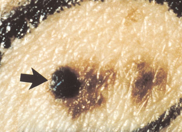
National Cancer Institute Services
  This is only one of many free booklets for
people with cancer.
  You may want more information for yourself,
your family, and your doctor.
   NCI offers comprehensive research-based
information for patients and their families, health
professionals, cancer researchers, advocates, and
the public.
  • Call NCI’s Cancer Information Service at
    1–800–4–CANCER (1–800–422–6237)
  • Visit us at http://www.cancer.gov or
    http://www.cancer.gov/espanol
  • Chat using LiveHelp, NCI’s instant
    messaging service, at http://www.cancer.gov/
    livehelp
  • E-mail us at cancergovstaff@mail.nih.gov
  • Order publications at http://www.cancer.gov/
    publications or by calling 1–800–4–CANCER
  • Get help with quitting smoking at
    1–877–44U–QUIT (1–877–448–7848)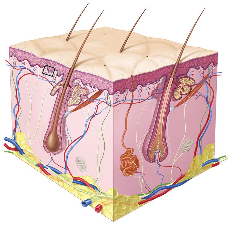
Contents

  About This Booklet           1
  The Skin     3
  Cancer Cells        5
  Types of Skin Cancer             6
  Risk Factors      7
  Symptoms         12
  Diagnosis      16
  Staging     17
  Treatment      21
  Second Opinion          32
  Taking Part in Cancer Research            34
  Follow-up Care          35
  Prevention       36
  How To Check Your Skin               37
  Sources of Support           38
  Dictionary       40
  National Cancer Institute Publications         53

 U.S. DEPARTMENT OF
 HEALTH AND HUMAN SERVICES
 National Institutes of Health
 National Cancer Institute
About This Booklet

  This National Cancer Institute (NCI) booklet is for
people diagnosed with the most common types of skin
cancer:*
• Melanoma
• Basal cell skin cancer
• Squamous cell skin cancer
   Skin cancer is the most common type of cancer in
the United States. Each year, more than 68,000
Americans are diagnosed with melanoma, and another
48,000 are diagnosed with an early form of the disease
that involves only the top layer of skin. Also, more
than 2 million people are treated for basal cell or
squamous cell skin cancer each year. Basal cell skin
cancer is several times more common than squamous
cell skin cancer.
   Learning about medical care for skin cancer can
help you take an active part in making choices about
your care. This booklet tells about:
• Diagnosis and staging
• Treatment
• Follow-up care
• How to prevent another skin cancer from forming
• How to do a skin self-exam
   This booklet has lists of questions that you may
want to ask your doctor. Many people find it helpful to
take a list of questions to a doctor visit. To help
remember what your doctor says, you can take notes.

*Words in italics are in the Dictionary on page 40. The Dictionary
explains these terms. It also shows how to pronounce them.

                                1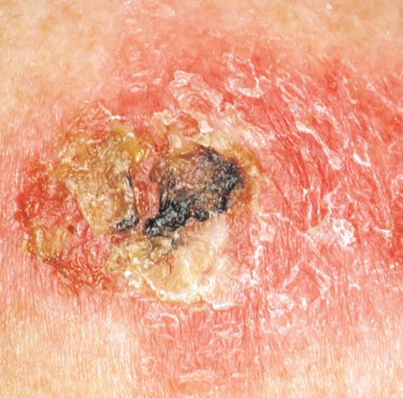
You may also want to have a family member or friend
go with you when you talk with the doctor—to take
notes, ask questions, or just listen.
   For the latest information about skin cancer, please
visit our Web site at http://www.cancer.gov/
cancertopics/types/skin. For information about
melanoma, go to http://www.cancer.gov/
cancertopics/types/melanoma.
   Also, NCI’s Cancer Information Service can answer
your questions about skin cancer. We can also send you
NCI booklets and fact sheets. Call 1–800–4–CANCER
(1–800–422–6237) or chat with us online using
LiveHelp, NCI’s instant messaging service at
http://www.cancer.gov/livehelp.
   This booklet does not describe rare types of skin
cancer, such as Merkel cell carcinoma. Also, this
booklet does not discuss melanoma that begins in the
eye, the digestive tract, or other areas of the body.
NCI’s Cancer Information Service can provide
information about rare skin cancers and melanoma that
begins in areas other than the skin.

                           2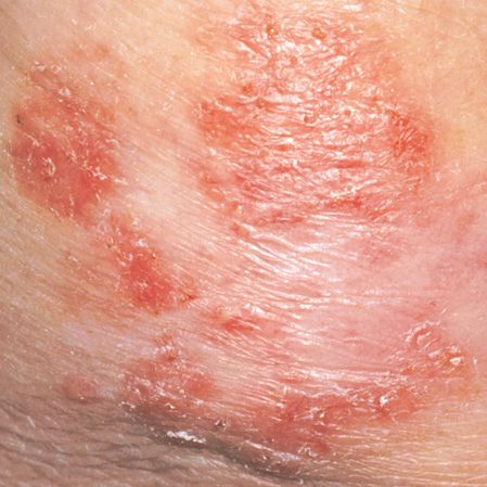
The Skin
Your skin protects your body from heat, injury, and
infection. It also protects your body from damage
caused by ultraviolet (UV) radiation (such as from the
sun or sunlamps).
Your skin stores water and fat. It helps control body
heat. Also, your skin makes vitamin D.
The picture on the next page shows the two main
layers of the skin:
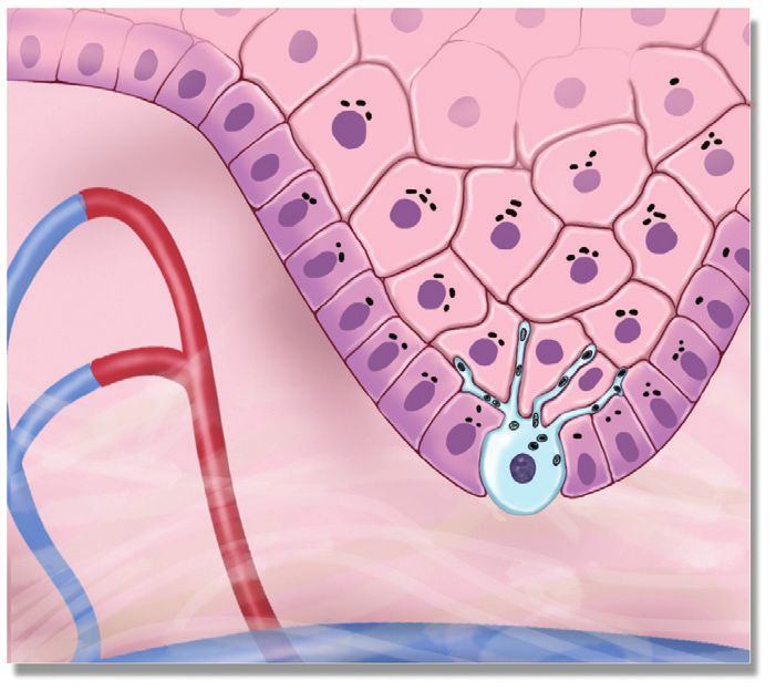
• Epidermis: The epidermis is the top layer of your
skin. It’s mostly made of flat cells called squamous
cells.
Below the squamous cells deeper in the epidermis
are round cells called basal cells.
Cells called melanocytes are scattered among the
basal cells. They are in the deepest part of the
epidermis. Melanocytes make the pigment (color)
found in skin. When skin is exposed to UV
radiation, melanocytes make more pigment, causing
the skin to darken, or tan.
• Dermis: The dermis is the layer under the epidermis.
The dermis contains many types of cells and
structures, such as blood vessels, lymph vessels, and
glands. Some of these glands make sweat, which
helps cool your body. Other glands make sebum.
Sebum is an oily substance that helps keep your skin
from drying out. Sweat and sebum reach the surface
of your skin through tiny openings called pores.
3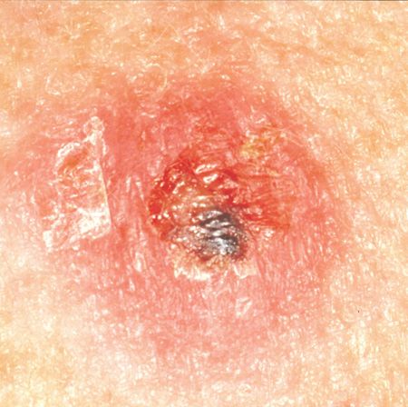
Epidermis          Squamous
  Dermis             cells

                                       Basal cell

    Blood vessels                      Melanocyte

                                       Hair shaft
                                       Oil gland
                                         Epidermis

                                         Dermis
                                                       U.S. Govt. has certain rights
                                                       © 2010 Terese Winslow

                                    Lymph vessel
                              Sweat gland
                         Fatty tissue
This picture shows the layers of the skin—the
epidermis and dermis. At the top, the close-up shows
a squamous cell, basal cell, and melanocyte.

                          4
Cancer Cells
Cancer begins in cells, the building blocks that make
up tissues. Tissues make up the skin and other organs
of the body.
Normal cells grow and divide to form new cells as
the body needs them. When normal cells grow old or
get damaged, they usually die, and new cells take their
place.
But sometimes this process goes wrong. New cells
form when the body doesn’t need them, and old or
damaged cells don’t die as they should. The buildup of
extra cells often forms a mass of tissue called a growth
or tumor.
Growths on the skin can be benign (not cancer) or
malignant (cancer). Benign growths are not as harmful
as malignant growths.
• Benign growths (such as moles):
—Are rarely a threat to life
—Generally can be removed and usually don’t grow
back
—Don’t invade the tissues around them
—Don’t spread to other parts of the body
• Malignant growths (such as melanoma, basal cell
cancer, or squamous cell cancer):
—May be a threat to life
—Often can be removed but sometimes grow back
—May invade and damage nearby organs and
tissues
—May spread to other parts of the body
5Types of Skin Cancer
Skin cancers are named for the type of cells that
become malignant (cancer). The three most common
types are:
• Melanoma: Melanoma begins in melanocytes
(pigment cells). Most melanocytes are in the skin.
See the picture on page 4 of a melanocyte and other
skin cells.
Melanoma can occur on any skin surface. In men,
it’s often found on the skin on the head, on the neck,
or between the shoulders and the hips. In women,
it’s often found on the skin on the lower legs or
between the shoulders and the hips.
Melanoma is rare in people with dark skin. When it
does develop in people with dark skin, it’s usually
found under the fingernails, under the toenails, on
the palms of the hands, or on the soles of the feet.
• Basal cell skin cancer: Basal cell skin cancer
begins in the basal cell layer of the skin. It usually
occurs in places that have been in the sun. For
example, the face is the most common place to find
basal cell skin cancer.
In people with fair skin, basal cell skin cancer is the
most common type of skin cancer.
• Squamous cell skin cancer: Squamous cell skin
cancer begins in squamous cells. In people with
dark skin, squamous cell skin cancer is the most
common type of skin cancer, and it’s usually found
in places that are not in the sun, such as the legs or
feet.
However, in people with fair skin, squamous cell
skin cancer usually occurs on parts of the skin that
have been in the sun, such as the head, face, ears,
and neck.
6Unlike moles, skin cancer can invade the normal
tissue nearby. Also, skin cancer can spread throughout
the body. Melanoma is more likely than other skin
cancers to spread to other parts of the body. Squamous
cell skin cancer sometimes spreads to other parts of the
body, but basal cell skin cancer rarely does.
When skin cancer cells do spread, they break away
from the original growth and enter blood vessels or
lymph vessels. The cancer cells may be found in
nearby lymph nodes. The cancer cells can also spread
to other tissues and attach there to form new tumors
that may damage those tissues.
The spread of cancer is called metastasis. See the
Staging section on page 17 for information about skin
cancer that has spread.
Risk Factors
When you’re told that you have skin cancer, it’s
natural to wonder what may have caused the disease.
The main risk factor for skin cancer is exposure to
sunlight (UV radiation), but there are also other risk
factors. A risk factor is something that may increase the
chance of getting a disease.
People with certain risk factors are more likely than
others to develop skin cancer. Some risk factors vary
for the different types of skin cancer.
7Risks for Any Type of Skin Cancer
Studies have shown that the following are risk
factors for the three most common types of skin
cancer:
• Sunlight: Sunlight is a source of UV radiation. It’s
the most important risk factor for any type of skin
cancer. The sun’s rays cause skin damage that can
lead to cancer.
—Severe, blistering sunburns: People who have
had at least one severe, blistering sunburn are at
increased risk of skin cancer. Although people
who burn easily are more likely to have had
sunburns as a child, sunburns during adulthood
also increase the risk of skin cancer.
—Lifetime sun exposure: The total amount of sun
exposure over a lifetime is a risk factor for skin
cancer.
—Tanning: Although a tan slightly lowers the risk
of sunburn, even people who tan well without
sunburning have a higher risk of skin cancer
because of more lifetime sun exposure.
Sunlight can be reflected by sand, water, snow, ice,
and pavement. The sun’s rays can get through
clouds, windshields, windows, and light clothing.
In the United States, skin cancer is more common
where the sun is strong. For example, more people
in Texas than Minnesota get skin cancer. Also, the
sun is stronger at higher elevations, such as in the
mountains.
Doctors encourage people to limit their exposure to
sunlight. See the Prevention section on page 36 for
ways to protect your skin from the sun.
8• Sunlamps and tanning booths: Artificial sources
of UV radiation, such as sunlamps and tanning
booths, can cause skin damage and skin cancer.
Health care providers strongly encourage people,
especially young people, to avoid using sunlamps
and tanning booths. The risk of skin cancer is
greatly increased by using sunlamps and tanning
booths before age 30.
• Personal history: People who have had melanoma
have an increased risk of developing other
melanomas. Also, people who have had basal cell or
squamous cell skin cancer have an increased risk of
developing another skin cancer of any type.
• Family history: Melanoma sometimes runs in
families. Having two or more close relatives
(mother, father, sister, brother, or child) who have
had this disease is a risk factor for developing
melanoma. Other types of skin cancer also
sometimes run in families. Rarely, members of a
family will have an inherited disorder, such as
xeroderma pigmentosum or nevoid basal cell
carcinoma syndrome, that makes the skin more
sensitive to the sun and increases the risk of skin
cancer.
• Skin that burns easily: Having fair (pale) skin that
burns in the sun easily, blue or gray eyes, red or
blond hair, or many freckles increases the risk of
skin cancer.
• Certain medical conditions or medicines: Medical
conditions or medicines (such as some antibiotics,
hormones, or antidepressants) that make your skin
more sensitive to the sun increase the risk of skin
cancer. Also, medical conditions or medicines that
suppress the immune system increase the risk of skin
cancer.
9Other Risk Factors for Melanoma
The following risk factors increase the risk of
melanoma:
• Dysplastic nevus: A dysplastic nevus is a type of
mole that looks different from a common mole. A
dysplastic nevus may be bigger than a common
mole, and its color, surface, and border may be
different. It’s usually wider than a pea and may be
longer than a peanut. A dysplastic nevus can have a
mixture of several colors, from pink to dark brown.
Usually, it’s flat with a smooth, slightly scaly or
pebbly surface, and it has an irregular edge that may
fade into the surrounding skin.
A dysplastic nevus is more likely than a common
mole to turn into cancer. However, most do not
change into melanoma. A doctor will remove a
dysplastic nevus if it looks like it might have
changed into melanoma.
• More than 50 common moles: Usually, a common
mole is smaller than a pea, has an even color (pink,
tan, or brown), and is round or oval with a smooth
surface. Having many common moles increases the
risk of developing melanoma.
10Other Risk Factors for Both Basal Cell and
Squamous Cell Skin Cancers
The following risk factors increase the risk of basal
cell and squamous cell skin cancers:
• Old scars, burns, ulcers, or areas of inflammation
on the skin
• Exposure to arsenic at work
• Radiation therapy
Other Risk Factors for Squamous Cell Cancer
The risk of squamous cell skin cancer is increased
by the following:
• Actinic keratosis: Actinic keratosis is a type of flat,
scaly growth on the skin. It is most often found on
areas exposed to the sun, especially the face and the
backs of the hands. The growth may appear as a
rough red or brown patch on the skin. It may also
appear as cracking or peeling of the lower lip that
does not heal. Without treatment, this scaly growth
may turn into squamous cell skin cancer.
• HPV (human papillomavirus): Certain types of
HPV can infect the skin and may increase the risk of
squamous cell skin cancer. These HPVs are different
from the HPV types that cause cervical cancer and
other cancers in the female and male genital areas.
11Symptoms
Symptoms of Melanoma
Often the first sign of melanoma is a change in the
shape, color, size, or feel of an existing mole.
Melanoma may also appear as a new mole. Thinking
of “ABCDE” can help you remember what to look for:
• Asymmetry: The shape of one half does not match
the other half.
• Border that is irregular: The edges are often
ragged, notched, or blurred in outline. The pigment
may spread into the surrounding skin.
• Color that is uneven: Shades of black, brown, and
tan may be present. Areas of white, gray, red, pink,
or blue may also be seen.
• Diameter: There is a change in size, usually an
increase. Melanomas can be tiny, but most are larger
than the size of a pea (larger than 6 millimeters or
about 1/4 inch).
• Evolving: The mole has changed over the past few
weeks or months.
Melanomas can vary greatly in how they look.
Many show all of the ABCDE features. However,
some may show changes or abnormal areas in only one
or two of the ABCDE features.
In more advanced melanoma, the texture of the
mole may change. The skin on the surface may break
down and look scraped. It may become hard or lumpy.
The surface may ooze or bleed. Sometimes the
melanoma is itchy, tender, or painful.
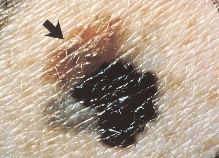
12This photo shows an asymmetric
melanoma with irregular and scalloped
borders. The color varies from gray to
brown to black. The melanoma is about
1.2 centimeters.
This photo shows a dysplastic nevus
with an arrow pointing to a new black
bump that was not there 18 months
earlier. The black bump is a melanoma
that is about 3 millimeters.
13Symptoms of Basal Cell and Squamous Cell
Skin Cancers
A change on the skin is the most common sign of
skin cancer. This may be a new growth, a sore that
doesn’t heal, or a change in an old growth. Not all skin
cancers look the same. Usually, skin cancer is not
painful.
Common symptoms of basal cell or squamous cell
skin cancer include:
• A lump that is small, smooth, shiny, pale, or waxy
• A lump that is firm and red
14• A sore or lump that bleeds or develops a crust or a
scab
• A flat red spot that is rough, dry, or scaly and may
become itchy or tender
• A red or brown patch that is rough and scaly
15Diagnosis
If you have a change on your skin, your doctor must
find out whether or not the problem is from cancer.
You may need to see a dermatologist, a doctor who has
special training in the diagnosis and treatment of skin
problems.
Your doctor will check the skin all over your body
to see if other unusual growths are present.
If your doctor suspects that a spot on the skin is
cancer, you may need a biopsy. For a biopsy, your
doctor may remove all or part of the skin that does not
look normal. The sample goes to a lab. A pathologist
checks the sample under a microscope. Sometimes it’s
helpful for more than one pathologist to check the
tissue for cancer cells.
You may have the biopsy in a doctor’s office or as
an outpatient in a clinic or hospital. You’ll probably
have local anesthesia.
There are four common types of skin biopsies:
• Shave biopsy: The doctor uses a thin, sharp blade to
shave off the abnormal growth.
• Punch biopsy: The doctor uses a sharp, hollow tool
to remove a circle of tissue from the abnormal area.
• Incisional biopsy: The doctor uses a scalpel to
remove part of the growth.
• Excisional biopsy: The doctor uses a scalpel to
remove the entire growth and some tissue around it.
This type of biopsy is most commonly used for
growths that appear to be melanoma.
16You may want to ask your doctor these
questions before having a biopsy:
• Which type of biopsy do you suggest for me?
• How will the biopsy be done?
• Will I have to go to the hospital?
• How long will it take? Will I be awake? Will it
hurt?
• Will the entire growth be removed?
• Are there any risks? What are the chances of
infection or bleeding after the biopsy?
• Will there be a scar? If so, what will it look
like?
• How soon will I know the results?
• If I do have cancer, who will talk with me
about treatment?
Staging
If the biopsy shows that you have skin cancer, your
doctor needs to learn the stage (extent) of the disease to
help you choose the best treatment.
The stage is based on:
• The size (width) of the growth
• How deeply it has grown beneath the top layer of
skin
• Whether cancer cells have spread to nearby lymph
nodes or to other parts of the body
17When skin cancer spreads from its original place to
another part of the body, the new tumor has the same
kind of abnormal cells and the same name as the
primary (original) tumor. For example, if skin cancer
spreads to the lung, the cancer cells in the lung are
actually skin cancer cells. The disease is metastatic
skin cancer, not lung cancer. For that reason, it’s
treated as skin cancer, not as lung cancer. Doctors
sometimes call the new tumor “distant” disease.
Blood tests and an imaging test such as a chest
x-ray, a CT scan, an MRI, or a PET scan may be used
to check for the spread of skin cancer. For example, if
a melanoma growth is thick, your doctor may order
blood tests and an imaging test.
For squamous cell skin cancer or melanoma, the
doctor will also check the lymph nodes near the cancer
on the skin. If one or more lymph nodes near the skin
cancer are enlarged (or if the lymph node looks
enlarged on an imaging test), your doctor may use a
thin needle to remove a sample of cells from the lymph
node (fine-needle aspiration biopsy). A pathologist will
check the sample for cancer cells.
Even if the nearby lymph nodes are not enlarged,
the nodes may contain cancer cells. The stage is
sometimes not known until after surgery to remove the
growth and one or more nearby lymph nodes. For thick
melanoma, surgeons may use a method called sentinel
lymph node biopsy to remove the lymph node most
likely to have cancer cells. Cancer cells may appear
first in the sentinel node before spreading to other
lymph nodes and other places in the body.
18Stages of Melanoma
These are the stages of melanoma:
• Stage 0: The melanoma involves only the top layer
of skin. It is called melanoma in situ.
• Stage I: The tumor is no more than 1 millimeter
thick (about the width of the tip of a sharpened
pencil.) The surface may appear broken down. Or,
the tumor is between 1 and 2 millimeters thick, and
the surface is not broken down.
• Stage II: The tumor is between 1 and 2 millimeters
thick, and the surface appears broken down. Or, the
thickness of the tumor is more than 2 millimeters,
and the surface may appear broken down.
• Stage III: The melanoma cells have spread to at
least one nearby lymph node. Or, the melanoma
cells have spread from the original tumor to tissues
nearby.
• Stage IV: Cancer cells have spread to the lung or
other organs, skin areas, or lymph nodes far away
from the original growth. Melanoma commonly
spreads to other parts of the skin, tissue under the
skin, lymph nodes, and lungs. It can also spread to
the liver, brain, bones, and other organs.
19Stages of Other Skin Cancers
These are the stages of basal cell and squamous cell
skin cancers:
• Stage 0: The cancer involves only the top layer of
skin. It is called carcinoma in situ.
Bowen disease is an early form of squamous cell
skin cancer. It usually looks like a reddish, scaly or
thickened patch on the skin. If not treated, the
cancer may grow deeper into the skin.
• Stage I: The growth is as large as 2 centimeters
wide (more than three-quarters of an inch or about
the size of a peanut).
• Stage II: The growth is larger than 2 centimeters
wide.
• Stage III: The cancer has invaded below the skin to
cartilage, muscle, or bone. Or, cancer cells have
spread to nearby lymph nodes. Cancer cells have not
spread to other places in the body.
• Stage IV: The cancer has spread to other places in
the body. Basal cell cancer rarely spreads to other
parts of the body, but squamous cell cancer
sometimes spreads to lymph nodes and other organs.
20Treatment
Treatment for skin cancer depends on the type and
stage of the disease, the size and place of the tumor,
and your general health and medical history. In most
cases, the goal of treatment is to remove or destroy the
cancer completely. Most skin cancers can be cured if
found and treated early.
Sometimes all of the skin cancer is removed during
the biopsy. In such cases, no more treatment is needed.
If you do need more treatment, your doctor can
describe your treatment choices and what to expect.
You and your doctor can work together to develop a
treatment plan that meets your needs.
21Surgery is the usual treatment for people with skin
cancer. In some cases, the doctor may suggest
chemotherapy, photodynamic therapy, or radiation
therapy. People with melanoma may also have
biological therapy.
You may have a team of specialists to help plan
your treatment. Your doctor may refer you to a
specialist, or you may ask for a referral. Specialists
who treat skin cancer include dermatologists and
surgeons. Some people may also need a reconstructive
or plastic surgeon.
People with advanced skin cancer may be referred
to a medical oncologist or radiation oncologist. Your
health care team may also include an oncology nurse, a
social worker, and a registered dietitian.
Because skin cancer treatment may damage healthy
cells and tissues, unwanted side effects sometimes
occur. Side effects depend mainly on the type and
extent of the treatment. Side effects may not be the
same for each person. Before treatment starts, your
health care team will tell you about possible side
effects and suggest ways to help you manage them.
Many skin cancers can be removed quickly and
easily. But some people may need supportive care to
control pain and other symptoms, to relieve the side
effects of treatment, and to help them cope with the
feelings that a diagnosis of cancer can bring.
Information about such care is available on NCI’s Web
site at http://www.cancer.gov/cancertopics/coping
and from NCI’s Cancer Information Service at
1–800–4–CANCER (1–800–422–6237) or at
LiveHelp.
You may want to talk with your doctor about taking
part in a clinical trial, a research study of new
treatment methods. See the Taking Part in Cancer
Research section on page 34.
22You may want to ask your doctor these
questions before you begin treatment:
• What is the stage of the disease? Has the
cancer spread? Do any lymph nodes or other
organs show signs of cancer?
• What are my treatment choices? Which do you
suggest for me? Why?
• What are the expected benefits of each kind of
treatment?
• What can I do to prepare for treatment?
• Will I need to stay in the hospital? If so, for
how long?
• What are the risks and possible side effects of
each treatment? How can side effects be
managed?
• Will there be a scar? Will I need a skin graft or
plastic surgery?
• What is the treatment likely to cost? Will my
insurance cover it?
• How will treatment affect my normal
activities?
• Would a research study (clinical trial) be a
good choice for me?
• How often should I have checkups?
23Surgery
In general, the surgeon will remove the cancerous
growth and some normal tissue around it. This reduces
the chance that cancer cells will be left in the area.
There are several methods of surgery for skin
cancer. The method your doctor uses depends mainly
on the type of skin cancer, the size of the cancer, and
where it was found on your body.
Your doctor can further describe these methods of
surgery:
• Excisional skin surgery: This is a common
treatment to remove any type of skin cancer. After
numbing the area of skin, the surgeon removes the
growth (tumor) with a scalpel. The surgeon also
removes a border (a margin) of normal skin around
the growth. The margin of skin is examined under a
microscope to be certain that all the cancer cells
have been removed. The thickness of the margin
depends on the size of the tumor.
• Mohs surgery (also called Mohs micrographic
surgery): This method is often used for basal cell
and squamous cell skin cancers. After numbing the
area of skin, a specially trained surgeon shaves away
thin layers of the tumor. Each layer is examined
under a microscope. The surgeon continues to shave
away tissue until no cancer cells can be seen under
the microscope. In this way, the surgeon can remove
all the cancer and only a small bit of healthy tissue.
Some people will have radiation therapy after Mohs
surgery to make sure all of the cancer cells are
destroyed.
24• Electrodesiccation and curettage: This method is
often used to remove a small basal cell or squamous
cell skin cancer. After the doctor numbs the area to
be treated, the cancer is removed with a sharp tool
shaped like a spoon (called a curette). The doctor
then uses a needle-shaped electrode to send an
electric current into the treated area to control
bleeding and kill any cancer cells that may be left.
This method is usually fast and simple. It may be
performed up to three times to remove all of the
cancer.
• Cryosurgery: This method is an option for an early-
stage or a very thin basal cell or squamous cell skin
cancer. Cryosurgery is often used for people who are
not able to have other types of surgery. The doctor
applies liquid nitrogen (which is extremely cold)
directly to the skin growth to freeze and kill the
cancer cells. This treatment may cause swelling. It
also may damage nerves, which can cause a loss of
feeling in the damaged area. The NCI fact sheet
Cryosurgery in Cancer Treatment has more
information.
For people with cancer that has spread to the lymph
nodes, the surgeon may remove some or all of the
nearby lymph nodes. Additional treatment may be
needed after surgery. See the Staging section on page
17 for information about finding cancer in lymph
nodes.
If a large area of tissue is removed, the surgeon may
do a skin graft. The doctor uses skin from another part
of the body to replace the skin that was removed. After
numbing the area, the surgeon removes a patch of
healthy skin from another part of the body, such as the
upper thigh. The patch is then used to cover the area
where skin cancer was removed. If you have a skin
graft, you may have to take special care of the area
until it heals.
25The time it takes to heal after surgery is different for
each person. You may have pain for the first few days.
Medicine can help control your pain. Before surgery,
you should discuss the plan for pain relief with your
doctor or nurse. After surgery, your doctor can adjust
the plan if you need more pain relief.
Surgery nearly always leaves some type of scar. The
size and color of the scar depend on the size of the
cancer, the type of surgery, the color of your skin, and
how your skin heals.
For any type of surgery, including skin grafts or
reconstructive surgery, follow your doctor’s advice on
bathing, shaving, exercise, or other activities.
You may want to ask your doctor these
questions before having surgery:
• What kind of surgery do you recommend for
me? Why?
• Will you remove lymph nodes? Why?
• Will I need a skin graft?
• What will the scar look like? Can anything be
done to help reduce the scar? Will I need
plastic surgery or reconstructive surgery?
• How will I feel after surgery?
• If I have pain, how will you control it?
• Will I need to stay in the hospital? If so, for
how long?
• Am I likely to have infection, swelling,
blistering, or bleeding, or to get a scab where
the cancer was removed?
• Will I have any long-term side effects?
26Chemotherapy
Chemotherapy uses drugs to kill cancer cells. Drugs
for skin cancer can be given in many ways.
Put directly on the skin
A cream or lotion form of chemotherapy may be
used to treat very thin, early-stage basal cell or
squamous cell skin cancer (Bowen disease). It may
also be used if there are several small skin cancers. The
doctor will show you how to apply the cream or lotion
to the skin one or two times a day for several weeks.
The cream or lotion contains a drug that kills cancer
cells only in the top layer of the skin:
• Fluorouracil (another name is 5-FU): This drug is
used to treat early-stage basal cell and squamous
cell cancers.
• Imiquimod: This drug is used to treat early-stage
basal cell cancer.
These drugs may cause your skin to turn red or
swell. Your skin also may itch, ooze, or develop a rash.
Your skin may be sore or sensitive to the sun after
treatment. These skin changes usually go away after
treatment is over.
A cream or lotion form of chemotherapy usually
does not leave a scar. If healthy skin becomes too red
or raw when the skin cancer is treated, your doctor
may stop treatment.
Swallowed or injected
People with melanoma may receive chemotherapy
by mouth or through a vein (intravenous). You may
receive one or more drugs. The drugs enter the
bloodstream and travel throughout the body.
27If you have melanoma on an arm or leg, you may
receive drugs directly into the bloodstream of that
limb. The flow of blood to and from the limb is
stopped for a while. This allows a high dose of drugs
in the area with the melanoma. Most of the
chemotherapy remains in that limb.
You may receive chemotherapy in an outpatient part
of the hospital, at the doctor’s office, or at home. Some
people need to stay in the hospital during treatment.
The side effects depend mainly on which drugs are
given and how much. Chemotherapy kills fast-growing
cancer cells, but the drugs can also harm normal cells
that divide rapidly:
• Blood cells: When drugs lower the levels of healthy
blood cells, you’re more likely to get infections,
bruise or bleed easily, and feel very weak and tired.
Your health care team will check for low levels of
blood cells. If your levels are low, your health care
team may stop the chemotherapy for a while or
reduce the dose of the drug. There are also
medicines that can help your body make new blood
cells.
• Cells in hair roots: Chemotherapy may cause hair
loss. If you lose your hair, it will grow back after
treatment, but the color and texture may be changed.
• Cells that line the digestive tract: Chemotherapy
can cause a poor appetite, nausea and vomiting,
diarrhea, or mouth and lip sores. Your health care
team can give you medicines and suggest other
ways to help with these problems. They usually go
away when treatment ends.
You may want to read the NCI booklet
Chemotherapy and You.
28You may want to ask your doctor these
questions about chemotherapy:
• Why do I need this treatment?
• Which drug or drugs will I have?
• How do the drugs work?
• Do I need to take special care when I put
chemotherapy on my skin? What do I need to
do? Will I be sensitive to the sun?
• When will treatment start? When will it end?
• Will I have any long-term side effects?
Photodynamic Therapy
Photodynamic therapy (PDT) uses a drug along with
a special light source, such as a laser light, to kill
cancer cells. PDT may be used to treat very thin, early-
stage basal cell or squamous cell skin cancer (Bowen
disease).
The drug is either rubbed into the skin or injected
intravenously. The drug is absorbed by cancer cells. It
stays in cancer cells longer than in normal cells.
Several hours or days later, a special light is focused on
the cancer. The drug becomes active and destroys the
cancer cells.
The side effects of PDT are usually not serious.
PDT may cause burning or stinging pain. It also may
cause burns, swelling, or redness. It may scar healthy
tissue near the growth. If you have PDT, you will need
to avoid direct sunlight and bright indoor light for at
least 6 weeks after treatment.
The NCI fact sheet Photodynamic Therapy for
Cancer has more information.
29You may want to ask your doctor these
questions about PDT:
• Will I need to stay in the hospital while the
drug is in my body?
• Will I need to have the treatment more than
once?
Biological Therapy
Some people with advanced melanoma receive a
drug called biological therapy. Biological therapy for
melanoma is treatment that may improve the body’s
natural defense (immune system response) against
cancer.
One drug for melanoma is interferon. It’s injected
intravenously (usually at a hospital or clinic) or
injected under the skin (at home or in a doctor’s
office). Interferon can slow the growth of melanoma
cells.
Another drug used for melanoma is interleukin-2.
It’s given intravenously. It can help the body destroy
cancer cells. Interleukin-2 is usually given at the
hospital.
Other drugs may be given at the same time to
prevent side effects. The side effects differ with the
drug used, and from person to person. Biological
therapies commonly cause a rash or swelling. You may
feel very tired during treatment. These drugs may also
cause a headache, muscle aches, a fever, or weakness.
You may find it helpful to read the NCI booklet
Biological Therapy. You may also wish to read the
NCI fact sheet Biological Therapies for Cancer.
30You may want to ask your doctor these
questions about biological therapy:
• What is the goal of treatment?
• When will treatment start? When will it end?
• Will I need to stay in the hospital for
treatment? If so, how long will I be in the
hospital?
Radiation Therapy
Radiation therapy uses high-energy rays to kill
cancer cells. The radiation comes from a large
machine outside the body. It affects cells only in the
treated area. You will go to a hospital or clinic several
times for this treatment.
Radiation therapy is not a common treatment for
skin cancer. But it may be used for skin cancer in areas
where surgery could be difficult or leave a bad scar.
For example, you may have radiation therapy if you
have a growth on your eyelid, ear, or nose. Radiation
therapy may also be used after surgery for squamous
cell carcinoma that can’t be completely removed or
that has spread to the lymph nodes. And it may be used
for melanoma that has spread to the lymph nodes,
brain, bones, or other parts of the body.
Although radiation therapy is painless, it may cause
other side effects. The side effects depend mainly on
the dose of radiation and the part of your body that is
treated. It’s common for the skin in the treated area to
become red, dry, tender, and itchy. Your health care
team can suggest ways to relieve the side effects of
radiation therapy.
You may find it helpful to read the NCI booklet
Radiation Therapy and You.
31You may want to ask your doctor these
questions about radiation therapy:
• How will I feel after treatment?
• Am I likely to have infection, swelling,
blistering, or bleeding after radiation therapy?
• Will I get a scar on the treated area?
• How should I take care of the treated area?
Second Opinion
Before starting treatment, you might want a second
opinion from another doctor about your diagnosis and
treatment plan. Some people worry that their doctor
will be offended if they ask for a second opinion.
Usually the opposite is true. Most doctors welcome a
second opinion. And many health insurance companies
will pay for a second opinion if you or your doctor
requests it. Some companies require a second opinion.
If you get a second opinion, the doctor may agree
with your first doctor’s diagnosis and treatment plan.
Or the second doctor may suggest another approach.
Either way, you’ll have more information and perhaps a
greater sense of control. You may also feel more
confident about the decisions you make, knowing that
you’ve looked carefully at all of your options.
It may take some time and effort to gather your
medical records and see another doctor. Usually it’s not
a problem if it takes you several weeks to get a second
opinion. In most cases, the delay in starting treatment
will not make treatment less effective. To make sure,
32you should discuss this possible delay with your
doctor. Some people with skin cancer need treatment
right away.
There are many ways to find a doctor for a second
opinion. You can ask your doctor, a local or state
medical society, or a nearby hospital or a medical
school for names of specialists.
Also, you can get information about treatment
centers near you from NCI’s Cancer Information
Service at 1–800–4–CANCER (1–800–422–6237)
or at LiveHelp (http://www.cancer.gov/livehelp).
Other sources can be found in the NCI fact sheet
How To Find a Doctor or Treatment Facility If You
Have Cancer.
33Taking Part in Cancer Research
Doctors all over the country are conducting many
types of clinical trials (research studies in which people
volunteer to take part). Clinical trials are designed to
find out whether new treatments are safe and effective.
Doctors are trying to find better ways to care for
people with skin cancer. They are studying many types
of treatment, such as surgery, chemotherapy, biological
therapy, and combinations of treatment. For example,
doctors are studying the use of a cancer treatment
vaccine after surgery for people with advanced
melanoma. For more information about cancer
vaccines, you may want to read the NCI fact sheet
Cancer Vaccines.
Even if the people in a trial do not benefit directly,
they may still make an important contribution by
helping doctors learn more about skin cancer and how
to control it. Although clinical trials may pose some
risks, doctors do all they can to protect their patients.
If you’re interested in being part of a clinical trial,
talk with your doctor. You may want to read the NCI
booklet Taking Part in Cancer Treatment Research
Studies. It describes how treatment studies are carried
out and explains their possible benefits and risks.
NCI’s Web site includes a section on clinical trials at
http://www.cancer.gov/clinicaltrials. It has general
information about clinical trials as well as detailed
information about specific ongoing studies of skin
cancer. Information specialists at 1–800–4–CANCER
(1–800–422–6237) or at LiveHelp at http://www.
cancer.gov/livehelp can answer questions and provide
information about clinical trials.
34Follow-up Care
After treatment for skin cancer, you’ll need regular
checkups (such as every 3 to 6 months for the first year
or two). Your doctor will monitor your recovery and
check for any new skin cancers. Regular checkups help
ensure that any changes in your health are noted and
treated if needed.
During a checkup, you’ll have a physical exam.
People with melanoma may have x-rays, blood tests,
and scans of the chest, liver, bones, and brain.
People who have had melanoma have an increased
risk of developing a new melanoma, and people with
basal or squamous cell skin cancers have a risk of
developing another skin cancer of any type. It’s a good
idea to get in a routine for checking your skin for new
growths or other changes. Keep in mind that changes
are not a sure sign of skin cancer. Still, you should tell
your doctor about any changes right away. You’ll find
a guide for checking your skin on page 37.
Follow your doctor’s advice about how to reduce
your risk of developing skin cancer again.
You may find it helpful to read the NCI booklet
Facing Forward: Life After Cancer Treatment. You
may also want to read the NCI fact sheet Follow-up
Care After Cancer Treatment.
35Prevention
People with skin cancer are at risk of developing
another skin cancer. Limit your time in the sun and
stay away from sunlamps and tanning booths. Keep in
mind that getting a tan may increase your risk of
developing another skin cancer.
The best way to prevent skin cancer is to protect
yourself from the sun:
• Avoid outdoor activities during the middle of the
day. The sun’s rays are the strongest between 10
a.m. and 4 p.m. When you must be outdoors, seek
shade when you can.
• Protect yourself from the sun’s rays reflected by
sand, water, snow, ice, and pavement. The sun’s rays
can go through light clothing, windshields,
windows, and clouds.
• Wear long sleeves and long pants. Tightly woven
fabrics are best.
• Wear a hat with a wide brim all around that shades
your face, neck, and ears. Keep in mind that
baseball caps and some sun visors protect only parts
of your skin.
• Wear sunglasses that absorb UV radiation to protect
the skin around your eyes.
• Use sunscreen lotions with a sun protection factor
(SPF) of at least 15. (Some doctors will suggest
using a lotion with an SPF of at least 30.) Apply the
product’s recommended amount to uncovered skin
30 minutes before going outside, and apply again
every two hours or after swimming or sweating.
36Sunscreen lotions may help prevent some skin
cancers. It’s important to use a broad-spectrum
sunscreen lotion that filters both UVB and UVA
radiation. But you still need to avoid the sun during
the middle of the day and wear clothing to protect your
skin.
How To Check Your Skin
Your doctor or nurse may suggest that you do a
regular skin self-exam to check for the development of
a new skin cancer.
The best time to do this exam is after a shower or
bath. Check your skin in a room with plenty of light.
Use a full-length mirror and a hand-held mirror.
It’s best to begin by learning where your birthmarks,
moles, and other marks are and their usual look and
feel.
Check for anything new:
• A new mole (that looks different from your other
moles)
• A new red or darker color flaky patch that may be a
little raised
• A new flesh-colored firm bump
• A change in the size, shape, color, or feel of a mole
• A sore that doesn’t heal
Check yourself from head to toe:
• Look at your face, neck, ears, and scalp. You may
want to use a comb or a blow dryer to move your
hair so that you can see better. You also may want to
have a relative or friend check through your hair. It
may be hard to check your scalp by yourself.
37• Look at the front and back of your body in the
mirror. Then, raise your arms and look at your left
and right sides.
• Bend your elbows. Look carefully at your
fingernails, palms, forearms (including the
undersides), and upper arms.
• Examine the back, front, and sides of your legs.
Also look around your genital area and between
your buttocks.
• Sit and closely examine your feet, including your
toenails, your soles, and the spaces between your
toes.
By checking your skin regularly, you’ll learn what is
normal for you. It may be helpful to record the dates of
your skin exams and to write notes about the way your
skin looks. If your doctor has taken photos of your
skin, you can compare your skin to the photos to help
check for changes. If you find anything unusual, see
your doctor.
Sources of Support
Learning that you have skin cancer can change your
life and the lives of those close to you. These changes
can be hard to handle. It’s normal for you, your family,
and your friends to need help coping with the feelings
that such a diagnosis can bring.
Concerns about treatments and managing side
effects, hospital stays, and medical bills are common.
You may also worry about caring for your family,
keeping your job, or continuing daily activities.
38Here’s where you can go for support:
• Doctors, nurses, and other members of your health
care team can answer questions about treatment,
working, or other activities.
• Social workers, counselors, or members of the
clergy can be helpful if you want to talk about your
feelings or concerns. Often, social workers can
suggest resources for financial aid, transportation,
home care, or emotional support.
• Support groups also can help. In these groups,
people with skin cancer or their family members
meet with other patients or their families to share
what they have learned about coping with the
disease and the effects of treatment. Groups may
offer support in person, over the telephone, or on the
Internet. You may want to talk with a member of
your health care team about finding a support group.
• NCI’s Cancer Information Service at
1–800–4–CANCER (1–800–422–6237) and at
LiveHelp (http://www.cancer.gov/livehelp) can
help you locate programs, services, and NCI
publications. They can send you a list of
organizations that offer services to people with
cancer.
For tips on coping, you may want to read the NCI
booklet Taking Time: Support for People With Cancer.
39Dictionary
Definitions of thousands of terms are on NCI’s Web
site in NCI’s Dictionary of Cancer Terms. You can
access it at http://www.cancer.gov/dictionary.
Actinic keratosis (ak-TIH-nik KAYR-uh-TOH-sis): A
thick, scaly patch of skin that may become cancer. Also
called senile keratosis and solar keratosis.
Basal cell (BAY-sul SEL): A small, round cell found in
the lower part (or base) of the epidermis, the outer
layer of the skin.
Benign (beh-NINE): Not cancerous. Benign tumors
may grow larger but do not spread to other parts of the
body. Also called nonmalignant.
Biological therapy (BY-oh-LAH-jih-kul THAYR-uh-
pee): Treatment to boost or restore the ability of the
immune system to fight cancer, infections, and other
diseases. Also used to lessen certain side effects that
may be caused by some cancer treatments. Agents used
in biological therapy include monoclonal antibodies,
growth factors, and vaccines. These agents may also
have a direct antitumor effect. Also called biological
response modifier therapy, biotherapy, BRM therapy,
and immunotherapy.
Biopsy (BY-op-see): The removal of cells or tissues for
examination by a pathologist. The pathologist may
study the tissue under a microscope or perform other
tests on the cells or tissue. There are many different
types of biopsy procedures. The most common types
include: (1) incisional biopsy, in which only a sample
of tissue is removed; (2) excisional biopsy, in which an
entire lump or suspicious area is removed; and (3)
needle biopsy, in which a sample of tissue or fluid is
removed with a needle. When a wide needle is used,
40the procedure is called a core biopsy. When a thin
needle is used, the procedure is called a fine-needle
aspiration biopsy.
Blood vessel: A tube through which the blood
circulates in the body. Blood vessels include a network
of arteries, arterioles, capillaries, venules, and veins.
Bowen disease (BOH-en dih-ZEEZ): A skin disease
marked by scaly or thickened patches on the skin and
often caused by prolonged exposure to arsenic. The
patches often occur on sun-exposed areas of the skin
and in older white men. These patches may become
malignant (cancer). Also called precancerous dermatitis
and precancerous dermatosis.
Cancer (KAN-ser): A term for diseases in which
abnormal cells divide without control and can invade
nearby tissues. Cancer cells can also spread to other
parts of the body.
Carcinoma in situ (KAR-sih-NOH-muh in SY-too): A
group of abnormal cells that remain in the place where
they first formed. They have not spread. These
abnormal cells may become cancer and spread into
nearby normal tissue. Also called stage 0 disease.
Cartilage (KAR-tih-lij): A tough, flexible tissue that
lines joints and gives structure to the nose, ears, larynx,
and other parts of the body.
Cell (sel): The individual unit that makes up the tissues
of the body. All living things are made up of one or
more cells.
Chemotherapy (KEE-moh-THAYR-uh-pee):
Treatment with drugs that kill cancer cells.
Clinical trial (KLIH-nih-kul TRY-ul): A type of
research study that tests how well new medical
approaches work in people. These studies test new
methods of screening, prevention, diagnosis, or
treatment of a disease. Also called clinical study.
41Cryosurgery (KRY-oh-SER-juh-ree): A procedure in
which tissue is frozen to destroy abnormal cells. This is
usually done with a special instrument that contains
liquid nitrogen or liquid carbon dioxide. Also called
cryoablation and cryosurgical ablation.
CT scan: A series of detailed pictures of areas inside
the body taken from different angles. The pictures are
created by a computer linked to an x-ray machine. Also
called CAT scan, computed tomography scan,
computerized axial tomography scan, and
computerized tomography.
Curettage (kyoo-reh-TAHZH): Removal of tissue with
a curette (a spoon-shaped instrument with a sharp
edge).
Curette (kyoo-RET): A spoon-shaped instrument with
a sharp edge.
Dermatologist (der-muh-TAH-loh-jist): A doctor who
has special training to diagnose and treat skin
problems.
Dermis (DER-mis): The inner layer of the two main
layers of the skin. The dermis has connective tissue,
blood vessels, oil and sweat glands, nerves, hair
follicles, and other structures.
Dysplastic nevus (dis-PLAS-tik NEE-vus): A type of
nevus (mole) that looks different from a common mole.
A dysplastic nevus is often larger with borders that are
not easy to see. Its color is usually uneven and can
range from pink to dark brown. Parts of the mole may
be raised above the skin surface. A dysplastic nevus
may develop into melanoma (a type of skin cancer).
Electrodesiccation (ee-LEK-troh-deh-sih-KAY-shun):
The drying of tissue by a high-frequency electric
current applied with a needle-shaped electrode.
Epidermis (EH-pih-DER-mis): The outer layer of the
two main layers of the skin.
42Excisional biopsy (ek-SIH-zhun-al BY-op-see): A
surgical procedure in which an entire lump or
suspicious area is removed for diagnosis. The tissue is
then examined under a microscope.
Excisional skin surgery (ek-SIH-zhun-al SER-juh-
ree): A surgical procedure used to remove moles, cysts,
skin cancer, and other skin growths using local
anesthesia. To treat skin cancer, the doctor uses a
scalpel to remove the entire tumor and some of the
healthy tissue around it.
Fine-needle aspiration biopsy (AS-pih-RAY-shun
BY-op-see): The removal of tissue or fluid with a thin
needle for examination under a microscope. Also
called FNA biopsy.
Fluorouracil (floor-oh-YOOR-uh-sil): A drug used to
treat symptoms of cancer of the colon, breast, stomach,
and pancreas. It is also used in a cream to treat certain
skin conditions. Also called 5-fluorouracil and 5-FU.
Gland:An organ that makes one or more substances,
such as hormones, digestive juices, sweat, tears, saliva,
or milk.
Graft: Healthy skin, bone, or other tissue taken from
one part of the body and used to replace diseased or
injured tissue removed from another part of the body.
Human papillomavirus (HYOO-mun PA-pih-LOH-
muh-VY-rus): Also called HPV. A type of virus that
can cause abnormal tissue growth (for example, warts)
and other changes to cells. Infection for a long time
with certain types of HPV can cause cervical cancer.
HPV may also play a role in some other types of
cancer, such as anal, vaginal, vulvar, penile,
oropharyngeal, and squamous cell skin cancers.
Imiquimod (ih-MIH-kwee-mod): A drug used to treat
early basal cell skin cancer and certain other skin
conditions. It is being studied in the treatment of other
types of cancer. Also called Aldara.
43Immune system (ih-MYOON SIS-tem): The complex
group of organs and cells that defends the body against
infections and other diseases.
Incisional biopsy (in-SIH-zhun-al BY-op-see): A
surgical procedure in which a portion of a lump or
suspicious area is removed for diagnosis. The tissue is
then examined under a microscope to check for signs
of disease.
Infection: Invasion and multiplication of germs in the
body. Infections can occur in any part of the body and
can spread throughout the body. The germs may be
bacteria, viruses, yeast, or fungi. They can cause a
fever and other problems, depending on where the
infection occurs. When the body’s natural defense
system is strong, it can often fight the germs and
prevent infection. Some cancer treatments can weaken
the natural defense system.
Inflammation (IN-fluh-MAY-shun): Redness,
swelling, pain, and/or a feeling of heat in an area of the
body. This is a protective reaction to injury, disease, or
irritation of the tissues.
Interferon (in-ter-FEER-on): A biological response
modifier (a substance that can improve the body’s
natural response to infections and other diseases).
Interferons interfere with the division of cancer cells
and can slow tumor growth. There are several types of
interferons, including interferon-alpha, -beta, and
-gamma. The body normally produces these
substances. They are also made in the laboratory to
treat cancer and other diseases.
Interleukin-2 (in-ter-LOO-kin): One of a group of
related proteins made by leukocytes (white blood cells)
and other cells in the body. Aldesleukin (interleukin-2
made in the laboratory) is being used as a biological
response modifier to boost the immune system in
cancer therapy. Also called IL-2.
44Intravenous (IN-truh-VEE-nus): Into or within a vein.
Intravenous usually refers to a way of giving a drug or
other substance through a needle or tube inserted into a
vein. Also called IV.
Local anesthesia (LOH-kul A-nes-THEE-zhuh): A
temporary loss of feeling in one small area of the body
caused by special drugs or other substances called
anesthetics. The patient stays awake but has no feeling
in the area of the body treated with the anesthetic.
Lymph node (limf node): A rounded mass of
lymphatic tissue that is surrounded by a capsule of
connective tissue. Lymph nodes filter lymph
(lymphatic fluid), and they store lymphocytes (white
blood cells). They are located along lymphatic vessels.
Also called lymph gland.
Lymph vessel (limf): A thin tube that carries lymph
(lymphatic fluid) and white blood cells through the
lymphatic system. Also called lymphatic vessel.
Malignant (muh-LIG-nunt): Cancerous. Malignant
tumors can invade and destroy nearby tissue and
spread to other parts of the body.
Margin:The edge or border of the tissue removed in
cancer surgery. The margin is described as negative or
clean when the pathologist finds no cancer cells at the
edge of the tissue, suggesting that all of the cancer has
been removed. The margin is described as positive or
involved when the pathologist finds cancer cells at the
edge of the tissue, suggesting that all of the cancer has
not been removed.
Medical oncologist (MEH-dih-kul on-KAH-loh-jist):
A doctor who specializes in diagnosing and treating
cancer using chemotherapy, hormonal therapy,
biological therapy, and targeted therapy. A medical
oncologist often is the main health care provider for
someone who has cancer. A medical oncologist also
45gives supportive care and may coordinate treatment
given by other specialists.
Melanocyte (mel-AN-o-site): A cell in the skin and
eyes that produces and contains the pigment called
melanin.
Melanoma (MEH-luh-NOH-muh): A form of cancer
that begins in melanocytes (cells that make the pigment
melanin). It may begin in a mole (skin melanoma), but
can also begin in other pigmented tissues, such as in
the eye or in the intestines.
Melanoma in situ (MEH-luh-NOH-muh in SY-too):
Abnormal melanocytes (cells that make melanin, the
pigment that gives skin its color) are found in the
epidermis (outer layer of the skin). These abnormal
melanocytes may become cancer and spread into
nearby normal tissue. Also called stage 0 melanoma.
Merkel cell carcinoma (MER-kel sel KAR-sih-NOH-
muh): A rare type of cancer that forms on or just
beneath the skin, usually in parts of the body that have
been exposed to the sun. It is most common in older
people and in people with weakened immune systems.
Also called Merkel cell cancer, neuroendocrine
carcinoma of the skin, and trabecular cancer.
Metastasis (meh-TAS-tuh-sis): The spread of cancer
from one part of the body to another. A tumor formed
by cells that have spread is called a “metastatic tumor”
or a “metastasis.” The metastatic tumor contains cells
that are like those in the original (primary) tumor. The
plural form of metastasis is metastases (meh-TAS-tuh-
SEEZ).
Metastatic (meh-tuh-STA-tik): Having to do with
metastasis, which is the spread of cancer from the
primary site (place where it started) to other places in
the body.
46You can also read


