Upregulation of the Fas Receptor Death-Inducing Signaling Complex after Traumatic Brain Injury in Mice and Humans
←
→
Page content transcription
If your browser does not render page correctly, please read the page content below
The Journal of Neuroscience, May 1, 2002, 22(9):3504–3511
Upregulation of the Fas Receptor Death-Inducing Signaling
Complex after Traumatic Brain Injury in Mice and Humans
Jianhua Qiu,1* Michael J. Whalen,1* Pedro Lowenstein,2 Gary Fiskum,3 Brenda Fahy,3 Ribal Darwish,3
Bizhan Aarabi,4 Junying Yuan,5 and Michael A. Moskowitz1
1Neuroscience Center, Massachusetts General Hospital, Harvard Medical School, Charlestown, Massachusetts 02129,
2Gene Therapeutics Research Institute, Cedars-Sinai Medical Center, and Department of Medicine, University of
California at Los Angeles, Los Angeles, California 90048, Departments of 3Anesthesiology and 4Neurosurgery, University
of Maryland School of Medicine, Baltimore, Maryland 21201, and 5Department of Cell Biology, Harvard Medical School,
Boston, Massachusetts 02115
Recent studies have implicated Fas in the pathogenesis of age of poly(ADP-ribose) polymerase at 12 hr. Fas pathways
inflammatory, ischemic, and traumatic brain injury (TBI); were also stimulated by TBI in human brain, because Fas
however, a direct link between Fas activation and caspase- expression plus Fas–procaspase-8 interaction were robust in
mediated cell death has not been established in injured contused cortical tissue samples surgically removed be-
brain. We detected Fas–Fas ligand binding and assembly of tween 2 and 30 hr after injury. To address whether Fas
death-inducing signaling complexes (DISCs) [Fas, Fas- functions as a death receptor in brain cells, cultured embry-
associated protein with death domain, and procaspase-8 or onic day 17 cortical neurons were transfected with an ad-
procaspase-10; receptor interacting protein (RIP)–RIP- enoviral vector containing the gene encoding Fas ligand.
associated interleukin-1 converting enzyme and CED-3 ho- After 48 hr in culture, Fas ligand expression and Fas–
molog-1/Ced 3 homologous protein with a death domain– procaspase-8 DISC assembly increased, and by 72 hr, cell
procaspase-2] by immunoprecipitation and immunoblotting death was pronounced. Cell death was decreased by ⬃50%
within mouse parietal cortex after controlled cortical impact. after pan-caspase inhibition (Z-Val-ALa-Asp(Ome)-fluoro-
At the time of DISC assembly, procaspase-8 was cleaved methylketone). These data suggest that Fas-associated
and the cleavage product appeared at 48 hr in terminal DISCs assemble in neurons overexpressing Fas ligand as well as
deoxynucleotidyl transferase-mediated biotinylated UTP within mouse and human contused brain after TBI. Therefore, Fas
nick end labeling-positive neurons. Cleavage of caspase-8 may function as a death receptor after brain injury.
was accompanied by caspase-3 processing detected at 48 Key words: traumatic brain injury; Fas; death-inducing sig-
hr by immunohistochemistry, and by caspase-specific cleav- naling complex; caspases; human; adenoviral vectors
Traumatic brain injury (TBI) causes acute as well as delayed, tutes an important cell-death mechanism. However, the mecha-
progressive cell death mediated in part by excess extracellular nisms initiating cell death after TBI are not well understood.
glutamate and derangements in intracellular calcium (Faden et al., Fas receptor is a prototype member of the tumor necrosis
1989). Cell death is also mediated by caspases, a family of cysteine factor/nerve growth factor receptor superfamily of death re-
proteases that cleave cellular proteins specifically at aspartate res- ceptors identified in brain (Nagata, 1999). After Fas ligation,
idues (for review, see Raghupathi et al., 2000). Caspase-1 and Fas receptor associates with Fas-associated protein with death
caspase-3 are activated in contused rodent brain (Yakovlev et al., domain (FADD) and initiator procaspases such as pro-
1997; Beer et al., 2000b; Clark et al., 2000) and in contused brain caspase-8, procaspase-10, or procaspase-2 to form a “death
from patients with severe TBI (Clark et al., 1999). In addition, inducing signaling complex” (DISC). Recruitment of initiator
pharmacological inhibition or genetic deletion of caspases reduces
procaspases to this complex results in their autoactivation (Boldin
cell death and improves functional outcome after experimental
et al., 1995; Chinnaiyan et al., 1995; Kischkel et al., 1995;
TBI (Yakovlev et al., 1997; Raghupathi et al., 1998; Fink et al.,
Medema et al., 1997). Caspase-8 may process and activate effec-
1999; Nakamura et al., 1999; Clark et al., 2000). Based on these
tor caspases, such as caspase-3, directly. Alternatively, caspase-8
studies, it seems likely that activation of caspases after TBI consti-
may activate caspase-3 indirectly by cleaving and activating the
cytosolic bid that promotes cytochrome c release, apoptosome
Received Nov. 5, 2001; revised Feb. 21, 2002; accepted Feb. 22, 2002.
formation, and activation of caspase-9 and caspase-3. Thus, DISC
This work was supported by National Institutes of Health Grants KO8
NS41969-01 (M.J.W.) and 2 RO1 NS34152 (G.F.) and by Stroke Program Project 5 formation is a key upstream event that links activation of death
P50 NS10828. We acknowledge Kristy Kikly and Frank Barone (SmithKline receptors with initiation of caspase-mediated cell death.
Beecham, King of Prussia, PA) for use of caspase-8 antibodies. We also acknowledge
Rosemary Russo for technical assistance.
Several lines of evidence suggest that death receptors partici-
*J.Q. and M.J.W. contributed equally to this work. pate in neuronal death after traumatic and ischemic CNS injury
Correspondence should be addressed to Dr. Michael A. Moskowitz, Massachu- (Cheema et al., 1999; Raoul et al., 1999, 2000; Felderhoff-Mueser
setts General Hospital, 149 13th Street, Room 6403, Charlestown, MA 02129.
E-mail: Moskowitz@helix.mgh.harvard.edu. et al., 2000; Rosenbaum et al., 2000; Martin-Villalba et al., 2001).
Copyright © 2002 Society for Neuroscience 0270-6474/02/223504-08$15.00/0 However, a direct link between death-receptor activation, DISC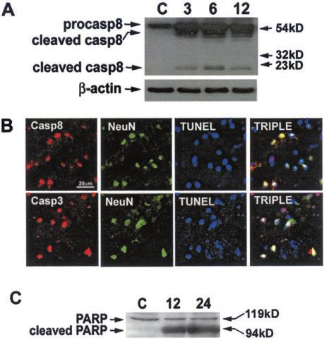
Qiu et al. • Fas and Traumatic Brain Injury J. Neurosci., May 1, 2002, 22(9):3504–3511 3505
formation, and activation of caspases after brain injury remains to (C -2–10) monoclonal antibody was obtained from Alexis Biochemicals
be established. (San Diego, CA). Anti-neutrophil antibodies were purchased from Sero-
tec (Raleigh, NC). The specificity of the antisera to cleaved (active)
The aim of this study was to test the hypothesis that TBI fragments of caspase-8 (SK440) and caspase-3 (SK398) has been charac-
induces Fas death receptors and DISC assembly and promotes terized previously (Velier et al., 1999; Matsushita et al., 2000). Specificity
the initiation of caspase cascades that lead to cell death. Using a of the commercially available antibodies was confirmed by detection of a
mouse controlled cortical impact (CCI) model and contused brain major band of the appropriate molecular weight on Western blot.
samples removed from patients with severe TBI, we show that Immunohistochemistr y and T UNEL. Coronal brain sections placed on
poly-L-lysine-coated slides were fixed in 100% ethanol at ⫺20°C for 10
death-receptor activation and DISC formation are upregulated min and then washed in PBS, pH 7.4, containing 0.1% Triton X-100
and temporally associated with activation of initiator and effector (PBST). Sections were blocked for 1 hr in PBST containing 5% normal
caspases in injured brain. Furthermore, we show that overexpres- goat serum and then incubated for 1–3 d with rabbit polyclonal primary
sion of Fas ligand (FasL) in cultured neurons using transfection antibodies at 4°C. Sections were washed in PBST and incubated with goat
with an adenoviral vector induces DISC assembly and cell death anti-rabbit IgG– C y3 conjugate (Jackson ImmunoResearch, West Grove,
PA) for 60 min. After washing in PBST, sections were incubated with
that is attenuated by a broad-spectrum caspase inhibitor. The mouse monoclonal anti-mouse NeuN (1:300) for 60 min and then reacted
data indicate that death receptors may constitute an important with goat anti-mouse IgG–bodipy (1:300; Molecular Probes, Eugene,
initiating mechanism of cell death after TBI. OR) for 30 min. For TUN EL staining, sections were incubated with TdT
buffer (in mM: 30 Tris, 140 sodium cacodylate, 1 cobalt chloride, pH 7.2)
MATERIALS AND METHODS containing TdT (0.5 U/ml) and biotin-16 – deoxyUTP (dUTP) (0.04
mol / l) (all reagents from Boehringer Mannheim, Indianapolis, I N) for 1
Mouse controlled cortical impact model. The mouse CCI model (refers
hr at 37°C. The reaction was terminated by washing in PBS. Biotin-16 –
specifically to the murine experimental TBI model) was used as de-
dUTP incorporated into tissue was reacted with streptavidin – C y5 (1:
scribed previously (Whalen et al., 1999a,b) with minor modifications. The
1000, Jackson ImmunoResearch) for 5 min for immunofluorescence
trauma protocol was approved by the Massachusetts General Hospital
Institutional Animal C are and Use Committee and complied with the microscopy. After washing in PBS, sections were dehydrated in an
National Institutes of Health Guide for the Care and Use of Laborator y ascending ethanol series, immersed in xylene, and coverslipped with
Animals. Mice were anesthetized with 2% isoflurane, N2O and O2 (2:1) Permount (Fisher Scientific, Pittsburgh, PA). Triple-labeled sections
using a nose cone. Mice were positioned in a stereotaxic frame and a were analyzed on a Leica (Nussloch, Germany) DMRB/ Bio-Rad (Her-
brain temperature probe (Physitemp Corp., C lifton, NJ) was inserted cules, CA) MRC 1024 krypton – argon laser-scanning confocal micro-
through a burr hole into the left frontal cortex. Body temperature was scope. E xcitation /emission filters were 488/522 nm for bodipy, 568/585
monitored with a rectal probe and maintained at 36 –38°C with a heating nm for C y3, and 650/670 nm for C y5, respectively. Double-labeled
pad. A 5 mm craniotomy was made using a portable drill and a 5 mm sections were analyzed on a Nikon (Tokyo, Japan) Eclipse T300 fluores-
trephine over the left parietotemporal cortex and the bone flap was cence microscope. Negative controls included incubation with rabbit
removed. Brain temperature was maintained at 36°C for 1 min. Mice serum instead of primary antibodies or omission of secondary antibodies,
were then subjected to CCI using a pneumatic cylinder with a 3 mm biotin-16 – dUTP, or C y5. Specificity controls also included varying the
flat-tip impounder, a velocity of 6 m /sec, and a depth of 0.6 mm. The order of reaction with the different antibodies and TUN EL.
bone flap was immediately replaced and the scalp was sutured closed. Immunoblotting. Brain tissue or cultured neurons were homogenized
Anesthesia was discontinued and the mice were allowed to recover in on ice in buffer A (10 mM H EPES buffer, pH 7.6, 42 mM KC l, 5 mM
room air until able to ambulate (⬃5 min) and were then returned to their MgC l2, 1% SDS, 1 mM phenylmethylsulfonylfluoride, 1 mM EDTA, 1 mM
cages. EGTA, 1 mM dithiothreitol, 1.5 M pepstatin, 2 M leupeptin, and 0.7 M
Animal protocols. For light microscopy studies [terminal deoxynucleo- aprotinin). The lysate was cleared by centrif ugation at 20,800 ⫻ g for 30
tidyl transferase (TdT)-mediated biotinylated UTP nick end labeling min at 4°C. The protein content of the supernatant was assayed (Bio-
(TUN EL), hematoxylin and eosin (H&E), and immunohistochemical Rad), and proteins were size fractionated on 10% or 10 –20% SDS-
staining for leukocytes], mice subjected to CCI were killed by decapita- polyacrylamide gels and blotted onto a Hybond nitrocellulose membrane
tion under isoflurane anesthesia at 0, 3, 6, 24, or 48 hr after injury. Brains (Amersham Biosciences, Arlington Heights, IL) overnight. The blot was
were removed, frozen in isopentane at ⫺40 to ⫺50°C, and stored at blocked for 1 hr in 5% milk in PBST and then incubated overnight at 4°C
⫺80°C. Within 1 week the brains were sectioned on a cryostat (10 m) in primary antibodies diluted according to the recommendations of the
and stained. manufacturer. Membranes were washed in PBST and 1% milk and then
For immunofluorescence histochemistry, mice were killed at 0, 6, 12, incubated for 1 hr with the appropriate horseradish peroxidase-
24, or 48 hr after CCI. Cryostat brain sections were prepared as described conjugated secondary antibody at room temperature. Proteins of interest
above and stored at ⫺80°C before staining. were detected using the enhanced chemiluminescence (ECL) Western
Because immunoprecipitation and Western blots require the use of blotting detection system kit (Amersham Biosciences, Buckinghamshire,
different homogenization buffers, separate groups of mice were used for UK) and Hyperfilm (Amersham Biosciences, Oakville, Ontario, C an-
each. For immunoprecipitation studies, mice were decapitated under ada) and analyzed by densitometry using an M4 imaging system (Imaging
isoflurane anesthesia at 0, 3, 6, 12, or 24 hr after CCI. Contused cortical Research, Inc., St. C atherines, Ontario, C anada).
tissue was caref ully removed, immediately frozen in liquid nitrogen, and Immunoprecipitation. Physical interaction between proteins was deter-
stored at ⫺80°C. For Western blotting, mice were killed as described mined by immunoprecipitation analysis of (1) cortical tissue from mice
above at 0, 0.5, 3, 6, 12, 24, 48, or 72 hr after CCI. Brain tissue was frozen killed at various times after CCI; (2) cortical tissue removed from
in liquid nitrogen and stored at ⫺80°C. patients with severe TBI, refractory seizure disorders, or postmortem; or
For all experiments, three to four animals were used for each time (3) cultured neurons after Fas ligand overexpression. Brain tissue was
point. lysed, homogenized, and pelleted by centrif ugation in buffer containing
Antibodies. Anti-Fas antibodies (M-20 and C -20), anti-FasL antibodies 20 mM Tris-HC l, pH 7.5, 140 mM NaC l, 1% Triton X-100, 2 mM EDTA,
(N-20 and C178), anti-caspase-8 antibodies (H-134 and D-8), anti- 1 mM p-amidinophenyl methanesulfonyl fluoride hydrochloride, 50 mM
caspase-10 antibody (H-131), and anti-receptor interacting protein NaF, 0.7 M aprotinin, and 10% glycerol. The supernatants were pre-
(RI P)-associated interleukin-1 converting enzyme and CED-3 cleared by incubation with protein G-agarose and normal rabbit IgG for
homolog-1 (ICH)/C ed 3 homologous protein with a death domain 2 hr at 4°C and were incubated with 2 g of anti-Fas antibody (M-20),
(R AI DD) (FL -199) antibody were purchased from Santa Cruz Biotech- anti-caspase-8 antibody (H-134), and anti-R AI DD (FL -199) or anti-Fas
nology (Santa Cruz, CA). Anti-FADD polyclonal antibody was pur- ligand (C -178) antibody overnight at 4°C. Lysates were then incubated
chased from Chemicon International Inc. (Temecula, CA). Monoclonal with protein G-agarose for 2 hr at 4°C. The immunoprecipitates were
antibody against Fas ligand /CD95L (clone 33), Fas/CD95/Apo-1 (clone washed three times with buffer containing 50 mM Tris-HC l, pH 7.5, 0.1%
13), and RI P (clone 38) were obtained from Transduction Laboratories SDS, 0.5% deoxycorticosterone, 1% N P-40, and 62.5 mM NaC l and
(Lexington, K Y). Anti-caspase-10/a monoclonal antibody was supplied subsequently dissolved in denaturing sample buffer.
by R&D Systems (Minneapolis, M N). Mouse anti-caspase-2 (ICH-1) The immunoprecipitates were separated by 10 –20% SDS-PAGE and
(clone G310-1248) antibody was purchased from PharMingen Interna- transferred to an Immobilon-P membrane (Millipore, Bedford, M A).
tional Inc. (San Diego, CA). Anti-poly(ADP-ribose) polymerase (PARP) After blocking with 5% skim milk in TBS with 0.05% T ween 20 (Sigma,3506 J. Neurosci., May 1, 2002, 22(9):3504–3511 Qiu et al. • Fas and Traumatic Brain Injury
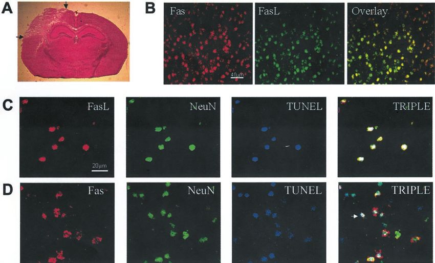
Figure 1. Anatomic localization of cortical contusion and colocalization of Fas ligand and Fas in cortical neurons after CCI. Mice subjected to CCI were
killed after 6 hr, and cryostat brain sections were processed by H&E staining ( A) or by immunohistochemistry ( B–D). A, Anatomic location in the
parietal cortex of the lesion produced by CCI. B, Colocalization of Fas and Fas ligand in normal mouse cortical neurons. Fas ligand was labeled with
mouse monoclonal antibody (clone 33) and goat anti-mouse IgG–Cy2 ( green). Fas receptor was labeled with rabbit polyclonal antibodies (M-20) and goat
anti-rabbit IgG–Cy3 (red). Brain sections were analyzed by fluorescence microscopy. Double-labeled cells appear yellow. C, D, Colocalization of Fas and
Fas ligand with TUNEL-positive neurons. Fas ligand ( C) and Fas receptor ( D) were labeled with rabbit polyclonal antibodies N-20 and M-20,
respectively, and with Cy3 (red). Neurons were detected using mouse monoclonal anti-mouse NeuN and visualized with goat anti-mouse bodipy ( green);
TUNEL-positive cells were labeled with biotin-16 –dUTP–streptavidin–Cy5 (blue). TUNEL-positive neurons colabeled with anti-Fas ligand or anti-Fas
stain white (arrow in D).
St. L ouis, MO), the membranes were incubated with anti-caspase-8 pf u /cell) for up to 3 d at 37°C. As a control, neurons were also incubated
(D-2), anti-Fas (clone-13), or anti-Fas ligand (clone-33) antibody fol- with rAd– C M V–lacZ at 40 pf u /cell. Dead cells were determined by
lowed by reaction with the appropriate horseradish peroxidase- exclusion of trypan blue and Hoechst staining. C ells were collected by
conjugated secondary antibody. Protein was detected using the ECL scraping at day 2 after transfection for analysis by immunoprecipitation
system and Hyperfilm (both from Amersham) and semiquantitated by and Western blot.
densitometry with the M4 imaging system (Imaging Research). To determine the sensitivity of cell death to a caspase inhibitor,
Anal ysis of human brain for Fas upregulation and Fas–procaspase-8 cultured neurons were transfected with adenovirus containing Fas ligand
interaction. Contused brain samples surgically removed from patients in the presence of 500 M Z-Val-ALa-Asp(Ome)-fluoromethylketone
between 2 and 30 hr after severe TBI were analyzed by Western blot and (Z VAD-fmk) in 0.1% dimethylsulfoxide (DMSO) or vehicle alone. At 72
immunoprecipitation (above) for expression of Fas receptor and for hr after transfection, dead cells were counted using trypan blue staining.
interaction between Fas and procaspase-8. The patients were admitted to A total of three wells with three areas per well and 100 cells/area were
the R. Adams Cowley Shock Trauma C enter of the University of Mary- analyzed for each condition.
land Medical C enter and the use of tissue samples was approved by the Statistical anal yses. t tests were used to determine differences in den-
Institutional Review Board. C erebral contusion, present in all patients, sitometry measurements between groups and differences in neuronal
was documented by computed tomography scan. Control brain samples survival in vitro. p ⬍ 0.05 was considered significant.
were taken within 24 hr postmortem from adults who died of non-C NS
related causes (n ⫽ 1) and from cerebral cortex excised from patients
with intractable seizure disorders (n ⫽ 5). Brain tissue was stored at RESULTS
⫺80°C until use. Histopathology of contused cortex after CCI in mice
Primar y neuronal culture and Fas ligand overe xpression. Primary cul-
tures of cerebral cortical neurons were prepared from embryonic day 16 The spatiotemporal distribution of cell death in the mouse model
(E16) or E17 C57BL /6 mice (Charles River Laboratories, Wilmington, was similar to that reported by others after CCI in mice (Smith et
M A). C ells were isolated using trypsin and cultivated in neurobasal al., 1995; Whalen et al., 1999a) and in rats (Colicos et al., 1996).
medium supplemented with 2% B27, 0.5 mM glutamine, and 25 M The anatomic location of the contusion produced by CCI is
glutamate (Invitrogen, San Diego, CA). C ells were seeded at a density of
1 ⫻ 10 6 cells/ well in six well plates coated with poly-D-lysine and then shown in Figure 1 A. Within 3– 4 hr of CCI, the cortical contusion
incubated in a humidified atmosphere of 5% C O2 at 37°C. On day 3, the was characterized by edema, hemorrhage, and hypereosinophilic
cultures were incubated with 10 M cytosine arabinoside for 24 hr to staining of cells with the morphologic appearance of neurons.
suppress the growth of glial cells. One-half of the medium in each well These “red neurons” were observed in ipsilateral but not con-
was changed every 4 d. At day 4, glutamate was withdrawn from the
medium. Neurons cultured for 1 week were used in all experiments.
tralateral cortex and hippocampus and were distributed among
Cultured cortical neurons were incubated with recombinant adenovi- normal-appearing cells in the impact zone. By 6 –12 hr, the
rus (rAd)– cytomegalovirus (C M V)–Fas ligand (Morelli et al., 1999) (40 number of red neurons in injured cortex was markedly increasedQiu et al. • Fas and Traumatic Brain Injury J. Neurosci., May 1, 2002, 22(9):3504–3511 3507
Figure 2. Expression of Fas and Fas ligand ( A) or Fas–Fas ligand Figure 3. DISC assembly after CCI. A, Cortical samples were analyzed
interaction ( B) after CCI in mice over time [in hours ( h)]. Contused or by immunoprecipitation using anti-FADD, anti-Fas, anti-caspase-8, or
normal (uninjured control; C) cortex was subjected to Western blot anti-caspase-10 antibodies, and were subjected to SDS-PAGE. Immuno-
analysis using polyclonal anti-Fas (M-20) or anti-Fas ligand (N-20). Fas blots were probed with antibodies against caspase-8 (H-134) or Fas
and Fas ligand were constitutively expressed in uninjured cortex. Expres- receptor (clone 13). Interaction between Fas receptor and FADD,
sion of Fas receptor was increased after CCI, whereas there was no change procaspase-8, and procaspase-10 was apparent after CCI. Bands corre-
in Fas ligand expression. Bands corresponding to -actin were of equal sponding to light-chain IgG were of equal intensity in all lanes, suggesting
intensity in all lanes, suggesting equal protein loading. B, Fas–FasL equal protein loading (data not shown). B, Traumatic brain injury pro-
interaction is increased after CCI. Fas ligand or Fas receptor was immu- motes Fas–RIP–RAIDD–caspase-2 interaction. Mice were subjected to
noprecipitated from cortical supernatant homogenates with the corre- CCI and killed at the indicated times (in hours). Brain tissue from
sponding polyclonal antibodies and subjected to SDS-PAGE. Western contused cortex was then analyzed by immunoprecipitation using anti-
blots were probed with monoclonal anti-Fas or anti-Fas ligand, respec- RAIDD. Immunoprecipitants were probed on an immunoblot using
tively. A time-dependent increase in Fas–Fas ligand interaction was anti-RIP and anti-caspase-2 antibodies. Interaction of RIP–RAIDD–
found. Robust interaction between Fas and Fas ligand was observed caspase-2 increased at 3 hr after injury. Bands corresponding to light-
regardless of whether anti-Fas or anti-Fas ligand was used to immunopre- chain IgG were of equal intensity in all lanes, suggesting equal antibody
cipitate. Bands corresponding to light-chain IgG were of equal intensity in loading (data not shown). C, Control (normal) mouse cortex; IP, immu-
all lanes, suggesting equal antibody loading. IP, Immunoprecipitation noprecipitation antibody; WB, immunoblotting antibody.
antibody; IB, immunoblotting antibody.
Fas–Fas ligand binding is increased after CCI
both in the center as well as at the margins of the contusion. A Binding of specific ligands to death receptors induces receptor
similar distribution of cells with DNA damage, as assessed by trimerization and activation. We reasoned that if TBI caused Fas
TUNEL staining, was observed at both early (3– 4 hr) and later activation, then we should detect a time-dependent increase in
times after injury. Neutrophils were first detected within contused Fas–Fas ligand interaction. Fas–Fas ligand interaction was de-
cortex at 24 hr after CCI, and increased at 24 – 48 hr in five of six tected in injured cortex early after CCI (Fig. 2 B). -actin was not
animals. Most neutrophils were found scattered throughout the detected in the same immunoprecipitant (data not shown),
contusion, but some were distributed in the perivascular space as thereby suggesting that our result was most likely attributable to
well. Cell death assessed by H&E and TUNEL staining appeared specific Fas–Fas ligand binding.
maximal at 24 – 48 hr and was largely complete after 72 hr. At 7 d,
DISC is formed by Fas, FADD, and procaspase-8 or
a cavitary lesion was present. procaspase-10 after CCI
Increased expression of Fas and Fas ligand early Fas ligand engagement promotes association of the cytosolic
after CCI domain of Fas with a cytosolic adapter FADD, which in turn
We first examined whether Fas and Fas ligand were expressed in recruits initiator procaspases. To examine whether DISC forma-
normal mouse brain and upregulated after CCI. Both Fas and Fas tion accompanies TBI, we used immunoprecipitation and West-
ligand colocalized with nearly all NeuN-positive cells (data not ern blots to detect interaction between Fas, FADD, and
shown), and both Fas and Fas ligand colocalized with each other in procaspase-8 or procaspase-10. Figure 3A shows marked, robust
nearly all cells with neuronal morphology in normal mouse cortex upregulation of Fas–FADD–procaspase-8 and Fas–procaspase-10
(Fig. 1 B). Fas and Fas ligand were detected in the cytosol of interaction as early as 3 hr after CCI. Interaction between Fas and
TUNEL-positive neurons at 6 hr after CCI (Fig. 1C,D). During procaspase-8 was either not detected at all or only weakly de-
the first 12 hr after CCI, only a minor fraction of the total number tected in normal mouse cortex. Hence, Fas receptor contributes
of cells expressing Fas and Fas ligand was NeuN-negative. to rapid and robust DISC assembly early after CCI. Because we
Constitutive expression of Fas and FasL was detected in homog- observed red blood cells in contused brain at 3– 6 hr after CCI, we
enates of mouse parietal cortex (Fig. 2 A). As reported previously assessed the possible contribution of activated blood cells to
after CCI in rats (Beer et al., 2000a), Fas expression increased as DISC formation in the postmortem brain. First, arterialized
early as 30 min (data not shown) and was sustained for up to 72 hr blood was withdrawn by cardiac puncture from a naive mouse or
after injury. Fas expression was increased fourfold versus controls a mouse 6 hr after CCI. Five microliters of fresh whole blood was
at 3 hr after CCI and at all subsequent times examined ( p ⬍ 0.05). then injected into the cortex (left hemisphere) of a freshly perfused
FasL expression remained at control levels and did not increase (saline) normal mouse brain. DISC assembly was assessed as de-
when examined up to 72 hr in our model (Fig. 2 A; data not shown). scribed above. In both cases, the results of Fas–procaspase-8 coim-3508 J. Neurosci., May 1, 2002, 22(9):3504–3511 Qiu et al. • Fas and Traumatic Brain Injury
munoprecipitation did not differ from controls (data not shown).
These data suggest that blood contaminants do not contribute
significantly to DISC formation in the traumatically injured brain.
They do not rule out a contribution from blood at later times when
leukocyte populations in the brain are more common.
RIP–RAIDD–caspase-2 interaction after CCI in mice
Fas signaling may also activate the initiator procaspase-2 by
interacting with RIP, a serine/threonine kinase that associates
with RAIDD, a FADD-like cytosolic adapter protein. The Fas–
RIP–RAIDD complex can form a DISC by recruiting
procaspase-2. In addition, RIP has been shown to mediate necro-
sis in non-neuronal cells, through mechanisms that remain un-
known (Holler et al., 2000). We demonstrated a marked increase
in RIP–RAIDD and RAIDD–procaspase-2 interaction at 3–12
hr after CCI (Fig. 3B). Little or no interaction between RIP–
RAIDD or RAIDD–procaspase-2 was detected in normal brain.
Activation of caspase-8 and caspase-3 after CCI
in mice
Because DISC formation does not imply significant downstream
caspase activation, we looked for processing of caspase-8 and
caspase-3 in injured brain. The p18 fragment of processed
Figure 4. Caspase activation after traumatic brain injury in mice. A,
caspase-8 was detected in mouse brain as early as 3 hr after TBI Cleavage of caspase-8 early after CCI. Cortical homogenates from con-
but was absent in uninjured brain (Fig. 4 A), whereas the proform tused mouse cortex were analyzed by Western blot using an antibody
was detected in normal brain homogenate. Processed caspase-8 (H-138) that reacts with the proform (p55) and active fragment (p18) of
and caspase-3 were also detected in TUNEL-positive neurons by caspase-8. Cleavage of p55 to p18, as well as another caspase-8 cleavage
immunohistochemistry at 48 hr after CCI (Fig. 4 B). We did not product (p43) was observed by 3 hr after injury. -actin was also probed
to confirm equal protein loading. C, Control (normal) mouse cortex. B,
detect cleaved caspase-8 and caspase-3 in neurons at early time Immunohistochemical detection of activated caspase-8 and caspase-3 in
points using immunohistochemistry (data not shown). Neverthe- TUNEL-positive cortical neurons 48 hr after CCI. Cryostat brain sections
less, these data suggest that activation of initiator and effector (10 m) taken from the center of the contusion were processed by
caspases are events that are downstream after DISC formation. immunohistochemistry. Caspase-8 (p18) or caspase-3 (p20) was detected
with rabbit polyclonal antisera SK440 or SK398, respectively, and Cy3
To determine whether caspase activity is present after TBI, we (red). Neurons were identified using monoclonal anti-mouse NeuN and
examined contused brain for the 85 kDa cleavage fragment of visualized with goat anti-mouse bodipy ( green). TUNEL-positive cells
PARP. The p85 PARP cleavage fragment, specific for cleavage by were labeled with biotin-16 –dUTP–streptavidin–Cy5 (blue). TUNEL-
caspases, was detected in contused cortex at 12–24 hr after CCI positive neurons colabeled with p18 or p20 appear white. At 48 hr after
but not in normal cortex (Fig. 4C). CCI, many of the TUNEL-positive neurons in the injured cortex cola-
beled with p18 and p20. C, PARP cleavage in brain after CCI. Cortical
homogenates were analyzed by Western blot at the times indicated. The
Neuronal cell death is induced by overexpression of
p85 PARP fragment, specific for caspase-mediated proteolysis of PARP,
Fas ligand in vitro was robust at 12 and 24 hr after CCI. Bands corresponding to -actin were
To establish that Fas is a functional death receptor in neurons, we of equal intensity in all lanes, suggesting equal protein loading.
overexpressed Fas ligand in cultured neurons and assessed Fas–
procaspase-8 coimmunoprecipitation as well as whether Fas li-
gand overexpression induced caspase-mediated cell death. Cells scale scores of 3–12) suffered head injury after motor vehicle
transfected with adenoviral vectors containing the gene encoding accidents (n ⫽ 2), falls (n ⫽ 2), or assault (n ⫽ 1). Compared with
Fas ligand overexpressed Fas ligand protein twofold compared controls, Fas receptor was increased twofold ( p ⬍ 0.05 vs control)
with cells transfected with control vector ( p ⬍ 0.05) (Fig. 5A). (Fig. 6 A,B) and Fas–procaspase-8 coimmunoprecipitation was in-
Moreover, specific Fas and procaspase-8 protein–protein interac- creased ⬃75% ( p ⬍ 0.02 vs control) (Fig. 6C,D) in brains from TBI
tions were present in these neurons but not in controls (Fig. 5B). patients. Notably, Fas–procaspase-8 interaction was only modestly
Between 48 and 72 hr, Fas ligand-expressing neurons showed increased in human brain samples containing large amounts of
morphologic features of apoptosis by Hoechst staining (data not blood versus those containing lesser amounts (as judged from the
shown), and by 72 hr, 80% of neurons were dead ( p ⬍ 0.05 vs amount of heme in the homogenate), arguing against a relationship
control). When cultured neurons overexpressing Fas ligand were between blood content and Fas–procaspase-8 DISC assembly (data
treated with a pan-caspase inhibitor, ZVAD-fmk (500 M), cell not shown). Thus, Fas is associated with DISC formation during
death was decreased to ⬃50% of control levels ( p ⬍ 0.05) (Fig. the pathogenesis of human TBI.
5C). These data indicate that Fas functions as a death receptor in
cultured neurons, and that caspases constitute one mechanism DISCUSSION
mediating neuronal death induced by Fas ligand. Fas-induced cell killing has been implicated as a major mecha-
nism of neuronal death in the developing brain (Cheema et al.,
Fas forms a DISC after TBI in human brain 1999; Raoul et al., 1999; Felderhoff-Mueser et al., 2000), after
Based on our results in a mouse model, we hypothesized that Fas cerebral ischemia (Martin-Villalba et al., 1999; Felderhoff-
contributes to DISC formation after human TBI. Five male pa- Mueser et al., 2000; Rosenbaum et al., 2000; Martin-Villalba et
tients (16 –75 years of age) with severe TBI (initial Glasgow coma al., 2001; Northington et al., 2001), and after TBI (Beer et al.,Qiu et al. • Fas and Traumatic Brain Injury J. Neurosci., May 1, 2002, 22(9):3504–3511 3509
Figure 6. Fas expression and DISC formation is increased in contused
human brain. A, Western blot analysis of brain tissue homogenates from
representative patients with severe TBI (lanes 3 and 4 ) or refractory
seizures (lanes 1 and 2) or of postmortem tissue from patients dying of
non-CNS causes (n ⫽ 1). Brain homogenates were separated by SDS-
PAGE and Western blots were probed with anti-Fas antibody (C-20). Fas
receptor expression was increased in patients with TBI. -actin was also
Figure 5. Fas ligand overexpression in enriched cultured cortical neu- probed to confirm equal protein loading. B, Densitometric analysis of data
rons. A, Expression of Fas ligand and Fas receptor in cortical neurons from all patients with TBI (n ⫽ 5), seizures (n ⫽ 5), and non-CNS causes
transfected with a gene encoding Fas ligand (see Materials and Methods). of death (n ⫽ 1). *p ⬍ 0.05 versus control. C, Fas–procaspase-8 interaction
E16 cortical neurons were transfected with a recombinant adenoviral assessed by immunoprecipitation of homogenates from patients with
vector with or without Fas ligand. At 48 hr after transfection, cell lysates severe TBI (lanes 3 and 4 ) or refractory seizure disorders (lanes 1 and 2)
were subjected to Western blot using an anti-Fas ligand antibody (N-20) as described in A. Cortical homogenates were incubated with anti-
or an anti-Fas antibody (M-20). Fas receptor was constitutively expressed procaspase-8, and the immunoprecipitants were subjected to SDS-PAGE.
and did not change, whereas Fas ligand was upregulated in neurons Immunoblots were probed with antibodies against human Fas receptor
transfected with vector containing the gene encoding Fas ligand (rAd– (clone 33). Coimmunoprecipitation of Fas–procaspase-8 is apparent in
FasL) but not by control vector (rAd–LacZ). B, Coimmunoprecipitation patients with TBI. Bands corresponding to light-chain IgG were of equal
of Fas–procaspase-8 in cultured cortical neurons transfected with Fas intensity in all lanes, suggesting equal antibody loading. D, Densitometric
ligand. E16 neurons were transfected with adenoviral vectors as in A. At analysis of Fas–procaspase-8 immunoprecipitants was performed as in B.
48 hr after transfection, cell lysates were incubated with anti-caspase-8 Fas–procaspase-8 coimmunoprecipitation is increased in brains taken
antibody (H-134), and the immunoprecipitants were separated by SDS- from patients with TBI (*p ⬍ 0.05 vs control). IP, Immunoprecipitation
PAGE and probed with anti-Fas antibody (clone 13). Fas–procaspase-8 antibody; WB, immunoblotting antibody.
coimmunoprecipitation was increased in overexpressing cortical neurons.
IP, Immunoprecipitation antibody. C, Neuronal death induced by Fas
ligand in vitro is mediated in part by caspases. Cell death was assessed at and DISC assembly. Infiltrating leukocytes can be a source of Fas
72 hr in cortical neurons overexpressing Fas ligand (trypan blue staining). and Fas ligand and DISC proteins, but we did not detect neutro-
Eighty percent of neurons transfected with Fas ligand died (vertical
stripes) versus control vector (diagonal stripes) (*p ⬍ 0.05). Pretreatment phils at early times of DISC assembly in contused brain (3– 6 hr),
with ZVAD-fmk (500 M; open bar) reduced Fas ligand-induced cell and lymphocyte accumulation in contused brain occurs after 24
death by ⬃50% at 72 hr after transfection compared with vehicle (0.5% hr (Holmin et al., 1995). Furthermore, DISC assembly was always
DMSO; solid bar) (**p ⬍ 0.05 vs control). greater in contused mouse brain than in brain injected with an
equivalent amount of blood. Finally, the magnitude of Fas–
2000a). The present study is the first to demonstrate linkage be- procaspase-8 immunoprecipitant did not relate in any simple way
tween ligation and activation of Fas, Fas-associated DISC assem- to blood content in the human TBI samples (data not shown).
bly, and activation of initiator and effector caspases after acute Nevertheless, our data do not rule out other sites of Fas expres-
TBI. We found that Fas receptors were expressed by neurons after sion and DISC assembly. Both Fas and Fas ligand expressed on
CCI in mice, and also found evidence for Fas–Fas ligand interac- microglia and astrocytes may play important roles in cell killing in
tion and formation of Fas–FADD–procaspase-8, Fas–FADD– the brain (Saas et al., 1999; Aquaro et al., 2000; Lee et al., 2000).
procaspase-10, and Fas–RIP–RAIDD–procaspase-2 complexes Thus, Fas ligand induced on glia could cause Fas-mediated auto-
preceding the onset of significant cell death. Early DISC assembly crine cell death of glia or paracrine neuronal cell death. Our data
was followed by processing of caspase-8 and caspase-3 and both showing colocalization of Fas and Fas ligand in neurons are
Fas and Fas ligand colocalized to TUNEL-positive neurons af- consistent with an autocrine mechanism of neuronal cell death.
ter CCI. DISC formation was found in cultured neurons when Additional study is required to determine the role of glia in
Fas ligand was overexpressed. A majority of the cells were killed Fas-mediated cell death after TBI.
by 72 hr, and cell death was significantly inhibited by the appli- The most notable finding here is that DISC assembles early and
cation of a pan-caspase inhibitor. The coimmunoprecipitate of is robust in contused brain after both experimental and human
Fas–procaspase-8 was also found in human brain after severe TBI. DISC formation does not appear to be a stereotyped response
traumatic brain injury, suggesting common cell-death mecha- to acute CNS injury but may reflect unique features of TBI. For
nisms in more than a single mammalian species. Together, the example, the robustness of DISC assembly after TBI appeared far
data suggest that Fas may function as a death receptor in mam- greater than that found in ischemic spinal cord (Matsushita et al.,
malian brain, and that death-receptor activation may provide an 2000) or brain after ischemia/reperfusion, even when assessed
important initiating mechanism of neuronal death after TBI. during the evolution of ischemic injury (M. A. Moskowitz, unpub-
Neurons appear to be the most likely site for Fas expression lished observations). Such differences may reflect unique aspects of3510 J. Neurosci., May 1, 2002, 22(9):3504–3511 Qiu et al. • Fas and Traumatic Brain Injury
pathophysiology such as specific gene expression and protein syn- al., 1997, 2000; Haviv et al., 1998). However, neurons lacking
thesis or recruitment and activation of distinct cell types between caspase-2 remain sensitive to cerebral ischemia and facial nerve
TBI and ischemia. Moreover, we and others have reported rapid axotomy in vivo and trophic deprivation in vitro (Bergeron et al.,
upregulation of Fas receptor within minutes after CCI (Beer et al., 1998), likely because of a compensatory overexpression of
2000a) (data not shown), but only after several hours of reperfu- caspase-9 (Troy et al., 2001). Whether caspase-2 plays a role in
sion after cerebral or spinal cord ischemia in mice (Martin-Villalba post-traumatic brain cell death requires additional study.
et al., 1999; Matsushita et al., 2000; Rosenbaum et al., 2000). Our data confirm that Fas and Fas ligand are constitutively
Mechanisms governing the unusually rapid increase in Fas receptor expressed in the adult mouse brain (Park et al., 1998), and that Fas
expression after TBI remain to be elucidated. and Fas ligand colocalize to neurons after CCI (Beer et al., 2000a).
DISC assembly was associated with robust cleavage of caspase-8 To pursue mechanisms of Fas-mediated neuronal cell death in
to its 18 kDa (active) fragment as detected in brain homogenates by more detail, we overexpressed Fas ligand using an adenoviral
3 hr and in TUNEL-positive neurons by 48 hr after CCI (Fig. 4). vector to determine whether Fas receptor is engageable by neuro-
Furthermore, we detected cytochrome c release at later times nally expressed ligand and capable of promoting cell death via
(12– 48 hr) after CCI (data not shown). We and others detected the DISC formation. Compared with treatment with empty vector,
18 kDa caspase-8 fragment in spinal motoneurons at 1.5 hr after overexpression of Fas ligand induced Fas–FADD–procaspase-8
transient ischemia (Matsushita et al., 2000), in rat cortical neurons DISC assembly and a fourfold increase in cell death mediated in
6 hr after permanent ischemia (Velier et al., 1999), and in rat brain part by caspases. In contrast, addition of soluble Fas ligand to
homogenates within several hours of hypoxic/ischemic brain injury cultured neurons failed to cause cell death (preliminary data not
(Northington et al., 2001) and fluid percussion TBI (Keane et al., shown), suggesting that activation of neuronal Fas by extrinsic Fas
2001). In agreement with our results, caspase-3 cleavage, as as- ligand may be less efficient than when endogenously expressed.
sessed by immunohistochemistry, appeared early but was maximal We also identified death-receptor mechanisms in TBI that are
at 48 hr after CCI in rats (Beer et al., 2001). Our data are consistent common to both apoptosis and necrosis. RIP is a protein kinase
with a cell-death mechanism early on in which caspase-8 induces that is activated by Fas signaling with both proapoptotic and
cell death by directly cleaving and activating caspase-3 (type I) anti-apoptotic actions. Transient RIP overexpression promotes
rather than via release of cytochrome c from mitochondria (type apoptosis (Stanger et al., 1995), but RIP may also promote
II) (Scaffidi et al., 1998, 1999). Type I cell death may develop in necrosis in non-neuronal cell types, presumably by phosphorylat-
ischemic spinal motoneurons (Matsushita et al., 2000). Type II cell ing key regulatory proteins (Holler et al., 2000). Caspases are
death requires mitochondrial amplification of caspase-8 cleavage important in Fas-mediated cell killing (Cheema et al., 1999;
by a positive feedback loop involving caspase-8-mediated process- Raoul et al., 1999); however, Fas signaling can also kill cells by
ing of bid, release of mitochondrial cytochrome c, and activation of caspase-independent mechanisms that resemble necrosis (Kawa-
caspase-3, which then may cleave and activate caspase-8. Indirect hara et al., 1998; Vercammen et al., 1998; Matsumura et al., 2000).
evidence for type II cell death was reported after TBI (Keane et Necrosis induced by Fas ligand in T lymphocytes requires func-
al., 2001) and derives from studies showing that overexpression of tional FADD and RIP (Holler et al., 2000). Because necrosis and
Bcl-2, an anti-apoptotic protein that inhibits cytochrome c release, apoptosis cause cell death after TBI, death receptor-mediated
reduces contusion volume after CCI in transgenic mice (Raghu- events that converge at the level of the DISC might activate both
pathi et al., 1998; Nakamura et al., 1999). apoptotic and necrotic cell-death mechanisms in injured neurons.
We detected increased Fas protein and marked upregulation of If so, then inhibition of DISC formation or function could poten-
Fas–procaspase-8 interaction in contused brain removed from tially impact multiple mechanisms governing post-traumatic and
patients with severe TBI, particularly at later times after injury. perhaps other forms of neuronal death.
Although the sample size limited more detailed analysis, Fas
upregulation and DISC formation were significantly greater in REFERENCES
contused brain than in brain removed from humans with refrac- Aquaro S, Panti S, Caroleo MC, Balestra E, Cenci A, Forbici F, Ippolito
G, Mastino A, Testi R, Mollace V, Calio R, Perno CF (2000) Primary
tory seizures or tissue removed postmortem from a patient who macrophages infected by human immunodeficiency virus trigger CD95-
died of non-CNS causes. Although both diseases are associated mediated apoptosis of uninfected astrocytes. J Leukoc Biol
68:429 – 435.
with death receptor-mediated mechanisms, the differences be- Beer R, Franz G, Schopf M, Reindl M, Zelger B, Schmutzhard E, Poewe
tween results from contused brain homogenates were striking. W, Kampfl A (2000a) Expression of Fas and Fas ligand after experi-
The data suggest that DISC formation is relevant to human TBI mental traumatic brain injury in the rat. J Cereb Blood Flow Metab
20:669 – 677.
and in part, validate studies in the mouse. The observation that Beer R, Franz G, Srinivasan A, Hayes RL, Pike BR, Newcomb JK, Zhao
Fas forms a DISC with procaspase-2, procaspase-8, and X, Schmutzhard E, Poewe W, Kampfl A (2000b) Temporal profile and
procaspase-10 suggests that Fas may activate other initiator cell subtype distribution of activated caspase-3 following experimental
traumatic brain injury. J Neurochem 75:1264 –1273.
caspases after TBI. Fibroblasts from caspase-8-deficient mice are Beer R, Franz G, Krajewski S, Pike BR, Hayes RL, Reed JC, Wang KK,
completely resistant to death induced by Fas, tumor necrosis Klimmer C, Schmutzhard E, Poewe W, Kampfl A (2001) Temporal
and spatial profile of caspase 8 expression and proteolysis after exper-
factor receptor 1, and death receptor 3 signaling, suggesting a imental traumatic brain injury. J Neurochem 78:862– 873.
central role for caspase-8 in non-neuronal cell types. Whether Bergeron L, Perez GI, Macdonald G, Shi L, Sun Y, Jurisicova A,
caspase 8 mediates cell death after CNS injury cannot be studied Varmuza S, Latham KE, Flaws JA, Salter JC, Hara H, Moskowitz MA,
Li E, Greenberg A, Tilly JL, Yuan J (1998) Defects in regulation of
in mutant mice directly, because the caspase-8 null mutation is apoptosis in caspase-2-deficient mice. Genes Dev 12:1304 –1314.
lethal at day 12.5 of embryogenesis (Varfolomeev et al., 1998). Boldin MP, Varfolomeev EE, Pancer Z, Mett IL, Camonis JH, Wallach
Humans with loss-of-function mutations in caspase-10 have au- D (1995) A novel protein that interacts with the death domain of
Fas/APO1 contains a sequence motif related to the death domain.
toimmune lymphoproliferative syndromes but no known CNS J Biol Chem 270:7795–7798.
phenotype in noninjured brain (Wang and Lenardo, 2000). Cheema ZF, Wade SB, Sata M, Walsh K, Sohrabji F, Miranda RC (1999)
Fas/Apo [apoptosis]-1 and associated proteins in the differentiating
Caspase-2 mediates the cell death induced by trophic deprivation cerebral cortex: induction of caspase-dependent cell death and activa-
and -amyloid in cultured neurons (Stefanis et al., 1997; Troy et tion of NF-B. J Neurosci 19:1754 –1770.Qiu et al. • Fas and Traumatic Brain Injury J. Neurosci., May 1, 2002, 22(9):3504–3511 3511 Chinnaiyan AM, O’Rourke K, Tewari M, Dixit VM (1995) FADD, a Park C, Sakamaki K, Tachibana O, Yamashima T, Yamashita J, Yone- novel death domain-containing protein, interacts with the death do- hara S (1998) Expression of fas antigen in the normal mouse brain. main of Fas and initiates apoptosis. Cell 81:505–512. Biochem Biophys Res Commun 252:623– 628. Clark RS, Kochanek PM, Chen M, Watkins SC, Marion DW, Chen J, Raghupathi R, Fernandez SC, Murai H, Trusko SP, Scott RW, Nishioka Hamilton RL, Loeffert JE, Graham SH (1999) Increases in Bcl-2 and WK, McIntosh TK (1998) BCL-2 overexpression attenuates cortical cleavage of caspase-1 and caspase-3 in human brain after head injury. cell loss after traumatic brain injury in transgenic mice. J Cereb Blood FASEB J 13:813– 821. Flow Metab 18:1259 –1269. Clark RS, Kochanek PM, Watkins SC, Chen M, Dixon CE, Seidberg NA, Raghupathi R, Graham DI, McIntosh TK (2000) Apoptosis after trau- Melick J, Loeffert JE, Nathaniel PD, Jin KL, Graham SH (2000) matic brain injury. J Neurotrauma 17:927–938. Caspase-3 mediated neuronal death after traumatic brain injury in rats. Raoul C, Henderson CE, Pettmann B (1999) Programmed cell death of J Neurochem 74:740 –753. embryonic motoneurons triggered through the Fas death receptor. Colicos MA, Dixon CE, Dash PK (1996) Delayed, selective neuronal J Cell Biol 147:1049 –1062. death following experimental cortical impact injury in rats: possible Raoul C, Pettmann B, Henderson CE (2000) Active killing of neurons role in memory deficits. Brain Res 739:111–119. during development and following stress: a role for p75(NTR) and Fas? Faden AI, Demediuk P, Panter SS, Vink R (1989) The role of excitatory Curr Opin Neurobiol 10:111–117. amino acids and NMDA receptors in traumatic brain injury. Science Rosenbaum DM, Gupta G, D’Amore J, Singh M, Weidenheim K, Zhang 244:798 – 800. H, Kessler JA (2000) Fas (CD95/APO-1) plays a role in the patho- Felderhoff-Mueser U, Taylor DL, Greenwood K, Kozma M, Stibenz D, physiology of focal cerebral ischemia. J Neurosci Res 61:686 – 692. Joashi UC, Edwards AD, Mehmet H (2000) Fas/CD95/APO-1 can Saas P, Boucraut J, Quiquerez AL, Schnuriger V, Perrin G, Desplat-Jego function as a death receptor for neuronal cells in vitro and in vivo and S, Bernard D, Walker PR, Dietrich PY (1999) CD95 (Fas/Apo-1) as a is upregulated following cerebral hypoxic-ischemic injury to the devel- oping rat brain. Brain Pathol 10:17–29. receptor governing astrocyte apoptotic or inflammatory responses: a Fink KB, Andrews LJ, Butler WE, Ona VO, Li M, Bogdanov M, Endres key role in brain inflammation? J Immunol 162:2326 –2333. M, Khan SQ, Namura S, Stieg PE, Beal MF, Moskowitz MA, Yuan J, Scaffidi C, Fulda S, Srinivasan A, Friesen C, Li F, Tomaselli KJ, Debatin Friedlander RM (1999) Reduction of post-traumatic brain injury and KM, Krammer PH, Peter ME (1998) Two CD95 (APO-1/Fas) signal- free radical production by inhibition of the caspase-1 cascade. Neuro- ing pathways. EMBO J 17:1675–1687. science 94:1213–1218. Scaffidi C, Schmitz I, Zha J, Korsmeyer SJ, Krammer PH, Peter ME Haviv R, Lindenboim L, Yuan J, Stein R (1998) Need for caspase-2 in (1999) Differential modulation of apoptosis sensitivity in CD95 type I apoptosis of growth-factor-deprived PC12 cells. J Neurosci Res and type II cells. J Biol Chem 274:22532–22538. 52:491– 497. Smith DH, Soares HD, Pierce JS, Perlman KG, Saatman KE, Meaney Holler N, Zaru R, Micheau O, Thome M, Attinger A, Valitutti S, Bodmer DF, Dixon CE, McIntosh TK (1995) A model of parasagittal con- JL, Schneider P, Seed B, Tschopp J (2000) Fas triggers an alternative, trolled cortical impact in the mouse: cognitive and histopathologic caspase-8-independent cell death pathway using the kinase RIP as effects. J Neurotrauma 12:169 –178. effector molecule. Nat Immunol 1:489 – 495. Stanger BZ, Leder P, Lee TH, Kim E, Seed B (1995) RIP: a novel Holmin S, Mathiesen T, Shetye J, Biberfeld P (1995) Intracerebral in- protein containing a death domain that interacts with Fas/APO-1 flammatory response to experimental brain contusion. Acta Neurochir (CD95) in yeast and causes cell death. Cell 81:513–523. (Wien) 132:110 –119. Stefanis L, Troy CM, Qi H, Greene LA (1997) Inhibitors of trypsin-like Kawahara A, Ohsawa Y, Matsumura H, Uchiyama Y, Nagata S (1998) serine proteases inhibit processing of the caspase Nedd-2 and protect Caspase-independent cell killing by Fas-associated protein with death PC12 cells and sympathetic neurons from death evoked by withdrawal domain. J Cell Biol 143:1353–1360. of trophic support. J Neurochem 69:1425–1437. Keane RW, Kraydieh S, Lotocki G, Alonso OF, Aldana P, Dietrich WD Troy CM, Stefanis L, Greene LA, Shelanski ML (1997) Nedd2 is re- (2001) Apoptotic and antiapoptotic mechanisms after traumatic brain quired for apoptosis after trophic factor withdrawal, but not superoxide injury. J Cereb Blood Flow Metab 21:1189 –1198. dismutase (SOD1) downregulation, in sympathetic neurons and PC12 Kischkel FC, Hellbardt S, Behrmann I, Germer M, Pawlita M, Krammer cells. J Neurosci 17:1911–1918. PH, Peter ME (1995) Cytotoxicity-dependent APO-1 (Fas/CD95)- Troy CM, Rabacchi SA, Friedman WJ, Frappier TF, Brown K, Shelanski associated proteins form a death-inducing signaling complex (DISC) ML (2000) Caspase-2 mediates neuronal cell death induced by with the receptor. EMBO J 14:5579 –5588. -amyloid. J Neurosci 20:1386 –1392. Lee SJ, Zhou T, Choi C, Wang Z, Benveniste EN (2000) Differential Troy CM, Rabacchi SA, Hohl JB, Angelastro JM, Greene LA, Shelanski regulation and function of Fas expression on glial cells. J Immunol ML (2001) Death in the balance: alternative participation of the 164:1277–1285. caspase-2 and -9 pathways in neuronal death induced by nerve growth Martin-Villalba A, Herr I, Jeremias I, Hahne M, Brandt R, Vogel J, factor deprivation. J Neurosci 21:5007–5016. Schenkel J, Herdegen T, Debatin KM (1999) CD95 ligand (Fas-L/ Varfolomeev EE, Schuchmann M, Luria V, Chiannilkulchai N, Beck- APO-1L) and tumor necrosis factor-related apoptosis-inducing ligand mann JS, Mett IL, Rebrikov D, Brodianski VM, Kemper OC, Kollet O, mediate ischemia-induced apoptosis in neurons. J Neurosci Lapidot T, Soffer D, Sobe T, Avraham KB, Goncharov T, Holtmann H, 19:3809 –3817. Lonai P, Wallach D (1998) Targeted disruption of the mouse Caspase Martin-Villalba A, Hahne M, Kleber S, Vogel J, Falk W, Schenkel J, 8 gene ablates cell death induction by the TNF receptors, Fas/Apo1, Krammer PH (2001) Therapeutic neutralization of CD95-ligand and and DR3 and is lethal prenatally. Immunity 9:267–276. TNF attenuates brain damage in stroke. Cell Death Differ 8:679 – 686. Velier JJ, Ellison JA, Kikly KK, Spera PA, Barone FC, Feuerstein GZ Matsumura H, Shimizu Y, Ohsawa Y, Kawahara A, Uchiyama Y, Nagata (1999) Caspase-8 and caspase-3 are expressed by different populations S (2000) Necrotic death pathway in fas receptor signaling. J Cell Biol of cortical neurons undergoing delayed cell death after focal stroke in 151:1247–1256. the rat. J Neurosci 19:5932–5941. Matsushita K, Wu Y, Qiu J, Lang-Lazdunski L, Hirt L, Waeber C, Hyman BT, Yuan J, Moskowitz MA (2000) Fas receptor and neuronal Vercammen D, Brouckaert G, Denecker G, Van de Craen M, Declercq cell death after spinal cord ischemia. J Neurosci 20:6879 – 6887. W, Fiers W, Vandenabeele P (1998) Dual signaling of the Fas recep- Medema JP, Scaffidi C, Kischkel FC, Shevchenko A, Mann M, Krammer tor: initiation of both apoptotic and necrotic cell death pathways. J Exp PH, Peter ME (1997) FLICE is activated by association with the Med 188:919 –930. CD95 death-inducing signaling complex (DISC). EMBO J Wang J, Lenardo MJ (2000) Roles of caspases in apoptosis, develop- 16:2794 –2804. ment, and cytokine maturation revealed by homozygous gene deficien- Morelli AE, Larregina AT, Smith-Arica J, Dewey RA, Southgate TD, cies. J Cell Sci 113:753–757. Ambar B, Fontana A, Castro MG, Lowenstein PR (1999) Neuronal Whalen MJ, Carlos TM, Dixon CE, Schiding JK, Clark RS, Baum E, Yan and glial cell type-specific promoters within adenovirus recombinants HQ, Marion DW, Kochanek PM (1999a) Effect of traumatic brain restrict the expression of the apoptosis-inducing molecule Fas ligand to injury in mice deficient in intercellular adhesion molecule-1: assess- predetermined brain cell types, and abolish peripheral liver toxicity. ment of histopathologic and functional outcome. J Neurotrauma J Gen Virol 80:571–583. 16:299 –309. Nagata S (1999) Fas ligand-induced apoptosis. Annu Rev Genet Whalen MJ, Clark RS, Dixon CE, Robichaud P, Marion DW, Vagni V, 33:29 –55. Graham SH, Virag L, Hasko G, Stachlewitz R, Szabo C, Kochanek PM Nakamura M, Raghupathi R, Merry DE, Scherbel U, Saatman KE, (1999b) Reduction of cognitive and motor deficits after traumatic brain McIntosh TK (1999) Overexpression of Bcl-2 is neuroprotective after injury in mice deficient in poly(ADP-ribose) polymerase. J Cereb experimental brain injury in transgenic mice. J Comp Neurol Blood Flow Metab 19:835– 842. 412:681– 692. Yakovlev AG, Knoblach SM, Fan L, Fox GB, Goodnight R, Faden AI Northington FJ, Ferriero DM, Flock DL, Martin LJ (2001) Delayed (1997) Activation of CPP32-like caspases contributes to neuronal ap- neurodegeneration in neonatal rat thalamus after hypoxia-ischemia is optosis and neurological dysfunction after traumatic brain injury. apoptosis. J Neurosci 21:1931–1938. J Neurosci 17:7415–7424.
You can also read