Elastin-Like Recombinamer Hydrogels for Improved Skeletal Muscle Healing Through Modulation of Macrophage Polarization - Frontiers
←
→
Page content transcription
If your browser does not render page correctly, please read the page content below
ORIGINAL RESEARCH
                                                                                                                                                 published: 14 May 2020
                                                                                                                                          doi: 10.3389/fbioe.2020.00413

                                            Elastin-Like Recombinamer
                                            Hydrogels for Improved Skeletal
                                            Muscle Healing Through Modulation
                                            of Macrophage Polarization
                                            Arturo Ibáñez-Fonseca 1† , Silvia Santiago Maniega 2† , Darya Gorbenko del Blanco 1† ,
                                            Benedicta Catalán Bernardos 3 , Aurelio Vega Castrillo 2 , Ángel José Álvarez Barcia 4 ,
                                            Matilde Alonso 1 , Héctor J. Aguado 2 and José Carlos Rodríguez-Cabello 1*
                                            1
                                             BIOFORGE (Group for Advanced Materials and Nanobiotechnology), CIBER-BBN, University of Valladolid, Valladolid, Spain,
                                            2
                                             Servicio de Traumatología, Hospital Clínico de Valladolid, Valladolid, Spain, 3 Servicio de Neurofisiología, Hospital Clínico
                             Edited by:
                                            de Valladolid, Valladolid, Spain, 4 Servicio de Investigación y Bienestar Animal, University of Valladolid, Valladolid, Spain
                  Dimitrios I. Zeugolis,
 National University of Ireland Galway,
                                 Ireland    Large skeletal muscle injuries, such as a volumetric muscle loss (VML), often result in
                      Reviewed by:          an incomplete regeneration due to the formation of a non-contractile fibrotic scar tissue.
             Johannes Von den Hoff,
Radboud University Nijmegen Medical
                                            This is, in part, due to the outbreak of an inflammatory response, which is not resolved
                 Centre, Netherlands        over time, meaning that type-1 macrophages (M1, pro-inflammatory) involved in the
                 Marie-noelle Giraud,
                                            initial stages of the process are not replaced by pro-regenerative type-2 macrophages
  Université de Fribourg, Switzerland
                                            (M2). Therefore, biomaterials that promote the shift from M1 to M2 are needed to achieve
                  *Correspondence:
       José Carlos Rodríguez-Cabello        optimal regeneration in VML injuries. In this work, we used elastin-like recombinamers
                roca@bioforge.uva.es        (ELRs) as biomaterials for the formation of non- (physical) and covalently (chemical)
     † These   authors have contributed     crosslinked bioactive and biodegradable hydrogels to fill the VML created in the tibialis
                    equally to this work
                                            anterior (TA) muscles of rats. These hydrogels promoted a higher infiltration of M2 within
                    Specialty section:      the site of injury in comparison to the non-treated control after 2 weeks (p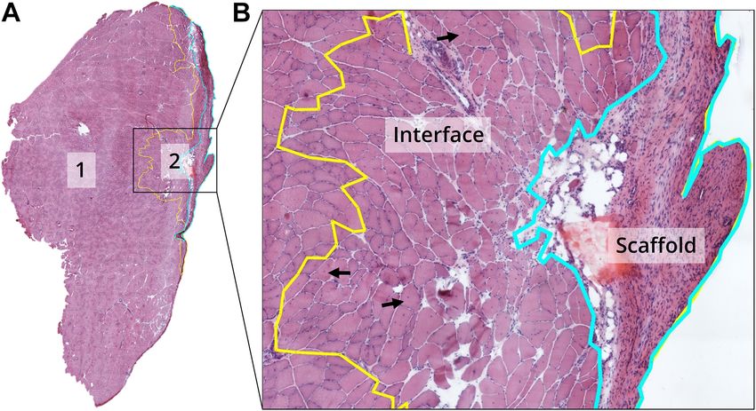
Ibáñez-Fonseca et al.                                                                                       ELR Hydrogels for Skeletal Muscle Healing

INTRODUCTION                                                               and hence a high reproducibility, although they usually lack
                                                                           biological activity (O’Brien, 2011).
Large skeletal muscle injuries are the result of high energy                   Within the different types of synthetic biomaterials, we
traumatisms as a consequence of different events, such as car              can identify recombinant polymers (recombinantly expressed
accidents or explosions, being very common in clinics (Zalavras            structural proteins with repetitive domains) (Cappello et al., 1990;
and Patzakis, 2003; Corona et al., 2015). They usually involve             Tirrell et al., 1991), such as elastin-like recombinamers (ELRs)
a volumetric muscle loss (VML), which implies an impairment                (Rodríguez-Cabello et al., 2009). These molecules derive from the
of muscle function, and their treatment is a challenge for                 repetition of the L-Val-L-Pro-Gly-X-Gly (VPGXG) pentapeptide
the orthopedic surgeon (Grogan et al., 2011; Greising et al.,              found in natural elastin, where X can be any amino acid
2018). Currently, the gold standard is scar tissue debridement             except L-Pro, and are able to self-assemble through hydrophobic
or autologous muscle transfer, which substantially increases               interactions above the so-called transition temperature (T t ) (Urry
donor site morbidity and can cause severe problems, such as                et al., 1976; Urry, 2006; Ibáñez-Fonseca et al., 2019). Due to
infections or non-functional transfers (Lin et al., 2004, 2007;            their recombinant nature, they can be precisely engineered at
Klinkenberg et al., 2013).                                                 the DNA level to bear specific amino acids (Rodríguez-Cabello
    After acute injuries, like VML, a complex process is activated         et al., 2009; Girotti et al., 2015). For instance, the introduction
in order to restore muscle structure and function, mainly due              of lysines with amine groups that can be modified for covalent
to the activation, proliferation and differentiation of a quiescent        crosslinking through “click chemistry” strategies, like strain-
population of resident muscle progenitor stem cells known as               promoted alkyne-azide cycloaddition (SPAAC), may allow the
satellite cells (SCs) (Tedesco et al., 2010; Lepper et al., 2011).         formation of chemical hydrogels (González de Torre et al., 2014;
These steps are orchestrated by the inflammatory response:                 Madl et al., 2016). On the other hand, physical hydrogels can
during the first hours post-damage, circulating monocytes start            be achieved by the inclusion of amino acid sequences able to
differentiating into pro-inflammatory type-1 macrophages (M1)              form stable non-covalent interactions (e.g., H-bonds), such as
that activate the proliferation of SCs from the surrounding                the repetitive domains found in silk fibroin from Bombyx mori
tissue (Tidball and Villalta, 2010; Saclier et al., 2013). Then,           silkworm that, in combination with the elastin-like building
as the muscle repair process advances, the phenotype of the                blocks, form hydrogels through a concomitant self-assembly
macrophages shifts to anti-inflammatory type-2 macrophages                 above the T t (Fernández-Colino et al., 2014; Ibáñez-Fonseca
(M2), which express and secrete cytokines that stimulate                   et al., 2020). Furthermore, the genetic fusion of bioactive
myogenic differentiation of the SCs toward myofibers, thus being           sequences, including cell adhesion domains, like the L-Arg-
essential for a successful healing (Tidball and Villalta, 2010;            Gly-L-Asp (RGD) tripeptide (Ruoslahti, 1996), or protease-
Saclier et al., 2013). A dysregulated macrophage response leads            sensitive sequences for improved biodegradation (Flora et al.,
to a chronic inflammation that induces the formation of a non-             2019; Contessotto et al., under review), permits the obtaining of
contractile fibrotic tissue due to the activation and recruiting of        hydrogels with acquired functionalities. In this last regard, the
fibroblasts that secrete extracellular matrix (ECM) components,            inclusion of motifs sensitive to matrix metalloproteinase (MMP)-
mainly collagen, to fill the void generated by the VML before it           2, 9 and 13 provides a multipurpose platform able to be degraded
can be repopulated by new myofibers, hence leading to functional           in different in vivo environments (Lutolf et al., 2003; Chung et al.,
deficits (Järvinen et al., 2005; Shin et al., 2014).                       2006; Contessotto et al., under review). Therefore, due to their
    During the past few years, several strategies have been                intrinsic properties of high biocompatibility (Ibáñez-Fonseca
proposed to modulate the inflammatory response to enhance                  et al., 2018), mechanical stability (Fernández-Colino et al., 2014;
skeletal muscle repair, many of them involving the use of                  González de Torre et al., 2014), injectability (Martín et al., 2010;
biological (mainly decellularized porcine ECM) (Greising et al.,           Fernández-Colino et al., 2014), and acquired bioactivity (Girotti
2017) or biomaterial-based scaffolds (Grasman et al., 2015).               et al., 2004; Ibáñez-Fonseca et al., 2017), ELR-based hydrogels
Interestingly, some of them have shown to promote a M2-                    have found several uses in tissue engineering and regenerative
balanced response (Sicari et al., 2014; Boersema et al., 2016; Wang        medicine (Coletta et al., 2017; Pescador et al., 2017; Staubli et al.,
et al., 2019). These scaffolds are intended to give mechanical             2017; Contessotto et al., under review), specially within the field
and biochemical support to the different types of cells involved           of in situ tissue regeneration (Lee et al., 2016).
in the regeneration process, such as SCs, and they need to                     In this work, we propose the use of chemical and physical
be biodegradable to give space for the formation of new                    ELR-based hydrogels, both of them biodegradable, to improve
myofibers (Wolf et al., 2015; Bartolacci et al., 2019). One specific       the healing of skeletal muscle injuries. Our hypothesis is that
type of scaffolds are injectable hydrogels, which are made up              ELR hydrogels will be able to modulate the macrophage response
of polymeric biomaterials that form 3D networks with high                  and facilitate the shift to pro-regenerative M2 macrophages.
water content and permeability and that can be applied in a                Moreover, the ELR hydrogels will provide a cell-friendly and
minimally invasive way. While some of them are made of natural             biodegradable environment that will prevent the formation of
biomaterials, e.g., alginate or chitosan, meaning that they are            fibrotic tissue in the area of the defect, and that will allow the
extracted from natural sources, some others are made of synthetic          development of new myofibers. Therefore, the objective of this
ones (Qazi et al., 2015; Wolf et al., 2015). These synthetic               study was to quantitatively analyze macrophage polarization and
biomaterials offer a better control on the chemical composition,           its effects on muscle healing, in terms of collagen deposition

Frontiers in Bioengineering and Biotechnology | www.frontiersin.org    2                                            May 2020 | Volume 8 | Article 413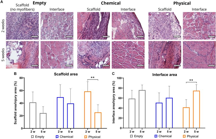
Ibáñez-Fonseca et al.                                                                                      ELR Hydrogels for Skeletal Muscle Healing

(fibrosis) and muscle morphology, following ELR hydrogel                   14 to 16 modified lysines, out of 24), and giving a HRGD6-N3 .
treatment in a rat model of VML.                                           On the other hand, the ELR comprising MMP-sensitive motifs
                                                                           (HE5) was modified similarly, in this case to bear cyclooctyne
                                                                           (activated alkyne) groups, resulting in a 30–40% of modification
MATERIALS AND METHODS                                                      (from 3 to 4 modified lysines, out of 9), and named HE5-C.
                                                                           These modified ELRs were used for the formation of covalently
ELRs Biosynthesis and Characterization                                     crosslinked “click” ELR-based hydrogels (chemical hydrogels).
The ELRs used in this work were biosynthesized through
recombinant DNA technology as described elsewhere
(Rodríguez-Cabello et al., 2012). Briefly, the genes encoding              ELR-Based Hydrogel Preparation and
for the recombinamers were cloned into a pET-25b(+) plasmid
vector (Novagen, Merck, Germany) that was used to transform a              in vivo Administration
BLR(DE3) strain of Escherichia coli (Novagen, Merck, Germany).             In this work, two different types of biodegradable hydrogels
An ELR-expressing clone was cultured in a 15-L bioreactor                  were used to evaluate their influence on muscle healing:
(Applikon Biotechnology B.V., Netherlands) and the ELR was                 non-covalently crosslinked SELR-based hydrogels (physical
purified by several cooling and heating cycles with centrifugation         hydrogels) and covalently crosslinked “click” ELR-based
steps. Then, the highly pure ELR solution was dialyzed against             hydrogels (chemical hydrogels), formed through SPAAC
ultra-pure water and filtered through 0.22 µm filters (Nalgene,            (González de Torre et al., 2014). In both cases, the
Thermo Fisher Scientific, United States) for sterilization. Finally,       recombinamers were dissolved in cold 1× PBS for 16–24 h
the solution was freeze-dried prior to storage.                            at 4◦ C and the hydrogels were formed in situ just after the
   Two of the ELRs used in this work, namely HRGD6 and                     creation of the muscle defect.
HE5, i.e., the ones used for the formation of chemically                      In the case of the “click” ELRs, the HRGD6-N3 and the HE5-C
crosslinked hydrogels (or simply chemical hydrogels), were                 were dissolved separately and mixed prior to injection in a 1:1.8
previously described (Costa et al., 2009; Contessotto et al.,              ratio, since this was found to be the optimal proportion, taking
under review). HRGD6 has six cell adhesion RGD sequences                   into account the different molecular weights and modification
per molecule, embedded within the lysine-containing elastin-like           percentage of the ELRs, finally giving 50 mg/mL hydrogels. The
backbone, whereas the HE5 includes MMP-sensitive domains                   mixture was left for 8 min in an ice bath and afterward it was
for biodegradation and lysine-rich crosslinking domains within             placed in the injury site with a pipette, where the hydrogel
a glutamic acid-containing elastin-like backbone. The presence             formation process was completed.
of lysines in both ELRs makes them suitable for chemical                      For the physical hydrogel (IKRS-MMP), the mono-
modification and subsequent covalent crosslinking via “click               component solution was left in an ice bath until its administration
chemistry” for the formation of chemical hydrogels (see below).            in the site of the defect with a pipette, similarly to the chemical
   On the other hand, the silk-elastin-like recombinamer (SELR)            hydrogel. In this case, the gelation was triggered by the
used for the formation of physically crosslinked hydrogels,                change in the temperature of the solution that leads to an
the so-called IKRS-MMP, was based on a previously designed                 inverse temperature transition (ITT) and to the formation of a
SELR (Ibáñez-Fonseca et al., 2020), to which a MMP-sensitive               network through hydrophobic interactions. The hydrogel was
domain, similar to the one included in the HE5, was included               further stabilized by the folding of silk domains into β-sheets,
for biodegradation.                                                        which results in crystallization (Fernández-Colino et al., 2014;
   The characterization methods for every ELR batch included               Ibáñez-Fonseca et al., 2020).
sodium dodecyl sulfate polyacrylamide gel electrophoresis (SDS-               In both cases, administration was easily performed with a
PAGE) and matrix-assisted laser desorption/ionization time-                pipette, taking advantage of the injectability of both hydrogels,
of-flight (MALDI-TOF) for the evaluation of the purity and                 and the gelation was instantaneous, which avoided the dilution
the molecular weight, HPLC to determine the amino acid                     of the hydrogel once implanted. Moreover, due to the inclusion
composition and differential scanning calorimetry (DSC) for                of a MMP-sensitive amino acid sequence in the HE5 and IKRS-
the calculation of the transition temperature. Furthermore,                MMP, both types of hydrogels, i.e., chemical and physical,
the endotoxin levels were assessed by the limulus amebocyte                were biodegradable.
lysate assay with the Endosafe -PTS system (Charles River
                                          R

Laboratories, Inc., United States) and were always below 1
endotoxin unit/mg of ELR.                                                  Animal Experiments
                                                                           Ethical Statement
ELR Chemical Modification                                                  All animal experiments were conducted in accordance with the
The chemical modification of the ELRs was performed as                     institutional guidelines for the care and use of experimental
previously described (González de Torre et al., 2014). On                  animals of the University of Valladolid (Spain) in accordance
one hand, the ELR containing RGD cell-adhesion domains                     with Directive 2010/63/EU. The protocol was approved by the
(HRGD6) was chemically modified with azide groups through                  Committee of Ethics in Animal Experimentation and Welfare
the transformation of the ε-amine group found in the side chain            (CEEBA, for its Spanish acronym) of the University of Valladolid
of lysine residues, achieving a 55–65% of modification (from               (protocol number 5402485).

Frontiers in Bioengineering and Biotechnology | www.frontiersin.org    3                                           May 2020 | Volume 8 | Article 413
Ibáñez-Fonseca et al.                                                                                      ELR Hydrogels for Skeletal Muscle Healing

Animal Care                                                                the hydrogel preparation section. Subsequently, the continuous
A total of 19 three-month-old male Wistar rats were used in this           suture was tensed and knotted. A self-adhesive bandage of
study. The average weight was 400 g at the time of surgery. The            both legs was made so that the rats could not contaminate
animals were kept in cages with a light:dark cycle of 12:12 and            or bite the wound.
provided with ad libitum food and water. An identification chip               Animals were provided with postoperative analgesia in food
was placed in the interscapular region, inaccessible to the animals,       and drink. Specifically, ibuprofen was dissolved in water at a
to ensure masking during the study.                                        concentration of 10 mL/L of water for 3 days, and tramadol was
                                                                           administered masked in commercial hazelnut cocoa spread at a
Experimental Groups                                                        concentration of 1 mg/kg/day, calculated for 48 h.
Four different groups were established through randomized
classification: non-treated or empty (n = 11), treated with                Euthanasia
chemical biodegradable hydrogels (n = 11) or physical                      Animals were euthanized 2 and 5 weeks post-injury by
biodegradable hydrogels (n = 11), and non-injury or                        intracardiac injection of phenobarbital, after being anesthetized
healthy group (n = 5). In the empty group, the VML was                     to avoid suffering. Then, whole TA muscles were extracted for
left untreated, whereas for the treated groups chemical and                processing and analysis as described below.
physical biodegradable hydrogels were placed in the injury area.
No surgical procedure was done in the non-injury group.
                                                                           Histological Processing
Anesthesia                                                                 The TA muscles were harvested and divided into two halves
                                                                           in the middle area of the defect, and they were used to
All surgical procedures were carried out under proper anesthesia.
                                                                           achieve both longitudinal and cross-sections (see Supplementary
Intraperitoneal anesthesia of the animals was performed with
                                                                           Figure 1B for a schematic representation). To this end, the
ketamine-medetomidine at a dose of 0.125 mL per 100 g
                                                                           samples were mounted on cork discs according to the direction
of animal weight.
                                                                           of the muscle fibers using a small amount of optimum cutting
Tibialis Anterior VML Injury                                               temperature (OCT) mounting medium (VWR, United States).
                                                                           Then, the samples were frozen in 2-Methylbutane (Thermo
Our goal was to create a defect of at least a 20% of the weight of
                                                                           Fisher Scientific, Belgium) previously chilled in liquid nitrogen.
the tibialis anterior (TA) muscle, which was calculated according
                                                                           After freezing, the samples were stored at −80◦ C until further
to the equation described by Wu et al. (2012), giving an average
                                                                           processing. Subsequently, 6 µm cross-section slices were cut in
defect weight of 121 mg. A scheme of the TA VML injury has been
                                                                           a cryostat (Thermo Fisher Scientific, United States) and stained
included in Supplementary Figure 1A.
                                                                           with Harris’ hematoxylin (Merck, Germany)/eosin-Y (Sigma-
   First, the inferior limbs of the animal were shaved using an
                                                                           Aldrich, United States) (HE staining) and Picrosirius red (Abcam,
electric shaver to facilitate subsequent procedures. Then, the
                                                                           United Kingdom) following the manufacturers’ protocols.
animal was placed in the supine position on a heat blanket (to
prevent hypothermia), and it was covered with a sterile drape,
so that only the legs were accessible. Subsequently, a longitudinal        Histomorphometry
incision was made in the skin from the knee to the ankle following         Histomorphometry methods were used to quantify areas, number
the course of the TA muscle with a sterile no. 11 blade scalpel.           and size of myofibers, and collagen percentage. For this
The fascia was sectioned independently and separated from the              purpose, HE images of the whole muscle sections were obtained
muscle using blunt dissection to completely uncover the anterior           using a Nikon Eclipse 80i microscope (Nikon Corporation,
surface of the TA.                                                         Japan) coupled to an automated stage (Prior, United Kingdom)
   Afterward, a transversal mark was made in the exposed muscle            and a DS-Fi1 camera (Nikon Corporation, Japan), which
with a sterile marker, measuring 1cm from the tibial tuberosity            were controlled with the NIS-Elements AR software (Nikon
with a rule. This mark would be the proximal limit of the defect.          Corporation, Japan).
A second mark was made parallel to the first, 1 cm from it. In this            Within the samples, three different regions were differentiated:
way, the proximal and distal limits of the defect were delimited.          the remaining native tissue, the interface (tissue newly
Regarding the width of the VML, we left a margin of about 2 mm             regenerated between the remaining muscle and the scaffold
from the medial and lateral margins of the TA muscle. In this way,         area, characterized by the presence of myofibers with internal
both the length and width of the defect were precisely delimited,          nuclei) and the scaffold area (without myofibers). The sum of
whereas the depth was adjusted according to the volume of                  these last two areas give the area corresponding to the potentially
muscle needed to achieve the above calculated weight.                      injured tissue, i.e., the region where the VML was created
   Once the defect was created, the corresponding treatment                (Figure 1), although it may not represent the wound area to
was applied, according to the groups defined above. For the                its full extent. The three different areas were traced manually
administration of the hydrogel, the skin was first sutured with            and measured using the Fiji distribution of the ImageJ software
Vicryl rapid 2/0, and the skin was left partially open during              (Schindelin et al., 2012). The percentage of injury (interface and
the operation to minimize mobilization of the hydrogel during              scaffold) area was normalized to the entire muscle area, whereas
closure. Both chemical and physical hydrogels were formed                  the percentage of the interface and scaffold areas were obtained
instantaneously once implanted, as previously described in                 by normalizing to the injury area.

Frontiers in Bioengineering and Biotechnology | www.frontiersin.org    4                                           May 2020 | Volume 8 | Article 413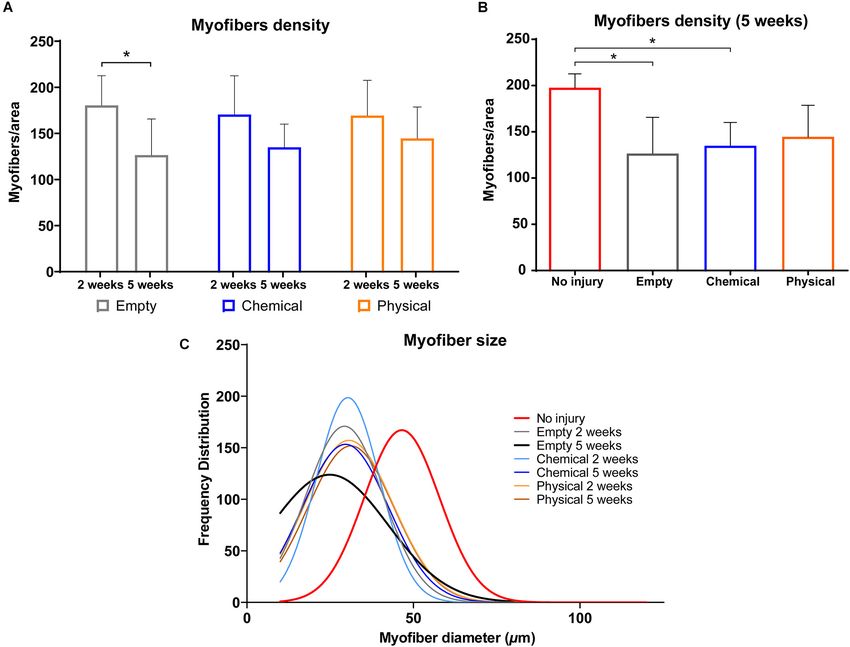
Ibáñez-Fonseca et al. ELR Hydrogels for Skeletal Muscle Healing
Nikon DS-2MBWc digital camera (Nikon Corporation, Japan).
The quantification of the number of cells in each image was
obtained manually with Fiji (ImageJ).
Statistical Analysis
All the results are presented as means ± SD (n = 5, unless
otherwise stated in figure caption). p-values were calculated
using the one-way (differences between more than 2 groups)
or 2-way ANOVA (including time-dependence) with Tukey’s
multiple comparisons test using GraphPad Prism 6.0 software.
All p-values < 0.05 were considered significant. ∗ p < 0.05,
∗∗ p < 0.01, ∗∗∗ p < 0.001, ∗∗∗∗ p < 0.0001, nsd, not
FIGURE 1 | Example of TA muscle cross-section and visual explanation of the
significantly different.
different areas used for the quantifications. (A) Example picture of the
hematoxylin–eosin (HE) staining of the entire TA muscle (×20 magnification),
where we identified and manually traced the remaining native tissue (1) and RESULTS
the injury area (2). (B) Visual explanation of the division of the injury area into
two clearly differentiated regions: interface (showing newly formed myofibers
The VML injury was successfully created in the rat TA muscle
with central nuclei, highlighted with black arrows) and scaffold (no visible
myofibers are present). Therefore, both the scaffold and interface areas as described above, and muscle samples were harvested at 2
conform the potentially injured/wounded region. and 5 weeks post-injury. No post-implantation mortality was
detected and no implant rejection was observed. No significant
differences between groups were found as regards initial animal
body weight, defect weight and percentage of excised TA muscle
Automatic myofiber counting was performed using a
(Supplementary Table 1).
customized macro that included the use of the Trainable Weka
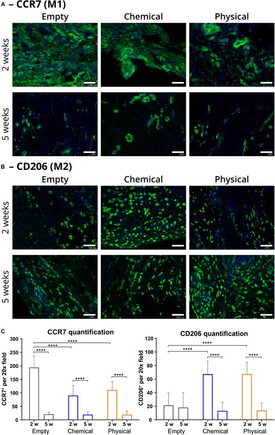
To assess our hypothesis, we first performed immunostaining
Segmentation tool in Fiji (ImageJ) (Arganda-Carreras et al.,
toward M1 (CCR7+ ) and M2 (CD206+ ) macrophages
2017). Myofiber size was determined by measuring the lesser
(Figures 2A,B, respectively) and quantified the number of
diameter (minimal Feret’s, defined as the closest distance between
each type of immune cells, observing that the injury areas of
the two parallel tangents of the muscle fiber) of 100 fibers
the samples treated with either the chemical or the physical
manually in four different locations (Briguet et al., 2004; Dubach-
hydrogels showed a significantly greater quantity of M2 than
Powell et al., 2008; Pertl et al., 2013) in the interface region of each
the empty (non-treated) samples (p < 0.0001) at 2 weeks
sample (n = 5 per group/time point). Data were represented as
post-injury (Figure 2C). Moreover, the presence of M1 in the
frequency distribution of size ranges with Gaussian distribution
hydrogel-treated samples was significantly lower than in the
with GraphPad Prism 6.0 software. The quantification of the
empty samples at this timepoint (p < 0.0001), thus giving a much
percentage of myofibers with internal nuclei was performed
higher M2/M1 ratio for the hydrogel-treated samples. These
similarly by counting the nuclei of a total of 100 fibers in four
ratios were 0.75 ± 0.38 and 0.61 ± 0.23 for the groups treated
different locations within the interface region of each sample
with the chemical and with the physical hydrogel, respectively,
(n = 5 per group/time point).
while it was 0.11 ± 0.10 for the empty group. On the other hand,
The entire muscle section was analyzed for collagen staining
the quantity of M1 and M2 macrophages decreased for every
with Picrosirius red under bright-field and polarized light. The
group after 5 weeks, giving similar values for all of them (nsd).
birefringent staining under polarized light is highly specific for
Nevertheless, the difference in M2 for the empty group between
type I and III mature collagen fibrils (Junqueira et al., 1979),
2 and 5 weeks was not significant, meaning that the quantity of
and it was used for collagen quantification with a custom macro
this type of macrophages did not peak at 2 weeks, contrarily to
in Fiji (ImageJ).
what we observed for the hydrogel-treated samples.
Since one of the main outcomes of a balanced macrophage
Immunofluorescence Staining response is the healing of the damaged tissue with a lesser amount
Immunofluorescence stainigs were performed as previously of fibrosis, we performed Picrosirius red histological staining
described (Aurora et al., 2016) to detect macrophages: type- to observe collagen deposition in the muscle samples. For this
1 or pro-inflammatory macrophages (anti-CCR7 antibody, purpose, we used polarized light (see Supplementary Figure 2 for
1:200; ab32527, Abcam, United Kingdom) (Corona et al., the bright-field pictures) to specifically differentiate and quantify
2013) and type-2 or anti-inflammatory macrophages (anti- collagen within the muscle sections for comparison between
mannose receptor (CD206) antibody, 1:200; ab64693, Abcam, groups (Figure 3A). In particular, there were not significant
United Kingdom) (Lankford et al., 2018). Alexa Fluor 488-labeled differences in collagen deposition in the interface area (injury
secondary antibody (1:500; ab150077, Abcam, United Kingdom) region with newly formed myofibers) between 2 and 5 weeks
was used for final immunostaining in both cases. At least 16 post-injury, although in the empty and physical hydrogel groups,
non-overlapping images (20× magnification) of the injury region but not in the chemical hydrogel one, there was a tendency
were randomly taken with a Nikon Eclipse Ti-E coupled to a toward increasing levels along time (Figure 3B). On the other
Frontiers in Bioengineering and Biotechnology | www.frontiersin.org 5 May 2020 | Volume 8 | Article 413Ibáñez-Fonseca et al. ELR Hydrogels for Skeletal Muscle Healing FIGURE 2 | Immunofluorescence representative images and quantification of macrophage populations in the injury area (scaffold and interface regions). (A) Type-1 macrophages (M1 or pro-inflammatory; in green) present in the injury area of samples from the empty (left), chemical (center), and physical (right) groups after 2 (top) and 5 weeks (bottom), labeled with an anti-CCR7 antibody. (B) Type-2 macrophages (M2 or anti-inflammatory; in green) present in the injury area of samples from the empty (left), chemical (center), and physical (right) groups after 2 (top) and 5 weeks (bottom), labeled with an anti-CD206 antibody. (C) Quantification of macrophage populations in the injury area (number of CCR7- or CD206-positive cells per 20× field) at 2 (2 w) and 5 weeks (5 w) post-injury. All the samples were counterstained with DAPI for nuclei. Scale bar = 50 µm. n = at least 16, which are the number of 20× fields used for the quantification. ****p < 0.0001. Frontiers in Bioengineering and Biotechnology | www.frontiersin.org 6 May 2020 | Volume 8 | Article 413
Ibáñez-Fonseca et al.                                                                                                         ELR Hydrogels for Skeletal Muscle Healing

  FIGURE 3 | Representative images and quantification for collagen staining with Picrosirius red. (A) Polarized light images from muscle samples of empty (left),
  chemical (center), and physical (right) groups for the scaffold (area without newly formed myofibers) and interface (area with newly formed myofibers) regions at 2
  (top) and 5 weeks (bottom) post-injury. Scale bar = 100 µm. (B,C) Percentage of collagen in the interface (including no injury group at 5 weeks) and scaffold regions,
  normalized to their areas, after 2 and 5 weeks. *p < 0.05, ***p < 0.001.

hand, there was a significant increase in collagen deposition in                        a greater remodeling of the muscle tissue during this time.
the scaffold area (injury region without newly formed myofibers)                        Moreover, we found an almost complete absence of myofibers
from 2 to 5 weeks for the empty and physical hydrogel groups                            with internal nuclei in the healthy tissue (Supplementary
(p < 0.05, respectively), whereas this was not observed for                             Figure 4), which suggests that the healing process only takes
the chemical hydrogel-treated samples (Figure 3C). Moreover,                            place in the interface and scaffold areas that represent the
the interface (remodeling) area of the samples treated with the                         potentially injured region.
chemical hydrogel after 5 weeks showed similar levels of collagen                          Further parameters, such as myofiber density, were also
compared to the total percentage of collagen presented in the                           quantified. In this case, we found that the density in the interface
non-injured (healthy) samples (Figure 3B), while the empty and                          area of samples from the empty group showed a significant
physical hydrogel groups presented significantly higher collagen                        decrease from 2 to 5 weeks (p < 0.05) (Figure 5A), which
levels (p
Ibáñez-Fonseca et al.                                                                                                           ELR Hydrogels for Skeletal Muscle Healing

  FIGURE 4 | (A) Representative images of the hematoxylin–eosin (HE) staining of sample cross-sections of the scaffold (area without newly formed myofibers) and
  interface (area with newly formed myofibers) regions (left and right for each group, respectively) of samples from the empty (left), chemical (center), and physical
  (right) groups at 2 (top) and 5 weeks (bottom) post-injury. Scale bar = 100 µm. (B,C) Percentage of the interface and scaffold areas, with respect to the injury area at
  2 and 5 weeks post-injury. **p < 0.01.

more similar to the size of the healthy ones than the cells in the                       is indicative of a lesser fibrosis. Similarly, histological results
empty samples at 5 weeks post-injury (Figure 5C), although all                           through hematoxylin-eosin staining showed that the morphology
the injured groups presented smaller myofibers at 2 and 5 weeks                          of the muscle treated with the ELR hydrogels is more similar to
than the uninjured samples.                                                              the healthy one than the untreated control at 5 weeks post-injury,
                                                                                         which is clearly evident when measuring specific parameters,
                                                                                         such as myofiber density and diameter. This effect is consistent
DISCUSSION                                                                               with the higher M2 response observed, which induces the
                                                                                         maturation of the myofibers to give a more healthy-like muscle,
The results obtained in this study show that the ELR hydrogels,                          while untreated samples, where the presence of M2 is much lower
both chemical and physical, promote a shift in macrophage                                than in the treated samples at 2 weeks post-injury, show smaller
polarization toward anti-inflammatory M2, highlighted by the                             myofibers, suggesting a lack of complete myogenic differentiation
higher M2/M1 ratio in comparison with the untreated control at                           and muscle growth.
2 weeks post-injury. As aforementioned, M2 are known to secrete                              In this work, we show how the use of a bioactive and
anti-inflammatory cytokines, while also regulating myogenic                              dynamic synthetic scaffold promotes an enhanced healing of
differentiation. Therefore, the increase in M2 for the hydrogel-                         injured skeletal muscle by itself, through the immunomodulation
treated samples at 2 weeks post-injury should promote a better                           of the macrophage response that guides the repair process. The
regulation of the skeletal muscle repair in comparison with the                          therapeutic strategy presented herein belongs to the field of in situ
untreated control, resulting in a morphology more similar to the                         tissue regeneration, where a host cell recruitment is achieved
healthy muscle and with a less amount of fibrotic/scar tissue.                           (Lee et al., 2016), in this case without the use of external cells
In this last regard, we studied collagen content by Picrosirius                          or growth factors, hence preventing potential side effects (Baldo,
red staining, and it revealed that there was less collagen in                            2014). The immunomodulation shown by ELR hydrogels has
the hydrogel-treated samples in comparison to the untreated                              also been observed in other works that used biological scaffolds,
control, especially in the case of the chemical hydrogel, which                          mainly decellularized urinary bladder matrix (UBM), to improve

Frontiers in Bioengineering and Biotechnology | www.frontiersin.org                  8                                                   May 2020 | Volume 8 | Article 413
Ibáñez-Fonseca et al. ELR Hydrogels for Skeletal Muscle Healing FIGURE 5 | Histomorphometric characterization of the HE-stained samples. (A) Myofiber density at 2 and 5 weeks post-injury for the empty (gray), chemical (blue) and physical (orange) groups. (B) Myofiber density after 5 weeks for the empty (gray), chemical (blue) and physical (orange) groups in comparison with no injury (healthy) samples (red). (C) Myofiber size distribution (number of myofibers with a specific diameter) for the different groups used in this study, i.e., empty, chemical, and physical, after 2 and 5 weeks, in comparison with the no injury (healthy) group. *p < 0.05. the healing of VML injuries. In some of them, authors do not RGD promotes a physiological interaction with the scaffolds, observe a shift from a M1 to a M2 response, and, as they state, that hence resemble some of the properties of the native this leads to an impaired muscle healing (Aurora et al., 2015, ECM. Another reason that may provide an explanation to 2016; Greising et al., 2017), whereas some others report dissimilar the findings of this work is the fact that the ELR hydrogels results (Sicari et al., 2014; Dziki et al., 2016; Lee et al., 2019). used herein are biodegradable. This means that they act as a On the other hand, it has been suggested that the treatment transient ECM-like scaffold, providing a dynamic environment with synthetic biomaterial-based scaffolds, comparable to the that evolves as required by the cells involved in the healing ELR hydrogels used in this work, also promote a M2- process, which secrete MMPs relevant for the proteolysis of balanced immune response, with different examples using a the hydrogels [mainly macrophages (Turner and Badylak, photoresponsive hyaluronan hydrogel (Wang et al., 2019), a 2012)] and make space for the formation of regenerated tissue. biohybrid pNIPAAm and UBM hydrogel (Zhu et al., 2018), Indeed, we found in a preliminary study that biodegradable keratin (Passipieri et al., 2017), or fibrin by itself (Tanaka ELR hydrogels are completely necessary for the healing of the et al., 2019). This strategy has several advantages in comparison VML injury, which otherwise is permanently occupied by the with biological scaffolds, since the controlled synthesis of the scaffold (Supplementary Figure 5). Previous studies have shown biomaterials used for their fabrication does not rely on methods the importance of the biodegradation of ELR hydrogels with like decelullarization. potential application in tissue engineering, influencing, for Regarding the immunomodulation mediated by ELR instance, vascularization (Staubli et al., 2017; Flora et al., 2019), hydrogels, we suggest that, on one hand, the presence of which is considered one of the main events to achieve a successful Frontiers in Bioengineering and Biotechnology | www.frontiersin.org 9 May 2020 | Volume 8 | Article 413
Ibáñez-Fonseca et al.                                                                                                                    ELR Hydrogels for Skeletal Muscle Healing

healing of damaged tissues. In addition, the filling of the void                                 ETHICS STATEMENT
created through the VML with the ELR hydrogels impedes the
formation of a large scar tissue (fibrosis) that usually impairs                                 The animal study was reviewed and approved by the Comité
muscle healing (Järvinen et al., 2005; Turner and Badylak, 2012).                                de Ética en Experimentación y Bienestar Animal (CEEBA) de la
Therefore, the use of biodegradable ELR hydrogels could provide                                  Universidad de Valladolid (protocol number 5402485).
a niche that promotes vascularization and replacement by newly
formed skeletal muscle, resulting in an efficient healing.
    This investigation has a main limitation, which is the lack of
functional characterization of the TA muscles. Nevertheless, we                                  AUTHOR CONTRIBUTIONS
aimed to delve into the effect of ELR hydrogels in the healing of
                                                                                                 AI-F, SS, DG, MA, HA, and JR-C designed the study. AI-F, SS,
VML injuries in terms of cell and molecular biology, in order
                                                                                                 DG, BC, ÁÁ, and HA performed the experiments. AI-F, SS, and
to set the basis for future works with larger animal models
                                                                                                 DG wrote the manuscript. AV, MA, HA, and JR-C revised the
(Pollot and Corona, 2016). These models will not only be more
                                                                                                 manuscript. All authors approved the final version.
relevant as regards the future application of ELR hydrogels in the
treatment of skeletal muscle injuries in humans, but they will also
allow the use of non-invasive techniques already implemented in
clinics for the determination of muscle function.                                                FUNDING
                                                                                                 This work was funded by the Spanish Government (MAT2016-
CONCLUSION                                                                                       78903-R, RTI2018-096320-B-C22), Junta de Castilla y León
                                                                                                 (VA015U16, VA317P18), Interreg V A España Portugal POCTEP
In conclusion, this study demonstrates that bioactive and
                                                                                                 (0624_2IQBIONEURO_6_E) and Centro en Red de Medicina
biodegradable ELR hydrogels regulate the macrophage response
                                                                                                 Regenerativa y Terapia Celular de Castilla y León.
by inducing M2 polarization after a VML injury. This
immunomodulation results in an enhanced skeletal muscle
healing, with a reduced collagen deposition and a muscle
morphology more similar to the healthy tissue. Therefore,                                        ACKNOWLEDGMENTS
we confirmed that ELR hydrogels provide a cell-friendly and
dynamic environment that induces M2 shift and supports an                                        The authors acknowledge the support of the technicians from
enhanced healing, especially in the case of the chemically                                       the Servicio de Investigación y Bienestar Animal (SIBA) of the
crosslinked ELR hydrogel, which showed a reduced fibrosis. The                                   University of Valladolid, and of Rocío García for the help in the
work presented here paves the way for future studies in more                                     bioproduction of the ELRs used in this work.
relevant large animal models of VML treated with the chemical
ELR hydrogel that include functional characterization.
                                                                                                 SUPPLEMENTARY MATERIAL
DATA AVAILABILITY STATEMENT
                                                                                                 The Supplementary Material for this article can be found
Data associated with this study is available upon request to the                                 online at: https://www.frontiersin.org/articles/10.3389/fbioe.
corresponding author.                                                                            2020.00413/full#supplementary-material

REFERENCES                                                                                          M. Mozafari, F. Sefat, and A. Atala (New York, NY: Woodhead Publishing),
                                                                                                    245–258.
Arganda-Carreras, I., Kaynig, V., Rueden, C., Eliceiri, K. W., Schindelin, J.,                   Boersema, G. S. A., Grotenhuis, N., Bayon, Y., Lange, J. F., and Bastiaansen-
   Cardona, A., et al. (2017). Trainable weka segmentation: a machine learning                      Jenniskens, Y. M. (2016). The effect of biomaterials used for tissue regeneration
   tool for microscopy pixel classification. Bioinformatics 33, 2424–2426. doi: 10.                 purposes on polarization of macrophages. Biores. Open Access 5, 6–14. doi:
   1093/bioinformatics/btx180                                                                       10.1089/biores.2015.0041
Aurora, A., Corona, B. T., and Walters, T. J. (2016). A porcine urinary bladder                  Briguet, A., Courdier-Fruh, I., Foster, M., Meier, T., and Magyar, J. P. (2004).
   matrix does not recapitulate the spatiotemporal macrophage response of muscle                    Histological parameters for the quantitative assessment of muscular dystrophy
   regeneration after volumetric muscle loss injury. Cells Tissues Organs 202,                      in the mdx-mouse. Neuromuscul. Disord. 14, 675–682. doi: 10.1016/j.nmd.2004.
   189–201. doi: 10.1159/000447582                                                                  06.008
Aurora, A., Roe, J. L., Corona, B. T., and Walters, T. J. (2015). An acellular                   Cappello, J., Crissman, J., Dorman, M., Mikolajczak, M., Textor, G., Marquet, M.,
   biologic scaffold does not regenerate appreciable de novo muscle tissue in                       et al. (1990). Genetic engineering of structural protein polymers. Biotechnol.
   rat models of volumetric muscle loss injury. Biomaterials 67, 393–407. doi:                      Prog. 6, 198–202. doi: 10.1021/bp00003a006
   10.1016/j.biomaterials.2015.07.040                                                            Chung, E. H., Gilbert, M., Virdi, A. S., Sena, K., Sumner, D. R., and Healy, K. E.
Baldo, B. A. (2014). Side effects of cytokines approved for therapy. Drug Saf. 37,                  (2006). Biomimetic artificial ECMs stimulate bone regeneration. J. Biomed.
   921–943. doi: 10.1007/s40264-014-0226-z                                                          Mater. Res. A 79A, 815–826. doi: 10.1002/jbm.a.30809
Bartolacci, J., Dziki, J., and Badylak, S. F. (2019). “12 - Scaffolds for skeletal muscle        Coletta, D. J., Ibáñez-Fonseca, A., Missana, L. R., Jammal, M. V., Vitelli,
   tissue engineering,” in Handbook of Tissue Engineering Scaffolds, Vol. One, eds                  E. J., Aimone, M., et al. (2017). Bone regeneration mediated by a bioactive

Frontiers in Bioengineering and Biotechnology | www.frontiersin.org                         10                                                    May 2020 | Volume 8 | Article 413
Ibáñez-Fonseca et al. ELR Hydrogels for Skeletal Muscle Healing and biodegradable extracellular matrix-like hydrogel based on elastin-like control of self-assembly on the microstructure evolution of silk-elastin-like recombinamers. Tissue Eng. A 23, 1361–1371. doi: 10.1089/ten.tea.2017.0047 recombinamer hydrogels. Small doi: 10.1002/smll.202001244 Corona, B. T., Garg, K., Ward, C. L., McDaniel, J. S., Walters, T. J., and Rathbone, Ibáñez-Fonseca, A., Ramos, T. L., González de Torre, I., Sánchez-Abarca, L. I., C. R. (2013). Autologous minced muscle grafts: a tissue engineerin therapy Muntión, S., Arias, F. J., et al. (2018). Biocompatibility of two model elastin-like for the volumetric loss of skeletal muscle. Am. J. Physiol. Cell Physiol. 305, recombinamer-based hydrogels formed through physical or chemical cross- C761–C775. doi: 10.1152/ajpcell.00189.2013 linking for various applications in tissue engineering and regenerative medicine. Corona, B. T., Rivera, J. C., Owens, J. G., Wenke, J. C., and Rathbone, C. R. (2015). J. Tissue Eng. Regen. Med. 12, e1450–e1460. doi: 10.1002/term.2562 Volumetric muscle loss leads to permanent disability following extremity Järvinen, T. A. H., Järvinen, T. L. N., Kääriäinen, M., Kalimo, H., and Järvinen, M. trauma. J. Rehabil. Res. Dev. 52, 785–792. doi: 10.1682/JRRD.2014.07.0165 (2005). Muscle injuries: biology and treatment. Am. J. Sports Med. 33, 745–764. Costa, R. R., Custódio, C. A., Testera, A. M., Arias, F. J., Rodríguez-Cabello, doi: 10.1177/0363546505274714 J. C., Alves, N. M., et al. (2009). Stimuli-responsive thin coatings using elastin- Junqueira, L. C. U., Bignolas, G., and Brentani, R. R. (1979). Picrosirius staining like polymers for biomedical applications. Adv. Funct. Mater. 19, 3210–3218. plus polarization microscopy, a specific method for collagen detection in tissue doi: 10.1002/adfm.200900568 sections. Histochem. J. 11, 447–455. doi: 10.1007/bf01002772 Dubach-Powell, J., Erb, M., Van Putten, M., Thirion, C., Barton, E., and Rüegg, Klinkenberg, M., Fischer, S., Kremer, T., Hernekamp, F., Lehnhardt, M., and M. A. (2008). Quantitative Determination of Muscle Fiber Diameter (Minimal Daigeler, A. (2013). Comparison of anterolateral thigh, lateral arm, and Feret’s Diameter) and Percentage of Centralized Nuclei. Available online at: parascapular free flaps with regard to donor-site morbidity and aesthetic and www.treat-nmd.eu/downloads/file/sops/dmd/MDX/DMD_M.1.2.001.pdf functional outcomes. Plast. Reconstr. Surg. 131, 293–302. doi: 10.1097/PRS. (accessed April 12, 2020). 0b013e31827786bc Dziki, J. L., Sicari, B. M., Wolf, M. T., Cramer, M. C., and Badylak, S. F. (2016). Lankford, K. L., Arroyo, E. J., Nazimek, K., Bryniarski, K., Askenase, P. W., and Immunomodulation and mobilization of progenitor cells by extracellular Kocsis, J. D. (2018). Intravenously delivered mesenchymal stem cell-derived matrix bioscaffolds for volumetric muscle loss treatment. Tissue Eng. Part A 22, exosomes target M2-type macrophages in the injured spinal cord. PLoS One 1129–1139. doi: 10.1089/ten.TEA.2016.0340 13:e0190358. doi: 10.1371/journal.pone.0190358 Fernández-Colino, A., Arias, F. J., Alonso, M., and Rodríguez-Cabello, J. C. (2014). Lee, H., Ju, Y. M., Kim, I., Elsangeedy, E., Lee, J. H., Yoo, J. J., et al. (2019). A Self-Organized ECM-mimetic model based on an amphiphilic multiblock silk- novel decellularized skeletal muscle-derived ECM scaffolding system for in situ elastin-like corecombinamer with a concomitant dual physical gelation process. muscle regeneration. Methods 171, 77–85. doi: 10.1016/j.ymeth.2019.06.027 Biomacromolecules 15, 3781–3793. doi: 10.1021/bm501051t Lee, S. J., Yoo, J. J., and Atala, A. (2016). “Chapter 1 - Fundamentals of In Situ Flora, T., González de Torre, I., Alonso, M., and Rodríguez-Cabello, J. C. (2019). Tissue Regeneration,” in In Situ Tissue Regeneration, eds S. J. Lee, J. J. Yoo, and Use of proteolytic sequences with different cleavage kinetics as a way to generate A. Atala (Boston: Academic Press), 3–17. hydrogels with preprogrammed cell-infiltration patterns imparted over their Lepper, C., Partridge, T. A., and Fan, C.-M. (2011). An absolute requirement given 3D spatial structure. Biofabrication 11:035008. doi: 10.1088/1758-5090/ for Pax7-positive satellite cells in acute injury-induced skeletal muscle ab10a5 regeneration. Development 138, 3639–3646. doi: 10.1242/dev.067595 Girotti, A., Orbanic, D., Ibáñez-Fonseca, A., Gonzalez-Obeso, C., and Rodríguez- Lin, C.-H., Lin, Y.-T., Yeh, J.-T., and Chen, C.-T. (2007). Free functioning muscle Cabello, J. C. (2015). Recombinant technology in the development of materials transfer for lower extremity posttraumatic composite structure and functional and systems for soft-tissue repair. Adv. Healthc. Mater. 4, 2423–2455. doi: defect. Plast. Reconstr. Surg. 119, 2118–2126. doi: 10.1097/01.prs.0000260595. 10.1002/adhm.201500152 85557.41 Girotti, A., Reguera, J., Rodríguez-Cabello, J. C., Arias, F. J., Alonso, M., Lin, S.-H., Chuang, D. C.-C., Hattori, Y., and Chen, H.-C. (2004). Traumatic and Testera, A. M. (2004). Design and bioproduction of a recombinant major muscle loss in the upper extremity: reconstruction using functioning multi(bio)functional elastin-like protein polymer containing cell adhesion free muscle transplantation. J. Reconstr. Microsurg. 20, 227–235. doi: 10.1055/ sequences for tissue engineering purposes. J. Mater. Sci. Mater. Med. 15, s-2004-823110 479–484. doi: 10.1023/B:JMSM.0000021124.58688.7a Lutolf, M. P., Weber, F. E., Schmoekel, H. G., Schense, J. C., Kohler, T., Müller, González de Torre, I., Santos, M., Quintanilla, L., Testera, A., Alonso, M., R., et al. (2003). Repair of bone defects using synthetic mimetics of collagenous and Rodríguez Cabello, J. C. (2014). Elastin-like recombinamer catalyst- extracellular matrices. Nat. Biotechnol. 21, 513–518. doi: 10.1038/nbt818 free click gels: characterization of poroelastic and intrinsic viscoelastic Madl, C. M., Katz, L. M., and Heilshorn, S. C. (2016). Bio-orthogonally crosslinked, properties. Acta Biomater. 10, 2495–2505. doi: 10.1016/j.actbio.2014. engineered protein hydrogels with tunable mechanics and biochemistry for cell 02.006 encapsulation. Adv. Funct. Mater. 26, 3612–3620. doi: 10.1002/adfm.201505329 Grasman, J. M., Zayas, M. J., Page, R. L., and Pins, G. D. (2015). Biomimetic Martín, L., Arias, F. J., Alonso, M., García-Arévalo, C., and Rodríguez-Cabello, J. C. scaffolds for regeneration of volumetric muscle loss in skeletal muscle injuries. (2010). Rapid micropatterning by temperature-triggered reversible gelation of Acta Biomater. 25, 2–15. doi: 10.1016/j.actbio.2015.07.038 a recombinant smart elastin-like tetrablock-copolymer. Soft Matter. 6, 1121– Greising, S. M., Rivera, J. C., Goldman, S. M., Watts, A., Aguilar, C. A., and Corona, 1124. doi: 10.1039/B923684H B. T. (2017). Unwavering pathobiology of volumetric muscle loss injury. Sci. O’Brien, F. J. (2011). Biomaterials & scaffolds for tissue engineering. Mater. Today Rep. 7:13179. doi: 10.1038/s41598-017-13306-2 14, 88–95. doi: 10.1016/S1369-7021(11)70058-X Greising, S. M., Warren, G. L., Southern, W. M., Nichenko, A. S., Qualls, A. E., Passipieri, J. A., Baker, H. B., Siriwardane, M., Ellenburg, M. D., Vadhavkar, M., Corona, B. T., et al. (2018). Early rehabilitation for volumetric muscle loss injury Saul, J. M., et al. (2017). Keratin hydrogel enhances in vivo skeletal muscle augments endogenous regenerative aspects of muscle strength and oxidative function in a rat model of volumetric muscle loss. Tissue Eng. Part A 23, capacity. BMC Musculoskelet. Disord. 19:173. doi: 10.1186/s12891-018- 556–571. doi: 10.1089/ten.tea.2016.0458 2095-6 Pertl, C., Eblenkamp, M., Pertl, A., Pfeifer, S., Wintermantel, E., Lochmüller, H., Grogan, B. F., Hsu, J. R., and Skeletal Trauma Research Consortium (2011). et al. (2013). A new web-based method for automated analysis of muscle Volumetric muscle loss. J. Am. Acad. Orthop. Surg. 19(Suppl. 1), S35–S37. histology. BMC Musculoskelet. Disord. 14:26. doi: 10.1186/1471-2474-14-26 doi: 10.5435/00124635-201102001-00007 Pescador, D., Ibáñez-Fonseca, A., Sánchez-Guijo, F., Briñón, J. G., Arias, F. J., Ibáñez-Fonseca, A., Alonso, M., Arias, F. J., and Rodríguez-Cabello, J. C. (2017). Muntión, S., et al. (2017). Regeneration of hyaline cartilage promoted Förster resonance energy transfer-paired hydrogel forming silk-elastin-like by xenogeneic mesenchymal stromal cells embedded within elastin-like recombinamers by recombinant conjugation of fluorescent proteins. Bioconjug. recombinamer-based bioactive hydrogels. J. Mater. Sci. Mater. Med. 28:115. Chem. 28, 828–835. doi: 10.1021/acs.bioconjchem.6b00738 doi: 10.1007/s10856-017-5928-1 Ibáñez-Fonseca, A., Flora, T., Acosta, S., and Rodríguez-Cabello, J. C. (2019). Pollot, B. E., and Corona, B. T. (2016). “Volumetric muscle loss,” in Skeletal Muscle Trends in the design and use of elastin-like recombinamers as biomaterials. Regeneration in the Mouse: Methods and Protocols, ed. M. Kyba (New York, NY: Matrix Biol. 84, 111–126. doi: 10.1016/j.matbio.2019.07.003 Springer), 19–31. Ibáñez-Fonseca, A., Orbanic, D., Arias, F. J., Alonso, M., Zeugolis, D. I., and Qazi, T. H., Mooney, D. J., Pumberger, M., Geißler, S., and Duda, G. N. (2015). Rodríguez-Cabello, J. C. (2020). Influence of the thermodynamic and kinetic Biomaterials based strategies for skeletal muscle tissue engineering: existing Frontiers in Bioengineering and Biotechnology | www.frontiersin.org 11 May 2020 | Volume 8 | Article 413
Ibáñez-Fonseca et al.                                                                                                                       ELR Hydrogels for Skeletal Muscle Healing

   technologies and future trends. Biomaterials 53, 502–521. doi: 10.1016/j.                       Tirrell, D. A., Fournier, M. J., and Mason, T. L. (1991). Genetic engineering of
   biomaterials.2015.02.110                                                                           polymeric materials. MRS Bull. 16, 23–28. doi: 10.1557/S0883769400056505
Rodríguez-Cabello, J. C., Girotti, A., Ribeiro, A., and Arias, F. J. (2012). “Synthesis            Turner, N. J., and Badylak, S. F. (2012). Regeneration of skeletal muscle. Cell Tissue
   of Genetically Engineered Protein Polymers (Recombinamers) as an Example of                        Res. 347, 759–774. doi: 10.1007/s00441-011-1185-7
   Advanced Self-Assembled Smart Materials,” in Nanotechnology in Regenerative                     Urry, D. W. (2006). What Sustains Life? Consilient Mechanisms for Protein-Based
   Medicine: Methods and Protocols, eds M. Navarro and J. A. Planell (Totowa, NJ:                     Machines and Materials. Boston: Birkhäuser Boston.
   Humana Press), 17–38. doi: 10.1007/978-1-61779-388-2_2                                          Urry, D. W., Okamoto, K., Harris, R. D., Hendrix, C. F., and Long, M. M.
Rodríguez-Cabello, J. C., Martín, L., Alonso, M., Arias, F. J., and Testera, A. M.                    (1976). Synthetic, cross-linked polypentapeptide of tropoelastin: an anisotropic,
   (2009). “Recombinamers” as advanced materials for the post-oil age. Polymer                        fibrillar elastomer. Biochemistry 15, 4083–4089. doi: 10.1021/bi00663a026
   50, 5159–5169. doi: 10.1016/j.polymer.2009.08.032                                               Wang, H., Morales, R.-T. T., Cui, X., Huang, J., Qian, W., Tong, J., et al.
Ruoslahti, E. (1996). RGD and other recognition sequences for integrins. Annu.                        (2019). A Photoresponsive Hyaluronan Hydrogel Nanocomposite for Dynamic
   Rev. Cell Dev. Biol. 12, 697–715. doi: 10.1146/annurev.cellbio.12.1.697                            Macrophage Immunomodulation. Adv. Healthc. Mater. 8:1801234. doi: 10.
Saclier, M., Yacoub-Youssef, H., Mackey, A. L., Arnold, L., Ardjoune, H., Magnan,                     1002/adhm.201801234
   M., et al. (2013). Differentially activated macrophages orchestrate myogenic                    Wolf, M. T., Dearth, C. L., Sonnenberg, S. B., Loboa, E. G., and Badylak,
   precursor cell fate during human skeletal muscle regeneration. Stem Cells 31,                      S. F. (2015). Naturally derived and synthetic scaffolds for skeletal muscle
   384–396. doi: 10.1002/stem.1288                                                                    reconstruction. Adv. Drug Deliv. Rev 84, 208–221. doi: 10.1016/j.addr.2014.
Schindelin, J., Arganda-Carreras, I., Frise, E., Kaynig, V., Longair, M., Pietzsch, T.,               08.011
   et al. (2012). Fiji: an open-source platform for biological-image analysis. Nat.                Wu, X., Corona, B. T., Chen, X., and Walters, T. J. (2012). A standardized rat model
   Methods 9:676. doi: 10.1038/nmeth.2019                                                             of volumetric muscle loss injury for the development of tissue engineering
Shin, E. H., Caterson, E. J., Jackson, W. M., and Nesti, L. J. (2014). Quality of                     therapies. Biores. Open Access 1, 280–290. doi: 10.1089/biores.2012.0271
   healing: defining, quantifying, and enhancing skeletal muscle healing. Wound                    Zalavras, C. G., and Patzakis, M. J. (2003). Open fractures: evaluation and
   Repair Regen. 22, 18–24. doi: 10.1111/wrr.12163                                                    management. JAAOS J. Am. Acad. Orth. Surg. 11, 212–219.
Sicari, B. M., Dziki, J. L., Siu, B. F., Medberry, C. J., Dearth, C. L., and Badylak, S. F.        Zhu, Y., Hideyoshi, S., Jiang, H., Matsumura, Y., Dziki, J. L., LoPresti, S. T., et al.
   (2014). The promotion of a constructive macrophage phenotype by solubilized                        (2018). Injectable, porous, biohybrid hydrogels incorporating decellularized
   extracellular matrix. Biomaterials 35, 8605–8612. doi: 10.1016/j.biomaterials.                     tissue components for soft tissue applications. Acta Biomater. 73, 112–126.
   2014.06.060                                                                                        doi: 10.1016/j.actbio.2018.04.003
Staubli, S. M., Cerino, G., Gonzalez de Torre, I., Alonso, M., Oertli, D., Eckstein,
   F., et al. (2017). Control of angiogenesis and host response by modulating                      Conflict of Interest: The authors declare that the research was conducted in the
   the cell adhesion properties of an Elastin-Like Recombinamer-based hydrogel.                    absence of any commercial or financial relationships that could be construed as a
   Biomaterials 135, 30–41. doi: 10.1016/j.biomaterials.2017.04.047                                potential conflict of interest.
Tanaka, R., Saito, Y., Fujiwara, Y., Jo, J.-I., and Tabata, Y. (2019). Preparation
   of fibrin hydrogels to promote the recruitment of anti-inflammatory                             Copyright © 2020 Ibáñez-Fonseca, Santiago Maniega, Gorbenko del Blanco, Catalán
   macrophages. Acta Biomater. 89, 152–165. doi: 10.1016/j.actbio.2019.03.011                      Bernardos, Vega Castrillo, Álvarez Barcia, Alonso, Aguado and Rodríguez-Cabello.
Tedesco, F. S., Dellavalle, A., Diaz-Manera, J., Messina, G., and Cossu, G. (2010).                This is an open-access article distributed under the terms of the Creative Commons
   Repairing skeletal muscle: regenerative potential of skeletal muscle stem cells.                Attribution License (CC BY). The use, distribution or reproduction in other forums
   J. Clin. Invest. 120, 11–19. doi: 10.1172/JCI40373                                              is permitted, provided the original author(s) and the copyright owner(s) are credited
Tidball, J. G., and Villalta, S. A. (2010). Regulatory interactions between muscle and             and that the original publication in this journal is cited, in accordance with accepted
   the immune system during muscle regeneration. Am. J. Physiol. Regul. Integr.                    academic practice. No use, distribution or reproduction is permitted which does not
   Comp. Physiol. 298, R1173–R1187. doi: 10.1152/ajpregu.00735.2009                                comply with these terms.

Frontiers in Bioengineering and Biotechnology | www.frontiersin.org                           12                                                      May 2020 | Volume 8 | Article 413
You can also read