Could de-stressing the brain be the solution for long-term weight loss?
←
→
Page content transcription
If your browser does not render page correctly, please read the page content below
Viewpoint
                                                                                    www.cell-stress.com

Could de-stressing the brain be the solution for long-term
weight loss?
Florian Seyfried1 and Mohammed K. Hankir2,*
1
  Department of General, Visceral, Vascular and Pediatric Surgery, University Hospital Wuerzburg, Wuerzburg, 97080 Bavaria, Ger-
many.
2
  Department of Experimental Surgery, University Hospital Wuerzburg, Wuerzburg, 97080 Bavaria, Germany.
* Corresponding Author:
Dr. Mohammed K. Hankir, PhD, Centre of Operative Medicine, Oberduerrbacherstraße 6, Wuerzburg, 97080 Bavaria, Germany;
Tel.: +49 9312 01 31728; E-mail: hankir_m@ukw.de

ABSTRACT The obese brain is stressed and inflamed. This is mainly at the lev-                doi: 10.15698/cst2019.02.174
el of neurons and glial cells in the hypothalamus: a brain region where the                  Received originally: 24.09.2018
                                                                                             in revised form: 16.01.2019,
adipokine leptin acts to control feeding and body weight. Relieving hypotha-
                                                                                             Accepted 22.01.2019,
lamic neuronal endoplasmic reticulum (ER) stress with the natural small mol-                 Published 25.01.2019.
ecule drugs celastrol or withaferin-A reverses the leptin resistance commen-
surate with obesity, producing a degree of weight loss found only with bari-
atric surgery. Here, recent evidence from rodent models of vertical sleeve                   Keywords: endoplasmic reticulum
gastrectomy (VSG) is brought to the fore which suggests that this particular                 stress, gliosis, hypothalamus,
                                                                                             inflammation, leptin resistance,
bariatric surgical procedure may work in a similar fashion to celastrol and
                                                                                             obesity, vertical sleeve gastrectomy.
withaferin-A alongside remedying hypothalamic inflammation and gliosis.
Thus, restoring and preserving healthy hypothalamic neuronal and glial cell
function, be it by pharmacological or surgical means, ensures a negative ener-               Abbreviations:
gy balance in an environment constructed to promote a one - possibly                         eIF2a – elongation initiation factor 2
                                                                                             alpha,
through re-establishing communication between adipose tissue and the brain.                  ER – endoplasmic reticulum,
                                                                                             FXR – farnesoid X receptor,
                                                                                             GABA – gamma amino butyric acid,
                                                                                             GLP-1 – glucagon-like peptide 1,
                                                                                             IKK-β – inhibitor of kappa beta kinase
                                                                                             beta,
                                                                                             IL – interleukin,
                                                                                             HSF1 – heat shock factor 1,
                                                                                             NF-Кβ – necrosis factor kappa beta,
                                                                                             PERK – protein kinase R (PKR)-like ER
                                                                                             kinase,
                                                                                             PTP1B – protein tyrosine phosphatase 1B,
                                                                                             PGC-1α – peroxisome proliferator-
                                                                                             activated receptor gamma coactivator 1-
                                                                                             alpha,
                                                                                             SOCS3 – suppressor of cytokine signaling
                                                                                             3,
                                                                                             TGR5 – Takeda G-protein 5 receptor,
                                                                                             TLR4 – toll-like receptor 4,
                                                                                             TNF-α – tumor necrosis factor alpha,
                                                                                             UCP1 – uncoupling protein 1,
                                                                                             VSG – vertical sleeve gastrectomy,
                                                                                             vWAT – visceral white adipose tissue.

INTRODUCTION                                                         generally less active way of life and superimposed on a
One just needs to take a look at a typical vending machine           susceptible genetic background has fueled a steep rise in
(often found in hospitals) to appreciate what has happened           global obesity prevalence. This in turn has directly contrib-
to our food and drink. The relatively sudden widespread              uted to the increased incidence of chronic debilitating con-
ease of access to energy-dense meals, combined with a                ditions such as type II diabetes, atherosclerosis, cardiovas-

OPEN ACCESS | www.cell-stress.com                           29                          Cell Stress | FEBRUARY 2019 | Vol. 3 No. 2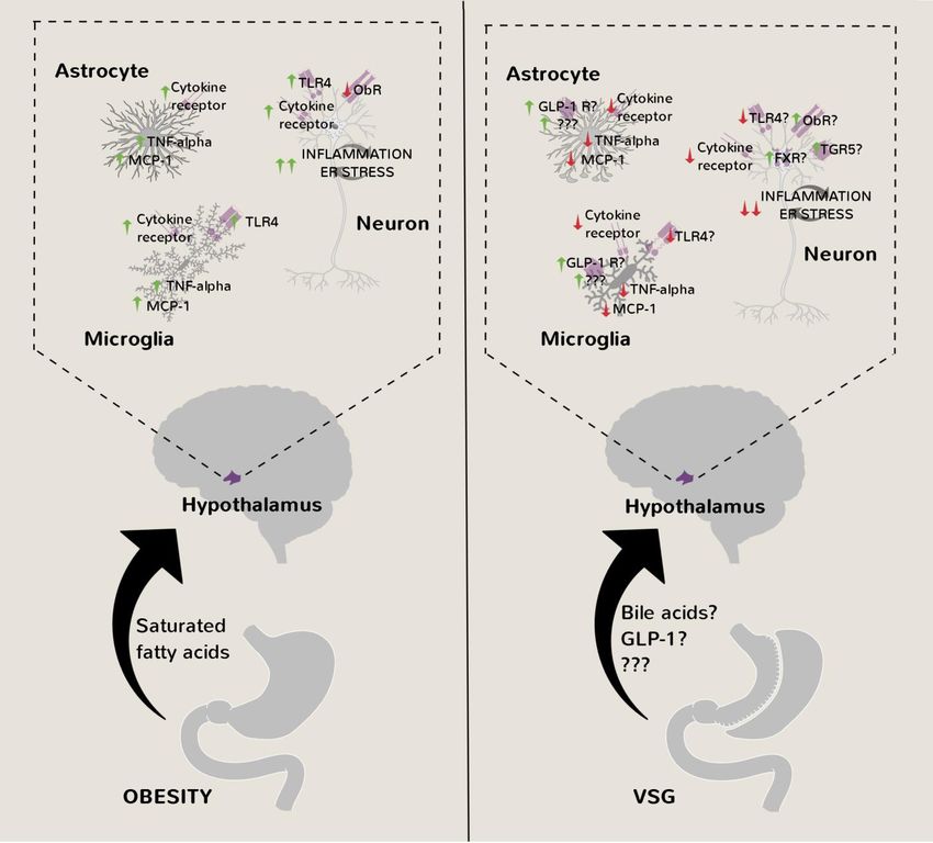
F. Seyfried and M.K. Hankir (2019)                                                          Hypothalamic inflammation in weight loss and gain

cular disease and certain cancers which are all major caus-               ic CD8+ and Th1 T cells, which secrete the pro-
es of premature death.                                                    inflammatory and intestinal barrier-disrupting cytokine
                                                                          interferon gamma (IFN-γ) [8]. The loss of gut barrier integ-
ADIPOSE TISSUE AND INTESTINAL INFLAMMATION AS                             rity itself in obesity results in systemic endotoxemia which
A CAUSE OF INSULIN RESISTANCE                                             too contributes to vWAT macrophage activation, possibly
Early efforts to disentangle the close relationship between               through bacterially-derived lipopolysaccharide (LPS) acting
obesity and type II diabetes focused on the pro-                          on local toll-like receptor 4 (TLR4) [8, 9]. Thus, we now
inflammatory cytokine tumor necrosis factor alpha (TNF-α)                 know a great deal about the molecular and cellular under-
in visceral white adipose tissue (vWAT) [1]. A pronounced                 pinnings of obesity-induced insulin resistance, although
increase was found in various rodent models of obesity [1]                this has yet to be translated into an effective immune-
and this marked the beginning of a powerful narrative in                  based therapy for type II diabetes in humans.
which increased visceral adiposity causes a state of chronic,
low-grade systemic inflammation involving both the adap-                  HYPOTHALAMIC INFLAMMATION AS A CAUSE OF
tive and innate immune systems. Subsequent rodent stud-                   LEPTIN RESISTANCE
ies revealed in reverse chronological order that during                   At about the same time that inflammation in the vWAT of
obesity progression, antigen-presenting and IgG-releasing                 severely obese and diabetic ob/ob mice was discovered [1],
B cells are the first to arrive in vWAT, possibly attracted by            the ob gene encoding the adipokine leptin was itself cloned
chemokines released from swelling adipocytes sensed by                    [10]. This generated considerable excitement about the
the mechanoreceptor transient receptor potential vanilloid                prospects of a new and more effective obesity pharma-
4 (TRPV4) [2], followed by the recruitment of cytotoxic                   cotherapy. However, it was soon realized that individuals
CD8+ T cells and then by M2 macrophages [3, 4 ,5]. These                  with obesity are refractory to the appetite suppressing and
white blood cells all release a plethora of pro-inflammatory              weight lowering effects of exogenous leptin treatment [11].
cytokines including macrophage-derived TNF-α [6] that                     Analogous to insulin resistance, inflammatory processes in
interfere with insulin receptor signaling both locally in                 the brain, specifically in hypothalamic neurons, would pro-
vWAT and distally in peripheral tissues such as the liver and             vide a link between obesity and the relatively newly coined
skeletal muscle [7] (Figure 1).                                           term leptin resistance [12]. Furthermore, hypothalamic
    It is now increasingly recognized that a complex im-                  neuronal endoplasmic reticulum (ER) stress was also
mune response also takes hold in the gut from chronic                     shown to develop upon chronic high-fat food consumption
consumption of a high-fat diet due to shifts in resident                  in mice, being both a cause and a consequence of pro-
microbiota species. This engenders the downregulation of                  inflammatory inhibitor of kappa beta kinase beta (IKK-β)
regulatory T cells (Treg ) and innate lymphoid cells (ILCs),              signaling [12]. That acute brain overload of the saturated
which normally secrete the anti-inflammatory/intestinal                   fatty acid oleate was sufficient to increase hypothalamic
barrier-protective cytokines interleukin 10 (IL-10) and IL-22,            necrosis factor kappa beta (NF-Кβ) transcriptional activity
respectively, with the concomitant upregulation of cytotox-               supported the concept that high-fat feeding first promotes

FIGURE 1: Molecular and cellular processes linking obesity with systemic insulin resistance. Swelling (hyperplasic) visceral adipocytes in
obesity have hyperactivated TRPV4 which leads to changes in gene expression (through extracellular related kinase 1/2 signaling) including
an increase in chemokine production. This sets into motion a sequence of molecular and cellular events ultimately leading to insulin re-
sistance in hepatocytes and myocytes in part through inhibitory serine 120 and serine 210 phosphorylation of insulin receptor substrate 1
in these cells by TNF-α. CXCL1 - chemokine (C-X-C motif) ligand 1, IFN-gamma – interferon gamma, MCP-1 - monocyte chemoattractant
protein 1, TNFα – tumor necrosis factor alpha, TRPV4 - transient receptor potential vanilloid 4.

OPEN ACCESS | www.cell-stress.com                               30                            Cell Stress | FEBRUARY 2019 | Vol. 3 No. 2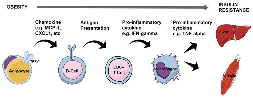
F. Seyfried and M.K. Hankir (2019) Hypothalamic inflammation in weight loss and gain
hypothalamic neuronal inflammation and ER stress, fol- tandem to disrupt whole-body energy balance regulation
lowed by leptin resistance. Consequently, the rise in circu- (Figure 3).
lating leptin levels as fat mass increases fails to act as a
negative feedback signal to maintain a stable body weight. HYPOTHALAMIC ER STRESS RELIEVERS ARE POTENT
Contrary to what might be expected however, ER stress WEIGHT LOSS COMPOUNDS
does not affect leptin receptor folding in the ER and traf- Because of the pivotal role hypothalamic ER stress plays in
ficking to the plasma membrane [13]. While a unifying leptin resistance and obesity development [12, 13, 19, 30],
mechanism for hypothalamic inflammation, ER stress and a screen was performed to identify small molecules which
diminished leptin receptor signaling in obesity is still miss- might promote weight loss through its amelioration [31].
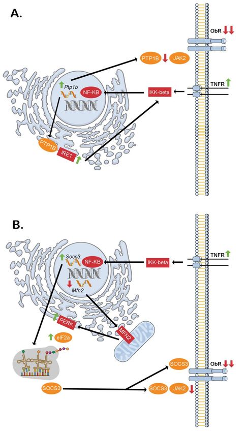
ing, the increased expression of protein tyrosine phospha- By comparing the mouse hypothalamic transcriptomic re-
tase 1B (PTP1B) caused by NF-Кβ is a likely candidate [14]. sponse to obesity and ER stress-relieving chemical chaper-
This is because beyond the established inhibitory role of ones with that of human cell lines treated with a panel of
PTP1B in dephosphorylating the leptin receptor effector FDA-approved drugs and other bioactive compounds [32],
protein janus kinase 2 (JAK2) at the cell membrane [15], it the thunder god vine root extract celastrol emerged as one
also potentiates the inositol requiring enzyme 1 (IRE1) arm that caused the most similar absolute changes. Next, in a
of the ER stress response through its phosphatase activity three week feeding study performed on high-fat diet-
at the ER [16] (Figure 2A). Additionally/alternatively, NF-Кβ induced obese mice, once daily intraperitoneal celastrol
could increase suppressor of cytokine signaling 3 (SOCS3) injections produced a striking 30% weight loss largely
[12] and decrease mitofusion 2 [17] expression to directly through reduced food intake. This is far greater than the 5-
interfere with leptin receptor signaling [18] and cause hy- 10% typically observed with currently prescribed obesity
pothalamic ER stress [19], respectively. Also, through the medications such as the 5 hydroxytryptamine 2C (5-HT2C)
protein kinase R (PKR)-like ER kinase (PERK) and eukaryotic receptor agonist lorcaserin or the glucagon-like peptide 1
elongation initiation factor 2 alpha (eIF2-α) arm of the ER (GLP-1) analogue liraglutide and approaches that found
stress response, a more stable SOCS3 isoform is produced with bariatric surgeries such as vertical sleeve gastrectomy
by alternative translation which would serve to further (VSG) and Roux-en-Y gastric bypass (RYGB). That weight
exacerbate leptin resistance [20] (Figure 2B). loss did not occur in leptin-deficient ob/ob or leptin recep-
The brain's support and immune cells would then be tor-deficient db/db mice treated with celastrol provided
added to the mix when it was shown that hypothalamic strong evidence that it acts as an endogenous leptin sensi-
astrocytes and microglia become activated within days tizer. Accordingly, hypothalamic leptin receptor signaling
after placing rats and mice on a high-fat diet [21]. Subse- was enhanced in wild-type mice after celastrol treatment
quent and prior studies suggested that elevated circulating alongside reduced ER stress although experiments with
saturated fatty acids themselves act as pro-inflammatory celastrol administered to mice lacking functional leptin
signaling molecules on hypothalamic neurons and micro- receptors specifically in various nuclei of the hypothalamus
glia through TLR4 [22-24]. In contrast, astrocytes appear to [33] still need to be performed to draw definitive conclu-
be activated by saturated fatty acids through a bystander sions. Celastrol also impressively prevented weight gain in
effect [22, 24]. Contributions of IKK-β in hypothalamic neu- mice placed on a high-fat diet for a year and was well tol-
rons [12] and microglia [25] to promoting leptin resistance erated.
and obesity are now clear. Furthermore, hypothalamic Motivated by this success, the same group of research-
microglial IKK-β signaling promotes the recruitment of cir- ers went on to search for compounds which produce a
culating CD169+ monocytes into the hypothalamus, which similar gene expression profile as celastrol in mouse em-
then adopt a microglia-like phenotype to further aggravate bryonic fibroblasts [34]. The winter cherry plant extract
inflammation and perpetuate leptin resistance [25]. This, withaferin-A emerged as the best hit. Comparable to celas-
although controversial [26], could be mediated in part trol, withaferin-A produced approximately 20% weight loss
through the release of the chemokine fractalkine from in high-fat diet-induced obese mice in a three week feeding
hypothalamic neurons consequential to receiving TNF-α study largely by reducing food intake. Again, withaferin-A
from neighboring glial cells [27]. Interestingly, IKK-β signal- was minimally effective in ob/ob and db/db mice and rein-
ing in hypothalamic astrocytes seems to serve a different stated the appetite suppressing effects of exogenous leptin
kind of function by shortening their fine processes in the treatment in otherwise leptin resistant, high-fat diet-
face of a high-fat diet leading to reduced gamma amino induced obese mice. Finally, as with celastrol, hypothalam-
butyric acid (GABA) reuptake from the extra-synaptic space ic leptin receptor signaling was enhanced and ER stress was
[28]. As a result, GABAB receptors are activated in nearby reduced by withaferin-A. Notably, precisely how celastrol
neurons decreasing their production of anorexigenic brain- and withaferin A reverse hypothalamic ER stress in obesity
derived neurotrophic factor (BDNF) which ultimately caus- remains unknown. For celastrol at least, this may be from
es hyperphagia and obesity [28, 29] Thus, the hypothalam- direct inhibition of IKK-β catalytic activity through targeting
ic molecular and cellular perturbations in response to cysteine 179 in the activation loop [35] and/or non-
chronic high-fat diet consumption are multifaceted, involv- competitive inhibition of PTP1B [36]. Interestingly, unlike
ing a complex array of signaling molecules and cell types celastrol, withaferin-A does not inhibit PTP1B catalytic ac-
originating both peripherally and centrally which act in tivity [36] which may explain why the former is the superi-
OPEN ACCESS | www.cell-stress.com 31 Cell Stress | FEBRUARY 2019 | Vol. 3 No. 2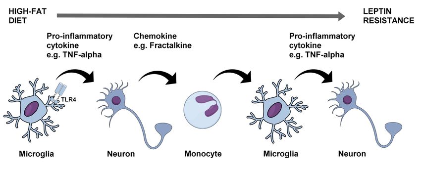
F. Seyfried and M.K. Hankir (2019)                                               Hypothalamic inflammation in weight loss and gain

                                                                                         FIGURE 2: Proposed intracellular
                                                                                         signaling cascades liking inflamma-
                                                                                         tion, ER stress and hypothalamic
                                                                                         neuronal leptin resistance in obesi-
                                                                                         ty. (A) Through the dual phosphatase
                                                                                         action of PTP1B at the ER (stimulato-
                                                                                         ry on IRE1) and cell membrane (in-
                                                                                         hibitory on JAK2) downstream of
                                                                                         TNF-α receptor activation, hypotha-
                                                                                         lamic neuronal leptin receptor sig-
                                                                                         naling may be blunted contributing
                                                                                         to increased food intake and obesity.
                                                                                         (B) Similarly, through the dual action
                                                                                         of NF-KB of increasing Socs3 tran-
                                                                                         scription and decreasing Mfn2 tran-
                                                                                         scription, hypothalamic neuronal
                                                                                         leptin receptor signaling may be
                                                                                         blunted contributing to increased
                                                                                         food intake and obesity. This would
                                                                                         be through decreased mitochondrial
                                                                                         MFN2 leading to reduced ER-
                                                                                         mitochondrial contacts thereby caus-
                                                                                         ing ER stress. The PERK-eIF2α arm of
                                                                                         this response mediates alternative
                                                                                         translation of Socs3 mRNA of a more
                                                                                         stable SOCS3 variant, which lacks an
                                                                                         amino terminus tail containing a
                                                                                         lysine residue that is normally ubiq-
                                                                                         uitinated sending the full-length
                                                                                         SOCS3 to the proteasome for degra-
                                                                                         dation. eIF2a - elongation initiation
                                                                                         factor 2 alpha, IKK-beta - inhibitor
                                                                                         of kappa beta kinase beta, IRE1 -
                                                                                         inositol requiring enzyme 1, JAK2 -
                                                                                         janus kinase 2, Mfn2 - Mitofusin-2,
                                                                                         NK-κB - necrosis factor kappa beta,
                                                                                         ObR – leptin receptor, PERK - protein
                                                                                         kinase R (PKR)-like ER kinase, PTP1B -
                                                                                         protein tyrosine phosphatase 1B,
                                                                                         Socs3 - suppressor of cytokine signal-
                                                                                         ing 3, TNFR - tumor necrosis factor
                                                                                         receptor.

or weight loss compound. It is also still unclear what effects   genesis [37]. This is thought to be from the stabilizing ef-
both these molecules have on hypothalamic glial cells.           fect of celastrol on the protein-protein interaction be-
    In addition to its central mode of action in suppressing     tween heat shock factor 1 (HSF1) and peroxisome prolifer-
energy (food) intake, celastrol has also been proposed to        ator-activated receptor gamma coactivator 1-alpha (PGC-
promote a negative energy balance by increasing energy           1α), two transcription factors that induce a thermogenic
expenditure through stimulating adipose tissue thermo-           gene expression program in adipocytes by binding to the

OPEN ACCESS | www.cell-stress.com                           32                     Cell Stress | FEBRUARY 2019 | Vol. 3 No. 2
F. Seyfried and M.K. Hankir (2019) Hypothalamic inflammation in weight loss and gain
Pgc1α promoter [37]. Indeed, normal weight mice placed diet. After 16 weeks, half of the high-fat group was ran-
on a high-fat diet for two weeks and treated with low dos- domized to receive VSG whereas the remaining rats re-
es of celastrol were protected from weight gain associated ceived sham surgery to control for the stress of laparotomy.
with increased energy expenditure but no reductions in The VSG-operated rats lost approximately 25% of their
food intake [37]. Furthermore, the marked upregulation of body weight after eight weeks, which is comparable to that
thermogenic genes in adipose tissue caused by celastrol of the human procedure, while the sham-operated groups
was not seen in HSF1-deficient mice. This peripheral mode continued to gain weight during this time period. Immuno-
of action for celastrol was however not supported by sub- histochemical analysis was then performed on hypotha-
sequent findings from mice deficient in uncoupling protein lamic sections. Levels of the chemokine monocyte chemo-
1 (UCP1), the principal thermogenic effector in adipose attractant protein 1 (MCP1) were decreased in the VSG-
tissue [38]. It is nevertheless still possible that UCP1- operated group compared to the sham-operated group on
independent thermogenesis contributes to celastrol’s ef- a high-fat diet and approached the levels found in the
fects on body weight. These issues notwithstanding, there sham-operated group on the low-fat chow diet. Similar
is genuine hope that natural, safe and effective obesity findings were made on the levels of pro-inflammatory
treatments are in the horizon. However, the effects of both phosphorylated signal transducer and activator of tran-
celastrol and withaferin-A need to be evaluated in human scription 3 (pSTAT3) specifically in hypothalamic microglial
individuals with obesity first before metabolic researchers cells.
will need to hang up their lab coats. In the study of McGavigan et al. [40], high-fat diet-
induced obese C57BL/6J mice received either VSG or sham
VSG RELIEVES HYPOTHALAMIC INFLAMMATION, surgeries. A subgroup of sham-operated mice was then
GLIOSIS, AND ER STRESS weight-matched to the VSG group by chronic caloric re-
If the pharmacological and surgical forms of obesity treat- striction – an important control to ensure any changes
ment described above cause comparable magnitudes of seen are not simply due to weight loss. All groups lost
weight loss, then it can be argued that they both have simi- weight during the first two weeks postoperatively, high-
lar mechanisms of action. Indeed, recent studies on rats lighting the sensitivity of mice to surgical stress. However,
and mice in the context of obesity-associated infertility and by the 10th week, VSG-operated mice weighed approxi-
hypertension have shown that VSG reduces hypothalamic mately 10% less than the sham-operated ad libitum fed
inflammation [39, 40], gliosis [39], and ER stress [40]. In- mice, consuming significantly less food during this time
terestingly, there is also evidence from mice [41, 42], rats, period. Hypothalamic lysates were then prepared for
[43] and humans [44] that like celastrol, VSG induces a Western Blot analysis. Levels of phosphorylated (activated)
thermogenic program in adipocytes although the function- PERK and eIF2-α were reduced in VSG mice, as was TNF-α.
al relevance of this remains unclear. This was not found in the weight-matched control group
In the study of Xiang et al. [39], Sprague Dawley rats suggesting that reduced hypothalamic ER stress and in-
were placed either on a standard chow diet or on a high-fat flammation are effects specific to VSG. A selective reduc-
FIGURE 3: Molecular and cellular processes linking chronic high-fat diet consumption with leptin resistance and obesity. The rise in cir-
culating saturated fatty acids from chronic consumption of a high-fat diet leads to activation of TLR4 in hypothalamic microglia which then
release pro-inflammatory cytokines such as TNF-α. This in turn leads to the release of the chemokine fractalkine from adjacent neurons
which recruits circulating monocytes into the hypothalamus and which then differentiate into activated microglia. A vicious cycle is thus
initiated which progressively worsens leptin resistance in hypothalamic neurons contributing to increased food intake and obesity. TLR4 –
toll-like receptor 4, TNF-alpha – tumor necrosis factor alpha.
OPEN ACCESS | www.cell-stress.com 33 Cell Stress | FEBRUARY 2019 | Vol. 3 No. 2F. Seyfried and M.K. Hankir (2019) Hypothalamic inflammation in weight loss and gain
tion in Adlercreutzia microbiota after VSG was proposed to [46]. Furthermore, VSG-operated rats are more responsive
provide the link between changes in gut anatomy and brain to the acute appetite suppressing effects of exogenous
cellular pathology. leptin treatment than pair-fed rats [45]. On the other hand,
The studies of Xiang et al. and McGavigan et al., de- unlike celastrol [24] or withaferin-A [26] treatments, leptin
spite only being associational in nature, collectively provide receptor-deficient fa/fa Zucker rats [47] and db/db mice
persuasive evidence that VSG reduces hypothalamic in- [48] still lose weight after VSG, suggesting that leptin is a
flammation, gliosis and/or ER stress in obesity (Figure 4). dispensable/redundant mediator of its effects on whole-
By extension, it can be reasonably inferred that this partic- body energy balance. One potential way to resolve these
ular bariatric surgical procedure restores leptin sensitivity inconsistencies is to induce hypothalamic ER stress and
to cause marked and lasting weight loss. In support of this leptin resistance post-abdominal surgeries in rodents
idea, VSG-operated rats and mice have lower circulating through intracerebroventricular administration of tuni-
leptin levels compared to pair-fed controls with similar camycin and to assess changes in feeding and body weight
adiposity indicative of decreased leptin resistance [45], [13].
FIGURE 4: Amelioration of hypothalamic inflammation, ER stress and gliosis after VSG. After VSG, activation of FXR/TGR5 in hypothalam-
ic neurons from the rise in circulating bile acids, and GLP-1 receptors in hypothalamic glial cells from the rise in circulating GLP-1, may
contribute to amelioration of inflammatory processes, ER stress, gliosis and leptin resistance in obesity, thereby potentially contributing to
reduced food intake and lasting weight loss. FXR - farnesoid X receptor, GLP-1 - glucagon-like peptide 1, ObR – leptin receptor, MCP-1 -
monocyte chemoattractant protein 1, TGR5 - Takeda G-protein 5 receptor, TLR4 – toll-like receptor 4, TNF-alpha – tumor necrosis factor
alpha, VSG - vertical sleeve gastrectomy.
OPEN ACCESS | www.cell-stress.com 34 Cell Stress | FEBRUARY 2019 | Vol. 3 No. 2F. Seyfried and M.K. Hankir (2019) Hypothalamic inflammation in weight loss and gain
Further questions still remain of course such as the de- VSG has an inhibitory effect. Furthermore, patients with
finitive nature of the anti-inflammatory/ER stress relieving higher levels of hypothalamic gliosis may respond better to
gut-derived factor(s) enhanced after VSG. Bile acids acting VSG which would take us one step closer to personalized
on hypothalamic farnesoid X receptors (FXR) and/or treatment options for individuals with severe obesity.
Takeda G-protein 5 receptors (TGR5) [49-52] are possible
candidates as they are essential for the reduced food in- ACKNOWLEDGMENTS
take and body weight postoperatively [42, 53, 54]. In this FS receives funding from the German Research Foundation
context, bile acids would conceivably be mediating their (DFG) grant number 271722282. MKH has received funding
effects through FXR and TGR5 in hypothalamic neurons from the DFG Collaborative Research Centre (CRC) 1052 in
and not glial cells [52, 55]. On the other hand, enhanced Obesity Mechanisms (Project A8). MKH is grateful to Prof.
GLP-1 receptor signaling in hypothalamic astrocytes [56] Matthias Blüher for continued support. The figures for this
and/or microglia [57] could explain their reduced activa- manuscript were produced using Mind the Graph software.
tion after VSG thereby contributing to weight loss but
again this is not supported by studies on germline GLP-1 CONFLICT OF INTEREST
receptor deficient mice [58]. Nevertheless, post-embryonic The authors have no conflicts of interest to declare.
hypothalamic microglial/astrocytic ablation approaches [25,
28, 29] may yield different findings. COPYRIGHT
© 2019 Seyfried and Hankir. This is an open-access article
CONCLUSIONS AND FUTURE DIRECTIONS released under the terms of the Creative Commons Attrib-
The fact that chemical hypothalamic ER stress relievers and ution (CC BY) license, which allows the unrestricted use,
VSG both can reverse a pathologic brain state in animal distribution, and reproduction in any medium, provided
models of obesity suggests that rather than just treating its the original author and source are acknowledged.
symptoms, they tackle it at one of its root causes. Future
work will be required to verify if the promising animal find-
ings can be translated to humans. For example, assessing Please cite this article as: Florian Seyfried and Mohammed K.
Hankir (2019). Could de-stressing the brain be the solution for
human hypothalamic gliosis through the use of T2-
long-term weight loss? Cell Stress 3(2): 29-37. doi:
weighted magnetic resonance imaging [59] or more direct- 10.15698/cst2019.02.174
ly with positron emission tomography [60] may reveal if
REFERENCES
1. Hotamisligil GS, Shargill NS, Spiegelman BM (1993). Adipose expres- 7. Hotamisligil GS, Peraldi P, Budavari A, Ellis R, White MF, Spiegelman
sion of tumor necrosis factor-alpha: direct role in obesity-linked insu- BM (1996). IRS-1-mediated inhibition of insulin receptor tyrosine
lin resistance. Science 259(5091):87-91. doi: 10.1126/science.7678183 kinase activity in TNF-alpha- and obesity-induced insulin resistance.
Science 271(5249):665-8. doi: 10.1126/science.271.5249.665
2. Ye L, Kleiner S, Wu J, Sah R, Gupta RK, Banks AS, Cohen P,
Khandekar MJ, Boström P, Mepani RJ, Laznik D, Kamenecka TM, Song 8. Luck H, Tsai S, Chung J, Clemente-Casares X, Ghazarian M, Revelo XS,
X, Liedtke W, Mootha VK, Puigserver P, Griffin PR, Clapham DE, Spie- Lei H, Luk CT, Shi SY, Surendra A, Copeland JK, Ahn J, Prescott D, Ras-
gelman BM (2012). TRPV4 is a regulator of adipose oxidative metabo- mussen BA, Chng MH, Engleman EG, Girardin SE, Lam TK, Croitoru K,
lism, inflammation, and energy homeostasis. Cell 151(1):96-110. doi: Dunn S, Philpott DJ, Guttman DS, Woo M, Winer S, Winer DA (2015).
10.1016/j.cell.2012.08.034 Regulation of obesity-related insulin resistance with gut anti-
inflammatory agents. Cell Metab 21(4):527-42. doi:
3. Winer DA, Winer S, Shen L, Wadia PP, Yantha J, Paltser G, Tsui H, 10.1016/j.cmet.2015.03.001
Wu P, Davidson MG, Alonso MN, Leong HX, Glassford A, Caimol M,
Kenkel JA, Tedder TF, McLaughlin T, Miklos DB, Dosch HM, Engleman 9. Shi H, Kokoeva MV, Inouye K, Tzameli I, Yin H, Flier JS (2006). TLR4
EG (2011). B cells promote insulin resistance through modulation of T links innate immunity and fatty acid-induced insulin resistance. J Clin
cells and production of pathogenic IgG antibodies. Nat Med Invest 116(11):3015-25. doi: 10.1172/JCI28898
17(5):610-7. doi: 10.1038/nm.2353
10. Zhang Y, Proenca R, Maffei M, Barone M, Leopold L, Friedman JM
4. Nishimura S, Manabe I, Nagasaki M, Eto K, Yamashita H, Ohsugi M, (1994). Positional cloning of the mouse obese gene and its human
Otsu M, Hara K, Ueki K, Sugiura S, Yoshimura K, Kadowaki T, Nagai R homologue. Nature 372(6505):425-32. doi: 10.1038/372425a0
(2009). CD8+ effector T cells contribute to macrophage recruitment
and adipose tissue inflammation in obesity. Nat Med 15(8):914-20. 11. Heymsfield SB, Greenberg AS, Fujioka K, Dixon RM, Kushner R,
doi: 10.1038/nm.1964 Hunt T, Lubina JA, Patane J, Self B, Hunt P, McCamish M (1999) Re-
combinant leptin for weight loss in obese and lean adults: a random-
5. Duffaut C, Galitzky J, Lafontan M, Bouloumié A (2009). Unexpected ized, controlled, dose-escalation trial. JAMA 282(16):1568-75. doi:
trafficking of immune cells within the adipose tissue during the onset 10.1001/jama.282.16.1568
of obesity. Biochem Biophys Res Commun 384(4):482-5. doi:
10.1016/j.bbrc.2009.05.002 12. Zhang X, Zhang G, Zhang H, Karin M, Bai H, Cai D (2008). Hypotha-
lamic IKKbeta/NF-kappaB and ER stress link overnutrition to energy
6. Weisberg SP, McCann D, Desai M, Rosenbaum M, Leibel RL, Fer- imbalance and obesity. Cell 135(1):61-73. doi:
rante AW Jr (2003). Obesity is associated with macrophage accumula- 10.1016/j.cell.2008.07.043
tion in adipose tissue. J Clin Invest 112(12):1796-808.
doi:10.1172/jci19246 13. Ozcan L, Ergin AS, Lu A, Chung J, Sarkar S, Nie D, Myers MG Jr,
Ozcan U (2009). Endoplasmic reticulum stress plays a central role in
OPEN ACCESS | www.cell-stress.com 35 Cell Stress | FEBRUARY 2019 | Vol. 3 No. 2F. Seyfried and M.K. Hankir (2019) Hypothalamic inflammation in weight loss and gain
development of leptin resistance. Cell Metab 9(1):35-51. doi: 26. Dorfman MD, Krull JE, Douglass JD, Fasnacht R, Lara-Lince F, Meek
10.1016/j.cmet.2008.12.004 TH, Shi X, Damian V, Nguyen HT, Matsen ME, Morton GJ, Thaler JP
(2017). Sex differences in microglial CX3CR1 signalling determine
14. Zabolotny JM, Kim YB, Welsh LA, Kershaw EE, Neel BG, Kahn BB obesity susceptibility in mice. Nat Commun 8:14556. doi:
(2008). Protein-tyrosine phosphatase 1B expression is induced by 10.1038/ncomms14556
inflammation in vivo. J Biol Chem283(21):14230-41. doi:
10.1074/jbc.m800061200 27. Morari J, Anhe GF, Nascimento LF, de Moura RF, Razolli D, Solon C,
Guadagnini D, Souza G, Mattos AH, Tobar N, Ramos CD, Pascoal VD,
15. Zhang ZY, Dodd GT, Tiganis T (2015). Protein Tyrosine Phosphatas- Saad MJ, Lopes-Cendes I, Moraes JC, Velloso LA (2014). Fractalkine
es in Hypothalamic Insulin and Leptin Signaling. Trends Pharmacol Sci (CX3CL1) is involved in the early activation of hypothalamic inflamma-
36(10):661-674. doi:10.1016/j.tips.2015.07.003 tion in experimental obesity. Diabetes 63(11):3770-84. doi:
16. Gu F, Nguyên DT, Stuible M, Dubé N, Tremblay ML, Chevet E 10.2337/db13-1495
(2004). Protein-tyrosine phosphatase 1B potentiates IRE1 signaling 28. Zhang Y, Reichel JM, Han C, Zuniga-Hertz JP, Cai D (2017). Astro-
during endoplasmic reticulum stress. J Biol Chem 279(48):49689-93. cytic Process Plasticity and IKKβ/NF-κB in Central Control of Blood
doi: 10.1074/jbc.c400261200 Glucose, Blood Pressure, and Body Weight. Cell Metab 25(5):1091-
17. Andrews M, Arredondo M (2012). Hepatic and adipocyte cells 1102.e4. doi: 10.1016/j.cmet.2017.04.002
respond differentially to iron overload, hypoxic and inflammatory 29. Douglass JD, Dorfman MD, Fasnacht R, Shaffer LD, Thaler JP
challenge. Biometals 25(4):749-59. doi:10.1007/s10534-012-9543-9. (2017). Astrocyte IKKβ/NF-κB signaling is required for diet-induced
doi: 10.1007/s10534-012-9543-9 obesity and hypothalamic inflammation. Mol Metab 6(4):366-373.
18. Münzberg H, Myers MG Jr (2005). Molecular and anatomical de- doi: 10.1016/j.molmet.2017.01.010
terminants of central leptin resistance. Nat Neurosci 8(5):566-70. 30. Williams KW, Liu T, Kong X, Fukuda M, Deng Y, Berglund ED, Deng
doi:10.1038/nn1454 Z, Gao Y, Liu T, Sohn JW, Jia L, Fujikawa T, Kohno D, Scott MM, Lee S,
19. Schneeberger M, Dietrich MO, Sebastián D, Imbernón M, Castaño Lee CE, Sun K, Chang Y, Scherer PE, Elmquist JK (2014). Xbp1s in Pomc
C, Garcia A, Esteban Y, Gonzalez-Franquesa A, Rodríguez IC, Bortolozzi neurons connects ER stress with energy balance and glucose homeo-
A, Garcia-Roves PM, Gomis R, Nogueiras R, Horvath TL, Zorzano A, stasis. Cell Metab 20(3):471-82. doi: 10.1016/j.cmet.2014.06.002
Claret M (2013). Mitofusin 2 in POMC neurons connects ER stress with 31. Liu J, Lee J, Salazar Hernandez MA, Mazitschek R, Ozcan U (2015).
leptin resistance and energy imbalance. Cell 155(1):172-87. Treatment of obesity with celastrol. Cell 161(5):999-1011. doi:
doi:10.1016/j.cell.2013.09.003 10.1016/j.cell.2015.05.011
20. Sasaki A, Inagaki-Ohara K, Yoshida T, Yamanaka A, Sasaki M, Ya- 32. Lamb J, Crawford ED, Peck D, Modell JW, Blat IC, Wrobel MJ, Ler-
sukawa H, Koromilas AE, Yoshimura A (2003). The N-terminal truncat- ner J, Brunet JP, Subramanian A, Ross KN, Reich M, Hieronymus H,
ed isoform of SOCS3 translated from an alternative initiation AUG Wei G, Armstrong SA, Haggarty SJ, Clemons PA, Wei R, Carr SA, Lander
codon under stress conditions is stable due to the lack of a major ES, Golub TR (2006). The Connectivity Map: using gene-expression
ubiquitination site, Lys-6. J Biol Chem 278(4):2432-6. signatures to connect small molecules, genes, and disease. Science
doi:10.1074/jbc.C200608200 313(5795):1929-35. doi: 10.1126/science.1132939
21. Thaler JP, Yi CX, Schur EA, Guyenet SJ, Hwang BH, Dietrich MO, 33. Dhillon H, Zigman JM, Ye C, Lee CE, McGovern RA, Tang V, Kenny
Zhao X, Sarruf DA, Izgur V, Maravilla KR, Nguyen HT, Fischer JD, CD, Christiansen LM, White RD, Edelstein EA, Coppari R, Balthasar N,
Matsen ME, Wisse BE, Morton GJ, Horvath TL, Baskin DG, Tschöp MH, Cowley MA, Chua S Jr, Elmquist JK, Lowell BB (2006). Leptin directly
Schwartz MW (2012). Obesity is associated with hypothalamic injury activates SF1 neurons in the VMH, and this action by leptin is required
in rodents and humans. J Clin Invest 122(1):153-62. doi: for normal body-weight homeostasis. Neuron 49(2):191-203 doi:
10.1172/jci59660 10.1016/j.neuron.2005.12.021
22. Milanski M, Degasperi G, Coope A, Morari J, Denis R, Cintra DE, 34. Lee J, Liu J, Feng X, Salazar Hernández MA, Mucka P, Ibi D, Choi
Tsukumo DM, Anhe G, Amaral ME, Takahashi HK, Curi R, Oliveira HC, JW, Ozcan U (2016). Withaferin A is a leptin sensitizer with strong
Carvalheira JB, Bordin S, Saad MJ, Velloso LA (2009). Saturated fatty antidiabetic properties in mice. Nat Med 22(9):1023-32. doi:
acids produce an inflammatory response predominantly through the 10.1038/nm.4145
activation of TLR4 signaling in hypothalamus: implications for the
pathogenesis of obesity. J Neurosci 29(2):359-70. doi: 35. Lee JH, Koo TH, Yoon H, Jung HS, Jin HZ, Lee K, Hong YS, Lee JJ
10.1523/jneurosci.2760-08.2009 (2006). Inhibition of NF-kappa B activation through targeting I kappa B
kinase by celastrol, a quinone methide triterpenoid. Biochem Phar-
23. Kleinridders A, Schenten D, Könner AC, Belgardt BF, Mauer J, macol 72(10):1311-21. doi: 10.1016/j.bcp.2006.08.014
Okamura T, Wunderlich FT, Medzhitov R, Brüning JC (2009). MyD88
signaling in the CNS is required for development of fatty acid-induced 36. Kyriakou E, Schmidt S, Dodd GT, Pfuhlmann K, Simonds SE, Lenhart
leptin resistance and diet-induced obesity. Cell Metab 10(4):249-59. D, Geerlof A, Schriever SC, De Angelis M, Schramm KW, Plettenburg O,
doi: 10.1016/j.cmet.2009.08.013 Cowley MA, Tiganis T, Tschöp MH, Pfluger PT, Sattler M, Messias AC
(2018). Celastrol Promotes Weight Loss in Diet-Induced Obesity by
24. Valdearcos M, Robblee MM, Benjamin DI, Nomura DK, Xu AW, Inhibiting the Protein Tyrosine Phosphatases PTP1B and TCPTP in the
Koliwad SK (2014). Microglia dictate the impact of saturated fat con- Hypothalamus. J Med Chem 61(24): 11144-11157. doi:
sumption on hypothalamic inflammation and neuronal function. Cell 10.1021/acs.jmedchem.8b01224
Rep 9(6):2124-38. doi: 10.1016/j.celrep.2014.11.018
37. Ma X, Xu L, Alberobello AT, Gavrilova O, Bagattin A, Skarulis M, Liu
25. Valdearcos M, Douglass JD, Robblee MM, Dorfman MD, Stifler DR, J, Finkel T, Mueller E (2015). Celastrol Protects against Obesity and
Bennett ML, Gerritse I, Fasnacht R, Barres BA, Thaler JP, Koliwad SK Metabolic Dysfunction through Activation of a HSF1-PGC1α Transcrip-
(2018). Microglial Inflammatory Signaling Orchestrates the Hypotha- tional Axis. Cell Metab 22(4):695-708. doi:
lamic Immune Response to Dietary Excess and Mediates Obesity Sus- 10.1016/j.cmet.2015.08.005.
ceptibility. Cell Metab 26(1):185-197.e3. doi:
10.1016/j.cmet.2017.05.015 38. Pfuhlmann K, Schriever SC, Baumann P, Kabra DG, Harrison L,
Mazibuko-Mbeje SE, Contreras RE, Kyriakou E, Simonds SE, Tiganis T,
Cowley MA, Woods SC, Jastroch M, Clemmensen C, De Angelis M,
OPEN ACCESS | www.cell-stress.com 36 Cell Stress | FEBRUARY 2019 | Vol. 3 No. 2F. Seyfried and M.K. Hankir (2019) Hypothalamic inflammation in weight loss and gain
Schramm KW, Sattler M, Messias AC, Tschöp MH, Pfluger PT (2018). 50. La Frano MR, Hernandez-Carretero A, Weber N, Borkowski K,
Celastrol-Induced Weight Loss Is Driven by Hypophagia and Independ- Pedersen TL, Osborn O, Newman JW (2017) Diet-induced obesity and
ent From UCP1. Diabetes 67(11):2456-2465. doi: 10.2337/db18-0146 weight loss alter bile acid concentrations and bile acid-sensitive gene
expression in insulin target tissues of C57BL/6J mice. Nutr Res 46:11-
39. Xiang J, Bian C, Wan X, Zhang Q, Huang S, Wu D (2018). Sleeve 21. doi: 10.1016/j.nutres.2017.07.006
Gastrectomy Reversed Obesity-Induced Hypogonadism in a Rat Model
by Regulating Inflammatory Responses in the Hypothalamus and Tes- 51. Cummings BP, Bettaieb A, Graham JL, Kim J, Ma F, Shibata N,
tis. Obesity Surgery 28(8):2272-2280. doi: 10.1007/s11695-018-3150- Stanhope KL, Giulivi C, Hansen F, Jelsing J, Vrang N, Kowala M, Choui-
y nard ML, Haj FG, Havel PJ (2013). Bile-acid-mediated decrease in en-
doplasmic reticulum stress: a potential contributor to the metabolic
40. McGavigan AK, Henseler ZM, Garibay D, Butler SD, Jayasinghe S, benefits of ileal interposition surgery in UCD-T2DM rats. Dis Model
Ley RE, Davisson RL, Cummings BP (2017). Vertical sleeve gastrectomy Mech 6(2):443-56. doi: 10.1242/dmm.010421
reduces blood pressure and hypothalamic endoplasmic reticulum
stress in mice. Dis Model Mech 10(3):235-243. doi: 52. McMillin M, Frampton G, Tobin R, Dusio G, Smith J, Shin H, Newell-
10.1242/dmm.027474 Rogers K, Grant S, DeMorrow S (2015). TGR5 signaling reduces neu-
roinflammation during hepatic encephalopathy. J Neurochem
41. Ding L, Sousa KM, Jin L, Dong B, Kim BW, Ramirez R, Xiao Z, Gu Y, 135(3):565-76. doi: 10.1111/jnc.13243
Yang Q, Wang J, Yu D, Pigazzi A, Schones D, Yang L, Moore D, Wang Z,
Huang W (2016). Vertical sleeve gastrectomy activates GPBAR-1/TGR5 53. Ryan KK, Tremaroli V, Clemmensen C, Kovatcheva-Datchary P,
to sustain weight loss, improve fatty liver, and remit insulin resistance Myronovych A, Karns R, Wilson-Pérez HE, Sandoval DA, Kohli R, Bäck-
in mice. Hepatology 64(3):760-73. doi: 10.1002/hep.28689 hed F, Seeley RJ (2014) FXR is a molecular target for the effects of
vertical sleeve gastrectomy. Nature 509(7499):183-
42. Chen Y, Yang J, Nie X, Song Z, Gu Y (2018). Effects of Bariatric Sur- 8.doi:10.1038/nature13135
gery on Change of Brown Adipocyte Tissue and Energy Metabolism in
Obese Mice. Obesity Surgery 28(3):820-830. doi: 10.1007/s11695- 54. McGavigan AK, Garibay D, Henseler ZM, Chen J, Bettaieb A, Haj FG,
017-2899-8 Ley RE, Chouinard ML, Cummings BP (2017). TGR5 contributes to
glucoregulatory improvements after vertical sleeve gastrectomy in
43. Moncada R, Becerril S, Rodríguez A, Méndez-Giménez L, Ramírez B, mice. Gut 66(2):226-234. doi: 10.1136/gutjnl-2015-309871
Catalán V, Gómez-Ambrosi J, Gil MJ, Fernández S, Cienfuegos JA,
Valentí V, Frühbeck G (2016). Sleeve Gastrectomy Reduces Body 55. Albrecht S, Fleck AK, Kirchberg I, Hucke S, Liebmann M, Klotz L,
Weight and Improves Metabolic Profile also in Obesity-Prone Rats. Kuhlmann T (2017) Activation of FXR pathway does not alter glial cell
Obesity Surgery 26(7):1537-48. doi: 10.1007/s11695-015-1915-0 function. J Neuroinflammation 14(1):66. doi: 10.1186/s12974-017-
0833-6
44. Jahansouz C, Xu H, Hertzel AV, Kizy S, Steen KA, Foncea R, Serrot FJ,
Kvalheim N, Luthra G, Ewing K, Leslie DB, Ikramuddin S, Bernlohr DA 56. Iwai T, Ito S, Tanimitsu K, Udagawa S, Oka J (2006) Glucagon-like
(2018). Partitioning of adipose lipid metabolism by altered expression peptide-1 inhibits LPS-induced IL-1beta production in cultured rat
and function of PPAR isoforms after bariatric surgery. Int J astrocytes. Neurosci Res 55(4):352-60. doi
Obes42(2):139-146. doi: 10.1038/ijo.2017.197 10.1016/j.neures.2006.04.008
45. Stefater MA, Pérez-Tilve D, Chambers AP, Wilson-Pérez HE, Sand- 57. Activation of Glucagon-Like Peptide-1 Receptor Promotes Neuro-
oval DA, Berger J, Toure M, Tschöp M, Woods SC, Seeley RJ (2010). protection in Experimental Autoimmune Encephalomyelitis by Reduc-
Sleeve gastrectomy induces loss of weight and fat mass in obese rats, ing Neuroinflammatory Responses (2018). Lee CH, Jeon SJ, Cho KS,
but does not affect leptin sensitivity. Gastroenterology 138(7):2426- Moon E, Sapkota A, Jun HS, Ryu JH, Choi JW. Mol Neurobiol
36, 2436.e1-3. doi: 10.1053/j.gastro.2010.02.059 55(4):3007-3020. doi: 10.1007/s12035-017-0550-2
46. Arble DM, Schwartz AR, Polotsky VY, Sandoval DA, Seeley RJ 58. Wilson-Pérez HE, Chambers AP, Ryan KK, Li B, Sandoval DA, Stof-
(2019). Vertical sleeve gastrectomy improves ventilatory drive fers D, Drucker DJ, Pérez-Tilve D, Seeley RJ (2013) Vertical sleeve gas-
through a leptin-dependent mechanism. JCI Insight 4(1). pii: 124469. trectomy is effective in two genetic mouse models of glucagon-like
doi: 10.1172/jci.insight.124469 Peptide 1 receptor deficiency. Diabetes 62(7):2380-5. doi:
10.2337/db12-1498
47. Lopez PP, Nicholson SE, Burkhardt GE, Johnson RA, Johnson FK
(2009). Development of a sleeve gastrectomy weight loss model in 59. Schur EA, Melhorn SJ, Oh SK, Lacy JM, Berkseth KE, Guyenet SJ,
obese Zucker rats. The Journal of Surgical Research 157(2):243-50. Sonnen JA, Tyagi V, Rosalynn M, De Leon B, Webb MF, Gonsalves ZT,
doi: 10.1016/j.jss.2008.10.025 Fligner CL, Schwartz MW, Maravilla KR (2015). Radiologic evidence
that hypothalamic gliosis is associated with obesity and insulin re-
48. Li F, Sheng C, Song K, Zhang M, Bu L, Yang P, Sheng H, Li H, Qu S sistance in humans. Obesity 23(11):2142-8. doi: 10.1002/oby.21248
(2016). Preventative Sleeve Gastrectomy Contributes to Maintaining β
Cell Function in db/db Diabetic Mouse. Obesity Surgery 26(10):2402- 60. Sandiego CM, Gallezot J, Pittman B, Nabulsi N, Lim K, Lin S, Matus-
10. doi: 10.1007/s11695-016-2112-5 key D, Lee J, O’Connor KC, Huang Y, Carson RE, Hannestad J, Cosgrove
KP (2015). Imaging robust microglial activation after lipopolysaccha-
49. Gofflot F, Chartoire N, Vasseur L, Heikkinen S, Dembele D, Le Mer- ride administration in humans with PET. Proc Natl Acad Sci U S A
rer J, Auwerx J (2007). Systematic gene expression mapping clusters 112(40): 12468–12473. doi: 10.1073/pnas.1511003112
nuclear receptors according to their function in the brain. Cell
131(2):405-18. doi:10.1016/j.cell.2007.09.012
OPEN ACCESS | www.cell-stress.com 37 Cell Stress | FEBRUARY 2019 | Vol. 3 No. 2You can also read