Reactive Oxygen Species Mediate Activity-Regulated Dendritic Plasticity Through NADPH Oxidase and Aquaporin Regulation
←
→
Page content transcription
If your browser does not render page correctly, please read the page content below
BRIEF RESEARCH REPORT
                                                                                                                                              published: 05 July 2021
                                                                                                                                     doi: 10.3389/fncel.2021.641802

                                             Reactive Oxygen Species Mediate
                                             Activity-Regulated Dendritic
                                             Plasticity Through NADPH Oxidase
                                             and Aquaporin Regulation
                                             Serene Dhawan 1,2 , Philip Myers 1,3 , David M. D. Bailey 1 , Aaron D. Ostrovsky 4 ,
                                             Jan Felix Evers 4 and Matthias Landgraf 1*
                                             1
                                              Department of Zoology, University of Cambridge, Cambridge, United Kingdom, 2 Neural Circuits and Evolution Laboratory,
                                             The Francis Crick Institute, London, United Kingdom, 3 Neurobiology Division, MRC Laboratory of Molecular Biology,
                                             Cambridge, United Kingdom, 4 Centre for Organismal Studies, Heidelberg University, Heidelberg, Germany

                                             Neurons utilize plasticity of dendritic arbors as part of a larger suite of adaptive plasticity
                                             mechanisms. This explicitly manifests with motoneurons in the Drosophila embryo and
                                             larva, where dendritic arbors are exclusively postsynaptic and are used as homeostatic
                                             devices, compensating for changes in synaptic input through adapting their growth and
                                             connectivity. We recently identified reactive oxygen species (ROS) as novel plasticity
                            Edited by:
                                             signals instrumental in this form of dendritic adjustment. ROS correlate with levels of
                            Chun Han,
      Cornell University, United States      neuronal activity and negatively regulate dendritic arbor size. Here, we investigated
                       Reviewed by:          NADPH oxidases as potential sources of such activity-regulated ROS and implicate
                       Dion Dickman,         Dual Oxidase (but not Nox), which generates hydrogen peroxide extracellularly. We
     University of Southern California,
          Los Angeles, United States
                                             further show that the aquaporins Bib and Drip, but not Prip, are required for activity-
                        Jay Z. Parrish,      regulated ROS-mediated adjustments of dendritic arbor size in motoneurons. These
            University of Washington,
                                             results suggest a model whereby neuronal activity leads to activation of the NADPH
                         United States
                                             oxidase Dual Oxidase, which generates hydrogen peroxide at the extracellular face;
                  *Correspondence:
                    Matthias Landgraf        aquaporins might then act as conduits that are necessary for these extracellular ROS to
                 ml10006@cam.ac.uk           be channeled back into the cell where they negatively regulate dendritic arbor size.
                   Specialty section:        Keywords: reactive oxygen species, aquaporins, NADPH oxidases, dendrites, Drosophila, plasticity
        This article was submitted to
             Cellular Neurophysiology,
                a section of the journal    INTRODUCTION
    Frontiers in Cellular Neuroscience
       Received: 15 December 2020           Neurons are inherently plastic and their ability to respond to changes in synaptic transmission
           Accepted: 02 June 2021           or activity patterns is central to many processes, from learning and memory (Martin et al.,
           Published: 05 July 2021          2000; Stuchlik, 2014) to homeostatic adjustments that stabilize circuit function (Turrigiano and
                             Citation:      Nelson, 2000; Pozo and Goda, 2010; Wefelmeyer et al., 2016; Frank et al., 2020). We recently
    Dhawan S, Myers P, Bailey DMD,          identified reactive oxygen species (ROS) as novel signals required for activity-regulated plasticity.
          Ostrovsky AD, Evers JF and        ROS have long been known to affect neuronal development and function. Commonly associated
  Landgraf M (2021) Reactive Oxygen
                                            with pathological conditions, ageing and disease, the roles of ROS as signalling molecules
  Species Mediate Activity-Regulated
  Dendritic Plasticity Through NADPH
                                            under normal physiological conditions are much less understood (Milton et al., 2011; Oswald
   Oxidase and Aquaporin Regulation.        et al., 2018b; Peng et al., 2019). At present, hydrogen peroxide (H2 O2 ) is thought to be the
    Front. Cell. Neurosci. 15:641802.       species predominantly required for homeostatic maintenance of synaptic transmission at the
     doi: 10.3389/fncel.2021.641802         neuromuscular junction in Drosophila larvae, and for adaptive structural changes of synaptic

Frontiers in Cellular Neuroscience | www.frontiersin.org                           1                                           July 2021 | Volume 15 | Article 641802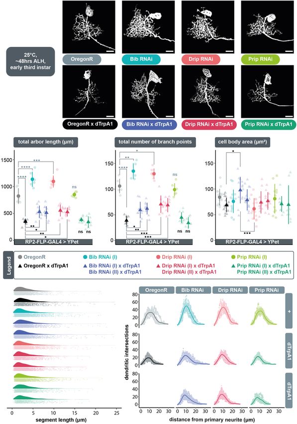
Dhawan et al.                                                                                                 Activity-Regulated Dendritic Plasticity

terminal arbors following periods of over-activation. H2 O2 is also       strains were used: OregonR (#2376, Bloomington Drosophila
sufficient to induce structural changes that largely phenocopy            Stock Center), UAS-dTrpA1 in attP16 (Hamada et al., 2008;
effects of over-activation, suggesting that ROS mediate plastic           FBtp0089791), UAS-Duox.RNAi (#32903, BDSC; FBtp0064955),
adjustments downstream of neuronal activity (Oswald et al.,               UAS-Nox.RNAi (Ha et al., 2005a; FBal0191562), UAS-bib.RNAi
2018a). Given that neuronal activity has a high energetic                 (I) (#57493, BDSC; FBtp0096443), UAS-bib.RNAi (II) (#27691
cost, correlating with metabolic demand (Attwell and Laughlin,            BDSC; FBtp0052515), UAS-Drip.RNAi (I) (#44661, BDSC;
2001; Zhu et al., 2012), a simplistic working model has been              FBtp0090566), UAS-Drip.RNAi (II) (#106911, Vienna Drosophila
proposed whereby ROS, largely generated as obligate by-products           Resource Centre; FBtp0045814), UAS-Prip.RNAi (I) (#50695,
of aerobic metabolism (Halliwell, 1992), regulate plasticity by           BDSC; FBtp0090659), UAS-Prip.RNAi (II) (#44464, BDSC;
providing ongoing feedback on a neuron’s activation status                FBtp0090258), and UAS-secreted human-catalase (FBal0190351;
(Hongpaisan et al., 2003, 2004; Oswald et al., 2018a).                    Ha et al., 2005b; Fogarty et al., 2016). Transgene expression
    In addition to mitochondria, NADPH oxidases are another               was targeted to 1–3 RP2 motoneurons per nerve cord using a
well documented source of activity-generated ROS (Hongpaisan              stochastic FLPout strategy, as detailed previously (Fujioka et al.,
et al., 2004; Massaad and Klann, 2011; Baxter and Hardingham,             2003; Ou et al., 2008). The GAL4 expression stock, termed “RP2-
2016; Hidalgo and Arias-Cavieres, 2016; Terzi and Suter, 2020;            FLP-GAL4 > YPet”, contains the following transgenes: RN2-
Terzi et al., 2021). Here, we focus on NADPH oxidases, a                  FLP, tub84B-FRT-CD2-FRT-GAL4, and 10XUAS-IVS-myr::YPet
family of differentially expressed multisubunit enzymes (Nox              in attP2. Briefly, yeast Flippase expression is directed to
1–5 and Dual Oxidase 1–2) that catalyse the transfer of an                RP2 motoneurons at embryonic stages only (at a lower
electron from cytosolic NADPH to oxygen to generate ROS                   level also to the aCC motoneuron and pCC interneuron)
at the extracellular face of the plasma membrane (Lambeth,                via RN2-FLP (Fujioka et al., 2003; Ou et al., 2008), then
2002; Panday et al., 2015). NADPH oxidases are commonly                   initiates permanent GAL4 expression in a subset of RP2
associated with immune responses, but more recently have also             motoneurons (as well as occasional aCC motoneurons) via
been shown to regulate aspects of nervous system development              FLP-conditional GAL4, tub84B-FRT-CD2-FRT-GAL4 (Pignoni
such as neuronal polarity (Wilson et al., 2015), growth cone              and Zipursky, 1997); this in turn initiates expression of the
dynamics (Munnamalai and Suter, 2009; Munnamalai et al.,                  membrane targeted myristoylated YPet morphological reporter
2014; Terzi et al., 2021), and intriguingly, synaptic plasticity          plus any additional UAS responder transgene. 10XUAS-IVS-
(Tejada-Simon et al., 2005).                                              myr::YPet in attP2 was generated by subcloning YPet (Nguyen
    Using the Drosophila locomotor network as an experimental             and Daugherty, 2005) into pJFRC12-10XUAS-IVS-myr::GFP,
model, we focused on the regulation of dendritic growth                   directly replacing GFP.
of identified motoneurons; a sensitive assay of structural
plasticity, whereby neuronal activity and associated ROS reduce           Larval Staging and Dissection
dendritic arbor size (Oswald et al., 2018a). Based on single              Eggs were collected at 25◦ C over an 8 h period on an apple
cell-specific targeting of RNAi knockdown constructs, our                 juice-based agar medium supplemented with a thin film of
data suggest that within the somato-dendritic compartment                 yeast paste and, following continued incubation at 25◦ C, were
the NADPH oxidase Dual Oxidase (Duox), but not Nox, is                    subsequently screened for freshly hatched larvae, selected against
required for activity-regulated generation of ROS. Because Duox           the presence of fluorescently marked (deformed-GMR-YFP)
generates H2 O2 at the outer face of the plasma membrane,                 balancer chromosomes. Larvae were transferred to a fresh agar
we tested a requirement for aquaporin channels as conduits                plate with yeast paste, incubated at 25◦ C (aquaporin/catalase
transporting extracellular ROS into the cytoplasm, as had                 experiments) or 27◦ C (NADPH oxidase experiments) and
been shown in other systems (Miller et al., 2010; Bertolotti              allowed to develop for 48 h to the third instar stage, followed
et al., 2013; Chakrabarti and Visweswariah, 2020). Indeed, we             by dissection in external saline (pH 7.15) (Marley and Baines,
found a requirement for two of three characterised Drosophila             2011). Nerve cords were transferred with a BSA coated glass
aquaporins, Bib and Drip, in activity-dependent regulation of             capillary onto a poly-L-lysine coated (Sigma-Aldrich) cover glass
dendritic arbor size, but not for the aquaporin Prip. Overall,            (22 × 22 mm), positioned dorsal side up. A clean cover glass
our data suggest that neuronal activity promotes Duox-mediated            was placed on top with two strips of electrical tape used as
generation of extracellular H2 O2 , which may be returned into            spacers.
the cytoplasm via aquaporin channels, where it inhibits dendritic
growth. Moreover, our findings imply that Duox-generated                  Image Acquisition
H2 O2 additionally acts non-autonomously on neighbouring                  Nerve cords were imaged within a 5 min window
synaptic terminals.                                                       from dissection using a custom-built spinning disk
                                                                          confocal microscope consisting of a CSU-22 field scanner
                                                                          (Yokagawa), mounted on a fixed stage upright Olympus
MATERIALS AND METHODS                                                     microscope frame (BX51-WI), equipped with a single
                                                                          objective piezo focusing device (Physik Instruments),
Fly Genetics                                                              a 60×/1.2 NA water immersion objective (Olympus),
Drosophila melanogaster strains were maintained on a standard             external filter wheel (Sutter) and programmable XY stage
apple juice-based agar medium at 25◦ C. The following fly                 (Prior). Images were acquired at an effective voxel size of

Frontiers in Cellular Neuroscience | www.frontiersin.org              2                                      July 2021 | Volume 15 | Article 641802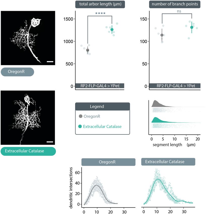
Dhawan et al.                                                                                                    Activity-Regulated Dendritic Plasticity

0.217 × 0.217 × 0.3 µm using a back-thinned Evolve EMCCD                    of a second source of ROS, generated at the plasma membrane by
camera (Photometrics), operated via MetaMorph software                      NADPH oxidases, during activity-regulated structural plasticity.
(Molecular Devices).                                                        Drosophila codes for only two NADPH oxidases; dDuox, an
                                                                            orthologue of vertebrate dual oxidase, and dNox, which is closely
Neuron Reconstruction                                                       related to human Nox5 (Kawahara et al., 2007). To investigate
Dendritic arbor reconstructions were carried out in Amira                   if either or both contributed to activity-regulated structural
6.5 (FEI). A deconvolution algorithm was used to reassign                   plasticity of dendrites, we targeted the expression of previously
photons from out-of-focus optical sections to their points                  tested RNAi constructs designed to knockdown dDuox or dNox
of origin, thus improving the signal-to-noise ratio of the                  (Ha et al., 2005a; Fogarty et al., 2016; Fujisawa et al., 2020)
image stack. Subsequently, thresholding of voxel grey values                to the well-characterized “RP2” motoneuron, with and without
was used to segment the fluorescent arbor from background.                  concomitant overactivation (Sink and Whitington, 1991; Baines
Structures, which did not require reconstruction, i.e., the                 et al., 1999; Landgraf et al., 2003). We then analysed RP2 dendritic
cell body and primary neurite, were manually removed. Post                  arbors morphometrically to quantify the extent to which these
segmentation, the Amira automatic reconstruction algorithm                  manipulations impacted their development.
was used to convert the centrelines of the user-defined                         Expression of UAS-dDuox.RNAi or UAS-dNox.RNAi
segmentation into a spatial graph structure. This structure                 transgenes alone under endogenous activity conditions, i.e.,
was manually reviewed and edited to correct for “loops”                     in the absence of dTrpA1 manipulation, did not produce
and other artefacts of the automatic reconstruction process.                significant differences in arbor characteristics (Figure 1A–C);
Quantification of cell body area was conducted in ImageJ                    though expression of UAS-dDuox.RNAi caused abnormal cell
(National Institutes of Health) by manually tracing around                  body morphology with supernumerary filopodial protrusions
individual cell bodies.                                                     (Figure 1A). In accordance with previous findings (Oswald
                                                                            et al., 2018a), neuronal overactivation by targeted dTrpA1
Data Handling and Statistical Analysis                                      misexpression in individual RP2 motoneurons resulted in
All data handling and statistical analyses were carried out                 significantly smaller dendritic arbors with reduced dendritic
in R. A Shapiro-Wilk test was used to confirm normality                     length and branch point number, as compared to non-
of all dendritic arbor reconstruction data presented. A one-                manipulated controls (Figure 1A–C). Co-expression of
way analysis of variance (ANOVA) followed by Tukey’s                        UAS-dDuox.RNAi along with UAS-dTrpA1 significantly
multiple comparisons test was used to compare experimental                  attenuated the activity-induced reduction of total dendritic
manipulations to the controls where ∗ p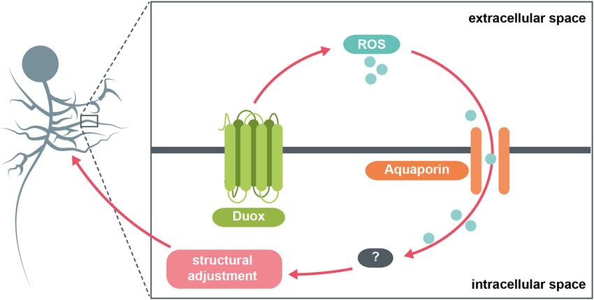
Dhawan et al. Activity-Regulated Dendritic Plasticity
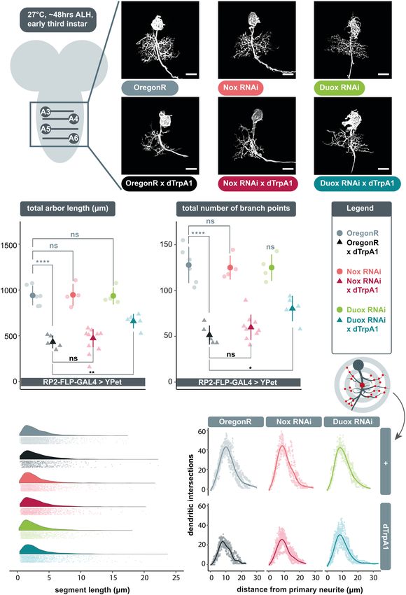
FIGURE 1 | Extracellular reactive oxygen species (ROS) generated by dDuox, but not dNox, are required for homeostatic structural plasticity in response to
increased neuronal activity. (A) Maximum intensity z-projections of representative RP2 motoneurons located within abdominal segments A3-6 in the ventral nerve
cord, from young third instar larvae raised at 27◦ C and dissected 48 h after larval hatching. GAL4 expression was elicited and maintained in individual RP2
motoneurons by crossing RP2-FLP-GAL4 > YPet males containing the transgenes RN2-FLP, tub84B-FRT-CD2-FRT-GAL4, 10xUAS-IVS-myr::YPet (a membrane
targeted YPet fluorophore) to virgins from wild type Oregon-Red (OregonR) flies (= controls; non-manipulated neurons) or from stocks containing UAS-dTrpA1 and/or
(Continued)
Frontiers in Cellular Neuroscience | www.frontiersin.org 4 July 2021 | Volume 15 | Article 641802Dhawan et al. Activity-Regulated Dendritic Plasticity
FIGURE 1 | Continued
UAS-dDuox.RNAi or UAS-dNox.RNAi transgenes. Note that cell body morphology is affected by expression of UAS-dDuox.RNAi, causing filopodial growth from the
soma. Scale bars: 15 µm. (B,C) Targeted expression of dTrpA1, known to cause neuronal over-activation at temperatures ≥ 25◦ C leads to reduced dendritic arbor
size. Co-expression of UAS-dDuox.RNAi, but not UAS-dNox.RNAi, significantly suppresses this activity-induced reduction in total dendritic arbor length and number
of branch points relative to UAS-dTrpA1 manipulated motoneurons. In absence of UAS-dTrpA1, RP2 dendrites expressing these RNAi constructs are comparable to
controls [analysis of variance (ANOVA), ns, not significant; *p < 0.05; **p < 0.01; ****p < 0.0001]. Comparisons with non-manipulated controls are shown directly
above data points (light grey) and comparisons with the UAS-dTrpA1 overactivation condition are shown directly below (black). (D) Irrespective of neuronal activity
regime, expression of UAS-dDuox.RNAi or UAS-dNox.RNAi does not alter the frequency distribution of dendritic segment lengths. A segment is defined as the
distance between two branch points, or, in the case of a terminal neurite, a branch point and the tip. Frequency density plot shown in darker color, with all individual
data points plotted below in a corresponding lighter shade. (E) Sholl analyses indicate that genetic manipulations do not obviously change the relative distribution of
dendrites in 3D space. Mean dendritic intersections as a function of distance from the midpoint of the primary neurite (E’) are shown as a solid coloured line, all
individual data points are shown in a corresponding lighter shade.
In summary, these data implicate dDuox, but not dNox, as targeted co-expression of UAS-Prip.RNAi (Chakrabarti and
necessary for structural plasticity of dendritic arbors in response Visweswariah, 2020) transgenes did not affect the UAS-dTrpA1
to elevated activity. mediated reduction of dendritic arbors, nor did UAS-Prip.RNAi
expression by itself have a measurable impact on dendritic
development in absence of TrpA1-mediated over-activation. In
Aquaporin Channel Proteins Bib and Drip contrast, mis-expression of UAS-bib.RNAi or UAS-Drip.RNAi
Regulate Dendritic Growth, Potentially alone, without concomitant dTrpA1 activity manipulation, was
by Functioning as Conduits for not phenotypically neutral, but produced a dendritic overgrowth
phenotype, of increased dendritic length (Figure 2B) and
Extracellular ROS
branching complexity (Figure 2C) in the ventral part of the
NADPH oxidases generate ROS extracellularly, which poses the
arbor. Throughout these manipulations, changes in dendritic
question of how such NADPH-generated ROS affect dendritic
growth appear to result from changes in the number rather than
growth. Do they do so by modifying extracellular components
length of dendritic segments (Figure 2E). Sholl analyses suggest
or via intracellular events? The latter would require entry into
that RP2 motoneurons target their normal neuropil territories
the cell, and we focused on investigating this scenario. Owing
irrespective of aquaporin knockdown manipulation (Figure 2F).
to the large dipole moments of key ROS, like H2 O2 , simple
We wondered whether UAS-bib.RNAi or UAS-Drip.RNAi
diffusion across the hydrophobic plasma membrane, as seen with
induced dendritic overgrowth might be caused by increased
small and non-polar molecules, is limited. Instead, evidence from
internal osmotic pressure, as a result of impaired aquaporin
other experimental systems points towards a model of facilitated
function, which in turn may stimulate mechanically sensitive
diffusion involving aquaporins (Bienert et al., 2007; Miller et al.,
proteins (Kerstein et al., 2015). However, we could not detect
2010; Bertolotti et al., 2013; Chakrabarti and Visweswariah,
evidence for cell body dilation as would be expected if expression
2020). Classically, the importance of these channel proteins has
of UAS-aquaporin.RNAi transgenes were to increase internal
been stressed in the process of transmembrane fluid transport.
osmotic pressure (Figure 2D).
However, some lines of research suggest that aquaporins can
In summary, these data suggest a requirement for the
regulate the downstream signaling pathways that rely on ROS
aquaporin channels Bib and Drip, but not Prip, in regulating
as a second messenger, by controlling entry of ROS into the
dendritic arbor size. However, since mis-expression of UAS-
cytosol (Miller et al., 2010; Bertolotti et al., 2013; Chakrabarti and
bib.RNAi or UAS-Drip.RNAi alone, in the absence of dTrpA1-
Visweswariah, 2020).
mediated overactivation, leads to a dendritic overgrowth
We postulated that following neuronal overactivation
phenotype, it is unclear if aquaporins and neuronal activity act
extracellular ROS generated by Duox are brought into the
in the same pathway or in parallel pathways with opposite effects
cell via aquaporin channels, where they can then trigger
on dendritic growth.
compensatory structural changes in dendritic arbor size. To test
this model, we overactivated individual RP2 motoneurons by
targeted expression of dTrpA1 whilst simultaneously expressing Extracellular ROS Act as Negative
RNAi constructs designed to knockdown genes that encode Regulators of Dendritic Growth
aquaporin channels: big brain (bib), Drosophila intrinsic proteins The above data suggest that extracellular ROS could provide
(Drip), or Pyrocoelia rufa integral proteins (Prip). For each a negative feedback signal to reduce dendritic arbor size, if
aquaporin encoding gene we used two independently generated those were channeled via aquaporins into the cytoplasm. To test
UAS-RNAi constructs, with at least one having previously this idea, we expressed in single RP2 motoneurons a secreted,
been shown to have specific effects. Under conditions of cell- extracellular form of catalase, to quench extracellular H2 O2 .
autonomous neuronal overactivation, targeted co-expression Compatible with the hypothesis, we found that expression of
of UAS-bib.RNAi (Djiane et al., 2013) or UAS-Drip.RNAi UAS-human-secreted-catalase (Ha et al., 2005b; Fogarty et al.,
(Bergland et al., 2012) transgenes resulted in a significant 2016) produced a dendritic overgrowth phenotype comparable
abrogation of activity-induced arbor reduction, similar to to that caused by expression of UAS-bib.RNAi or UAS-Drip.RNAi
co-expression of UAS-dDuox.RNAi (Figures 2A–C). In contrast, (Figures 2A, 3A). This overgrowth was characterized by dense
Frontiers in Cellular Neuroscience | www.frontiersin.org 5 July 2021 | Volume 15 | Article 641802Dhawan et al. Activity-Regulated Dendritic Plasticity FIGURE 2 | The aquaporins encoded by bib and Drip, but not Prip, are necessary for ROS and activity-induced dendritic plasticity. (A) Maximum intensity z-projections of representative RP2 motoneurons from young third instar larvae raised at 25◦ C and dissected 48 h after larval hatching, expressing GAL4 and the membrane-targeted cell morphology reporter, 10xUAS-IVS-myr::YPet. Controls were from crosses of RP2-FLP-GAL4 > YPet males containing the transgenes RN2-FLP, tub84B-FRT-CD2-FRT-GAL4, 10xUAS-IVS-myr::YPet (a membrane targeted YPet fluorophore) to virgins from wild type Oregon-Red (OregonR) flies (= controls; non-manipulated neurons) or from stocks containing UAS-dTrpA1 and/or UAS-aquaporin.RNAi transgenes. Expression of UAS-bib.RNAi or UAS-Drip.RNAi, but not UAS-Prip.RNAi, without concomitant dTrpA1 manipulation causes dendritic over-growth in RP2 motoneurons. Under conditions of Frontiers in Cellular Neuroscience | www.frontiersin.org 6 July 2021 | Volume 15 | Article 641802
Dhawan et al.                                                                                                                       Activity-Regulated Dendritic Plasticity

  FIGURE 2 | Continued
  cell-selective neuronal over-activation by UAS-dTrpA1 co-expression of UAS-bib.RNAi or UAS-Drip.RNAi, but not UAS-Prip.RNAi, leads to significant attenuation of
  dTrpA1-induced dendritic under-growth in RP2 motoneurons. Scale bars: 15 µm. (B,C) Targeted expression of UAS-bib.RNAi or UAS-Drip.RNAi, but not
  UAS-Prip.RNAi in RP2 motoneurons significantly attenuates the reduction in dendritic arbor length and number of branch points caused by UAS-dTrpA1-mediated
  neuronal overaction. Two independently generated RNAi constructs were tested for each aquaporin. Expression of UAS-bib.RNAi or UAS-Drip.RNAi without
  concomitant dTrpA1 manipulation produces a dendritic overgrowth phenotype characterized by increased arbor length and branching complexity relative to
  non-manipulated controls. (D) Quantification of cell body size, measured as the maximum area of the 2D cross-section through the centre of the soma, shows no
  consistent differences between controls and UAS-aquaporin.RNAi manipulations. (B–D) ANOVA, ns, not significant; *p < 0.05; **p < 0.01; ***p < 0.001;
  ****p < 0.0001. Comparisons with non-manipulated controls are shown directly above data points (light grey) and comparisons with overactivated controls are
  directly below (black). (E) Expression of RNAi transgenes designed to knock down specific aquaporins does not alter fundamental arbor structure under conditions
  of endogenous activity or chronic overactivation. Frequency density plot of dendritic segment lengths shown in darker color, with all individual data points plotted
  below in a corresponding lighter shade. (F) The branching topology of RP2 motoneurons is not affected by UAS-aquaporin-RNAi manipulations. Mean dendritic
  intersections as a function of distance from the midpoint of the primary neurite are shown as a solid coloured line, all individual data points are shown in a
  corresponding lighter shade.

dendritic arbors with significantly larger total arbor length as                       that the size and complexity of dendritic arbors correlates
compared to non-manipulated controls (Figure 3B). As with the                          with the range and amount of synaptic input received (Purves
other manipulations above, here too the relative distribution of                       and Lichtman, 1985; Ivanov and Purves, 1989). Similarly, in
dendritic segments and arbor topography remained comparable                            the Drosophila larval locomotor network we demonstrated
to controls (Figures 3C–E). Somewhat unexpected though, our                            that during development the dramatic growth of motoneuron
analysis indicates no change in branch point number despite                            dendritic arbors, which scales with overall body growth,
the increase in arbor size (Figure 3C). This is counter-intuitive                      facilitates increases in presynaptic input and thus also of the
and we think the most parsimonious explanation for that this is                        amount of synaptic drive necessary for appropriate levels of
an artefactual under-representation of segment number, caused                          muscle activation (Zwart et al., 2013). This kind of structural
by the high density of dendritic segments in these overgrown                           plasticity is not limited to periods of growth, but also evident
neurons, leading to failure of resolving all branch points during                      following activity manipulations. For example, changes in the
the reconstruction process.                                                            number of active presynaptic sites are compensated for by
   In conclusion, in this study we identified a selective                              complementary changes in postsynaptic dendritic arbor size,
requirement for the NADPH oxidase, Duox (but not Nox), in                              suggesting that neurons use their dendritic arbors as structural
activity-regulated adjustment of dendritic arbor growth during                         homeostatic devices (Tripodi et al., 2008). Similar structural
larval nervous system development. Thus generated extracellular                        homeostatic adjustments have also been documented in the
ROS could signal to neighbouring cells, as well as mediate                             developing visual system (Yuan et al., 2011; Sheng et al.,
autocrine signaling. We also identified a role for the aquaporins                      2018; Dombrovski et al., 2019). ROS are necessary for this
Bib and Drip (but not Prip), which we propose serve as                                 plasticity to occur, with ROS acting as brakes on dendritic arbor
conduits for channeling extracellular H2 O2 back into the cell,                        growth (Oswald et al., 2018a). Here, we identify the NADPH
from adjacent cells but likely also mediating autocrine signaling.                     oxidase, Duox, as a source of such activity-generated ROS.
Overall, extracellular ROS act as negative feedback signals that                       The topography of Duox, which is known to reside within
mediate homeostatic adjustment of dendritic arbor size. The                            the plasma membrane (Morand et al., 2009), is such that it
data suggest this process operates at physiological activity levels                    generates H2 O2 into extracellular space (Fogarty et al., 2016).
since manipulations where we expressed secreted Catalase to                            We previously showed that H2 O2 is required intracellularly, the
quench extracellular ROS in the immediate vicinity of a neuron                         effects of cell-specific over-activation rescued cell-autonomously
lead to dendritic overgrowth that is indistinguishable from                            by co-expression of cytoplasmic Catalase (Oswald et al., 2018a).
overgrowth phenotypes caused by cell-autonomously targeting                            This raised the question of how such extracellular ROS re-
the aquaporins Bib and Drip for RNAi knockdown. Such enlarged                          enter the neuronal cytoplasm. Our findings suggest that the
dendritic arbors would be predicted by our model (Figure 4), as                        aquaporin channels Bib and Drip, but not Prip, might function
a consequence of reduced influx of ROS, which act as a brake on                        as conduits for extracellular ROS, necessary for activity-regulated
dendritic growth.                                                                      dendritic structural remodelling (Figure 4). Specifically, targeting
                                                                                       bib or Drip for RNAi-mediated knockdown in individual
                                                                                       RP2 motoneurons significantly attenuates the overactivation
DISCUSSION                                                                             phenotype of smaller dendritic arbors. These observations
                                                                                       suggest that specific aquaporins located in the somato-dendritic
A prerequisite of flexible yet stable neural circuitry is the                          compartment of motoneurons act as conduits that facilitate
ability to detect and appropriately respond to changes in                              entry of extracellular H2 O2 into the cytoplasm, where it can
activity, particularly perturbations that push activity towards                        modulate dendritic growth pathways. This model is similar
extremes of quiescence or saturation (Turrigiano and Nelson,                           to one recently proposed by Chakrabarti and Visweswariah
2004; Yin and Yuan, 2014). Here, we focused on structural                              (2020) in Drosophila hemocytes, where in response to wounding
plasticity of dendrites in response to increased neuronal                              Duox is activated, generates extracellular H2 O2 , which partly
activation. Seminal comparative studies in mammals showed                              signals in an autocrine fashion with import back into the

Frontiers in Cellular Neuroscience | www.frontiersin.org                           7                                              July 2021 | Volume 15 | Article 641802
Dhawan et al. Activity-Regulated Dendritic Plasticity FIGURE 3 | Cell-specific expression of extracellular catalase produces a dendritic overgrowth phenotype comparable to that produced by UAS-bib.RNAi or UAS-Drip.RNAi expression. (A) Maximum intensity z-projections of representative RP2 motoneurons, from young third instar larvae raised at 25◦ C and dissected 48 h after larval hatching. To target GAL4 expression to RP2 motoneurons RP2-FLP-GAL4 > YPet males were crossed to virgins from wild type Oregon-Red (OregonR) flies (= controls; non-manipulated neurons) or flies containing UAS-human secreted catalase, here termed “extracellular Catalase”. Scale bars: 15 µm. (B,C) Targeted expression of UAS-human secreted catalase in RP2 motoneurons results in a significant increase or “overgrowth” of dendritic arbor length relative to otherwise non-manipulated neurons, but does not change the total number of branch points (unexpected and likely caused by poor signal-to-noise ratio resulting from very dense branching of these enlarged arbors) (ANOVA, ns, not significant; ****p < 0.0001). (D) UAS-human secreted catalase expression in RP2 motoneurons does not change the frequency distribution of dendritic segment lengths relative to overactivated or non-manipulated controls Frequency density plot shown in darker color, with all individual data points plotted below in a corresponding lighter shade. (E) Sholl analyses indicate that UAS-human secreted catalase expression does not obviously cause RP2 motoneurons to alter the placement or density of their dendrites. Mean dendritic intersections as a function of distance from the midpoint of the primary neurite are shown as a solid coloured line, all individual data points are shown in a corresponding lighter shade. hemocyte cytoplasm facilitated by the aquaporin Prip. Moreover, Based on our previous observations, we speculate that within expression of a secreted Catalase, IRC, modulates such H2 O2 the neuron H2 O2 acts, amongst others, on the cytoplasmic redox- signaling (Chakrabarti and Visweswariah, 2020). sensitive dimer DJ-1β, which we previously showed necessary Frontiers in Cellular Neuroscience | www.frontiersin.org 8 July 2021 | Volume 15 | Article 641802
Dhawan et al.                                                                                                                          Activity-Regulated Dendritic Plasticity

  FIGURE 4 | Model summary. The NADPH oxidase Duox generates ROS at the extracellular face of the plasma membrane in response to increases in neuronal
  activity. These ROS are brought back into the cytosol via specific aquaporin channel proteins. Here, they interact with various intracellular pathways, potentially
  involving the redox-sensitive dimer DJ-1β, which in turn mediate adaptive reductions in dendritic arbor size.

for structural and physiological changes in response to activity-                        possibility of an intercellular redox-based communication
generated ROS (Oswald et al., 2018a). Dendritic overgrowth, as                           network that coordinates homeostatic structural adjustments
caused by expression of a secreted form of Catalase, known to                            more widely, potentially within local volumes. Synaptic clefts
scavenge extracellular H2 O2 (Ha et al., 2005b; Fogarty et al., 2016;                    in Drosophila are approximately 10–20 nm in width (Prokop
Chakrabarti and Visweswariah, 2020), suggests that extracellular                         and Meinertzhagen, 2006). ROS such as O2 − and H2 O2
ROS from adjacent cells might contribute to structural plasticity                        have been reported to travel distances in the order of
regulation. Expression of a cytoplasmic Catalase, in contrast, did                       several micrometers within living tissue (Cuypers et al., 2016;
not have such an effect, at least not in aCC motoneurons at an                           Krumova and Cosa, 2016). It follows that extracellular ROS
earlier stage of 24 h after larval hatching (Oswald et al., 2018a).                      generated by Duox in postsynaptic dendrites could traverse
This might suggest stage-specific differences in levels of ROS or                        the synaptic cleft and act retrogradely on the surface of
differences in the efficacy of these two transgenes, either due to                       presynaptic partner terminals, e.g., on ion channels (Sah
expression levels or difficulty of cytoplasmic Catalase to diffuse                       et al., 2002; Sesti et al., 2010; Sahoo et al., 2014). Such
into small diameter dendritic branches.                                                  ROS could further enter the cytosol of presynaptic partners
   The partial penetrance of RNAi knockdown phenotypes,                                  via aquaporin channels and trigger compensatory structural
combined with the observation that two distinct aquaporin                                remodelling. In addition, in analogy to retrograde nitric oxide
encoding genes are involved, suggests that there may                                     signaling (Hardingham et al., 2013), Duox generated extracellular
be functional redundancy between these aquaporins.                                       ROS have the potential to modify pre- and postsynaptic
Such redundancy has previously been observed in the                                      terminals within a local volume of the neuropil, thus acting
tsetse fly, where simultaneous downregulation of multiple                                as regional activity-triggered modulators even between non-
aquaporins exacerbates the negative effects on female                                    synaptic neurons. Perhaps, such ROS may even act on adjacent
fecundity produced by individual aquaporin knockdowns                                    glia, potentially via redox-sensitive glial proteins such as transient
(Benoit et al., 2014).                                                                   receptor potential melastatin 2 (TRPM2), which have been
                                                                                         shown to modulate synaptic plasticity (Wang et al., 2016;
Extracellular Duox-Generated ROS as a                                                    Turlova et al., 2018).
Potential Means for Coordinating
Network-Wide Homeostatic Structural                                                      Downstream Effector Pathways of ROS
Adjustments                                                                              and Aquaporin-Dependent Structural
Whilst ROS typically operate as intracellular second messengers                          Plasticity
(Forman et al., 2014; Schieber and Chandel, 2014), their                                 Reactive oxygen species can regulate the activity of several
long-range effects have also been observed during paracrine                              protein kinases, including those implicated in canonical
H2 O2 signaling in vertebrate and invertebrate inflammatory                              neurodevelopmental pathways, either via modification of reactive
responses (Niethammer et al., 2009; Moreira et al., 2010). In                            amino acid residues on kinases or, indirectly, by redox-mediated
light of this, our finding that extracellular, activity-generated                        inhibition of counteracting phosphatases (Finkel and Holbrook,
ROS are necessary for structural plasticity raises the intriguing                        2000; Corcoran and Cotter, 2013; Holmström and Finkel, 2014).

Frontiers in Cellular Neuroscience | www.frontiersin.org                             9                                               July 2021 | Volume 15 | Article 641802
Dhawan et al. Activity-Regulated Dendritic Plasticity
Notably, previous work has implicated aquaporin channels DATA AVAILABILITY STATEMENT
in modulating the efficacy of ROS-regulated protein kinase
signaling. By altering a cell’s permeability to extracellularly- The raw data supporting the conclusions of this article will be
generated ROS, aquaporins can amplify or diminish the strength made available by the authors, without undue reservation.
of redox-dependent downstream pathways (Miller et al., 2010;
Bertolotti et al., 2013). For instance, mammalian Aquaporin 8,
which shares ∼33% of amino acid sequence identity with the
Drosophila aquaporin channels, controls the entry of NADPH- AUTHOR CONTRIBUTIONS
oxidase derived H2 O2 to increase growth factor signaling in
human leukaemia B-cells (Vieceli Dalla Sega et al., 2017). Of SD designed and executed the experiments, analysed the data,
particular interest are CaMKII and PKA signaling, which can and wrote the manuscript. PM carried out experiments. DMD,
be enhanced by elevated cytosolic ROS (Humphries et al., 2007; ADO and JFE generated reagents. ML conceived of the study and
Anderson, 2011), and which both pathways act to limit the wrote the manuscript. All authors contributed to the article and
elaboration of dendritic arbors in an activity-dependent manner. approved the submitted version.
For example, targeted inhibition of CaMKII or PKA in otherwise
non-manipulated neurons results in dendritic over-growth and
an increase in arbor size and complexity (Wu and Cline, 1998;
Zou and Cline, 1999; Tripodi et al., 2008). This is similar to
FUNDING
what we have seen following quenching of extracellular H2 O2 This work was supported by funding from the Biotechnology
or knockdown of the aquaporins Bib and Drip, suggesting that and Biological Sciences Research Council to ML (BB/R016666/1)
either CaMKII or PKA signaling might be downstream effectors and from the DFG to JFE (EV198/1-1). The work benefited from
of activity regulated, Duox-generated extracellular H2 O2 . the Imaging Facility, Department of Zoology, supported by Matt
Given the increasing number of signals involved in Wayland and funds from a Wellcome Trust Equipment Grant
anterograde and retrograde signaling between neurons one might (WT079204) with contributions by the Sir Isaac Newton Trust
ask how ROS contribute to these signaling pathways. It is possible in Cambridge, including Research Grant [18.07ii(c)].
that Duox acts as an integrator of multiple signaling pathways, in
that its activity is regulated by a number of pathways, including
the Rho GTPase Rac1 (Hordijk Peter, 2006) and calcium, via
its EF-hands (Kawahara et al., 2007). It will be interesting to ACKNOWLEDGMENTS
determine the range and temporal dynamics of Duox activity
following neuronal activation; whether Duox reports on low, The authors would like to thank members of the Landgraf lab
medium or high levels of neuronal activation, brief bursts or for feedback on the manuscript. The authors are grateful to
only following prolonged activation. Thus, it is conceivable Andreas Bergmann, Paul Garrity, Won-Jae Lee, Paul Martin,
that different inter-neuronal signaling pathways are utilised for Sean Sweeney, Helen Weavers, and Will Wood, as well as the
distinct contexts, in terms of their activation pattern and, equally, Bloomington Drosophila Stock Center and Vienna Drosophila
their spatio-temporal dynamics of signaling. Resource Centre for generously providing fly stocks.
REFERENCES function of an aquaporin. PLoS Genet. 8:e1002631. doi: 10.1371/journal.pgen.
1002631
Anderson, M. E. (2011). Pathways for CaMKII activation in disease. Heart Rhythm Bertolotti, M., Bestetti, S., García-Manteiga, J. M., Medraño-Fernandez, I., Dal Mas,
8, 1501–1503. doi: 10.1016/j.hrthm.2011.04.027 A., Malosio, M. L., et al. (2013). Tyrosine kinase signal modulation: a matter of
Attwell, D., and Laughlin, S. B. (2001). An energy budget for signaling in the H2O2 membrane permeability? Antioxid. Redox Signal. 19, 1447–1451. doi:
grey matter of the brain. J. Cereb. Blood Flow Metab. 21, 1133–1145. doi: 10.1089/ars.2013.5330
10.1097/00004647-200110000-00001 Bienert, G. P., Møller, A. L. B., Kristiansen, K. A., Schulz, A., Møller, I. M.,
Baines, R. A., Robinson, S. G., Fujioka, M., Jaynes, J. B., and Bate, M. (1999). Schjoerring, J. K., et al. (2007). Specific aquaporins facilitate the diffusion of
Postsynaptic expression of tetanus toxin light chain blocks synaptogenesis hydrogen peroxide across membranes. J. Biol. Chem. 282, 1183–1192. doi:
in Drosophila. Curr. Biol. 9, 1267–1270. doi: 10.1016/s0960-9822(99) 10.1074/jbc.m603761200
80510-7 Chakrabarti, S., and Visweswariah, S. S. (2020). Intramacrophage ROS primes the
Baxter, P. S., and Hardingham, G. E. (2016). Adaptive regulation of the brain’s innate immune system via JAK/STAT and toll activation. Cell Rep. 33:108368.
antioxidant defences by neurons and astrocytes. Free Radic. Biol. Med. 100, doi: 10.1016/j.celrep.2020.108368
147–152. doi: 10.1016/j.freeradbiomed.2016.06.027 Corcoran, A., and Cotter, T. G. (2013). Redox regulation of protein kinases. FEBS
Benoit, J. B., Hansen, I. A., Attardo, G. M., Michalková, V., Mireji, P. O., Bargul, J. 280, 1944–1965. doi: 10.1111/febs.12224
J. L., et al. (2014). Aquaporins are critical for provision of water during Cuypers, A., Hendrix, S., Amaral Dos Reis, R., De Smet, S., Deckers, J., Gielen,
lactation and intrauterine progeny hydration to maintain tsetse fly reproductive H., et al. (2016). Hydrogen peroxide, signaling in disguise during metal
success. PLoS Negl. Trop. Dis. 8:e2517. doi: 10.1371/journal.pntd.00 phytotoxicity. Front. Plant Sci. 7:470.
02517 Djiane, A., Krejci, A., Bernard, F., Fexova, S., Millen, K., and Bray, S. J. (2013).
Bergland, A. O., Chae, H. S., Kim, Y. J., and Tatar, M. (2012). Fine-scale mapping of Dissecting the mechanisms of Notch induced hyperplasia. EMBO J. 32, 60–71.
natural variation in fly fecundity identifies neuronal domain of expression and doi: 10.1038/emboj.2012.326
Frontiers in Cellular Neuroscience | www.frontiersin.org 10 July 2021 | Volume 15 | Article 641802Dhawan et al. Activity-Regulated Dendritic Plasticity Dombrovski, M., Kim, A., Poussard, L., Vaccari, A., Acton, S., Spillman, Lambeth, J. D. (2002). Nox/Duox family of nicotinamide adenine dinucleotide E., et al. (2019). A Plastic visual pathway regulates cooperative behavior (phosphate) oxidases. Curr. Opin. Hematol. 9, 11–17. doi: 10.1097/00062752- in Drosophila Larvae. Current Biology 29, 1866.e–1876.e. 1866– 200201000-00003 1876.e5, ∗ Landgraf, M., Jeffrey, V., Fujioka, M., Jaynes, J. B., and Bate, M. (2003). Embryonic Finkel, T., and Holbrook, N. J. (2000). Oxidants, oxidative stress and the biology of origins of a motor system: motor dendrites form a myotopic map in Drosophila. ageing. Nature 408, 239–247. doi: 10.1038/35041687 PLoS Biol. 1:E41. Fogarty, C. E., Diwanji, N., Lindblad, J. L., Tare, M., Amcheslavsky, A., Makhijani, Marley, R., and Baines, R. A. (2011). Dissection of Third-Instar Drosophila Larvae K., et al. (2016). Extracellular Reactive Oxygen Species Drive Apoptosis- for Electrophysiological Recording from Neurons. Cold Spring Harb. Protoc. Induced Proliferation via Drosophila Macrophages. Curr. Biol. 26, 575–584. 2011, db.rot065656. ∗ , doi: 10.1016/j.cub.2015.12.064 Martin, S. J., Grimwood, P. D., and Morris, R. G. M. (2000). Synaptic Plasticity and Forman, H. J., Ursini, F., and Maiorino, M. (2014). An overview of mechanisms of Memory: an Evaluation of the Hypothesis. Annu. Rev. Neurosci. 23, 649–711. redox signaling. J. Mol. Cell. Cardiol. 73, 2–9. doi: 10.1146/annurev.neuro.23.1.649 Frank, C. A., James, T. D., and Müller, M. (2020). Homeostatic control of Massaad, C. A., and Klann, E. (2011). Reactive oxygen species in the regulation Drosophila neuromuscular junction function. Synapse 74, e22133. of synaptic plasticity and memory. Antioxid. Redox Signal. 14, 2013–2054. Fujioka, M., Lear, B. C., Landgraf, M., Yusibova, G. L., Zhou, J., Riley, K. M., doi: 10.1089/ars.2010.3208 et al. (2003). Even-skipped, acting as a repressor, regulates axonal projections Miller, E. W., Dickinson, B. C., and Chang, C. J. (2010). Aquaporin-3 mediates in Drosophila. Development 130, 5385–5400. doi: 10.1242/dev.00770 hydrogen peroxide uptake to regulate downstream intracellular signaling. Proc. Fujisawa, Y., Shinoda, N., Chihara, T., and Miura, M. (2020). ROS Regulate Natl. Acad. Sci. U. S. A. 107, 15681–15686. doi: 10.1073/pnas.1005776107 Caspase-Dependent Cell Delamination without Apoptosis in the Milton, V. J., Jarrett, H. E., Gowers, K., Chalak, S., Briggs, L., Robinson, I. M., et al. Drosophila Pupal Notum. iScience 23, 101413. doi: 10.1016/j.isci.2020. (2011). Oxidative stress induces overgrowth of the Drosophila neuromuscular 101413 junction. Proc. Natl. Acad. Sci. U. S. A. 108, 17521–17526. doi: 10.1073/pnas. Ha, E.-M., Oh, C.-T., Bae, Y. S., and Lee, W.-J. (2005a). A direct role for dual 1014511108 oxidase in Drosophila gut immunity. Science 310, 847–850. doi: 10.1126/ Morand, S., Ueyama, T., Tsujibe, S., Saito, N., Korzeniowska, A., and Leto, science.1117311 T. L. (2009). Duox maturation factors form cell surface complexes with Duox Ha, E.-M., Oh, C.-T., Ryu, J.-H., Bae, Y. S., Kang, S. W., Jang, I.-H., et al. (2005b). affecting the specificity of reactive oxygen species generation. FASEB J. 23, An antioxidant system required for host protection against gut infection 1205–1218. doi: 10.1096/fj.08-120006 in Drosophila. Developmental Cell 8, 125–132. doi: 10.1016/j.devcel.2004. Moreira, S., Stramer, B., Evans, I., Wood, W., and Martin, P. (2010). Prioritization 11.007 of competing damage and developmental signals by migrating macrophages Halliwell, B. (1992). Reactive oxygen species and the central nervous in the Drosophila embryo. Curr. Biol. 20, 464–470. doi: 10.1016/j.cub.2010. system. J. Neurochem. 59, 1609–1623. doi: 10.1111/j.1471-4159.1992.tb 01.047 10990.x Munnamalai, V., and Suter, D. M. (2009). Reactive oxygen species regulate F-actin Hamada, F. N., Rosenzweig, M., Kang, K., Pulver, S. R., Ghezzi, A., Jegla, T. J., dynamics in neuronal growth cones and neurite outgrowth. J. Neurochem. 108, et al. (2008). An internal thermal sensor controlling temperature preference in 644–661. doi: 10.1111/j.1471-4159.2008.05787.x Drosophila. Nature 454, 217–220. doi: 10.1038/nature07001 Munnamalai, V., Weaver, C. J., Weisheit, C. E., Venkatraman, P., Agim, Z. S., Hardingham, N., Dachtler, J., and Fox, K. (2013). The role of nitric oxide in Quinn, M. T., et al. (2014). Bidirectional interactions between NOX2-type pre-synaptic plasticity and homeostasis. Front. Cell. Neurosci. 7:190. NADPH oxidase and the F-actin cytoskeleton in neuronal growth cones. Hidalgo, C., and Arias-Cavieres, A. (2016). Calcium, Reactive Oxygen Species, J. Neurochem. 130, 526–540. doi: 10.1111/jnc.12734 and Synaptic Plasticity. Physiology 31, 201–215. doi: 10.1152/physiol.000 Nguyen, A. W., and Daugherty, P. S. (2005). Evolutionary optimization of 38.2015 fluorescent proteins for intracellular FRET. Nature Biotechnology 23, 355–360. Holmström, K. M., and Finkel, T. (2014). Cellular mechanisms and physiological doi: 10.1038/nbt1066 consequences of redox-dependent signalling. Nat. Rev. Mol. Cell Biol. 15, Niethammer, P., Grabher, C., Look, A. T., and Mitchison, T. J. (2009). A tissue-scale 411–421. doi: 10.1038/nrm3801 gradient of hydrogen peroxide mediates rapid wound detection in zebrafish. Hongpaisan, J., Winters, C. A., and Andrews, S. B. (2003). Calcium-dependent Nature 459, 996–999. doi: 10.1038/nature08119 mitochondrial superoxide modulates nuclear CREB phosphorylation in Oswald, M. C., Brooks, P. S., Zwart, M. F., Mukherjee, A., West, R. J., Giachello, hippocampal neurons. Mol. Cell. Neurosci. 24, 1103–1115. doi: 10.1016/j.mcn. C. N., et al. (2018a). Reactive oxygen species regulate activity-dependent 2003.09.003 neuronal plasticity in Drosophila. Elife 7, doi: 10.7554/eLife.39393 ∗ , Hongpaisan, J., Winters, C. A., and Andrews, S. B. (2004). Strong calcium entry Oswald, M. C. W., Garnham, N., Sweeney, S. T., and Landgraf, M. (2018b). activates mitochondrial superoxide generation, upregulating kinase signaling Regulation of neuronal development and function by ROS. FEBS Lett. 592, in hippocampal neurons. J. Neurosci. 24, 10878–10887. doi: 10.1523/jneurosci. 679–691. doi: 10.1002/1873-3468.12972 3278-04.2004 Ou, Y., Chwalla, B., Landgraf, M., and van Meyel, D. J. (2008). Identification of Hordijk Peter, L. (2006). Regulation of NADPH Oxidases. Circ. Res. 98, 453–462. genes influencing dendrite morphogenesis in developing peripheral sensory doi: 10.1161/01.res.0000204727.46710.5e and central motor neurons. Neural Dev. 3, 16. doi: 10.1186/1749-81 Humphries, K. M., Pennypacker, J. K., and Taylor, S. S. (2007). Redox regulation 04-3-16 of cAMP-dependent protein kinase signaling: kinase versus phosphatase Panday, A., Sahoo, M. K., Osorio, D., and Batra, S. (2015). NADPH oxidases: an inactivation. J. Biol. Chem. 282, 22072–22079. doi: 10.1074/jbc.m7025 overview from structure to innate immunity-associated pathologies. Cell. Mol. 82200 Immunol. 12, 5–23. doi: 10.1038/cmi.2014.89 Ivanov, A., and Purves, D. (1989). Ongoing electrical activity of superior cervical Peng, J.-J., Lin, S.-H., Liu, Y.-T., Lin, H.-C., Li, T.-N., and Yao, C.-K. (2019). ganglion cells in mammals of different size. J. Comp. Neurol. 284, 398–404. A circuit-dependent ROS feedback loop mediates glutamate excitotoxicity to doi: 10.1002/cne.902840307 sculpt the Drosophila motor system. Elife 8, e47372. Kawahara, T., Quinn, M. T., and Lambeth, J. D. (2007). Molecular evolution of the Pignoni, F., and Zipursky, S. L. (1997). Induction of Drosophila eye development reactive oxygen-generating NADPH oxidase (Nox/Duox) family of enzymes. by decapentaplegic. Development 124, 271–278. doi: 10.1242/dev.124.2.271 BMC Evol. Biol. 7:109. doi: 10.1186/1471-2148-7-109 Pozo, K., and Goda, Y. (2010). Unraveling mechanisms of homeostatic synaptic Kerstein, P. C., Nichol, R. H. IV, and Gomez, T. M. (2015). Mechanochemical plasticity. Neuron 66, 337–351. doi: 10.1016/j.neuron.2010.04.028 regulation of growth cone motility. Front. Cell. Neurosci. 9:244. Prokop, A., and Meinertzhagen, I. A. (2006). Development and structure of Krumova, K., and Cosa, G. (2016). Chapter 1 Overview of Reactive Oxygen Species. synaptic contacts in Drosophila. Semin. Cell Dev. Biol. 17, 20–30. doi: 10.1016/ 1–21. ∗ . j.semcdb.2005.11.010 Frontiers in Cellular Neuroscience | www.frontiersin.org 11 July 2021 | Volume 15 | Article 641802
Dhawan et al.                                                                                                                                  Activity-Regulated Dendritic Plasticity

Purves, D., and Lichtman, J. W. (1985). Geometrical differences among                          Vieceli Dalla Sega, F., Prata, C., Zambonin, L., Angeloni, C., Rizzo, B., Hrelia,
   homologous neurons in mammals. Science 228, 298–302. doi: 10.1126/science.                     S., et al. (2017). Intracellular cysteine oxidation is modulated by aquaporin-
   3983631                                                                                        8-mediated hydrogen peroxide channeling in leukaemia cells. Biofactors 43,
Sah, R., Galeffi, F., Ahrens, R., Jordan, G., and Schwartz-Bloom, R. D. (2002).                   232–242. doi: 10.1002/biof.1340
   Modulation of the GABA(A)-gated chloride channel by reactive oxygen species.                Wang, J., Jackson, M. F., and Xie, Y.-F. (2016). Glia and TRPM2 Channels in
   J. Neurochem. 80, 383–391. doi: 10.1046/j.0022-3042.2001.00706.x                               Plasticity of Central Nervous System and Alzheimer’s Diseases. Neural Plast.
Sahoo, N., Hoshi, T., and Heinemann, S. H. (2014). Oxidative modulation of                        2016, 1680905.
   voltage-gated potassium channels. Antioxid. Redox Signal. 21, 933–952. doi:                 Wefelmeyer, W., Puhl, C. J., and Burrone, J. (2016). Homeostatic Plasticity of
   10.1089/ars.2013.5614                                                                          Subcellular Neuronal Structures: From Inputs to Outputs. Trends Neurosci. 39,
Schieber, M., and Chandel, N. S. (2014). ROS function in redox signaling and                      656–667. doi: 10.1016/j.tins.2016.08.004
   oxidative stress. Curr. Biol. 24, R453–R462.                                                Wilson, C., Núñez, M. T., and González-Billault, C. (2015). Contribution of
Sesti, F., Liu, S., and Cai, S.-Q. (2010). Oxidation of potassium channels by ROS:                NADPH oxidase to the establishment of hippocampal neuronal polarity in
   a general mechanism of aging and neurodegeneration? Trends Cell Biol. 20,                      culture. J. Cell Sci. 128, 2989–2995.
   45–51. doi: 10.1016/j.tcb.2009.09.008                                                       Wu, G. Y., and Cline, H. T. (1998). Stabilization of dendritic arbor structure in vivo
Sheng, C., Javed, U., Gibbs, M., Long, C., Yin, J., Qin, B., et al. (2018). Experience-           by CaMKII. Science 279, 222–226. doi: 10.1126/science.279.5348.222
   dependent structural plasticity targets dynamic filopodia in regulating dendrite            Yin, J., and Yuan, Q. (2014). Structural homeostasis in the nervous system:
   maturation and synaptogenesis. Nat. Commun. 9, 3362.                                           a balancing act for wiring plasticity and stability. Front. Cell. Neurosci.
Sink, H., and Whitington, P. M. (1991). Location and connectivity of abdominal                    8:439.
   motoneurons in the embryo and larva of Drosophila melanogaster. J. Neurobiol.               Yuan, Q., Xiang, Y., Yan, Z., Han, C., Jan, L. Y., and Jan, Y. N. (2011).
   22, 298–311. doi: 10.1002/neu.480220309                                                        Light-Induced Structural and Functional Plasticity in Drosophila
Stuchlik, A. (2014). Dynamic learning and memory, synaptic plasticity and                         Larval Visual System. Science 333, 1458–1462.                 doi: 10.1126/science.
   neurogenesis: an update. Front. Behav. Neurosci. 8:106.                                        1207121
Tejada-Simon, M. V., Serrano, F., Villasana, L. E., Kanterewicz, B. I., Wu, G.-Y.,             Zhu, X.-H., Qiao, H., Du, F., Xiong, Q., Liu, X., Zhang, X., et al. (2012). Quantitative
   Quinn, M. T., et al. (2005). Synaptic localization of a functional NADPH oxidase               imaging of energy expenditure in human brain. Neuroimage 60, 2107–2117.
   in the mouse hippocampus. Mol. Cell. Neurosci. 29, 97–106. doi: 10.1016/j.mcn.                 doi: 10.1016/j.neuroimage.2012.02.013
   2005.01.007                                                                                 Zou, D. J., and Cline, H. T. (1999). Postsynaptic calcium/calmodulin-dependent
Terzi, A., Roeder, H., Weaver, C. J., and Suter, D. M. (2021). Neuronal                           protein kinase II is required to limit elaboration of presynaptic and postsynaptic
   NADPH oxidase 2 regulates growth cone guidance downstream of slit2/robo2.                      neuronal arbors. J. Neurosci. 19, 8909–8918. doi: 10.1523/jneurosci.19-20-
   Developmental Neurobiology 81, 3–21. doi: 10.1002/dneu.22791                                   08909.1999
Terzi, A., and Suter, D. M. (2020). The role of NADPH oxidases in neuronal                     Zwart, M. F., Randlett, O., Evers, J. F., and Landgraf, M. (2013). Dendritic growth
   development. Free Radical Biology & Medicine 154, 33–47. doi: 10.1016/j.                       gated by a steroid hormone receptor underlies increases in activity in the
   freeradbiomed.2020.04.027                                                                      developing Drosophila locomotor system. Proc. Natl. Acad. Sci. U. S. A. 110,
Tripodi, M., Evers, J. F., Mauss, A., Bate, M., and Landgraf, M. (2008).                          E3878–E3887.
   Structural homeostasis: compensatory adjustments of dendritic arbor geometry
   in response to variations of synaptic input. PLoS Biol. 6:e260. doi: 10.1371/               Conflict of Interest: The authors declare that the research was conducted in the
   journal.pbio.0060260                                                                        absence of any commercial or financial relationships that could be construed as a
Turlova, E., Feng, Z.-P., and Sun, H.-S. (2018). The role of TRPM2 channels                    potential conflict of interest.
   in neurons, glial cells and the blood-brain barrier in cerebral ischemia and
   hypoxia. Acta Pharmacol. Sin. 39, 713–721. doi: 10.1038/aps.2017.194                        Copyright © 2021 Dhawan, Myers, Bailey, Ostrovsky, Evers and Landgraf. This is an
Turrigiano, G. G., and Nelson, S. B. (2000). Hebb and homeostasis in neuronal                  open-access article distributed under the terms of the Creative Commons Attribution
   plasticity. Curr. Opin. Neurobiol. 10, 358–364. doi: 10.1016/s0959-4388(00)                 License (CC BY). The use, distribution or reproduction in other forums is permitted,
   00091-x                                                                                     provided the original author(s) and the copyright owner(s) are credited and that the
Turrigiano, G. G., and Nelson, S. B. (2004). Homeostatic plasticity in the                     original publication in this journal is cited, in accordance with accepted academic
   developing nervous system. Nat. Rev. Neurosci. 5, 97–107. doi: 10.1038/                     practice. No use, distribution or reproduction is permitted which does not comply
   nrn1327                                                                                     with these terms.

Frontiers in Cellular Neuroscience | www.frontiersin.org                                  12                                                 July 2021 | Volume 15 | Article 641802
You can also read