Chagas Disease Cardiomyopathy - SciELO
←
→
Page content transcription
If your browser does not render page correctly, please read the page content below

International Journal of Cardiovascular Sciences. 2018;31(2)173-189
                                                                                                                                                                                    173

                                                                                                                                            REVIEW ARTICLE

Chagas Disease Cardiomyopathy
Marcus Vinicius Simões, Minna Moreira Dias Romano, André Schmidt, Káryta Suely Macedo Martins, José Antonio
Marin-Neto
Divisão de Cardiologia - Departamento de Clínica Médica - Faculdade de Medicina de Ribeirão Preto da Universidade de São Paulo, SP – Brazil

Abstract                                                                         patients in the chronic phase of the disease includes relevant
                                                                                 morbidity and mortality, in addition to being the main cause
    Chronic Chagas disease cardiomyopathy (CCC) is a result                      of nonischemic cardiomyopathy in Latin America.
of low-intensity, but incessant, focal fibrosing myocarditis,
caused by persistent T. cruzi infection associated with
                                                                                 Epidemiology
inflammation, mediated by adverse immune mechanisms.
About 30 percent of infected individuals have developed                             Chagas Disease transmission cycle has been based
throughout life the chronic cardiac form of Chagas' disease                      mostly on triatomine species as main vectors of the
with protean clinical manifestations, such as sudden                             disease. However, after several national campaigns and
death, signs and symptoms of heart failure, cardioembolic                        multinational initiatives, transmission by this means is
events, arrhythmia and angioid symptoms. Sudden                                  partially controlled. In 2006, Brazil was certified by the
death and the progression of heart failure (HF) are the                          World Health Organization (WHO) as an area free of ChD
most common mechanisms of death in this condition.                               vectorial transmission by the most important domiciled
The most relevant prognostic aspects are symptoms of                             vector, the Triatoma infestans.1,2 This in no way represents
advanced HF (NYHA Fc III-IV), cardiomegaly, LV systolic                          the disease eradication– an inherently unreachable goal
dysfunction and nonsustained ventricular tachycardia.                            – which continues to occur, through outbreaks mediated
Preventing cardioembolic events is an important aspect in                        by other transmission mechanisms, such as the oral route.
the management of patients with CCC. Oral anticoagulant                          From 1975 to 1995, the Southern Cone Initiative against
agents must be prescribed for high-risk patients according to                    Chagas' disease detected an 89% reduction in the disease
the presence of a set of risk factors: LV systolic dysfunction,                  transmission.3 Mortality rates secondary to Chagas' disease
apical aneurysm, altered ventricular repolarization by ECG                       have also been reduced to 75% since the 1990’s.4
and advanced age. The treatment of HF in patients with
                                                                                    However, WHO still estimates that 300,000 new
CCC follows the same principles applied to HF secondary
                                                                                 cases of the disease are diagnosed each year in Latin
to dilated cardiomyopathy of other etiologies.
                                                                                 America and believes that there are 8 million infected
                                                                                 people worldwide. CCC is considered a major public
Introduction
                                                                                 health problem in the endemic areas of Latin America,
                                                                                 and represents one of the greatest causes of heart failure
  Chagas disease (ChD) is caused by the protozoan parasite
                                                                                 and sudden death. Nowadays, due to globalization
Trypanosoma cruzi, which causes an acute myocarditis
                                                                                 and migratory currents, it is also an emergent disease
and subsequently a low-grade incessant chronic fibrosing
myocarditis, which produces progressive myocardial                               in nonendemic countries, such as the United States of
damage and later results in chronic cardiomyopathy of                            America, Canada, Spain, France, Switzerland, Italy,
chronic Chagas' disease (CCC). Cardiac impairment in                             Japan, and other countries in Asia and Oceania.1,5,6

Keywords                                                                         National history and evolutive stages
   Cardiomyopathies; Chagas Cardiomyopathy;                                        The natural history of ChD includes acute and
Trypanosoma Cruzi; Chagas Disease; Heart Failure.                                chronic phases. Most patients with the acute disease are

Mailing Address: Prof. Dr. Marcus Vinícius Simões, MD, PhD
Cardiology Division – Internal Medicine Department
Hospital das Clínicas da Faculdade de Medicina de Ribeirão Preto, 3900 Bandeirantes Avenue, Ribeirão Preto, SP – Brazil
e-mail: msimoes@fmrp.usp.br

DOI: 10.5935/2359-4802.20180011                                           Manuscript received September 08, 2017, revised manuscript October 27, 2017, accepted November 13, 2017
Simões et. al.                                                                                                                                                                         Int J Cardiovasc Sci. 2018;31(2)173-189
174   Chagas Disease Cardiomyopathy                                                                                                                                                                                  Review Article

      asymptomatic or have only the mild and non-specific                                                            CCC not only is the most common manifestation, but it is
      symptoms of an infectious syndrome, and rarely present                                                         also the most severe, with morbidity rates of up to 30%.9,10
      myocarditis or symptomatic meningoencephalitis.                                                                   Also, considering that late evolution of CCC
      As soon as the acute phase collapses, generally after 4 to                                                     involves the appearance of a clinical picture of
      8 weeks, the patient evolves to a chronic phase, which                                                         dilated cardiomyopathy, with global LV dysfunction,
      includes two forms of the disease: an indeterminate                                                            and heart failure syndrome, the Latin American
      (latent or preclinical) form, and a determined form,                                                           guidelines for the diagnosis and treatment of Chagas
      with clinical expression, which subdivides into cardiac,                                                       cardiomyopathy have proposed a clinical classification
      digestive or cardiodigestive. There may also be a direct                                                       of LV dysfunction in Chagas' disease which reflects
      evolution from the acute phase to the chronic phase,                                                           the progression of evolutionary stages of heart failure
      without the occurrence of the indeterminate form, in                                                           adopted in the international guidelines for this
      5 to 10% of cases.7 Reactivation of the chronic disease
                                                                                                                     syndrome. Thus, the chronic phase of CCC can be
      can also occur, presenting as an acute (exacerbated)
                                                                                                                     classified into 5 evolutionary stages (A, B1, B2, C and
      disease, in individuals with natural immunosuppression,
                                                                                                                     D) of LV dysfunction (Chart 1).
      due to diseases such as AIDS, or iatrogenically (e.g. in
      solid‑organ-transplanted patients). Figure 1 represents
      the natural evolution of the disease.                                                                          Etiopathogeny

         The indeterminate form includes patients with                                                                  In the acute phase, organic damage is clearly
      evidence of T. cruzi infection (positive serological                                                           associated with parasitic infestation and multiplication
      tests, based on the presence of antiparasite circulating                                                       in the myocardium, in addition to other commonly
      antibodies), but without clinical manifestations of cardiac                                                    impaired tissues, such as the nervous system and the
      or digestive tract disease. About 30 to 50% of patients                                                        digestive tract. Lymphadenopathy and enlargement of
      with the indeterminate form, which may usually last from                                                       the spleen and liver are a result of the systemic immune
      10 to 30 years, will develop CCC throughout their lives.8                                                      reaction and correlate with the high parasitemia.

                                                                                                   Human exposure to T cruzi
                                                                                             (vectorial, via transfusion, congenital,
                                                                                             oral, via organ transplant, accidental)

                                                                                                                                        No infection

                                                                                                                                                              < 5–10% of
                                                           Antiparasitic drug                                                                              symptomatic cases
                             Cure (50–80%                                                          Acute Chagas infection                                                                          Death from myocarditis
                               of cases)                                                       (asymptomatic or symptomatic)                                                                       or meningoencephallitis

                                      Antiparasitic drug                                               10–30 years later                                                Antiparasitic drug             Cure (lower proportion
              Cure (20–60%                                               Chronic phase in                                                    Chronic phase in
                                                                                                                                                                                                        of cases than for the
                of cases)                                               indeterminate form                                                   determinate form
                                                                                                                                                                                                        indeterminate form)

                                                                                                     Immunosuppression

                                                                                                         Reactivation

                                                                            Permanent
                                                                                                                   Cardiac                   Cardiodigestive           Digestive
                                                                        indeterminate form

            Figure 1 – Natural History of Chagas Disease. Reprinted from Rassi A.Jr et alii. Lancet. 2010:1388-402.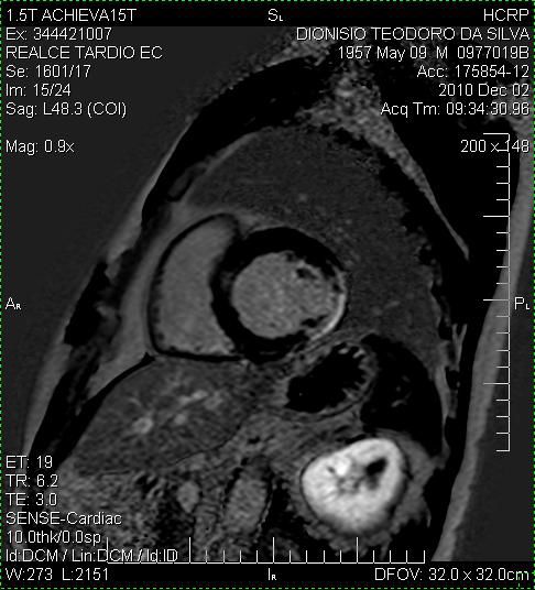
Int J Cardiovasc Sci. 2018;31(2)173-189                                                                                                                  Simões et. al.
Review Article                                                                                                                          Chagas Disease Cardiomyopathy     175

   Chart 1 – Clinical classification of left ventricular dysfunction in Chagas’ heart disease (apud Andrade J et alii)

                                                                                           Chronic phase

                                           Indeterminate        Cardiac form with no
   Acute phase                                                                                        Cardiac form with ventricular dysfunction
                                                form           ventricular dysfunction

                                                  A                      B1                          B2                    C                        D

                                                               Patients with structural
                                                                                                Patients with
                                           Patients at risk    cardiopathy, evidenced                               Patients with            Patients with
                                                                                                  structural
                                           for developing      by electrocardiographic                               ventricular              refractory
                                                                                                cardiopathy
  Patients with                            CHF. They have       or echocardiographic                                 dysfunction          symptoms of CHF
                                                                                              characterized by
  findings compatible                     positeve serology,      changes, but with                                  and current            at rest, despite
                                                                                             global ventricular
  with acute                              neither structural      normal global                                      or previous         optimized clinical
                                                                                              dysfunction, and
  Chagas disease                          cardiopathy nor       ventricular function                                symptoms of         treatment, requiring
                                                                                             neither current nor
                                      CHF symptoms.             and neither current                                CHF (NYHA FC               specialized
                                                                                             previous signs and
                                     No digestive changes      nor previous signs and                               I, II, III or IV)        interventions
                                                                                             symptoms of CHF
                                                                 symptoms of CHF

   With remission of the parasitemia and systemic                                         as “chagomas”, including the typical, but non-specific,
inflammatory reactions, the patient enters the chronic                                    Romaña’s sign, are a result of mucosal or cutaneous
phase of the disease, in which it is believed that, a process                             edema at the site of inoculation. Most patients present
of low-intensity, but incessant, focal myocarditis occurs                                 asymptomatic or show systemic infection symptoms (fever,
since the indeterminate form, which causes progressive                                    hepatosplenomegaly, diaphoresis, myalgia), accompanied
destruction of fibers and restorative myocardial fibrosis.                                by equally non-specific laboratory alterations, especially
This causes cumulative myocardial damage, and results                                     leukocytosis, with absolute lymphocytosis. A minority
in a picture of late dilated cardiomyopathy, usually                                      of patients present a clinical picture of myocarditis, with
accompanied by severe arrhythmias, thromboembolic                                         signs and symptoms similar to myocarditis of other causes:
complications and sudden death in a high proportion                                       dyspnea, fatigue and other commemorative symptoms
of cases. It is believed that chronic myocarditis in                                      of heart failure. In these cases, the ECG may show sinus
Chagas' disease is due to two main pathogenetic                                           tachycardia, ventricular ectopic beats, low voltage of the
processes: myocardial damage associated directly with                                     QRS complexes, branch block, diffuse disturbances of
inflammation caused by parasitized cardiac fibers, with                                   ventricular repolarization, first-degree or more advanced
multiple but low-intensity outbreaks; and myocardial                                      AV block. Chest x-ray may show increased cardiothoracic
aggression caused by adverse immune reaction directed,                                    ratio in more severe cases, which may be associated with
and continuously fed, by the reiterated presentation of                                   increased heart chambers and/or pericardial effusion.
antigens linked to persistent cardiac parasitism.                                         The echocardiogram often shows pericardial effusion,
                                                                                          segmental changes in parietal mobility and insufficiency
    In addition, there is evidence to support the idea that
                                                                                          of mitral and tricuspid valves and, less frequently, cavitary
there are still two auxiliary and amplifying mechanisms of
                                                                                          dilation and decreased global systolic performance.
the myocardial injury: myocardial perfusion disorders due
                                                                                          These abnormalities usually resolve in the majority of
to the presence of abnormalities in coronary microcirculation
                                                                                          patients over the first year of follow-up.12,13
and autonomic cardiac innervation (Figure 2).11

                                                                                            Diagnosis
Acute phase
                                                                                             Serological tests are usually negative in the first
                                                                                          weeks of infection. The diagnosis is made by detection
    Signs and Symptoms                                                                    of circulating parasites or their genetic material (PCR)
   The acute phase begins after an incubation period usually                              through a variety of methods, such as blood culture,
of 1-4 week after exposure to T. cruzi. The lesions known                                 direct visualization of the parasite in peripheral blood,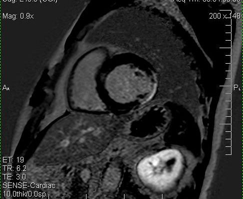
Simões et. al.                                                                                                               Int J Cardiovasc Sci. 2018;31(2)173-189
176   Chagas Disease Cardiomyopathy                                                                                                                       Review Article

                                                                         Low-grade chronic T. cruzi infection

                                                       Continuous antigen
                                                          presentation

                                                  Cross-reactive humoral
                                               and cellular immune response
                                                                         Autoimmune
                                                                          aggression

                                                                               Damaged cardiac fibers
                                                               Platelet
                                                             agrregation,
                                                             Thrombosis,                                  Nervous Tissue
                                                              endothelial                                    damage
                                                             dysfunction

                                                                                                            Autonomic
                                                                                                            denervation
                                                        Microvascular
                                                        Derangement

                                                                                              Impaired hemeometric regulation
                                                                  Myocardial                      Hemodynamic overload
                                                                   ischemia                      LV dilation and remodeling

                                                                                  Cardiac dilation
                                                                                    and failure

            Figure 2 – Scheme showing the relationship between etiopathogenic mechanisms in chronic chagasic myocarditis. A) main mechanisms B) auxiliary
            and amplifying mechanisms of the myocardial injury. Adapted from Marin-Neto et alii. Circulation. 2007;115(9):1109-23.

      xenodiagnosis, or the presence of parasite nests in                                mortality are higher. This is probably due to inoculation
      amastigote form revealed by biopsy with histopathology                             of high parasite load and ease of penetration through the
      of affected organs, or of cutaneous “chagomas”.                                    gastrointestinal mucosa, which is highly permeable to the
         Endomyocardial biopsy is rarely used for the                                    parasite, in these cases.
      diagnosis, but may be necessary, especially in cases of
      suspected reactivation of Chagas disease after heart                                   Treatment
      transplantation, in which a clear distinction from implant                            The treatment of clinical manifestations of myocarditis
      rejection is critical in patient management.14                                     and heart failure is similar to the one recommended for
                                                                                         cases of myocarditis of other etiologies, including intensive
           Clinical Course                                                               measures of circulatory support in more severe cases.
         The clinical course of the acute phase in Chagas disease is                        However, more specifically, although there is no
      often benign and the signs and symptoms typically resolve                          conclusive evidence concerning possible clinically
      spontaneously over 1 to 3 months. It is estimated that fatal                       relevant benefits that could be achieved, antiparasitic
      evolution occurs in < 5% of patients in the acute phase, when                      treatment with benznidazole or nifurtimox is indicated
      contaminated through classic vectorial transmission (via the                       for all cases of acute Chagas disease, independently of
      bite of a triatomine bug), predominantly in patients with                          the infection route or the reactivation mechanism, since
      refractory heart failure. However, in acute cases resulting                        it may decrease the severity of the symptoms, reduce
      from contamination by the oral route (for example after                            disease time and the duration of detectable parasitemia.
      ingestion of T cruzi contaminated sugar cane juice or açai),                       The occurrence of parasitological cure, besides clinical
      the acute disease is usually more severe and the rates of                          cure, is estimated in 60 to 85% of cases.15,16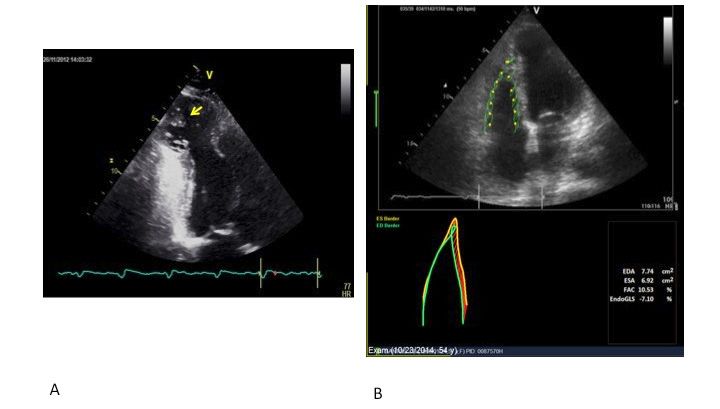
Int J Cardiovasc Sci. 2018;31(2)173-189                                                                                         Simões et. al.
Review Article                                                                                                 Chagas Disease Cardiomyopathy     177

Chronic phase                                                         conduction disturbance, changes in LV segmental
                                                                      parietal mobility on ECG or Holter arrhythmias. In these
                                                                      individuals, however, sudden death may occur due to
    Indeterminate form
                                                                      arrhythmic events, as evidenced by studies demonstrating
   The indeterminate form of the disease is classically               a worsen prognosis in individuals with alterations in the
defined as the clinical situation of an individual with               ECG, even when asymptomatic.24 In a 10-year cohort study
parasitological and/or serological evidence of chronic                of 885 seropositive individuals, it was shown that T. cruzi
T. cruzi infection, but without symptoms or physical                  infected individuals with normal ECG had a survival
signs of the disease, with normal ECG and chest X-ray
                                                                      of 97.4%, comparable to the survival of seronegative
and without digestive tract (esophagus and colon)
                                                                      individuals. On the other hand, survival of those with
impairment seen in radiological exams.
                                                                      abnormal ECG was 61.3%, with a nine-fold increased
   However, more accurate complementary exams                         risk in this group.25 It is estimated that 2 to 5% of patients
(i.e. echocardiography, nuclear angiocardiography,                    without any apparent cardiopathy will develop new ECG
hemodynamic study and autonomic cardiac assessment)                   alterations and evidence of cardiopathy each year.19,26,27
may demonstrate - usually subtle and of no prognostic
relevance - cardiac alterations in this group of patients
                                                                      Clinical manifestations
classified as indeterminate by classical criteria.17-19 In spite of
these minor abnormalities verified in many patients, those               The symptoms and physical signs present in the
classified as indeterminate by the classical criteria, while          chronic phase of the Chagas disease cardiomyopathy
maintaining their normal ECG status, present excellent                are a result of four essential syndromes that can often
prognosis and mortality rates comparable to those of the              coexist in the same patient: heart failure, arrhythmias,
control group of the same age without T. cruzi.4,20,21                thromboembolism and anginal manifestations.

  Guidelines for monitoring chagasic patients in                         Heart Failure Syndrome
the indeterminate form                                                    The clinical picture of CCC with ventricular dysfunction
   There are no formal guidelines in relation to the                  is described in a quite uniform manner in several literature
conduction of exams for early detection of left ventricular           reports, following the pioneering remarks of Chagas and
dysfunction in patients with the indeterminate form                   Villela.28 In the early stages of manifestation, the most
of Chagas disease. On the other hand, there are no                    frequent symptoms are fatigue and dyspnea on exertion,
identifiable factors in this phase that can distinguish the           but the registry of more intense symptoms of pulmonary
individuals who will develop the clinical cardiopathy                 congestion, such as paroxysmal nocturnal dyspnea and
from those who will remain asymptomatic during their                  decubitus with orthopnea, are uncommon. In the disease
whole lives, just keeping the serological positivity.                 evolution, there occur the symptoms of systemic venous
   It is suggested that the ECG be repeated every 1 or                congestion (jugular swelling, hepatomegaly, lower limb
2 years and a simple chest X-ray every 3 or 5 years.                  edema and ascites) and the evolution can still progress
Though it is more controversial, one may also suggest                 to anasarca, adynamia, or cardiac cachexia, similar to
that the transthoracic echocardiography can be                        what happens in other cardiopathies with advanced
performed initially, and later on at regular intervals as             ventricular dysfunction. At clinical examination, there
well, every 3 to 5 years.8,17,22,23                                   are also signs of cardiomegaly resulting from deviation
                                                                      of the ictus cordis, there may be a muffled S1 heart sound
                                                                      in the mitral area, fixed doubling of second heart sound
Cardiac Chronic Form
                                                                      due to RBBB, third heart sound and atrioventricular valve
                                                                      regurgitation murmur, which may occur secondarily
  Asymptomatic condition in Chronic Chagas'                           to the dilation of ventricular chambers. Signs of low
disease cardiomyopathy                                                systemic output may occur in advanced cases, such as
   The absence of symptoms is most marked in individuals              filiform pulse, slow and oliguria peripheral perfusion.
who are in incipient stages of the chronic disease, when              These signs are common to other clinical syndromes of
(discrete) myocardial injury can be detected only due                 heart failure. In contrast to these similarities, in heart
to alterations in complementary exams, such as ECG                    failure of Chagasic etiology, pulmonary congestion is
Simões et. al. Int J Cardiovasc Sci. 2018;31(2)173-189
178 Chagas Disease Cardiomyopathy Review Article
commonly mild in the advanced stages of the disease, A transthoracic and transesophageal echocardiography
compared to the more exuberant systemic congestion, study showed that thromboembolism frequently
and the pulmonary semiology may be more affected originated from the heart in 75 T. cruzi chronically infected
by the signs of pleural effusion than by crepitations, as patients without symptoms of heart failure, or with mild
well as by lower systemic pressure levels in this group symptoms. Left ventricular mural thrombi were found
of patients. The progression to acute pulmonary edema in 23% of patients and were associated with previous
in these cases is even more rare.20 These particularities in history of stroke. Apical aneurysm was identified in
the clinical presentation may relate to the most frequent 47% of patients and was significantly related to mural
concomitance of biventricular dysfunction, with right thrombosis and the occurrence of stroke. Left and right
ventricular heart failure, sometimes earlier and more atrial appendage thrombosis was present in 4 and 1 patient,
pronounced than the left one in T. cruzi infected patients. respectively. During the 24-month follow-up period,
1 non-fatal stroke event and 13 deaths were observed,
Thromboembolic manifestations 7 of which were sudden, 5 due to HF progression and
1 death by stroke.30,31
Pulmonary and systemic embolisms are common
manifestations in patients with CCC, as a result of Systematic revision of 8 observational studies,
murine thrombosis from cardiac chambers and systemic involving 4158 patients, addressed the association
vein thrombosis, and are a major cause of embolic stroke between CCC and the risk of stroke.32 The results indicate
and other morbidities. Thromboembolic accidents are that chronically T. cruzi infected patients, when compared
often the first manifestation of the disease and may to the non-infected, had an excess risk of stroke of about
occur in stages without ventricular dysfunction (Stage 70% (RR = 1.70; HF 95%: 1.06 to 2.71). When the analysis
B2). However, as in several other cardiopathies, cavity was limited to 3 studies, with more strict criteria for
stroke, an even higher excess risk was found (RR = 6.02;
dilation and HF syndrome are known associated risk
HF 95%: 1.86 to 19.49).
factors. Nevertheless, it is chronic regional ventricular
dyskinesia, mainly apical, such as the classic aneurysm The characteristics of stroke patients with CCC were
of the glove finger, which has shown a special explored in a study of 94 patients with acute ischemic
propensity for the formation of mural thrombi and the stroke, compared with the characteristics of a control group
consequent embolic events, particularly the systemic of patients without CCC. T. cruzi infected individuals
ones. As predicted, atrial fibrillation, even when present showed higher rates of cardioembolic stroke (56% versus
in the minority of this population, as a relatively late and 9%), left ventricular dilatation (23% versus 5%), LV mural
secondary manifestation to the ventricular dysfunction, thrombosis (12% versus 2%), apical aneurysm (37% versus
also constitutes an additional thrombogenic factor. 1%) and atrial fibrillation (14% versus 5%).23
Pulmonary embolization, which can originate from
peripheral venous thrombi and right cardiac cavities, Prevention of cardioembolic stroke in patients
is much less frequently clinically diagnosed, but its with CCC
incidence is certainly underestimated, compared to its The I Latin American Guideline for the Diagnosis
prevalence in necropsy material.29 and Treatment of Chagas Cardiopathy adopted
There is a clear lack of data to provide an estimate recommendations for estimation and prevention of
of the actual incidence of clinical thromboembolism in cardioembolic stroke risk through the use of oral
CCC, but series of autopsies and clinical studies indicate antithrombotic agents,1 based on a prospective cohorte
high rates of intracardiac thrombi and thromboembolic study of 1,043 patient with CCC. The total incidence
events in this population. In a revision of 1345 necropsies reported in this event was 3.0%, or 0.56%/year. In the
of patients with chronic chagasic cardiomiopathy, final risk model for cardioembolic stroke prediction, a
thromboemboli and/or intracardiac thrombi were score was calculated in which the presence of LV systolic
observed in 44% of cases. 29 Thrombi were equally dysfunction added two points, and apical aneurysm,
frequent in right and left cardiac cavities. Systemic alteration of the ventricular repolarization at the ECG
circulation thromboembolism was more frequent, but and age > 48 years added one point for each alteration.
more associated with fatal events. Considering the risk-benefit ratio, warfarin would beInt J Cardiovasc Sci. 2018;31(2)173-189 Simões et. al.
Review Article Chagas Disease Cardiomyopathy 179
indicated for patients with 4-5 points (in this subgroup, global left ventricular function, constituting the "isolated
there is incidence of 4.4% of stroke versus 2.0% of major arrhythmogenic form” of the disease. This characteristic,
bleeding per year). In the 3 point score group, stroke which distinguishes CCC from coronary artery disease
and major bleeding rates with OAC are equivalent, and in patients with ventricular dysfunction, as well as from
acetylsalicylic acid (ASA) or warfarin can be indicated.32 other cardiomyopathies, and makes T. cruzi infected
In 2 point patients with low incidence of stroke (1.2% patients especially susceptible to early sudden death,
per year), ASA was recommended, or no prophylaxis. derives from its pathophysiological peculiarities and
Patients with 0-1 point, with incidence close to zero, peculiar pathogenesis.
would not require prophylaxis.33 In fact, the mechanism of severe ventricular arrhythmia
in CCC is mainly associated with the presence of regional
Arrhythmic manifestations fibrosis (especially in the posterolateral regions of the LV)
CCC is essentially an arrhythmogenic and macroreentry circuits formation.35 Recent studies
cardiomyopathy, with pathophysiological peculiarities using cardiac magnetic ressonance have reinforced the
in this context, which makes it uniquely distinct from idea that the presence of regional fibrosis is a major factor
other cardiopathies. Virtually, all types of atrial and for the arrhythmic mechanism in this disease.36-38
ventricular arrhythmia may occur, including sinus Another relevant pathophysiological factor that
node dysfunction, intermittent or complete AV block potentially triggers severe ventricular arrhythmia and
and complex ventricular arrhythmias. The arrhythmias sudden death in CCC patients is regional myocardial
may course asymptomatic or present with non-specific extensive and early sympathetic denervation. In a study
malaise or sudden, fleeting and spontaneously resolved with patients with CCC and normal or slightly reduced
onset palpitation, at rest or by exertion. Symptoms of LV function, the presence of sustained ventricular
low cardiac output due to Stokes-Adams syndrome are tachycardia has been associated with more extensive
less common, but more ominous, including presyncope, areas of viable denervated myocardium, detected
lipotimia, or even syncope, which can occasionally be through I-MIBG myocardial scintigraphy123.39
preceded by palpitations. These episodes can either
correspond to (sustained or nonsustained) ventricular Sudden Death
tachycardia, with or without hemodynamic instability,
It is estimated that sudden death is the leading cause
or to bradyarrhythmias due to atrioventricular block.34
of mortality throughout the various phases of CCC,
In some cases, the standard 12-lead ECG with corresponding to 55 - 65% of deaths.22 Sudden death is
rhythm strip shows premature and ectopic ventricular often triggered by physical effort and may be caused
depolarization, and even ventricular tachycardia both by severe tachyarrhythmias, such as ventricular
outbreaks, in addition to atrial fibrillation or complete tachycardia and fibrillation (probably in 80-90% of cases),
atrioventricularblock (AVB). Tachycardic ventricular and (less frequently) by asystole or complete AV block.40
arrhythmias and AV conduction disorders leading to The detection of nonsustained and especially of sustained
periods of bradycardia can often alternate, frequently ventricular tachycardias increases the chance of sudden
coexisting during the same Holter recording. death, but it occurs mainly in patients with advanced
At clinical examination, it is possible to detect fixed ventricular dysfunction.
doubling of the second heart sound (at a pulmonary
focus), irregular heart rhythm, or even bradycardia, Anginal manifestations
often associated with typical signs of a-waves,
Complaints of precordialgia in patients with CCC
periodically incremented in the jugular venous pulse
are quite common. This pain has characteristics that
and reinforcement of the first heart sound, in cannon
are often atypical for myocardial ischemia, described as
waves, when there is a temporal correlation between
stabbing, fleeting, or, conversely, long lasting (hours or
atrial and ventricular systoles, which is suggestive of
even days), poorly located, usually not related to efforts,
complete atrioventricular block.
sometimes caused by emotional stress and with recurrent
The presence and density of arrhythmia correlate patterns throughout the day. However, sometimes, the
with the degree of ventricular dysfunction in many episodes may be more acute, with typically ischemic
cases, but can also occur in patients with preserved characteristics, making the diagnosis even moreSimões et. al. Int J Cardiovasc Sci. 2018;31(2)173-189
180 Chagas Disease Cardiomyopathy Review Article
difficult. Evidence of microcirculatory disorders it constitutes an element of strong prognostic value.
as causes of anginal manifestations in this group Symptoms suggestive of arrhythmic syndromes also
accumulates in the literature.11,41,42 make ECG Holter monitoring mandatory, since it allows
the assessment of episodes of both tachyarrhythmias
Clinical diagnosis and bradycardia for risk stratification in these patients.34
Recording for at least 24 hours allows determining the
The diagnosis of Chagas' heart disease must density of ventricular ectopy, detecting episodes of
be based on epidemiological criteria, clinical nonsustainable or sustained ventricular tachycardia, as
manifestations, serological tests and on the results of well as determining the duration of sinus pauses and of
some complementary tests. asystolia of different origins. It is worth recalling that, to
compose the Rassi score, used for predicting mortality
Serological tests in patients with CCC, the 24-hour Holter test is essential
Given the low parasitaemia in the chronic phase of to assess the prognostic criteria, with independent
the disease, the serological tests must be able to detect value, of nonsustained ventricular tachycardia, as it
antibodies against T. cruzi antigens. The most commonly will be seen later.
used tests are: immunoenzymatic assay (ELISA), indirect Other frequent changes in ECG at rest are: diffuse
immunofluorescence (IFI), and indirect hemagglutination T wave and ST segment abnormalities, pathological Q
(HAI). When all 3 are performed, agreement (90-98%) waves, prolongation of the QT interval and increased
was observed among them. Since ELISA and IFI have QT dispersion. The evaluation of fibrosis using the QRS
similar characteristics in terms of their accuracy, with score applied to the standard ECG correlates with the
higher sensivity, but slightly reduced specificity than NYHA functional class and with the extent of myocardial
the HAI assay, a positive result in two of these three tests fibrosis detected in Late Gadolinium Enhanced (LGE)
is recommended for the diagnosis. However, when the cardiac magnetic resonance imaging.45
first test is negative, in the current practice based on the Chest X-ray: Advanced stages of the disease are
high sensitivity of all serological tests, there is no need marked by (often massive) cardiomegaly, which
for a second test.43 may include signs of not only increased LV, but also
of increased RV and both atrial dilation; however,
Complementary cardiologic exams pulmonary congestion differs from other cardiopathies
The main complementary diagnostic tests used in because it is often discrete, in contrast to the degree of
Chagas' heart disease are briefly described below, with cardiothoracic ratio. Cardiomegaly is also an important
emphasis on those focusing on the characterization and prognostic factor in these patients, stratified by the Rassi
gradation of ventricular dysfunction. score, as discussed next.
Electrocardiography and Holter Monitoring: Echocardiography: Echocardiography is a non‑invasive
The most prevalent electrocardiographic alterations in imaging method that allows the geometrical and
patients with CCC are right bundle branch disorders functional diagnosis of both ventricles, which is essentially
and the left anterior hemiblock, and may reach 50% in important in CCC. Alterations in segmental mobility in
patients of this group.44 These changes in the cardiac the inferior and inferolateral regions of the LV are quite
conduction system may be evolutionary, such as AV common in patients with the cardiac chronic form.46-50
conduction delays. Sinus node dysfunction can also Even though the detection of LV apical aneurysm (a
cause bradycardia. However, atrial arrhythmias tend very common alteration in patients with CCC)51 may
to occur in the evolution of the heart disease with be subject to operational limitations, it is one of the
advanced ventricular dysfunction. It is crucial to typical changes found in the disease, and may be filled
observe that, although ventricular ectopic beats can with thrombus (Figure 3, A). Although early changes
be seen in normal individuals during the recording of in LV regional mobility can be detected on ECG, both
a standard ECG, when it is verified in patients with through conventional techniques17,52,53 and analysis of
CCC, the meaning of this alteration is completely myocardial deformation, in some patients classified as
different and it usually indicates that the ventricular having the indeterminate form, or even with CCC and
arrhythmia is an integral part of the syndrome and function preserved by other methods,18,53,54 the prognostic
value of these alterations is not well established yet.50,55Int J Cardiovasc Sci. 2018;31(2)173-189 Simões et. al.
Review Article Chagas Disease Cardiomyopathy 181
Two-dimensional echocardiographic evaluation of the Myocardial perfusion scintigraphy may be required
right ventricle can be performed through an acquisition for non-invasive investigation of Chagas disease
protocol with images dedicated to this investigation.56 patients with precordialgia. Negative findings for
New methods for the assessment of right ventricular myocardial ischemia virtually excludes the presence
systolic function, such as the analysis of myocardial of significant coronary artery disease, indicating a
deformation (Figura 3, B), have already shown to high negative predictive value. However, reversible
correlate quite often to the quantification of RV ejection perfusion defects have been detected in 30 to 50% of
fraction by other methods, such as magnetic resonance patients, in the absence of atherosclerotic epicardial
imaging, in groups of patients with Chagas’ disease. CAD.58-60 These perfusion changes have been attributed
Although three‑dimensional echocardiography presents to coronary microcirculation in CCC and it has been
the benefit of volumetric quantification of cavities and, as postulated that such ischemic changes can contribute to
a result, of ventricular ejection fractions, its role in patients regional myocardial damage in the chronic phase of the
with CCC has not been adequately established yet. cardiomyopathy.37,60 Fixed perfusion defects, on the other
Nuclear Medicine: Radioisotope ventriculography hand, are also frequently observed in patients with CCC
(RIV), also known as radionuclear angiocardiography, and, in general, represent areas of fibrosis caused by the
can be used as an alternative method to echocardiography typical pathophysiology of Chagas disease.42
to measure the LV ejection fraction (EF), and presents Iodine-123-labeled meta-iodobenzylguanidine
the advantage of being a quantitative method free from myocardial scintigraphy ( 123 I-MIBG) allows the
geometric inferences, thereby granting it the role of a real non‑invasive evaluation of the neuronal integrity of the
gold standard method in this context. When simultaneous cardiac sympathetic nervous system at a myocardial
measurement of the right and left ventricular ejection level. The use of this imaging technique allowed the
fraction (RVEF, LVEF) is required, RIV has been used identification of regional myocardial denervation in
successfully and may detect earlier and more severe RV early stages of chronic Chagas' disease in patients with
dysfunction, including in patients with the digestive form no apparent impairment of left ventricular function,
of Chagas' disease.57 involving mainly the basal parts of the posterolateral
Figure 3 – Chart A: Echocardiography of apical two-chamber view of the left ventricle shows an image suggestive of a large apical aneurysm
filled with thrombus (yellow arrow). Chart B: Point Tracking Technique (Speckle tracking) supporting the analysis of RV systolic function in a
patient with Chagas’ disease. EDA: end-diastole area; ESA: end-systole area; FAC: Fractional area change; GLS-endo: Global longitudinal strain
in the endocardial layer.Simões et. al. Int J Cardiovasc Sci. 2018;31(2)173-189
182 Chagas Disease Cardiomyopathy Review Article
and inferior walls and of the apical region.61 The results MRI for detecting the regions of myocardial fibrosis in
of these studies suggest that the extension/severity of patients with CCC and for being a potentially valuable
the regional myocardial denervation and the intensity non‑invasive risk prediction tool to assess sudden death
of the global derangement of sympathetic innervation risk in these patients, even in those with preserved left
(detected in planar images) correlate with the severity ventricular ejection fraction.38 The fibrosis pattern is
of LV systolic dysfunction.61 A more recent study has varied, with the presence of focal or diffusely distributed
shown that cardiac patients with preserved or slightly fibrosis, and even with transmural impairment, simulating
reduced systolic function and sustained ventricular a fibrosis area usually seen in myocardial infarction due
tachycardia (SVT) presented greater extension of to obstructive coronary disease (Figure 5).
sympathetic denervation evaluated by 123I-MIBG Electrophysiological study (EPS): The general
myocardial scintigraphy, compared to individuals indications for EPS apply for patients with CCC. The EPS
without SVT, which reinforces the idea that autonomic is required for the evaluation of the sinus node function
cardiac denervation can play an important role in the and AV conduction when the origin of symptoms,
arrhythmogenesis of this myocardial disease - Figure 4.39 particularly syncope, remains uncertain after noninvasive
Magnetic Resonance Imaging: MRI is a methodology evaluation. In most patients with preserved left
which allows analysis of morpho-functional parameters of ventricular function who have nonsustained ventricular
the heart with high degree of two-dimensional detailing, tachycardia or without spontaneous arrhythmia, the EPS
and can be quite elucidative, especially in cases where the does not provide any relevant additional prognostic
quality of the echocardiographic images is poor, or when information. The use of EPS has been proposed in
there are ventricular cavities with advanced geometric survivors of sudden cardiac death and those with SVT
changes, making it difficult to perform echocardiographic for prognostic evaluation and indication of drug therapy
measurement with the usual techniques. It is a method and implantation of antiarrhythmic devices, but the data
with great capacity for quantitative analysis of ventricular on the efficacy of this approach are still limited.68-70
volumes and accurate calculation of LV ejection fraction.62-66 Cardiac catheterization: CCC can mimic several
It can also be quite useful for specific analysis of the clinical aspects of ischemic heart disease. In fact, patients
right ventricular cavity, according to recent studies.67 with CCC may show precordial pain, electrocardiographic
More recent studies call attention for the potential of changes of the ST-T segment and pathological Q waves,
Figure 4 – Illustrative example of patients with CCC, presenting Sustained Ventricular Tachycardia (SVT, documented on the ECG of image B)
and presenting uptake defect in the inferior and posterolateral walls in the 123-I MIBG SPECT images (image C). Sestamibi Myocardial perfusion
images (SMPI) were normal, with myocardial viability in denervated myocardial segments that correlate topographically with the site of origin of SVT.
Reprinted from Gadioli, LP et al. Journal of Nuclear Cardiology March 23, 2016 (doi:10.1007/s12350-016-0556-6).Int J Cardiovasc Sci. 2018;31(2)173-189 Simões et. al.
Review Article Chagas Disease Cardiomyopathy 183
Figure 5 – Delayed enhanced images where fibrosis can be seen as the white area inserted in the (dark) muscle, indicated by arrows. Left panel
shows a small mesomyocardial area in the LV lateral wall in a four-chambered image. Right panel shows extensive transmural impairment of the
posterolateral wall.
in addition to changes in LV segmental parietal mobility. through basic assignment methods (Table 1). This score
Thus, the requirement of coronary angiography is not allows detecting relevant extracts on the risk of mortality
uncommon to rule out the presence of coronary artery in patients with CCC.
disease in patients with risk factors for this condition. Over about 10 years of follow-up, patients classified
As stressed above, in the vast majority of patients referred as low risk (score from 0 to 6) had mortality of 9 to 10%;
for cardiac catheterization, with CCC, the subepicardial those with intermediate risk (score between 7 and 11)
coronary arteries are essentially normal or have had mortality from 37 to 44% and those with high risk
non‑significant hemodynamically obstructive lesions.42,59 (score between 12 and 20) had mortality from 84 to 85%.
The combination of LV systolic function (even if only
Prognosis regional) and NSVT was associated with a particularly
elevated risk of mortality, of the order of 15.1 times.
The prognosis of CCC depends on various factors, The detection of NSVT alone was associated with a
among them the stage of the disease presented by each 2.15 times increase in death.
patient, as already described in this text.
Figure 6 reproduces the algorithm for risk stratification
In the chronic phase, in relation to the clinical form in patients with CCC, derived from a systematic review
with LV impairment, several observational series have of observational studies.73
shown worse prognosis in patients with CCC compared
to those with other heart diseases manifested by heart
Treatment
failure. In a recent prospective observational study,
including 456 patients with heart failure, the 68 patients
with CCC had lower survival compared to the ones with Etiological Treatment
other etiologies.71 Several pathophysiological factors can The role of antiparasitic agents in the treatment of
explain this difference, but some prognostic markers T. cruzi infection is considerably limited in the chronic
have already been defined as independent predictors, phase of Chagas heart disease, since much reversal of
among them LV contractile dysfunction, both evaluated established tissue damage should not be expected at
by echocardiography and suggested by cardiomegaly these advanced stages of the disease.76
on chest x-ray.4,72 The BENEFIT study, 77 released in 2015, was the
The Rassi score, used for mortality risk stratification in only large-scale clinical trial carried out on Chagas
patients with chronic chagasic cardiopathy,9,73-75 consists Disease. The study randomized 2854 patients, who
of points assigned to simple characteristics and obtained received benznidazole or placebo, with the essentialSimões et. al. Int J Cardiovasc Sci. 2018;31(2)173-189
184 Chagas Disease Cardiomyopathy Review Article
Table 1 – Rassi score for mortality risk stratification in patients with chronic chagasic cardiopathy
Clinical Characteristic Punctuation
Male Gender 2
ECG with low QRS voltage 2
Nonsustained ventricular tachycardia 3
Global LV alteration or LV segmental motion 3
Cardiomegaly on chest x-ray 5
Heart failure FC III-IV (NYHA) 5
objective of evaluating the efficacy of this treatment. However, certain particularities in the management of
Trypanosomicidal treatment was only effective to detect patients with HF secondary to CCC must be highlighted.
negative parasitological test results evaluated by the Several studies suggest that these patients exhibit a
polymerase chain reaction technique (PCR) (66% in the higher risk of symptomatic bradycardia and AV block
treatment group versus 34% in the control group) even with the use of beta-blockers, thus the heart rate in these
though a negative result did not correlate with clinical patients must be carefully monitored. This precaution
benefit over the 5-year follow-up.76 After the initial is especially applicable when, due to antiarrhythmic
publication of the results, supplementary analysis indication, amiodarone has already been initiated for
comparing the outcomes verified in Brazil with the the patient. In spite of this aspect, the results of a recent
ones obtained in the other four countries allows the prospective observational cohort study suggest that
hypothesis that, probably due to the predominance beta-blockers can have a positive effect on the survival
of T. cruzi lineage II, which is more sensitive to the in patients with chronic HF caused by CCC.79
trypanocidal action of benznidazole, in Brazil, the effect
of the etiological treatment may turn out to have a clinical Alternative therapies
benefit for chronically infected Brazilian patients without
Several clinical studies have shown that the efficacy of
very advanced heart disease.77 Thus, the treatment could
the cardiac resynchronization therapy, through multisite
be offered, on an individual basis, for patients with this
pacemaker implantation, depends on the presence of left
profile, in order to reach a potential decision following
bundle branch block on ECG, a pattern found in the vast
sharing models with the responsible physician.
majority of patients included in large multicenter studies
who have tested this therapy. However, due to evident
Heart Failure Treatment predominance of RBBB, the usefulness of CRT in patients
with CCC has not been demonstrated.
Drug Therapy Heart transplantation has been successfully used in
There is a clear lack of evidence supporting the patients with advanced HF secondary to CCC.80 In a study
clinical benefit of conventional systolic heart failure of 117 patients with CCC who received the transplant, the
drug therapy, based primarily on neurohormonal block, survival reported at 1, 4, 8 and 12 years after the procedure,
in patients with CCC. However, considering that the was 71, 57, 55 and 46%, respectively. These observational
general phenotype of HF caused by CCC is that of a studies show that the survival of patients with CCC was
dilated cardiomyopathy, the treatment for heart failure better than that observed in patients with HF of other
of other etiologies is empirically extrapolated for the etiologies,81 which seems to be a consequence of several
treatment of patients suffering from CCC. This position aspects, such as less advanced age and lower number of
was ratified by the recommendations of the Brazilian comorbidities in transplanted patients with CCC.
Guidelines on Diagnosis and Treatment of Chronic HF This series of cases has also shown that the reactivation of
according to which all treatment recommendations were the T cruzi infection is a common clinical problem, as a result
extended to the etiology of CCC.78 of post-transplantation immunosuppression, and sometimesInt J Cardiovasc Sci. 2018;31(2)173-189 Simões et. al.
Review Article Chagas Disease Cardiomyopathy 185
Patients with positive serologic test results
Normal EGG
NYHA
Not normal
classes III/IV
X-ray: normal NYHA
X-ray: Cardiomegaly
cardiac area classes I/II
ECG – normal LVEF ECG – Reduced LVEF
Holter Holter Holter Holter
without NSVT with NSVT without NSVT with NSVT
Low risk Intermediate risk High risk
Figure 6 – Algorithm for risk stratification in patients with Chagasic Chronic Cardiopathy. Adapted from Rassi et al. Circulation. 2007;115:1101-8
and reprinted from the I Latin American guideline for the diagnosis and treatment of Chagas cardiomyopathy.1
difficult to differentiate from organ rejection; however, results The ICD implantation is useful in the secondary
through the use of trypanosomicidal therapy were found. prevention of sudden cardiac death, in survivors
of sudden arrhythmic death or with sustained
Treatment of cardiac arrhythmias ventricular tachycardia, especially when accompanied
by hemodynamic instability. For those who are not
candidates for the implantation of this device, the use of
Bradyarrhythmias and AV block
amiodarone is recommended. In fact, there is acceptable
Patients with second- or third-degree AVB or evidence of potential benefit for this antiarrhythmic
symptomatic sinus node dysfunction require definitive drug in patients with ventricular arrhythmias of
pacemaker implantation. In this respect, CCC does not Chagas disease etiology. The concomitant use of
seem to differ from other etiologies, and usual guidelines amiodarone and beta-blockers is also recommended
for indicating these devices must be followed. routinely to reduce the number of therapies, even
when appropriate, due to ICD implantation in
Arritmias ventriculares e morte súbita arrítmica patients with CCC.
The optimal approach for the management of severe A multicenter randomized trial (CHAGASICS) is
ventricular arrhythmias and resuscitated sudden cardiac underway to assess the ICD benefit versus amiodarone,
death secondary to CCC is still uncertain due to absolute for primary prevention of sudden death in patients
lack of data. The first therapeutic measure in patients with CCC and high Rassi score. 82 Amiodarone can
with CCC under risk of malignant ventricular arrhythmia be used, ideally associated with a beta-blocker, for
is the optimization of drug therapy for those who also patients with CCC, Rassi risk score of ≥10 points and
have heart failure, preferably with the concomitant use nonsustained ventricular tachycardia detected on
of beta-blockers and amiodarone. Holter monitoring.Simões et. al. Int J Cardiovasc Sci. 2018;31(2)173-189
186 Chagas Disease Cardiomyopathy Review Article
Author contributions Potential Conflict of Interest
No potential conflict of interest relevant to this article
Conception and design of the research: Simões MV, was reported.
Romano MMD, Schmidt A, Martins KSM. Acquisition
of data: Simões MV, Romano MMD, Schmidt A,
Sources of Funding
Martins KSM. Analysis and interpretation of the data:
There were no external funding sources for this study.
Simões MV, Romano MMD, Marin-Neto JA. Writing of
the manuscript: Simões MV, Romano MMD, Schmidt A,
Martins KSM, Marin-Neto JA. Critical revision of Study Association
the manuscript for intellectual content: Simões MV, This study is not associated with any thesis or
Romano MMD, Schmidt A, Marin-Neto JA. dissertation work.
Erratum
In the manuscript “Chagas Disease Cardiomyopathy”, DOI number: : 10.5935/2359-4802.20180011, published in
the International Journal of Cardiovascular Sciences, 2018;31(2)173-189, on page: 183, Figure 5 – Delayed enhanced
images where fibrosis can be seen as the white area inserted in the (dark) muscle, indicated by arrows. Left panel
shows a small mesomyocardial area in the LV lateral wall in a four-chambered image. Right panel shows extensive
transmural impairment of the posterolateral wall.
Where it read:
Now it reads:Int J Cardiovasc Sci. 2018;31(2)173-189 Simões et. al.
Review Article Chagas Disease Cardiomyopathy 187
References
1. Andrade JP, Marin-Neto JA, Paola AA, Vilas-Boas F, Oliveira GM, Bacal 18. Garcia-Alvarez A, Sitges M, Regueiro A, Poyatos S, Jesus Pinazo M,
F, et al; Sociedade Brasileira de Cardiologia. [I Latin American guidelines Posada E, et al. Myocardial deformation analysis in Chagas heart
for the diagnosis and treatment of Chagas cardiomyopathy]. Arq Bras disease with the use of speckle tracking echocardiography. J Card Fail.
Cardiol. 2011;97(2 Suppl 3):1-48. PMID: 21952638. 2011;17(12):1028-34. doi: 10.1016/j.cardfail.2011.08.007.
2. Marin-Neto JA, Rassi A Jr. Update on Chagas heart disease on the 19. Dias JC. The indeterminate form of human chronic Chagas' disease: a
first centenary of its discovery. Rev Esp Cardiol. 2009;62(11):1211-6. clinical epidemiological review. Rev Soc Bras Med Trop. 1989;22(3):147-
PMID: 19889330. 56. http://dx.doi.org/10.1590/S0037-86821989000300007.
3. Wanderley DM, Correa FM. Epidemiology of Chagas' heart disease. Sao 20. Marin-Neto JA, Simoes MV, Sarabanda AV. Forma crônica cardíaca.
Paulo Med J. 1995;113(2):742-9. doi: http://dx.doi.org/10.1590/S1516- Trypanosoma Cruzi e Doença de Chagas. 2ª ed. São Paulo: Guanabara-
31801995000200003. Koogan; 2000. p. 266-96.
4. Rassi A Jr, Rassi A, Marin-Neto JA. Chagas disease. Lancet. 21. Ianni BM, Arteaga E, Frimm CC, Pereira Barretto AC, Mady C. Chagas'
2010;375(9723):1388-402. doi: 10.1016/S0140-6736(10)60061-X. heart disease: evolutive evaluation of electrocardiographic and
echocardiographic parameters in patients with the indeterminate form.
5. World Health Organization. (WHO). UNICEF/UNDP/World Bank/
Arq Bras Cardiol. 2001;77(1):59-62. doi: http://dx.doi.org/10.1590/
WHO Special Programme for Research and Training in Tropical
Diseases., Pan American Health Organization. Reporte del grupo de S0066-782X2001000700006.
trabajo científico sobre la enfermedad de Chagas : 17-20 de abril de 2005, 22. Rassi A Jr, Rassi SG, Rassi A. Sudden death in Chagas' disease. Arq
actualizado en julio de 2007, Buenos Aires (Argentina); 2007. Bras Cardiol. 2001;76(1):75-96. doi: http://dx.doi.org/10.1590/S0066-
6. World Health Organization. (WHO). First WHO Report on neglected 782X2001000100008.
tropical diseases: working to overcome the global impact of neglected 23. Carod-Artal FJ, Vargas AP, Horan TA, Nunes LG. Chagasic
tropical diseases. In: Organization WH. (editor): Geneva; 2010. p. 172. cardiomyopathy is independently associated with ischemic stroke
7. World Health Organization. (WHO). Control of Chagas' disease: second in Chagas disease. Stroke. 2005;36(5):965-70. doi: 10.1161/01.
report of the WHO expert committee. Geneva; 2002. (WHO tecnical report STR.0000163104.92943.50.
series, 905). 24. Maguire JH, Hoff R, Sherlock I, Guimaraes AC, Sleigh AC, Ramos NB, et
8. Marin-Neto JA, Almeida Filho OC, Pazin-Filho A, Maciel BC. al. Cardiac morbidity and mortality due to Chagas' disease: prospective
[Indeterminate form of Chagas' disease: proposal of new diagnostic electrocardiographic study of a Brazilian community. Circulation.
criteria and perspectives for early treatment of cardiomyopathy]. Arq 1987;75(6):1140-5. PMID: 3552307.
Bras Cardiol. 2002;79(6):623-7. PMID: 12532246..
25. Forichon E. Contribution aux estimations de morbité et de mortalité dans
9. Rassi Jr A, Rassi A, Marin-Neto JA. Chagas heart disease: pathophysiologic la maladie de Chagas. Toulouse (France): Univers. Paul Sabatier; 1974.
mechanisms, prognostic factors and risk stratification. Mem Inst Oswaldo
26. Nunes MC, Dones W, Morillo CA, Encina JJ, Ribeiro AL; Council on
Cruz. 2009;104 Suppl 1:152-8. doi: http://dx.doi.org/10.1590/S0074-
Chagas Disease of the Interamerican Society of Cardiology. Chagas
02762009000900021.
disease: an overview of clinical and epidemiological aspects. J Am Coll
10. Coura JR, De Abreu LL, Dubois LE, Lima FD, De Arruda Junior E, Willcox Cardiol. 2013;62(9):767-76. doi: 10.1016/j.jacc.2013.05.046.
HP, et al. [Morbidity of Chagas' disease. II - Sectional studies in 4 field
27. Manzullo EC, Darraidou MA, Libonatti O, Rozlosnik J, Bazzano AC.
areas in Brazil]. Mem Inst Oswaldo Cruz. 1984;79(1):101-24. doi: http://
Estudio longitudinal de la cardiopatia chagasica cronica. [Tese]. Centro
dx.doi.org/10.1590/S0074-02761984000100012.
de Chagas de la Catedra de Enfermedades Infecciosas de la Facultad de
11. Marin-Neto JA, Cunha-Neto E, Maciel BC, Simoes MV. Pathogenesis Ciencias Medicas de Buenos Aires. Buenos Aires; 1982.
of chronic Chagas heart disease. Circulation. 2007;115(9):1109-23. doi:
10.1161/CIRCULATIONAHA.106.624296. 28. Chagas C. A short chronicle of the discovery of Chagas' disease. Pacing
Clin Electrophysiol. 1988;11(7):1108-13. PMID: 2457892.
12. Dias E, Laranja FS, Miranda A, Nobrega G. Chagas' disease; a clinical,
epidemiologic, and pathologic study. Circulation. 1956;14(6):1035-60. 29. Samuel J, Oliveira M, Correa De Araujo RR, Navarro MA, Muccillo G.
PMID: 13383798. Cardiac thrombosis and thromboembolism in chronic Chagas' heart
disease. Am J Cardiol. 1983;52(1):147-51. PMID: 6858902.
13. Coura JR, Vinas PA, Brum-Soares LM, de Sousa AS, Xavier SS. Morbidity
of Chagas heart disease in the microregion of Rio Negro, Amazonian 30. Nunes Mdo C, Barbosa MM, Rocha MO. Peculiar aspects of cardiogenic
Brazil: a case-control study. Mem Inst Oswaldo Cruz. 2013;108(8):1009- embolism in patients with Chagas' cardiomyopathy: a transthoracic
13. doi: http://dx.doi.org/10.1590/0074-0276130425. and transesophageal echocardiographic study. J Am Soc Echocardiogr.
2005;18(7):761-7. doi: 10.1016/j.echo.2005.01.026.
14. Souza FF, Castro ES, Marin Neto JA, Sankarankutty AK, Teixeira AC,
Martinelli AL, et al. Acute chagasic myocardiopathy after orthotopic 31. Carod-Artal FJ, Gascon J. Chagas disease and stroke. Lancet Neurol.
liver transplantation with donor and recipient serologically negative 2010;9(5):533-42. doi: 10.1016/S1474-4422(10)70042-9.
for Trypanosoma cruzi: a case report. Transplant Proc. 2008;40(3):875-8.
32. Cardoso RN, Macedo FY, Garcia MN, Garcia DC, Benjo AM, Aguilar D, et
doi: 10.1016/j.transproceed.2008.02.032.
al. Chagas' cardiomyopathy is associated with higher incidence of stroke:
15. Wegner DH, Rohwedder RW. The effect of nifurtimox in acute a meta-analysis of observational studies. J Card Fail. 2014;20(12):931-8.
Chagas' infection. Arzneimittelforschung. 1972;22(9):1624-35. doi: 10.1016/j.cardfail.2014.09.003.
PMID: 4630485.
33. Sousa AS, Xavier SS, Freitas GR, Hasslocher-Moreno A. Prevention
16. Kirchhoff LV. Chagas disease. American trypanosomiasis. Infect Dis strategies of cardioembolic ischemic stroke in Chagas' disease. Arq
Clin North Am. 1993;7(3):487-502. PMID: 8254156. Bras Cardiol. 2008;91(5):306-10. doi: http://dx.doi.org/10.1590/S0066-
782X2008001700004.
17. Pazin-Filho A, Romano MM, Almeida-Filho OC, Furuta MS, Viviani LF,
Schmidt A, et al. Minor segmental wall motion abnormalities detected 34. Rassi Junior A, Gabriel Rassi A, Gabriel Rassi S, Rassi Junior L, Rassi
in patients with Chagas' disease have adverse prognostic implications. A. [Ventricular arrhythmia in Chagas disease. Diagnostic, prognostic,
Braz J Med Biol Res. 2006;39(4):483-7. doi: http://dx.doi.org/10.1590/ and therapeutic features]. Arq Bras Cardiol.1995;65(4):377-87.
S0100-879X2006000400008. PMID: 8728815.You can also read