Comorbidities and Cofactors of Anaphylaxis in Patients with Moderate to Severe Anaphylaxis. Analysis of Data from the Anaphylaxis Registry for ...
←
→
Page content transcription
If your browser does not render page correctly, please read the page content below
International Journal of
               Environmental Research
               and Public Health

Article
Comorbidities and Cofactors of Anaphylaxis in Patients with
Moderate to Severe Anaphylaxis. Analysis of Data from the
Anaphylaxis Registry for West Pomerania Province, Poland
Iwona Poziomkowska-G˛esicka 1, * , Magdalena Kostrzewska 2 and Michał Kurek 1

                                           1   Clinical Allergology Department, Pomeranian Medical University (PMU) in Szczecin,
                                               70-204 Szczecin, Poland; alergologia@spsk2-szczecin.pl
                                           2   Department of Pulmonology, Allergology and Respiratory Oncology, University of Medical Sciences,
                                               60-569 Poznan, Poland; m0304@tlen.pl
                                           *   Correspondence: iwona.poziomkowska@op.pl

                                           Abstract: Anaphylaxis is a severe, potentially life-threatening systemic hypersensitivity reaction that
                                           is still rarely diagnosed. For safety reasons, patients should visit an allergologist to identify potential
                                           causes and cofactors of this reaction. This paper presents the analysis of data from the Anaphylaxis
                                           Registry gathered over ten years at the Allergy Clinic, Pomeranian Medical University (PMU). A
                                           questionnaire-based survey was used for patients visiting the Allergy Clinic to identify potential
                                           augmentation factors/comorbidities and/or cofactors of anaphylaxis in patients with a history of
                                           moderate to severe anaphylaxis. The registry comprised patients with grade II or higher anaphylaxis.
                                           The gathered data concerned chronic comorbidities (cardiovascular diseases, respiratory diseases,

                                    and others), recurrence of anaphylaxis, and potential cofactors in anaphylaxis. In the analyzed
                                           group, the incidence rate of anaphylaxis was the highest for women aged 19–60 years. Most common
Citation: Poziomkowska-G˛esicka, I.;
                                           comorbidities in patients with moderate to severe anaphylaxis included: cardiovascular diseases,
Kostrzewska, M.; Kurek, M.
Comorbidities and Cofactors of
                                           respiratory tract diseases, features of atopy, and thyroid diseases. More than 30% of drug-induced
Anaphylaxis in Patients with               reactions were anaphylactic reactions due to the re-exposure to the same drug, which points to
Moderate to Severe Anaphylaxis.            the need for educational initiatives in this area. The incidence rate of anaphylaxis induced by
Analysis of Data from the                  Hymenoptera stings was comparable in patients who had a previous generalized reaction and those
Anaphylaxis Registry for West              who had good tolerance to the previous sting. It is important to take these cofactors into consideration
Pomerania Province, Poland. Int. J.        when evaluating patients with anaphylaxis as they may play a role in future anaphylactic reactions.
Environ. Res. Public Health 2021, 18,
333. https://doi.org/10.3390/              Keywords: anaphylaxis; epidemiology; cofactors; comorbidity
ijerph18010333

Received: 12 October 2020
Accepted: 31 December 2020
                                           1. Introduction
Published: 5 January 2021
                                                The term anaphylaxis was used for the first time in 1902 by Portier and Richet to
Publisher’s Note: MDPI stays neu-          describe a reaction opposite to prophylaxis. They described an experiment on dogs that
tral with regard to jurisdictional clai-   had tolerated a certain dose of a jellyfish toxin, but after the injection of a lower dose of the
ms in published maps and institutio-       same toxin, the dogs reacted with bronchospasm and cardiorespiratory arrest and died [1].
nal affiliations.                               Different definitions of anaphylaxis have been proposed since the first use of this
                                           term—Table 1. Contemporary definitions are presented in a paper by Turner in 2019 [2].

                                           Table 1. Definitions of anaphylaxis.
Copyright: © 2021 by the authors. Li-
censee MDPI, Basel, Switzerland.                                  A serious life-threatening generalized or systemic hypersensitivity reaction.
                                                 WAO [3]
This article is an open access article                            A serious allergic reaction that is rapid in onset and might cause death
distributed under the terms and con-                              A severe life-threatening generalized or systemic hypersensitivity reaction.
ditions of the Creative Commons At-             EAACI [4]
                                                                  An acute, potentially fatal, multi-organ system, allergic reaction.
tribution (CC BY) license (https://
creativecommons.org/licenses/by/
4.0/).

Int. J. Environ. Res. Public Health 2021, 18, 333. https://doi.org/10.3390/ijerph18010333                     https://www.mdpi.com/journal/ijerph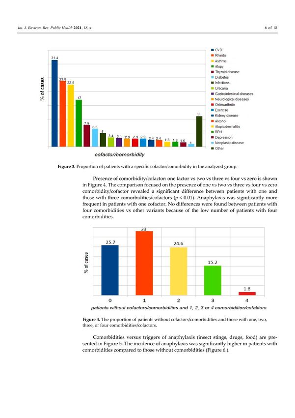
Int. J. Environ. Res. Public Health 2021, 18, 333                                                                                      2 of 17

                                                                                    Table 1. Cont.

                                                              An acute life-threatening systemic reaction with varied mechanisms, clinical
                                          AAAAI/ACAAI
                                                              presentations, and severity that results from the sudden release of mediators
                                              [5]
                                                              from mast cells and basophils.
                                                              Anaphylaxis is a serious, rapid-onset, allergic reaction that may cause death.
                                              ASCIA [6]       Severe anaphylaxis is characterized by life-threatening upper airway
                                                              obstruction, bronchospasm and/or hypotension.

                                             Generally, anaphylaxis is most commonly defined as an acute, severe, potentially life-
                                        threatening systemic hypersensitivity reaction [4] and remains a clinical diagnosis. There
                                        are also definitions without the word ‘acute’ or without the adjective ‘allergic’ reaction.
                                        Anaphylaxis may also be delayed, with the onset 4–6 h after the intake of food, or without
                                        the involvement of immunologic mechanisms. It should be kept in mind that the onset
                                        of anaphylaxis after stings or allergen injections is usually rapid; 70% begin in less than
                                        20 min, and 90% in less than 40 min [7]. Most healthcare professionals define anaphylaxis
                                        as a serious, generalized, allergic, or hypersensitivity reaction that can be life-threatening
                                        and even fatal [8–12].
                                             Depending on the pathomechanism, anaphylactic reactions are classified as aller-
                                        gic anaphylaxis (usually IgE-dependent), non-allergic anaphylaxis, and the so-called cy-
                                        tokine storm with the involvement of new G-coupled receptor MRGPRX2, located on
                                        mast cells [13]. In the literature, there are several classification systems, considering the
                                        severity of anaphylaxis and clinical symptoms, and proposed, for example, by Ring and
                                        Messmer [14], Muller [15], Brown [16], Muraro [17], and Mehl [18]. Table 2 below presents
                                        the classification of anaphylaxis severity by Ring and Messmer (adopted in this article).

                                        Table 2. Classification of anaphylaxis severity [14].

                                                                        Classification by Ring and Messmer
                                             Grade I        Generalized skin symptoms (e.g., flush, generalized urticaria, angioedema)
                                             Grade II      Mild to moderate pulmonary, cardiovascular, and/or gastrointestinal symptoms
                                             Grade III                       Anaphylactic shock, loss of consciousness
                                             Grade IV                                 Cardiac arrest, apnea

                                             Many publications, apart from the analysis of causes of anaphylaxis, present in-
                                        formation about patient-specific risk factors and cofactors amplifying anaphylactic reac-
                                        tion [19–27]. Multiple episodes of anaphylaxis following the consumption of unconnected
                                        foods should raise concerns about the possibility of a hidden allergen-induced or “summa-
                                        tion anaphylaxis” due to cofactor influence [28]. Skypala provides an overview of hidden
                                        allergens and the influence of cofactors in food-related anaphylaxis. An accurate clinical
                                        history with a high index of suspicion is paramount in making a correct diagnosis [29].
                                             Severe anaphylaxis is associated with older age, asthma, and chronic obstructive
                                        pulmonary disease (COPD), and pharmacotherapy [30]. According to different authors and
                                        reports, the role of cofactors in about 30% of anaphylactic reactions has been documented,
                                        from 25.6% in France to 39% in Germany [31]. Cofactors, including exercise, ethanol,
                                        acute infections, and stress potentially amplify anaphylaxis by decreasing the threshold of
                                        allergen exposure (the allergen “dose”) needed to trigger anaphylaxis in patients with low
                                        or borderline allergen sensitization [19,32–34]. An analysis of data from the European Ana-
                                        phylaxis Registry assessed factors increasing the risk for a severe anaphylactic reaction [35].
                                        The following augmentation factors or cofactors were listed by Worm [35]:
                                        1.     Non-modifiable/stable/independent/intrinsic
                                        2.     Modifiable/unstable/dependent/extrinsic
                                            Nonmodifiable factors include age, sex, comorbidities, basic tryptase level, and a
                                        previous reaction triggered by the same factor.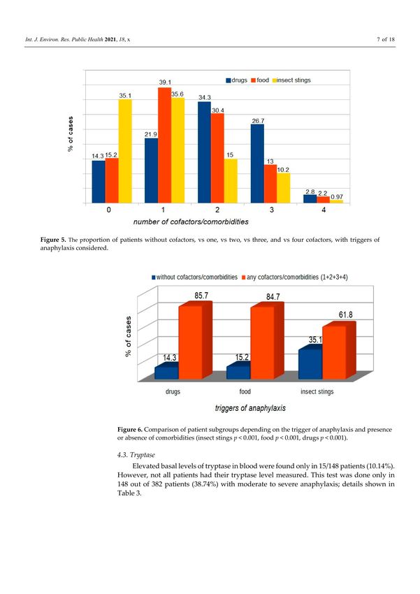
Int. J. Environ. Res. Public Health 2021, 18, 333                                                                                 3 of 17

                                             Modifiable factors include long-term pharmacotherapy, especially with non-steroidal
                                        anti-inflammatory drugs (NSAIDs), proton pump inhibitors (PPIs) [31], exercise, alcohol,
                                        and emotional stress.
                                             Acute infections, especially the early phase of infection, during specific immunother-
                                        apy (SIT) and food immunotherapy, are cofactors in anaphylaxis. It has been assumed
                                        that bacterial or viral products can be sensed by receptors on mast cells and basophils and,
                                        under certain conditions, trigger or enhance mast cell degranulation [31].
                                             Old age, combined with comorbidities such as cardiovascular disease (CVD) and
                                        asthma, especially allergic asthma, is an important risk factor for severe anaphylaxis with
                                        hospitalization, prolonged hospital stay, and fatality [20,21,23,36,37]. There are also reports
                                        on studies in a group of patients who have unexplained recurrent episodes of severe
                                        anaphylaxis with CVD and elevated basal tryptase levels (>11.4 mcg/L) [19,26].
                                             According to the literature on anesthesiology, factors that enhance the risk of anaphy-
                                        laxis include old age, female sex, lactation, asthma, fever, systemic mastocytosis, active
                                        infection, spinal anesthesia, pre-menstrual state, and emotional state [38].
                                             A review published in 2018 referenced many other cofactors, including menstruation,
                                        infection, extreme air temperatures, cannabis use, and medications other than NSAIDs,
                                        including angiotensin-converting enzyme inhibitors, beta-blockers, and antacids [39].
                                             Certain factors place some individuals at increased risk for more severe anaphylactic
                                        reactions: (1) history of an anaphylactic reaction; (2) history of asthma, especially if poorly
                                        controlled; (3) allergy to peanuts, nuts, fish, and shellfish; (4) teenage patients, and 5)
                                        patients on β-blockers or angiotensin-converting enzyme inhibitors [40].
                                             The most common cause of anaphylaxis mentioned in Polish and German registries
                                        are Hymenoptera stings [41,42]. Current German and European guidelines recommend
                                        Venom Immunotherapy (VIT) for all patients with grade II or higher reactions and for
                                        patients with a grade 1 reaction if they have any other risk factors or if their quality of life
                                        has been negatively impacted [43–46]. Other risk factors for severe anaphylaxis triggered
                                        by insect sting include male sex, older age, a large number of stings, a short time interval
                                        between stings, the location of the sting, the absence of skin symptoms, high baseline
                                        serum tryptase levels, as well as cardiovascular comorbidity [47]. Oropeza et al. [48] also
                                        reported other risk factors, such as asthma, rhinitis, atopic dermatitis, urticaria, and/or
                                        angioedema.
                                             The presence of cofactors is associated with a more severe anaphylactic reaction
                                        and reduces the amount of the allergen needed to trigger anaphylaxis. Reports from the
                                        United States emphasize the role of education on anaphylaxis, as the majority of patients
                                        experience subsequent episodes of anaphylaxis [7].

                                        2. Objective
                                             The objective of this study was to identify factors increasing the severity of anaphy-
                                        laxis/cofactors/comorbidities in patients with a history of moderate to severe anaphylactic
                                        reactions.

                                        3. Material and Methods
                                        3.1. Study Design and Data Collection
                                              In the retrospective analysis, we used a questionnaire-based survey completed by
                                        doctors specialized in allergology. Details on the study design are presented in another
                                        publication [42]. Of all 10,738 new patients examined at the Allergology Department in
                                        2006–2015, with suspicion of any allergy or non-allergic hypersensitivity, we found above
                                        490 patients with suspicion of moderate and severe anaphylaxis. One year after the first
                                        visit, each was analyzed again using the survey and additional results were collected. The
                                        study protocol did not require the approval of a Bioethics Committee.
                                              The basic questionnaire was a simplified version of the Network for Online-Registration
                                        of Anaphylaxis survey (NORA) from Berlin [42]; more information in the Appendix A.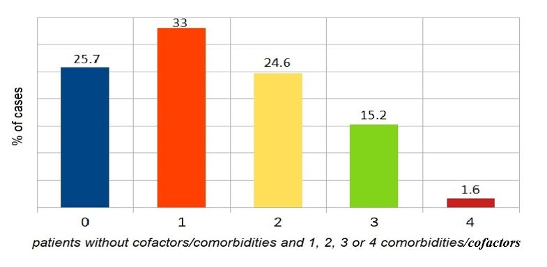
Int. J. Environ. Res. Public Health 2021, 18, 333                                                                                   4 of 17

                                             Patients were asked about their sex and age at the time of the anaphylactic episode, as
                                        well as about:
                                             Presence of chronic diseases affecting the cardiovascular system, lower respiratory
                                        tract, upper respiratory tract, thyroid, gastrointestinal tract, kidneys, as well as diabetes,
                                        features of atopy, and others. Definition of atopy by Johansson: “Atopy is a personal
                                        and/or familial tendency, usually in childhood or adolescence, to become sensitized and
                                        produce IgE antibodies in response to ordinary exposures to allergens, usually proteins. As
                                        a consequence, these persons can develop typical symptoms of asthma, rhinoconjunctivitis,
                                        or eczema.” At the first visit, the patients indicated whether they suspected or had a
                                        confirmed atopic disease (phenotypes listed above); later, one year after reporting, an
                                        allergist specialist either confirmed or removed the existence of atopic features (atopic
                                        disease, e.g., the reported allergic rhinitis (AR) turned out to be chronic non-allergic rhinitis).
                                        Other aspects were defined as follows:
                                             Recurrence of anaphylaxis, i.e., re-exposure to the same trigger or a pharmaceutical
                                        with a similar chemical structure or activity, after the initial reaction.
                                             Additional factors associated with anaphylaxis, e.g., alcohol intake, exercise, symp-
                                        toms of acute infection, and menstruation.

                                        3.2. Statistical Analysis
                                              Obtained data were analyzed using the Statistica 12 software package (StatSoft, Inc.,
                                        Cracow, Poland license, Tulsa, OK, USA). A basic statistics panel was used for data process-
                                        ing: for descriptive statistics and qualitative variables used in the analysis, non-parametric
                                        tests were used. The collected data were presented in the form of a multi-division table.
                                        For qualitative variables, we used Pearson’s chi-square test, the Wilcoxon non-parametric
                                        signed-rank test, other significance tests, and structural indicators. Statistical significance
                                        was adopted at a p-value of p < 0.05.

                                        4. Results
                                        4.1. Demographic Data
                                             Of all 10,738 new patients examined at the Allergology Department in 2006–2015, there
                                        were 382 cases of moderate and severe anaphylaxis (grades II-IV by Ring and Messmer
                                        classification), which accounted for 3.56% of new patients.
                                             There were 236 women (61.8%) and 146 men (38.2%) with anaphylaxis, p < 0.001. In
                                        This group was 50 children (13.1%): 29 girls (7.6%), 21 boys (5.5%). Adults vs. children
                                        p < 0.001. The mean age at the onset of anaphylaxis was 40.4 years (range 0–79) for women
                                        (n = 236), and 38.2 years (range 0–76) for men (n = 146). In the group of children and
                                        adolescents (max. age 18 years), the mean age at onset was 11 years (9.9 for boys and 12.1
                                        for girls). Most patients with moderate to severe anaphylaxis were in the age range 19–40
                                        and 41–60 years, which accounted for about 75% of all anaphylactic reactions in the registry.
                                        A detailed distribution of data for the analyzed group is presented in Figure 1.

                                        Figure 1. Distribution of patients by age range in the analyzed group.
Int. J. Environ. Res. Public Health 2021, 18, 333 5 of 17
There were no significant differences in the proportion of male and female patients
within age groups: females vs. males Figure 2, but there were significant differences in the
proportion of males and females between age groups.
Figure 2. Distribution of patients by age range and sex in the analyzed group.
4.2. Comorbidities
The most common comorbidities recorded in patients with moderate to severe ana-
phylaxis include cardiovascular disease, upper respiratory tract disease (mainly allergic
rhinitis), lower respiratory tract diseases (mainly asthma), features of atopy, thyroid disease,
diabetes mellitus (mainly type 2).
Data on comorbidities in the analyzed group are presented in Figure 3. Note, one
patient may have more than one cofactor and or comorbidity.
Figure 3. Proportion of patients with a specific cofactor/comorbidity in the analyzed group.
Presence of comorbidity/cofactor: one factor vs. two vs. three vs. four vs. zero is
shown in Figure 4. The comparison focused on the presence of one vs. two vs. three vs.
four vs. zero comorbidity/cofactor revealed a significant difference between patients with
one and those with three comorbidities/cofactors (p < 0.01). Anaphylaxis was significantly
more frequent in patients with one cofactor. No differences were found between patients
with four comorbidities vs. other variants because of the low number of patients with four
comorbidities.Int. J. Environ. Res. Public Health 2021, 18, 333 6 of 17
Figure 4. The proportion of patients without cofactors/comorbidities and those with one, two, three,
or four comorbidities/cofactors.
Comorbidities versus triggers of anaphylaxis (insect stings, drugs, food) are pre-
sented in Figure 5. The incidence of anaphylaxis was significantly higher in patients with
comorbidities compared to those without comorbidities (Figure 6).
Figure 5. The proportion of patients without cofactors, vs. one, vs. two, vs. three, and vs. four
cofactors, with triggers of anaphylaxis considered.Int. J. Environ. Res. Public Health 2021, 18, 333 7 of 17
Figure 6. Comparison of patient subgroups depending on the trigger of anaphylaxis and presence or
absence of comorbidities (insect stings p < 0.001, food p < 0.001, drugs p < 0.001).
4.3. Tryptase
Elevated basal levels of tryptase in blood were found only in 15/148 patients (10.14%).
However, not all patients had their tryptase level measured. This test was done only in 148
out of 382 patients (38.74%) with moderate to severe anaphylaxis; details shown in Table 3.
Table 3. The proportion of patients with an elevated basal tryptase level according to age ranges.
% of Whole
Age Range n Patients % of Tested n = 148 % of Age Group
Group n = 382
0–18 0 0 0 0
19–40 1 0.26 0.68 0.79
41–60 11 2.88 7.43 6.88
>60 3 0.79 2.03 6.67
Total 15 3.93 10.14 3.93
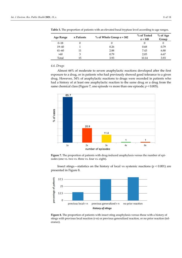
4.4. Drugs
Almost 66% of moderate to severe anaphylactic reactions developed after the first
exposure to a drug, or in patients who had previously showed good tolerance to a given
drug. However, 34% of anaphylactic reactions to drugs were recorded in patients who had
a history of at least one anaphylactic reaction to the same drug or a drug from the same
chemical class (Figure 7, one episode vs. more than one episode; p < 0.005).Int. J. Environ. Res. Public Health 2021, 18, 333 8 of 17
Figure 7. The proportion of patients with drug-induced anaphylaxis versus the number of episodes
(one vs. two vs. three vs. four vs. eight).
Insect stings—statistics on the history of local vs. systemic reactions (p < 0.001) are
presented in Figure 8.
Figure 8. The proportion of patients with insect sting anaphylaxis versus those with a history of stings
with previous local reaction (r-n) or previous generalized reaction, or no prior reaction (tolerance).
Exercise as a cofactor—11 patients (including five with Food-dependent exercise-
induced anaphylaxis (FDEIA)). In this group of patients, the exercise challenge test was
negative. FDEIA was diagnosed using molecular tests—Table 4 (these cases were previously
labeled as idiopathic anaphylaxis).
Table 4. Characteristics of patients with Food-dependent exercise-induced anaphylaxis (FDEIA).
Allergen Cofactor Patient Age Recurrence
Omega 5 gliadin Exercise 24 years 8×
Omega 5 gliadin Exercise + asthma 28 years 5×
Omega 5 gliadin Exercise + asthma + alcohol 28 years 4×
Omega 5 gliadin Exercise 27 years 2×
LPT Exercise + asthma 31 years 4×
5. Analysis of Results and Discussion
In our study, the incidence of moderate to severe anaphylaxis in the group of patients
referred for all causes to an allergy specialist was estimated at 0.35% per year. The annual
incidence rate for the West Pomerania province was 2.3/100,000, which is close to the
lower limit reported by Wolbing (3.2–68.4) [31]. This is also consistent with data reported
by Panesar et al. for the European population [4], where the incidence rate ranged from
1.5 to 7.9 per 100,000 person-years. In 2015, the prevalence rate of anaphylaxis in PolandInt. J. Environ. Res. Public Health 2021, 18, 333 9 of 17
(according to the National Health Fund of Poland, NHF) was much higher, but differed
considerably between regions and was estimated at 8.2 per 100,000 [49], and a similar rate
was reported by Tejedor-Alonso et al. [50]. In the general population of the United States,
the incidence rate was estimated at 1.6% [7].
Sex and age: Patients aged 19–60 years accounted for 75% of all anaphylaxis cases in
the analyzed registry. Anaphylaxis was more frequent in females than in males (p < 0.001),
which is explained by the promoting effect of estrogens in anaphylaxis. Similar data
were reported by other researchers, who indicated the reproductive age of women as
an augmenting factor in anaphylaxis [13]. Estrogen might also play a role by enhancing
endothelial expression of nitric oxide synthase and nitric oxide production, increasing
vascular permeability, and intensifying anaphylaxis severity [19,33]. In the analyzed group,
we found no increased incidence of anaphylaxis among adolescents and patients older
than 60 years, unlike Muñoz-Cano et al. [13], who emphasized the association between
old age and anaphylaxis due to comorbidities and increased use of medications. Increased
incidence of anaphylaxis in adolescents has been attributed to their “risky behavior” [13,51].
Data from the European and Korean registries suggest that the risk factors for severe
anaphylaxis include older age [35] and male sex [21], but this was not supported by
findings from our analysis. Ruëff et al. [52] and Chen et al. [53] investigated insect venom-
allergic patients and indicated male sex as a risk factor for severe anaphylaxis. Older
age [54] increases the risk of severe anaphylaxis, but in the presented material, the rate of
anaphylaxis in patients older than 60 years was similar to that in the age range 0–18 years
and significantly lower than in age ranges 19–40 (p < 0.005) or 41–60 years (p < 0.001).
Tryptase—basal level (not at the time of anaphylactic reaction): In the analyzed group,
the rate of patients with elevated (>11.4 µg/L) tryptase levels (not always with diagnosed
mastocytosis) was higher than 10%, vs. 1.64% of subjects with diagnosed mastocytosis
according to other anaphylaxis registers [35]. The highest percentage of patients (>6%)
with elevated tryptase levels has been observed in the age range 41–60 and in the group
of patients aged more than 60. According to Kucharewicz, in the group of patients with
Hymenoptera sting anaphylaxis, similar to our study, the rate of patients with elevated
tryptase level relative to all measurements was 11% [55], and comparable rates (7–11%)
have been reported by other authors [56–58].
Comorbidities. The following comorbidities/cofactors were identified in the analyzed
group of patients with moderate to severe anaphylaxis: CVD > upper respiratory tract
disease > lower respiratory tract disease > atopy > thyroid disease > diabetes mellitus type2
> infections > urticaria > gastrointestinal disease > exercise > osteoarthritis > neurological
disease > alcohol.
Literature data indicate that in patients with anaphylaxis, the presence of cardio-
vascular disease has been shown to predispose them to fatalities [13,19,20,23,37], and it
is probable that other chronic conditions such as renal and pulmonary problems would
do likewise [59]. Concomitant cardiac conditions were an important predictor of severe
anaphylaxis in the analysis of food-elicited reactions [35] and in patients with anaphylaxis
induced by Hymenoptera venom [47,60], which is consistent with findings from our anal-
ysis. Recent medical history in elderly patients consisted of significantly more frequent
cardiovascular, thyroid, and malignant diseases [61]. The above-mentioned studies suggest
that comorbidities alone are regarded as a risk factor for severe anaphylaxis, although
some researchers emphasized the significant effect of medications used [23]. Similar to
our findings, cardiovascular diseases and asthma were reported as risk factors for severe
anaphylaxis [54].
Asthma is associated with an increased incidence of anaphylaxis [51]. Contrasting
data were presented by Worm et al. [35], who found no such association and even indicated
that asthmatic patients had a lower risk of developing serious anaphylaxis (odds ratio (OR):
0.75, confidence interval (CI): 0.61–0.88).
Atopy was the fourth most common comorbidity in the analyzed group of patients
with moderate to severe anaphylaxis. It was identified in 17% of cases, which is consis-Int. J. Environ. Res. Public Health 2021, 18, 333 10 of 17
tent with data reported by Versluis et al. [62] and Aurich et al. [60]. According to other
researchers, atopic disease is identified in as many as 20–39% of patients with anaphy-
laxis [48,63–65]. It is clear that atopy increases the risk of systemic reactions because
patients with atopy are at risk for food allergy, but they also appear to be at risk for events
in general [59]. Similar conclusions were reached in a study on a population of beekeepers
in Turkey and patients with exercise-induced or latex-induced anaphylaxis [54].
Thyroid diseases were identified in 30/382 patients and were the fifth most common
comorbidity in the analyzed group. Perhaps this reflects the observed general increase
in the incidence of thyroid diseases in the general Polish population. Nevertheless, data
presented in the European anaphylaxis registry also indicate coexisting thyroid diseases in
anaphylaxis [35] as a risk factor for moderate anaphylaxis (with an incidence rate of 1.5),
and similar data were reported from the United States [20].
Infection—active infection was recorded as a cofactor in 19/382 cases of anaphylaxis
(ca. 5%). Literature data indicate the potential role of infection in 1.3% to 11% [31], and
even 29.8% of anaphylaxis cases [48]. On the other hand, in 257 cases (3.2%) recorded in
the European Anaphylaxis Registry, physicians reported an active infection concomitant to
anaphylaxis (e.g., upper respiratory tract infection or common cold) [35].
Menstruation, according to the available literature, is a cofactor of anaphylaxis in 8%
to 12.1% of cases [31]. In our study, we did not find such a high rate, perhaps because
patients could not remember details other than the symptoms of anaphylaxis. We recorded
only one case in 236 women where menstruation was a cofactor. Nevertheless, single cases
have been reported, indicating beyond any doubt, a significant effect of menstruation on
the onset of anaphylaxis [66].
Considering modifiable extrinsic factors/cofactors of anaphylaxis, exercise was the
most common one. In the analyzed material, this cofactor was identified in 11/382
cases. The second most common modifiable cofactor was alcohol, and it was identi-
fied in 9/382 cases. Awareness of the effect of these cofactors on the onset of anaphylaxis is
important since they can be easily eliminated.
Exercise was identified as a cofactor in 2.9% cases, and, similar to a study by Oropeza
et al. [48], FDEIA was diagnosed in five of these cases. An analysis conducted by Wölbing
et al. revealed that exercise was a cofactor in 0% to 20.4% of anaphylaxis cases [31], which
is consistent with our findings. The mechanism of action of this cofactor in anaphylaxis is
explained by the activation of tissue transglutaminase (tTG), which results in the formation
of large complexes of omega 5 gliadin and tTG. In addition, exercise increases the intestinal
absorption of allergens and hence the concentration of these substances in the blood [67–69].
Christensen et al. concluded that exercise lowers the threshold and increases the severity
of the reaction to the food [70].
Alcohol—In the analyzed registry, alcohol was a cofactor in 2.36% cases, while accord-
ing to other researchers, it was involved in 1–15.2% [31,48,60], and even in up to 15% of
cases of anaphylactic reaction according to some series [62,71]. It is also assumed that alco-
hol increases the gastrointestinal absorption of allergens [63], and induces modification in
the expression of the tight junction-associated proteins ZO-1 and claudin-1 of the intestinal
epithelium, thereby augmenting the permeability of the intestinal epithelial barrier [13].
According to the literature, only alcohol consumption could be implicated as a cofactor [72].
It is worth mentioning the importance of molecular diagnostics in allergology. Molec-
ular tests help identify the cause of anaphylaxis, especially in patients with idiopathic
anaphylaxis. In our analysis, the initial incidence rate of idiopathic anaphylaxis was 3.9%
(15 patients), but according to literature data, it may be up to 20% [19]. Tests with new
molecules for the determination of sIgE allowed for the identification of the direct cause of
anaphylaxis in five patients, which accounts for 33% of cases with the established cause
and initially labeled as idiopathic anaphylaxis. This rate is comparable to rates reported by
other researchers [73], where the actual cause was identified in 45% of the previously unrec-
ognized sensitizations. Moreover, recognized sensitization to heat-resistant molecules, e.g.,
lipid transfer proteins (LTP), has been reported as a predictive factor for severe anaphylacticInt. J. Environ. Res. Public Health 2021, 18, 333 11 of 17
reactions in the future [51,74,75]. Importantly, people with a diagnosed LTP allergy appear
to be more likely to have a reaction to foods when a cofactor is present [76,77].
Hymenoptera-previous stings. In our analysis, 38% of patients with moderate to
severe anaphylaxis following Hymenoptera stings previously had a generalized reaction to
stings. Therefore, a history of generalized reaction is a significant risk factor for another
anaphylaxis episode, compared to a previous severe local reaction (p < 0.001). Similar
data have been reported since 1988 and have been described in a Hymenoptera venom
study [52,78,79]. Even though 15% of patients with moderate to severe anaphylaxis had
prior large local reactions to stings, similar to observations by Bilo et al. [60], venom
immunotherapy is not recommended for large local reactions in either children [80,81]
or adults [82]. Other factors may influence the decision to initiate VIT. These include
occupations and/or hobbies where the risk of exposure is high, the culprit insect itself,
concomitant cardiovascular diseases, other pathologies, or psychological factors arising
from anxiety, which can seriously impair patient quality of life [83]. The natural history of
large local reactions to Hymenoptera stings allowed the estimation of the risk of developing
a systemic reaction after an initial large local reaction in about 4% of patients [84]; according
to other authors, it is 2–15% [82,85,86]. Severino observed that in patients who had a history
of a large local reaction, 24% did not experience any reactions, 52% reported a second
large local reaction, and 24% had systemic reactions [85].On the other hand, concerning
VIT, both American and European guidelines advise that it could be an acceptable option
or recommended, in recurrent and troublesome large local reaction (LLR), to reduce the
duration and size of future LLR, but only in special circumstances (i.e., frequent exposure,
lifestyle factors) and after evaluating the cost/benefit profile [44,86–89].
Drugs-prior anaphylaxis. In the analyzed population, about 35% of patients with
moderate to severe drug-induced anaphylaxis had a previous anaphylactic reaction to
the same drug or a drug from the same chemical class, which is consistent with other
reports [90,91]. This proves either low awareness among patients/doctors or the fact
that patients did not try to explain previous health problems, which is consistent with
observations made for a Polish population [49]. The risk factors for drug anaphylaxis are
previous cardiovascular morbidity and older age [92]. The female predisposition to drug
allergy can be explained by higher drug consumption, genetic factors, epigenetic changes,
and discrepant hormonal interactions with immune cells [93]. However, literature data
indicate that patients with a previous reaction, when re-exposed to the same drug, have a
21–60% risk of an immediate repeat reaction [94–96]. Among other conditions, atopy was
reported as a risk factor for both NSAIDs, and antibiotic allergies [97,98], Kurt et al. [99]
found that female sex, asthma, allergic rhinitis, and eczema diagnoses were associated with
drug hypersensitivity reactions. According to a study based on data from the European
Anaphylaxis Registry, 28% of elderly patients reported a previous allergic reaction to the
same elicitor [60], which again substantiates the need for educating patients, people from
their close environment, and healthcare professionals. Even though the first episode of
anaphylaxis is unpredictable, further episodes in the same patient are preventable, but still
happen [7,21].
5.1. Additional Material
The analysis of moderate and severe anaphylaxis cases was performed, excluding
the youngest patients (0–18 years of age) n = 332, owing to the underrepresented children
group (n = 50), which is emphasized in the Limitations section. Similar numerical values
were obtained, which did not change the final conclusions of the work.
Conclusions after excluding the children group:
Anaphylaxis occurred significantly more often in the age range 41–60; the only signif-
icant differences have been observed in women aged 19–40 vs. >60 and in women aged
41–60 vs. >60 as well as in men in the respective age ranges, people aged 19–60 constituted
86% of all the patients, women experienced anaphylaxis significantly more often, the most
common comorbidities present in the analyzed group were: CVD, rhinitis (mainly allergic),Int. J. Environ. Res. Public Health 2021, 18, 333 12 of 17
bronchial asthma, atopy, thyroid diseases, diabetes (mainly type 2). When comparing
the proportion of patients without comorbidities vs. one vs. two vs. three vs. four dis-
eases/cofactors, a significant difference (p < 0.05) between patients with one cofactor vs.
patients with three cofactors was observed. Analyzing the occurrence of anaphylaxis in
people without any comorbidities/cofactors vs. people with at least one disease/cofactor,
significant differences (p < 0.01) for each cause of anaphylaxis (drugs, food, insects) was
observed. Drug-induced anaphylaxis recurred in about 37% of patients after contact with
the same drug or a drug from the same group. Anaphylaxis after Hymenoptera stings
occurred significantly more often when a patient had already had a similar anaphylaxis
episode, as compared with a past local reaction.
5.2. Limitations
The population of the youngest children is underrepresented in the analyzed registry
since our Allergy Clinic is a reference center for children older than five years and adults.
Because this was a retrospective study, it may have been influenced by selection bias, and
patients may not have remembered certain facts related to anaphylaxis. Not all patients
completed all the investigations. Lack of detailed data on the history of anaphylaxis-
cofactors was due to self-reported data. Challenge tests were performed only in a few
cases, and the number of tests to measure tryptase levels were low.
The analysis was not conducted for individual grades of anaphylaxis severity but for
a pooled dataset of patients with moderate to severe anaphylaxis (grades II–IV). We did
not assess the effect of drugs used by patients on the onset of anaphylaxis since no drug-
related data were gathered. Another limitation is the lack of a corresponding control group.
Therefore, we cannot draw inferences on which factors increase the risk of developing
anaphylactic responses in the general population.
6. Conclusions
Anaphylaxis is an acute, severe, and life-threatening reaction. Cofactors were reported
more frequently in patients with moderate to severe anaphylaxis, and it is important to
take these factors into consideration when evaluating patients with anaphylaxis, as the
cofactors may play a role in future anaphylactic reactions.
The presence of cofactors may explain why the intake of some foods sometimes leads
to anaphylaxis, while in other cases, the same allergen induces a milder reaction or is
even tolerated. It is necessary to refer every patient, after anaphylaxis, to an allergist for
diagnosis.
Patients do not appear adequately equipped to deal with future episodes, which indi-
cates the need for public health initiatives to improve anaphylaxis recognition, treatment,
and prophylaxis.
Author Contributions: M.K. (Michał Kurek) conceived the idea for the study, approved the final
version of the manuscript. I.P.-G. contributed to the design of the research and was involved in data
collection. I.P.-G. analyzed the data. I.P.-G. coordinated funding for the project. M.K. (Magdalena
Kostrzewska) performed calculations and statistical analyses and approved the final version of the
manuscript. I.P.-G. edited and approved the final version of the manuscript. All authors have read
and agreed to the published version of the manuscript.
Funding: This research received no external funding.
Institutional Review Board Statement: Bioethics Committee of the Pomeranian Medical University
PMU—issued a statement that keeping the Anaphylaxis Register and retrospective analysis does
not require its consent. The study was conducted according to the guidelines of the Declaration of
Helsinki.
Informed Consent Statement: Not applicable.
Data Availability Statement: The data presented in this study are openly available in [repository
name e.g., FigShare] at [DOI:10.3390/ijerph17082787], reference number [42].Int. J. Environ. Res. Public Health 2021, 18, 333 13 of 17
Acknowledgments: The Anaphylaxis Registry is financed from the budget for statutory Research
of the Clinical Allergology Department, PMU, No. WLA-151-01/S/12 IPG. Special thanks to Artur
G˛esicki for help in editing responses to reviewers (tables, figures).
Conflicts of Interest: The authors declare that they have no conflicts of interest.
Appendix A
Supplemental material with a simplified Network for Online-Registration of Anaphy-
laxis (NORA) questionnaire- from article” Clinical Manifestations and Causes of Anaphy-
laxis. Analysis of 382 Cases from the Anaphylaxis Registry in West Pomerania Province in
Poland”.
The basis questionnaire—simplified version of the Network for Online-Registration
of Anaphylaxis survey (NORA) from Berlin.
1. Did Your Patients, after contact with any factor, or spontaneously experience: diffi-
culty breathing, wheezing, hypotension, cramping abdominal pain, diarrhea, vomit-
ing, loss of consciousness? If so, complete the data
2. Year of the patient’ birth
3. Date of anaphylaxis onset
4. Place of reaction
5. Gender
6. Mark the organ systems involved
6.1. Cutaneous: angioedema, flush, generalized erythema, generalized itching,
generalized urticaria
6.2. Respiratory: apnoea, dyspnoea, stridor
6.3. Gastrointestinal: abdominal pain, diarrhoea, nausea, vomiting, incontinence
6.4. Cardiovascular: loss of consciousness, drop in blood pressure, collapse, cardiac
arrest, dizziness, tachycardia, disorientation
7. Mark the diagnostic tests used Medical Interview, skin tests, sIgE, tryptase, provoca-
tion, other
8. Was the reaction the first time?
9. Is the trigger factor known?—is the identified trigger of anaphylaxis? Is this medicine?
substance Is this food? which one Is this venom? which one Other
10. Prevention
11. Please write other important details about this episode
References
1. Hepner, D.L.; Castells, M.C. Anaphylaxis during the Perioperative Period. Anesth. Analg. 2003, 97, 1381–1395. [CrossRef]
[PubMed]
2. Turner, P.J.; Worm, M.; Ansotegui, I.J.; El-Gamal, Y.; Rivas, M.F.; Fineman, S.; Geller, M.; Gonzalez-Estrada, A.; Greenberger, P.A.;
Tanno, L.K.; et al. Time to revisit the definition and clinical criteria for anaphylaxis? World Allergy Organ. J. 2019, 12, 100066.
[CrossRef] [PubMed]
3. Simons, F.E.R.; Ardusso, L.R.F.; Bilò, M.B. World allergy organization guidelines for the assessment and management of
anaphylaxis. World Allergy Organ. J. 2011, 4, 13–37. [CrossRef]
4. Panesar, S.S.; Javad, S.; De Silva, D.; Nwaru, B.I.; Hickstein, L.; Muraro, A.; Roberts, G.; Worm, M.; Bilò, M.B.; Cardona, V.; et al.
The epidemiology of anaphylaxis in Europe: A systematic review. Allergy 2013, 68, 1353–1361. [CrossRef]
5. Lieberman, P.; Nicklas, R.A.; Oppenheimer, J. The diagnosis and management of anaphylaxis practice parameter: 2010 update. J.
Allergy Clin. Immunol. 2010, 126, 477–480. [CrossRef]
6. Brown, S.G.; Mullins, R.J.; Gold, M.S. Anaphylaxis: Diagnosis and management. Med. J. Aust. 2006, 185, 283–289. [CrossRef]
7. Wood, R.A.; Camargo, C.A.; Lieberman, P.; Sampson, H.A.; Schwartz, L.B.; Zitt, M.; Collins, C.; Tringale, M.; Wilkinson, M.; Boyle,
J.; et al. Anaphylaxis in America: The prevalence and characteristics of anaphylaxis in the United States. J. Allergy Clin. Immunol.
2014, 133, 461–467. [CrossRef]
8. Tanno, L.K.; Bierrenbach, A.L.; Simons, F.E.R.; Cardona, V.; Bernard, T.Y.H.; Molinari, N.; Calderón, M.; Worm, M.; Chang, Y.-S.;
Papadopoulos, N.G.; et al. Critical view of anaphylaxis epidemiology: Open questions and new perspectives. Allergy Asthma Clin.
Immunol. 2018, 14, 1–11. [CrossRef]
9. Jimenez-Rodriguez, T.W.; Garcia-Neuer, M.; Alenazy, L.A.; Castells, M. Anaphylaxis in the 21st century: Phenotypes, endotypes,
and biomarkers. J. Asthma Allergy 2018, 11, 121–142. [CrossRef]Int. J. Environ. Res. Public Health 2021, 18, 333 14 of 17
10. Sampson, H.A.; Muñoz-Furlong, A.; Campbell, R.L.; Adkinson, N.F.; Bock, S.A.; Branum, A.; Brown, S.G.; Camargo, C.A.;
Cydulka, R.; Galli, S.J.; et al. Second symposium on the definition and management of anaphylaxis: Summary report—Second
National Institute of Allergy and Infectious Disease/Food Allergy and Anaphylaxis Network symposium. J. Allergy Clin. Immunol.
2006, 117, 391–397. [CrossRef]
11. Simons, F.E.R.; Ardusso, L.R.; Bilò, M.B.; Cardona, V.; Ebisawa, M.; El-Gamal, Y.M.; Lieberman, P.; Lockey, R.F.; Muraro, A.;
Roberts, G.; et al. International consensus on (ICON) anaphylaxis. World Allergy Organ. J. 2014, 7, 1–19. [CrossRef] [PubMed]
12. Muraro, A.; Roberts, G.; Worm, M.; Bilò, M.B.; Brockow, K.; Rivas, M.F.; Santos, A.F.; Zolkipli, Z.Q.; Bellou, A.; Beyer, K.;
et al. Anaphylaxis: Guidelines from the European Academy of Allergy and Clinical Immunology. Allergy 2014, 69, 1026–1045.
[CrossRef] [PubMed]
13. Muñoz-Cano, R.; Pascal, M.; Araujo, G.; Goikoetxea, M.J.; Valero, A.L.; Picado, C.; Bartra, J. Mechanisms, Cofactors, and
Augmenting Factors Involved in Anaphylaxis. Front. Immunol. 2017, 8, 1193. [CrossRef]
14. Ring, J.; Messmer, K. Incidence and severity of anaphylactoid reactions to colloid volume substituted. Lancet 1977, 1, 466–469.
[CrossRef]
15. Muller, H.L. Diagnosis and treatment of insect sensitivity. J. Asthma Res. 1966, 3, 331–333. [CrossRef]
16. Brown, S.G. Clinical features and severity gradning of anaphylaxis. J. Allergy Clin. Immunol. 2004, 114, 371–376. [CrossRef]
17. Muraro, A.; Roberts, G.; Clark, A.; Eigenmann, P.A.; Halken, S.; Lack, G.; Moneret-Vautrin, A.; Niggemann, B.; Rancé, F.; EAACI
Task Force on Anaphylaxis in Children. The management of anaphylaxis in childhood: Position paper of the European academy
of allergology and clinical immunology. Allergy 2007, 62, 857–871. [CrossRef]
18. Mehl, A.; Wahn, U.; Niggermann, B. Anaphylactic reactions in children—A questionnaire- based survey in Germany. Allergy
2005, 60, 1440–1445. [CrossRef]
19. Simons, F.E.R.; Ebisawa, M.; Sanchez-Borges, M.; Thong, B.Y.; Worm, M.; Tanno, L.K.; Lockey, R.F.; El-Gamal, Y.M.; Brown, S.G.A.;
Park, H.-S.; et al. 2015 update of the evidence base: World Allergy Organization anaphylaxis guidelines. World Allergy Organ. J.
2015, 8, 32. [CrossRef]
20. Clark, S.; Wei, W.; Rudders, S.A.; Camargo, C.A., Jr. Risk factors for severe anaphylaxis in patients receiving anaphylaxis
treatment in US emergency departments and hospitals. J. Allergy Clin. Immunol. 2014, 134, 1125–1130. [CrossRef]
21. Ye, Y.-M.; Kim, M.K.; Kang, H.-R.; Kim, T.-B.; Sohn, S.-W.; Koh, Y.-I.; Park, H.-K.; Jang, G.C.; Kim, C.-W.; Jee, Y.K.; et al. Predictors
of the severity and serious outcomes of anaphylaxis in Korean adults: A multicenter retrospective case study. Allergy Asthma
Immunol. Res. 2015, 7, 22–29. [CrossRef]
22. Vazquez-Ortiz, M.; Álvaro, M.; Piquer, M.; Giner, M.T.; Domínguez, O.; Lozano, J.; Jiménez-Feijoo, R.; Cambra, F.J.; Plaza, A.M.
Life-threatening anaphylaxis to egg and milk oral immunotherapy in asthmatic teenagers. Ann. Allergy Asthma Immunol. 2014,
113, 482–484. [CrossRef]
23. Triggiani, M.; Montagni, M.; Parente, R.; Ridolo, E. Anaphylaxis and cardiovascular diseases: A dangerous liaison. Curr. Opin.
Allergy Clin. Immunol. 2014, 14, 309–315. [CrossRef]
24. Rueff, F.; Vos, B.; Elberink, J.O.; Bender, A.; Chatelain, R.; Dugas-Breit, S.; Horny, H.-P.; Kuechenhoff, H.; Linhardt, A.; Mastnik, S.;
et al. Predictors of clinical effectiveness of Hymenoptera venom immunotherapy. Clin. Exp. Allergy 2014, 44, 736–746. [CrossRef]
25. Valent, P. Risk factors and management of severe life-threatening anaphylaxis in patients with clonal mast cell disorders. Clin.
Exp. Allergy 2014, 44, 914–920. [CrossRef]
26. Gülen, T.; Hägglund, H.; Sander, B.; Dahlén, B.; Nilsson, G. The presence of mast cell clonality in patients with unexplained
anaphylaxis. Clin. Exp. Allergy 2014, 44, 1179–1187. [CrossRef]
27. Álvarez-Twose, I.; Zanotti, R.; González-De-Olano, D.; Bonadonna, P.; Vega, A.; Matito, A.; Sánchez-Muñoz, L.; Morgado, J.M.;
Perbellini, O.; Montero, A.G.; et al. Nonaggressive systemic mastocytosis (SM) without skin lesions associated with insect-induced
anaphylaxis shows unique features versus other indolent SM. J. Allergy Clin. Immunol. 2014, 133, 520–528. [CrossRef] [PubMed]
28. Krishna, M.T.; Worm, M.; Bilo, M.B. Editorial: Anaphylaxis—A distinct immunological syndrome, but how much do we really
understand? Front. Immunol. 2019, 10, 2943. [CrossRef]
29. Skypala, I.J. Food-induced anaphylaxis: Role of hidden allergens and cofactors. Front. Immunol. 2019, 10, 673. [CrossRef]
30. Brown, S.G.A.; Stone, S.F.; Fatovich, D.M.; Burrows, S.; Holdgate, A.; Celenza, A.; Coulson, A.; Hartnett, L.; Nagree, Y.; Cotterell,
C.; et al. Anaphylaxis: Clinical patterns, mediator release, and severity. J. Allergy Clin. Immunol. 2013, 132, 1141–1149.e5.
[CrossRef]
31. Wölbing, F.; Fischer, J.; Köberle, M.; Kaesler, S.; Biedermann, T. About the role and underlying mechanisms of cofactors in
anaphylaxis. Allergy 2013, 68, 1085–1092. [CrossRef]
32. Fischer, J.; Hebsaker, J.; Caponetto, P.; Platts-Mills, T.A.; Biedermann, T. Galactose-alpha-1,3-galactose sensitization is a prerequisite
for pork-kidney allergy and cofactor-related mammalian meat anaphylaxis. J. Allergy Clin. Immunol. 2014, 134, 755–759.e1.
[CrossRef]
33. Hox, V.; Desai, A.; Bandara, G.; Gilfillan, A.M.; Metcalfe, D.D.; Olivera, A. Estrogen increases the severity of anaphylaxis in
female mice through enhanced endothelial nitric oxide synthase expression and nitric oxide production. J. Allergy Clin. Immunol.
2015, 135, 729–736.e5. [CrossRef]
34. Shadick, N.A.; Liang, M.H.; Partridge, A.J.; Bingham, C.; Wright, E.; Fossel, A.H.; Sheffer, A.L. The natural history of exercise-
induced anaphylaxis: Survey results from a 10-year follow-up study. J. Allergy Clin. Immunol. 1999, 104, 123–127. [CrossRef]Int. J. Environ. Res. Public Health 2021, 18, 333 15 of 17
35. Worm, M.; Francuzik, W.; Renaudin, J.-M.; Bilo, M.B.; Cardona, V.; Hofmeier, K.S.; Köhli, A.; Bauer, A.; Christoff, G.; Cichocka-
Jarosz, E.; et al. Factors increasing the risk for a severe reaction in anaphylaxis: An analysis of data from The European
Anaphylaxis Registry. Allergy 2018, 73, 1322–1330. [CrossRef]
36. Turner, P.J.; Gowland, M.H.; Sharma, V.; Ierodiakonou, D.; Harper, N.; Garcez, T.; Pumphrey, R.; Boyle, R.J. Increase in anaphylaxis-
related hospitalizations but no increase in fatalities: An analysis of United Kingdom national anaphylaxis data, 1992-2012. J.
Allergy Clin. Immunol. 2015, 135, 956–963.e1. [CrossRef] [PubMed]
37. Lieberman, P.; Simons, F.E.R. Anaphylaxis and cardiovascular disease: Therapeutic dilemmas. Clin. Exp. Allergy 2015, 45,
1288–1295. [CrossRef]
38. Valencia, B.; Inés, M. Perioperative anaphylaxis. Rev. Bras. Anestesiol. 2015, 65, 292–297.
39. Asaumi, T.; Ebisawa, M. How to manage food dependent exercise induced anaphylaxis (FDEIA). Curr. Opin. Allergy Clin.
Immunol. 2018, 18, 243–247. [CrossRef]
40. Sampson, H.A. Anaphylaxis and emergency treatment. Pediatrics 2003, 111, 1601–1608.
41. Worm, M.; Dölle, S.; Francuzik, W. Data from the anaphylaxis registry of the German-speaking countries. Revue Française
d’Allergologie 2015, 55, 452–455. [CrossRef]
42. Poziomkowska-Gesicka, I.; Kurek, M. Clinical manifestations and causes of anaphylaxis. Analysis of 382 cases from the
anaphylaxis registry in West Pomerania Province in Poland. Int. J. Environ. Res. Public Health 2020, 17, 2787. [CrossRef]
43. Przybilla, B.; Rueff, F.; Walker, A.I.; Räwer, H.-C.; Aberer, W.; Bauer, C.P.; Berdel, D.; Biedermann, T.; Brockow, K.; Forster, J.; et al.
Diagnose und Therapie der Bienen- und Wespengiftallergie. Allergo J. 2011, 20, 318–339. [CrossRef]
44. Sturm, G.J.; Varga, E.M.; Roberts, G.; Mosbech, H.; Bilò, M.B.; Akdis, C.A.; Antolín-Amérigo, D.; Cichocka-Jarosz, E.; Gawlik, R.;
Jakob, T.; et al. EAACI guidelines on allergen immunotherapy: Hymenoptera venom allergy. Allergy 2018, 73, 744–764. [CrossRef]
[PubMed]
45. Koschel, D. Impaired quality of life in patients with insect venom allergy. Allergo J. Int. 2017, 26, 88–92. [CrossRef]
46. Manmohan, M.; Müller, S.; Rauber, M.M.; Koberne, F.; Reisch, H.; Koster, J.; Böhm, R.; Messelken, M.; Fischer, M.; Jakob, T.
Current state of follow-up care for patients with Hymenoptera venom anaphylaxis in southwest Germany. Allergo J. Int. 2018, 27,
4–14. [CrossRef]
47. Fehr, D.; Micaletto, S.; Moehr, T.; Schmid-Grendelmeier, P. Risk factors for severe systemic sting reactions in wasp (Vespula spp.)
and honeybee (Apis mellifera) venom allergic patients. Clin. Transl. Allergy 2019, 9, 54. [CrossRef]
48. Oropeza, A.R.; Bindslev-Jensen, C.; Broesby-Olsen, S.; Kristensen, T.K.; Møller, M.B.; Vestergaard, H.; Kjaer, H.F.; Halken, S.;
Lassen, A.; Mortz, C.G. Patterns of anaphylaxis after diagnostic workup: A follow-up study of 226 patients with suspected
anaphylaxis. Allergy 2017, 72, 1944–1952. [CrossRef]
49. Jahnz-Rozyk, K.; Raciborski, F.; Śliwczyński, A.M.; Kłak, A.; Pinkas, J. Anaphylaxis in Poland the epidemiology and direct costs.
ADV Dermatol. Alergol. 2017, 34, 573–579. [CrossRef]
50. Tejedor-Alonso, M.A.; Moro-Moro, M.; Múgica-García, M.V. Epidemiology of anaphylaxis: Contributions from the last 10 years. J.
Investig. Allergol. Clin. Immunol. 2015, 25, 163–175.
51. Ewan, P.; Brathwaite, N.; Leech, S.; Luyt, D.; Till, S.; Nasser, S.M.; Powell, R.; Clark, A. Prescribing an adrenaline auto-injector—
Personalized care recommended. Clin. Exp. Allergy 2016, 46, 1621–1622. [CrossRef] [PubMed]
52. Rueff, F.; Przybilla, B.; Biló, M.B.; Müller, U.; Scheipl, F.; Aberer, W.; Birnbaum, J.; Bodzenta-Lukaszyk, A.; Bonifazi, F.; Bucher, C.;
et al. Predictors of severe systemic anaphylactic reactions in patients with Hymenoptera venom allergy: Importance of baseline
serum tryptase—A study of the European Academy of Allergology and Clinical Immunology Interest Group on Insect Venom
Hypersensitivity. J. Allergy Clin. Immunol. 2009, 124, 1047–1054. [CrossRef] [PubMed]
53. Chen, W.; Mempel, M.; Schober, W.; Behrendt, H.; Ring, J. Gender difference, sex hormones, and immediate type hypersensitivity
reactions. Allergy 2008, 63, 1418–1427. [CrossRef]
54. Shaker, M.; Wallace, D.V.; Golden, D.B.; Oppenheimer, J.; Bernstein, J.A.; Campbell, R.C.; Dinakar, C.; Ellis, A.; Greenhawt,
M.; Khan, D.A.; et al. Anaphylaxis—A 2020 practice parameter update, systematic review, and Grading of Recommendations,
Assessment, Development and Evaluation (GRADE) analysis. J. Allergy Clin. Immunol. 2020, 145, 1082–1123. [CrossRef]
55. Kucharewicz, I.; Bodzenta-Lukaszyk, A.; Szymanski, W.; Mroczko, B.; Szmitkowski, M. Basal serum tryptase level correlates with
severity of hymenoptera sting and age. J. Investig. Allergol. Clin. Immunol. 2007, 17, 65–69.
56. Haeberli, G.; Brönnimann, M.; Hunziker, T.; Müller, U. Elevated basal serum tryptase and hymenoptera venom allergy: Relation to
severity of sting reactions and to safety and efficacy of venom immunotherapy. Clin. Exp. Allergy 2003, 33, 1216–1220. [CrossRef]
57. Ludolph-Hauser, D.; Ruëff, F.; Fries, C.; Schöpf, P.; Przybilla, B. Constitutively raised serum concentrations of mast-cell tryptase
and severe anaphylactic reactions to Hymenoptera stings. Lancet 2001, 357, 361–362. [CrossRef]
58. Greenhawt, M.; Akin, C. Mastocytosis and allergy. Curr. Opin. Allergy Clin. Immunol. 2007, 7, 387–392. [CrossRef]
59. Lieberman, P.; Nicklas, R.A.; Randolph, C.; Oppenheimer, J.; Bernstein, D.I.; Bernstein, J.A.; Ellis, A.K.; Golden, D.B.; Greenberger,
P.A.; Kemp, S.; et al. Anaphylaxis—A practice parameter update 2015. Ann. Allergy Asthma Immunol. 2015, 115, 341–384.
[CrossRef]
60. Bilo, B.M.; Rueff, F.; Mosbech, H.; Bonifazi, F.; Oude-Elberink, J.N.G.; The EAACI Interest Group on Insect Venom Hypersensitivity.
Diagnosis of Hymenoptera venom allergy. Allergy 2005, 60, 1339–1349. [CrossRef]Int. J. Environ. Res. Public Health 2021, 18, 333 16 of 17
61. Aurich, S.; Dölle-Bierke, S.; Francuzik, W.; Bilo, M.B.; Christoff, G.; Fernandez-Rivas, M.; Hawranek, T.; Pföhler, C.; Poziomkowska-
Gȩsicka, I.; Renaudin, J.-M.; et al. Anaphylaxis in elderly patients—Data from the European Anaphylaxis Registry. Front. Immunol.
2019, 10, 750. [CrossRef] [PubMed]
62. Versluis, A.; Van Os-Medendorp, H.; Kruizinga, A.G.; Blom, W.M.; Houben, G.F.; Knulst, A.C. Cofactors in allergic reactions to
food: Physical exercise and alcohol are the most important. Immun. Inflamm. Dis. 2016, 4, 392–400. [CrossRef] [PubMed]
63. Helbling, A.; Hurni, T.; Mueller, U.R.; Pichler, W.J. Incidence of anaphylaxis with circulatory symptoms: A study over a 3-year
period comprising 940,000 inhabitants of the Swiss Canton Bern. Clin. Exp. Allergy 2004, 34, 285–290. [CrossRef]
64. Alvarez-Perea, A.; Tomás-Pérez, M.; Martínez-Lezcano, P.; Marco, G.; Pérez, D.; Zubeldia, J.M.; Baeza, M.L. Anaphylaxis in
adolescent/adult patients treated in the emergency department: Differences between initial impressions and the definitive
diagnosis. J. Investig. Allergol. Clin. Immunol. 2015, 25, 288–294.
65. Worm, M.; Edenharter, G.; Rueff, F.; Scherer, K.; Pföhler, C.; Mahler, V.; Treudler, V.; Lang, R.; Nemat, K.; Koehli, A. Symptom
profile and risk factors of anaphylaxis in Central Europe. Allergy 2012, 67, 691–698. [CrossRef]
66. De Silva, R.; Dasanayake, W.M.D.K.; Karunatilleke, C.; Malavige, G.N. Food dependant exercise induced anaphylaxis a retrospec-
tive study from 2 allergy clinics in Colombo, Sri Lanka. Allergy Asthma Clin. Immunol. 2015, 11, 22. [CrossRef]
67. Scherf, K.A.; Lindenau, A.-C.; Valentini, L.; Collado, M.C.; García-Mantrana, I.; Christensen, M.; Tomsitz, D.; Kugler, C.;
Biedermann, T.; Brockow, K. Cofactors of wheat-dependent exercise-induced anaphylaxis do not increase highly individual
gliadin absorption in healthy volunteers. Clin. Transl. Allergy 2019, 9, 1–12. [CrossRef]
68. Hompes, S.; Dölle, S.; Grünhagen, J.; Grabenhenrich, L.B.; Worm, M. Elicitors and co-factors in food-induced anaphylaxis in
adults. Clin. Transl. Allergy 2013, 3, 38. [CrossRef]
69. Aihara, M.; Miyazawa, M.; Osuna, H.; Tsubaki, K.; Ikebe, T.; Aihara, Y.; Ikezawa, Z. Food-dependent exercise-induced anaphylaxis:
Influence of concurrent aspirin administration on skin testing and provocation. Br. J. Dermatol. 2002, 146, 466–472. [CrossRef]
70. Christensen, M.; Eller, E.; Mortz, C.G.; Brockow, K.; Bindslev-Jensen, C. Exercise lowers threshold and increases severity, but
wheat-dependent, exercise-induced anaphylaxis can be elicited at rest. J. Allergy Clin. Immunol. Pract. 2018, 6, 514–520. [CrossRef]
71. Zogaj, D.; Ibranji, A.; Hoxha, M. Exercise–induced anaphylaxis: The role of cofactors. Mater. Socio Med. 2014, 26, 401–404.
[CrossRef] [PubMed]
72. Mateo-Borrega, M.; Garcia, B.; Larramendi, C.H.; Azofra, J.; González-Mancebo, E.; Alvarado, M.; De Durana, A.D.; Núñez-
Orjales, R.; Dieguez, M.C.; Guilarte, M.; et al. IgE-mediated sensitization to Galactose-α-1,3- Galactose (α-Gal) in urticaria and
anaphylaxis in Spain: Geographical variations and risk factors. J. Investig. Allergol. Clin. Immunol. 2019, 29, 436–443. [CrossRef]
[PubMed]
73. Heaps, A.; Carter, S.; Selwood, C.; Moody, M.; Unsworth, J.; Deacock, S.; Sumar, N.; Bansal, A.; Hayman, G.; El-Shanawany, T.;
et al. The utility of the ISAC allergen array in the investigation of idiopathic anaphylaxis. Clin. Exp. Immunol. 2014, 177, 483–490.
[CrossRef] [PubMed]
74. Cardona, V.; Guilarte, M.; Labrador-Horrillo, M. Molecular diagnosis usefulness for idiopathic anaphylaxis. Curr. Opin. Allergy
Clin. Immunol. 2020, 20, 248–252. [CrossRef]
75. Fiedler, E.-M.; Zuberbier, T.; Worm, M. A combination of wheat flour, ethanol and food additives inducing FDEIA. Allergy 2002,
57, 1090–1091. [CrossRef]
76. Romano, A.; Scala, E.; Rumi, G.; Gaeta, F.; Caruso, C.M.R.; Alonzi, C.; Maggioletti, M.; Ferrara, R.; Palazzo, P.; Palmieri, V.; et al.
Lipid transfer proteins: The most frequent sensitizer in Italian subjects with food-dependent exercise-induced anaphylaxis. Clin.
Exp. Allergy 2012, 42, 1643–1653. [CrossRef]
77. Pascal, M.; Muñoz-Cano, R.; Reina, Z.; Palacín, A.; Vilella, R.; Picado, C.; Juan, M.; Sánchez-López, J.; Rueda, M.; Salcedo, G.; et al.
Lipid transfer protein syndrome: Clinical pattern, cofactor effect and profile of molecular sensitization to plant-foods and pollens.
Clin. Exp. Allergy 2012, 42, 1529–1539. [CrossRef]
78. Solley, G.O. Stinging and biting insect allergy: An Australian experience. Ann. Allergy Asthma Immunol. 2004, 93, 532–537.
[CrossRef]
79. Lockey, R.F.; Turkeltaub, P.C.; Baird-Warren, I.A.; Olive, C.A.; Olive, E.S.; Peppe, B.C.; Bukantz, S.C. The Hymenoptera venom
study I, 1979–1982: Demographics and history-sting data. J. Allergy Clin. Immunol. 1988, 82, 370–381. [CrossRef]
80. Schuberth, K.C.; Lichtenstein, L.M.; Kagey-Sobatka, A.; Szklo, M.; Kwiterovich, K.A.; Valentine, M.D. Epidemiologic study of
insect allergy in children. II. Effect of accidental stings in allergic children. J. Pediatr. 1983, 102, 361–365. [CrossRef]
81. Graft, D.F.; Schuberth, K.C.; Kagey-Sobotka, A.; Kwiterovich, K.A.; Niv, Y.; Lichtenstein, L.M.; Valentine, M.D. A prospective
study of the natural history of large local reactions after Hymenoptera stings in children. J. Pediatr. 1984, 104, 664–668. [CrossRef]
82. Mauriello, P.; Barde, S.; Georgitis, J.; Reisman, R. Natural history of large local reactions from stinging insects. J. Allergy Clin.
Immunol. 1984, 74, 494–498. [CrossRef]
83. Bonifazi, F.; Jutel, M.; Bilo, B.M.; Birnbaum, J.; Muller, U.; The EAACI Interest Group on Insect Venom Hypersensitivity. Prevention
and treatment of hymenoptera venom allergy: Guidelines for clinical practice. Allergy 2005, 60, 1459–1470. [CrossRef]
84. Pucci, S.; Incorvaia, C.; Romano, A. Large local reaction to Hymenoptera stings: Sound studies are needed to change a shared
concept. Immun. Inflamm. Dis. 2019, 7, 258–259. [CrossRef]
85. Severino, M.; Bonadonna, P.; Passalacqua, G. Large local reactions from stinging insects: From epidemiology to management.
Curr. Opin. Allergy Clin. Immunol. 2009, 9, 334–337. [CrossRef]You can also read