Brain Delivery of Single-Domain Antibodies: A Focus on VHH and VNAR - MDPI
←
→
Page content transcription
If your browser does not render page correctly, please read the page content below
pharmaceutics

Review
Brain Delivery of Single-Domain Antibodies:
A Focus on VHH and VNAR
Elodie Pothin 1,2 , Dominique Lesuisse 2, * and Pierre Lafaye 1, *
 1   Antibody Engineering Platform, Structural Biology and Chemistry Department, Institut Pasteur,
     75015 Paris, France; elodie.pothin@pasteur.fr
 2   Tissue Barriers, Rare and Neurological Diseases TA Department, Sanofi, 91161 Chilly-Mazarin, France
 *   Correspondence: dominique.lesuisse@sanofi.com (D.L.); pierre.lafaye@pasteur.fr (P.L.)

 Received: 25 August 2020; Accepted: 25 September 2020; Published: 30 September 2020

 Abstract: Passive immunotherapy, i.e., treatment with therapeutic antibodies, has been increasingly
 used over the last decade in several diseases such as cancers or inflammation. However, these
 proteins have some limitations that single-domain antibodies could potentially solve. One of the main
 issues of conventional antibodies is their limited brain penetration because of the blood–brain barrier
 (BBB). In this review, we aim at exploring the different options single-domain antibodies (sDAbs)
 such as variable domain of heavy-chain antibodies (VHHs) and variable new antigen receptors
 (VNARs) have already taken to reach the brain allowing them to be used as therapeutic, diagnosis or
 transporter tools.

 Keywords: variable domain of heavy-chain antibody (VHH); variable new antigen receptor (VNAR);
 blood–brain barrier (BBB); drug delivery; single-domain antibody

1. Introduction
     Monoclonal antibodies have been of common use for several years now for the treatment of
several diseases such as cancers or inflammation. Their high affinity and selectivity for their targets,
along with their potential to reach intractable or difficult targets such as protein–protein interactions
or aggregated proteins, make them tools of choice in several indications. However, these proteins
also have limitations. As imaging tool, their long half-lives (around several days or weeks) make
them inappropriate because of their low clearance from the organism [1]. Diffusion of conventional
antibodies is restricted in tissues due to their large size (150 kDa) and in particular by the blood–tumor
barrier (BTB) limiting access to the tumor center [2]. More specifically, their use for brain diseases
(glioblastoma, neurodegeneration, etc.) has been hampered by the blood–brain barrier (BBB) that
limits their access to the brain [3]. The brain is a highly protected tissue, with extremely tightly sealed
endothelial cells equipped with many efflux transporters and metabolic systems, preventing molecules’
penetration, even more so large hydrophilic compounds such as antibodies [4,5]. Some of the limits
of antibodies could potentially be overcome by single-domain antibodies (sDAbs) such as variable
domain of heavy-chain antibodies (VHHs), also called Nanobodies® , or variable new antigen receptors
(VNARs) due to their much lower size and different pharmacokinetic properties. Several endocytic
mechanisms such as receptor-mediated transcytosis, adsorptive transcytosis or macropinocytosis have
been reported for immunoglobulin Gs (IgGs) [6], and several strategies have been used to increase brain
exposure of biotherapeutics [7]. A few recent reports have reviewed single-domain antibodies directed
against brain targets [8] and optimization of nanobodies to treat neurodegenerative disorders [9].
The object of the present review is to summarize the state of the art regarding brain exposure of
single-domain antibodies with a focus on VHHs and VNARs. We show that some VHHs can cross the
blood–brain barrier (BBB) directly or be delivered indirectly and act either on their own or by delivering

Pharmaceutics 2020, 12, 937; doi:10.3390/pharmaceutics12100937            www.mdpi.com/journal/pharmaceutics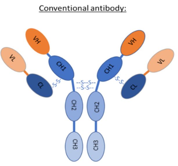
Pharmaceutics 2020, 12, 937 2 of 16
Pharmaceutics 2020, 12, x 2 of 16
either
an onpayload
active their own
intoorthe
bybrain.
delivering andescribe
We first active payload
VHHs andintoVNARs
the brain.
withWe first
their describe VHHs
characteristics and
making
VNARs
them withproteins.
unique their characteristics making
Then, we discuss thethem unique
different proteins. Then,
mechanisms VHHswe anddiscuss
VNARsthe candifferent
use to
mechanisms
reach VHHs
the brain. andwe
Finally, VNARs can
describe theuse
usetoofreach
brainthe brain. Finally,
penetrating sDAbsweas describe the diagnosis
therapeutic, use of brain
or
penetratingtools.
transporter sDAbs as therapeutic, diagnosis or transporter tools.
2.2. Single-Domain
Single-DomainAntibodies
Antibodiesand
andTheir
TheirProperties:
Properties: Variable
VariableDomain
Domainof
ofHeavy-Chain
Heavy-ChainAntibodies
Antibodies
(VHHs)
(VHHs)and andVariable
VariableNew
NewAntigen
AntigenReceptors
Receptors(VNARs)
(VNARs)
In
Inaddition
additiontotoconventional
conventionalantibodies,
antibodies,made
madeofoftwo
twoheavy
heavyand
andtwo
twolight
lightchains,
chains,camelids
camelidsand
and
sharks produce unusual antibodies composed only of heavy chains. Their variable
sharks produce unusual antibodies composed only of heavy chains. Their variable antigen-bindingantigen-binding
domain
domainisisformed
formedby byaasingle-domain.
single-domain.In InCamelidae,
Camelidae,ititisisdesignated
designatedbybyVHHs
VHHsfor forvariable
variabledomain
domainofof
heavy-chain
heavy-chainantibodies
antibodiesand in some
and cartilaginous
in some fishes
cartilaginous such such
fishes as sharks [10], it [10],
as sharks is designated by VNARs
it is designated by
for variable
VNARs for new antigen
variable newreceptor.
antigen receptor.
2.1. VHH
2.1. VHH
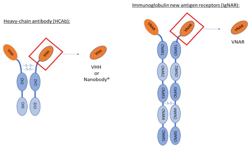
VHHs are the variable domains of heavy-chain antibodies (HCAb) found in Camelidae. This family
VHHs are the variable domains of heavy-chain antibodies (HCAb) found in Camelidae. This
is composed of Vicugna, Alpaca, Llama, Camel and Dromedary. As other mammals, they express
family is composed of Vicugna, Alpaca, Llama, Camel and Dromedary. As other mammals, they
conventional antibodies composed of two heavy chains and two light chains. In addition, they also
express conventional antibodies composed of two heavy chains and two light chains. In addition,
express non-conventional antibodies named heavy-chain antibodies devoid of CH1 domain and light
they also express non-conventional antibodies named heavy-chain antibodies devoid of CH1 domain
chain (Figure 1). HCAb are present for IgG2 and IgG3 isotypes. These antibodies are composed of two
and light chain (Figure 1). HCAb are present for IgG2 and IgG3 isotypes. These antibodies are
heavy chains divided in three domains each: CH3–CH2–VHH. The molecular weight of these HCAb is
composed of two heavy chains divided in three domains each: CH3–CH2–VHH. The molecular
90 kDa, smaller than the 150 kDa of conventional antibodies [11].
weight of these HCAb is 90 kDa, smaller than the 150 kDa of conventional antibodies [11].
Figure 1. Schematic representation of conventional antibody versus heavy-chain antibody found in
Figure 1. Schematic representation of conventional antibody versus heavy-chain antibody found in
Camelidae versus immunoglobulin new antigen receptor found in sharks. VHH is the variable
Camelidae versus immunoglobulin new antigen receptor found in sharks. VHH is the variable domain
domain of heavy-chain IgG2 and IgG3. The difference between these two IgG is the length of the
of heavy-chain IgG2 and IgG3. The difference between these two IgG is the length of the hinge (region
hinge (region between VHH and CH2). VNAR is the variable domain of IgNAR.
between VHH and CH2). VNAR is the variable domain of IgNAR.Pharmaceutics 2020, 12, 937 3 of 16
The variable domain of HCAb corresponding to the paratope recognizing the antigen is called
VHH. It is also named Nanobody® , a name registered by Ablynx, a Sanofi company working specifically
on these proteins (NANOBODY Trademark of ABLYNX N.V.–Registration Number 5098047–Serial
Number 85573029: Justia Trademarks Available online: http://trademarks.justia.com/855/73/nanobody-
85573029.html (accessed on 11 June 2020)). The mean molecular weight is 15 kDa, tenfold smaller than
a conventional antibody. This variable domain can be expressed on its own and still recognize the
antigen [12]. These proteins have specific properties making them unique tools. They have a high
identity rate with mouse and human variable domain of conventional antibodies heavy chain (VH)
around 80% [13], and, even if they come from a different species than the one treated, they are weakly
immunogenic in mice [14,15]. With this criterion, VHHs can be used in humans. In 2018, the first
VHH named caplacizumab was approved in Europe and then in the US. This VHH was developed
against von Willebrand factor and is available for patients with acquired thrombotic thrombocytopenic
purpura [16,17]. VHHs low immunogenicity has been evaluated with native VHHs. In this review, we
also deal with mutated or complexified VHHs that might have different immune responses that will
have to be checked before clinical use. Compared to conventional antibodies, VHHs are more stable
and can refold after denaturation [18]. They are also highly soluble owing to hydrophilic amino acids
mutated compared to VH [19]. Because of their small size, VHHs have a short half-life between 0.5 and
2 h [14,15] compared to several weeks for conventional antibodies [20]. To be used as therapeutics, they
can be linked to a life extension module [21,22]. The affinity is similar to antibodies, most of the time in
the nanomolar range [23]. In addition, VHHs can reach other epitopes than conventional antibodies.
For example, VHH can recognize cryptic epitopes [24] normally hidden from the immune system
and not recognized by immune cells in a normal process. VHH are easily produced in prokaryotic
or eukaryotic expression systems [25] and can be obtained in decent yield in Pichia Pastoris [26] or
Escherichia coli [27].
Their small size allows them to diffuse more than four times better in tissues [28,29] and
tumors [14] than antibodies. However, half-life-extension modules linked in some cases to the VHHs
might obliterate their small size benefit and negatively impact this diffusion and internalization.
Some VHHs have been shown to cross cell membranes and reach cytosolic targets. In this case, their
internalization has been linked to their basic isoelectric point and positive surface charges [30–33].
Even though the precise mechanism is still unknown, an adsorptive-mediated endocytosis could
be hypothesized. Brain penetration is going one step further than cell penetration as the molecule
must enter the endothelial cells forming the blood–brain barrier and then cross the membrane of the
abluminal endothelial cell surface to reach the brain parenchyma. This is the subject of this review.
2.2. VNAR
In 1995, heavy-chain antibodies (HCAb) were also discovered in sharks [34]. These antibodies are
composed of two heavy chains made of five constant domains (CNAR1, CNAR2, CNAR3, CNAR4
and CNAR5) (Figure 1). VNARs are the variable domain of these antibodies. As for VHHs, VNARs
bear full antigen recognition properties. The main difference of their variable domain is the absence
of complementary-determining region 2 (CDR2) leading to only two CDRs. Even though VNAR are
lacking one CDR, they can still recognize numerous antigens thanks to a higher variability in CDR1
and a longer CDR3 [35]. These characteristics make them the smallest antibodies with a molecular
weight of only 12 kDa.
VHHs and VNARs small sizes allow them to be good candidates as therapeutics, diagnostics
and transporters. Moreover, this small size allows them also to reach buried epitopes, facilitating
the discovery of mouse–human cross-species reactive sDAbs, a feature not always accessible with
conventional IgGs [36].Pharmaceutics 2020, 12, 937 4 of 16
3. Single-Domain Antibodies Crossing the Blood–Brain Barrier (BBB)
3.1. Mechanisms
Several mechanisms have been reported for VHHs that cross the BBB. These ways are either
direct by using endogenous brain transport mechanisms or indirect relying on BBB opening either in
disease-free or disease-dependent state.
3.1.1. Receptor-Mediated Transcytosis
The first mechanism used by some VHHs to go through the BBB is the same reported for some
conventional antibodies: receptor-mediated transcytosis (RMT). Receptors mostly used to deliver
conventional antibodies into the brain are transferrin and insulin receptors [37–39] but others such as
lipoprotein-related proteins [40,41] or IgF1 receptors have also been reported [42,43]. Fusions of insulin
and transferrin receptors antibodies to iduronidase lysosomal enzyme are currently evaluated in the
clinics against mucopolysaccharidosis [44,45]. The first VHHs found to perform receptor-mediated
transcytosis across the BBB are FC5 and FC44. These two VHHs discovered in 2001 were obtained
from a naive llama phage-displayed library followed by a panning on human endothelial cells forming
BBB [46]. The aim was to identify VHHs recognizing these cells and able to transmigrate across
them. Their brain uptakes have been quantified and shown to be more than 10 times higher than
controls [47]. The VHH FC5 internalization mechanism has been determined as a clathrin-dependent
receptor-mediated transcytosis [48]. The protein involved is the transmembrane domain protein 30 A
of α(2-3)-sialoglycoprotein and the use of this domain to obtain molecules for brain delivery has been
patented [49].
More recently, Stanimirovic et al. patented VHHs recognizing the insulin-like growth factor 1
receptor and transmigrating across the BBB by RMT [50]. The VHH name is insulin-like growth factor
1 receptor 5 (IGF1R5) and its transcytosis has been tested in vitro on a BBB model and in vivo on mice.
The VHH recognizes IGF1R at the luminal side of endothelial cells to transcytose across the cells and
be released in the brain environment on the abluminal side of the BBB. Its humanized and Fc-fusion
versions have shown an improved passage through the BBB. We show in the second part of this review
that this VHH can be used to deliver a cargo into the brain.
VHHs FC5 and IGF1R5 have been used as positive controls to validate the new human stem cell
BBB model described by Ribecco-Lutkiewicz et al. [51].
Several VNARs directed against transferrin receptor (TfR1) have also been designed by Ossianix,
to cross the BBB by receptor-mediated transcytosis. The two main VNARs developed against TfR1 are
B2 and TXB2. To increase their half-lives, they have been linked to Fc domains. B2-Fc [52] and TXB2-Fc
demonstrated up to 10–20-fold brain enhancement versus controls in PK studies. These sDAbs have
mostly been used as transporters of biotherapeutics into the brain demonstrating pharmacological
effects that are reviewed in the second part of this paper.
3.1.2. Adsorptive-Mediated Endocytosis
Two decades ago, positive charges addition at the protein surface was already reported as enabling
brain penetration [53]. This has been applied to IgGs after cationization by covalent coupling with
hexamethylenediamine or putrescine raising their isoelectric point largely above 7 favoring membrane
crossing through the process of adsorptive transcytosis [54]. One peculiar aspect of VHHs is probably
that, owing to their high content in exposed hydrophilic amino acids, they often appear to be produced
with largely basic isoelectric points. This is the case of VHH E9 [31] recognizing glial fibrillary
acidic protein (GFAP), an astrocyte marker, shown to cross the BBB after in vivo administration of a
fluorescent derivative. These results have been obtained on mice after intracarotid perfusion or lateral
tail vein injection with either VHH alone or VHH-eGFP. The VHH has been detected on astrocytes,
meaning that, after crossing the BBB, the VHH reaches cell cytosol and its target. The same team
has published another study in which two other VHHs cross the BBB [32]. Both VHHs have a basicPharmaceutics 2020, 12, 937 5 of 16
isoelectric point. The first VHH is directed against the peptide Aβ42 in amyloid plaques (VHH
R3VQ) and the other one against the phospho-tau (VHH TauA2). After intravenous injection with
10–20 mg/kg of VHH in PS2APP mice, in vivo two-photon microscopy revealed that amyloid plaques
and neurofibrillary tangles were stained. These two VHHs could be used in imaging Alzheimer’s
disease main lesions in a transgenic amyloid peptide precursor (APP) mouse model. A complementary
unpublished pharmacokinetics study has indicated that 2 h after intravenous injection, 0.5% of the
injected dose VHH TauA2 was found in the brain. Even though this percentage is higher than for
classical antibodies, which are found in the brain at 0.01–0.4% [55], this penetration rate remains low.
In addition, since VHHs half-lives are much shorter than the ones of conventional antibodies, higher or
more frequent dosing will be necessary to compensate these low rates. The precise mechanism has not
been determined but the importance of positive charges and basic isoelectric point has been noticed
such as for other VHH’s internalization [30,31]. Another anti-Aβ amyloid VHH carrying 18 positive
charges has been shown to cross the BBB using an active transport in an in vitro model [56].
3.1.3. Carrier-Mediated Transcytosis
Nanotechnologies have largely been reported to be able to cargo small molecules or
oligonucleotides into the brain if they are targeted to the brain by specific ligands [57] but examples
with antibodies are rare. The concept has been specifically applied to an anti-Aβ amyloid VHH after
encapsulation in liposomes decorated with glutathione (GSH) [58]. This tripeptide was believed to use
a specific carrier that was recently reported to be the N-Methyl-D-Aspartate receptor (NMDAR) [59].
This VHH pa2H encapsulated into liposomes was able to stain amyloid plaques into the brain after
intravenous administration. The brain exposure of the encapsulated VHH was improved compared to
the VHH alone from 0.001% of injected dose (ID) to 0.015% and 0.094% of ID in wild-type (WT) and
APP/PS1 transgenic mice, respectively [60]. GSH-liposome encapsulation also allowed decreasing the
clearance of the VHH from the blood.
3.1.4. BBB Opening
The two previous paragraphs report on sDAbs crossing the BBB by making use of endogenous
mechanisms by which the brain imports its proteins. This formidable challenge represented by BBB
can also be circumvented by other means. Low energy ultrasounds in parallel to injected microbubbles
have the potential to temporarily open the BBB [61]. Even though a few reports have applied the
technology to antibodies [62,63], we are not aware of application to sDAbs. Temporary BBB disruption
can also be induced by a few agents such as mannitol provoking an osmotic opening of the BBB which
can lead to increased brain exposure. This was already applied to Fabs and IgGs, showing that osmotic
BBB disruption significantly increased monoclonal antibody delivery to the brain [64]. However, the
extent of enhancement is modest. Osmotic disruption has been applied to the anti-gelsolin VHH
Nb11 [65] designed for the potential treatment of hereditary gelsolin amyloidosis, an autosomal
dominantly inherited amyloid disorder. PET imaging showed that mannitol BBB opening allowed an
increase of the VHH in the brain of around 2.5-fold, however the VHH was injected intraarterially [66].
This mode of administration has already demonstrated an advantage over intravenous administration
regarding brain exposure [67].
3.1.5. BBB Integrity Modified by Diseases
In some diseases such as in multiple sclerosis [68] or in cerebral malaria that is deadly [69], the
BBB can be altered. As with mannitol, this alteration might allow molecules to reach the brain. In
African trypanosomiasis late stage, also called sleeping sickness, Trypanosoma brucei parasite alters
the BBB and induces brain inflammation [70]. The VHH Nb-An33 recognizing a surface glycoprotein
of this parasite has been developed and tested on rat. The nanobody has been injected at 4 mg/kg in
normal rat versus rat with encephalitis comparable to late stage sleeping sickness. The VHH showed
capacity to reach the brain in both cases. However, in a rat brain from a late stage of encephalitis, VHHPharmaceutics 2020, 12, 937 6 of 16
found in hippocampal extracellular fluid was doubled compared to a normal rat brain. The VHH
concentration measured in the brain was 50 ± 21 ng/mL in control rat and 131 ± 63 ng/mL in the late
stage disease model [71], showing slightly better exposure linked to altered BBB.
3.1.6. Intranasal Delivery: Another Brain Delivery Option for VHH
Intranasal delivery of therapeutics involves spraying therapeutics into the upper part of the
nasal cavity to enable them to follow the olfactory axon bundles directly into the brain [72,73]. This
non-invasive, needle-free and painless method has already allowed brain exposure of a radiolabeled
IgG in rats [74]. It has also been used for VHH delivery into the brain. Ablynx has patented intranasal
delivery of therapeutic polypeptides and proteins including VHHs [75]. This strategy has been applied
to a VHH against transthyretin protein, used as a research tool, allowing a positive brain distribution
into several areas [76].
3.2. Single-Domain Antibodies as Therapeutics or Diagnostics in Central Nervous System (CNS) Diseases
Even though we show in the previous section that several sDAbs have demonstrated enhanced
brain exposure, no VHH or VNAR are presently in clinical development in a CNS application except
for one VHH currently in phase II development [77] in breast cancer brain metastasis. A preclinical
study had shown an increase of survival rate with this VHH CAM-H2 [78]. A phase I clinical study
has confirmed the safety of this radiolabeled VHH in patient, allowing its use as a therapeutic or as a
diagnostic tool [79]. An ongoing clinical trial aims at showing the use of this anti-HER2 VHH as a
diagnostic tool for human epidermal growth factor receptor 2 (HER2)-positive brain tumors [80]. This
might be enabled by local impairment of the BBB that can occur in brain cancers and in diseases such
as stroke, Parkinson’s disease, AD and multiple sclerosis even if the extent and duration of the leakage
is highly variable [81]. This can be the case also in some high-grade gliomas, where some degree of
parenchymal penetration of large molecules (such as antibodies) is likely in areas in which contrast
extravasation is detected by imaging, evidencing disruption of the BBB. However, this disruption is
heterogeneous and might mostly represent microscopic disease foci behind an intact BBB [82].
Several examples describing sDAbs used as either therapeutics, diagnostics, theranostics or
transporters are found in the literature and will be the object of the rest of this paper. We include in
this review only the examples where the sDabs designed for a CNS indication or imaging purpose
have also generated positive data after in vivo evaluation illustrating thereby that they have reached
the brain.
3.2.1. VHHs as Brain Therapeutic Tools
As rabies virus can target the brain leading to death-causing inflammation, a therapeutic molecule
able to reach the brain and neutralize the virus would be key. Conventional immunoglobulins are
currently used but an additional brain neutralizing effect would be desirable. VHHs recognizing
the glycoprotein of rabies virus have been developed and shown to be able to neutralize the virus
in vitro [83]. After extending their half-lives by coupling with an albumin binding VHH, they were
tested in vivo in mice. After preliminary validation of the effect upon direct brain injection, the
VHHs were tested by intraperitoneal injection. Compared to a conventional antibody that allowed a
prolonged survival of two days without rescue, these VHHs constructs allowed more than nine days
of prolonged survival with 43% of rescue. However, their brain exposure corresponded to 0.1% of
their plasma exposure, the same rate as for conventional antibodies. The authors concluded that this
difference could be due to the neutralizing power of the VHHs rather than their ability to cross the
BBB [84]. Even if the brain penetration could be optimized, this VHH could be a potential therapy for
rabies disease.
The VHH PrioV3, designed to target brain misfolded prion protein causing prion disease, has
been reported to cross the BBB in vitro and in vivo. After crossing the BBB, this VHH was able toPharmaceutics 2020, 12, 937 7 of 16
prevent prion replication by reaching neuron cytosol [85]. The mechanism of BBB crossing involves
prion protein at the surface cell membrane and clathrins [86].
3.2.2. VHHs as Brain Diagnostic Tools
In addition to a therapeutic use, VHHs reaching the brain can also be used as diagnostic tools.
Rutgers et al. [87] developed, from immune and non-immune libraries, VHHs recognizing either
parenchymal or vascular beta amyloid. They were evaluated to image brain lesions of Alzheimer’s
disease. Among the VHHs obtained, ni3A was actively passing through an in vitro model of BBB [56].
The BBB passage was further evaluated in vivo for VHH ni3A and pa2H obtained from the first
screening. The VHHs were radiolabeled with two different labels (111 In and 99m Tc) and their brain
uptakes monitored. 99m Tc-labeled VHH ni3A and pa2H crossed the BBB and were quantified in the
cerebellum at 0.053% ID/g and 0.04% ID/g, respectively. Even if 99m Tc-VHH allowed a quantification
into the brain, the rate was too low for imaging [88]. 111 In-pa2H quantification did not differ from the
control with ~0.001% ID/g. A better knowledge of transport mechanism of these VHH into the brain
would allow an optimization of BBB crossing to use as a diagnostic tool. The VHH pa2H has been
engineered with a Fc domain to increase half-life and decrease blood clearance. This new construction
allowed an increase half-life but did not improve brain uptake keeping the quantified VHH at 0.001%
ID/g [89]. However, the 111 In radiolabel that showed less brain uptake than 99m Tc in the previous
in vivo study was used. To improve brain exposure, the VHH was encapsulated into GSH-decorated
liposomes which led to increased exposure up to 0.025% ID [60]. Even though the rate of BBB passage
is not high, ni3A and pa2H could be used to image amyloid plaque as it has been shown on APP/PS1
amyloid transgenic mice [88].
VHH E9 and R3VQ, described previously as brain penetrant could also be used as a diagnosis
marker of Alzheimer’s disease as they recognize its main lesions [32]. VHH R3VQ recognizing
amyloid plaques has been coupled to gadolinium, a magnetic resonance imaging (MRI) contrast agent.
Immunohistochemistry showed positive staining of amyloid plaques validating the BBB passage [90].
Even if MRI visualization of amyloid plaques was obtained in vitro, no labeling of plaques was obtained
in vivo on live mice, suggesting that the amount of VHH in the brain was too low. However, these
results are promising for a potential use of R3VQ for amyloid plaques detection on patients.
A nanobody directed against vascular endothelium growth factor (VEGF) has shown a BBB
passage optimized by mannitol treatment and intra-arterial administration instead of intravenous
injection. The improved brain uptake allowed imaging of the brain by positron electron tomography
(PET) using 89 Zr as radioactive tracer [66].
The brain can also be imaged in the case of brain cancer. Iqbal et al. [91] used a VHH against
EGFR to image glioblastoma in the brain. Brain images are better when VHH named EG2 was coupled
to an Fc domain.
3.2.3. VHHs as Brain Theranostic Tools
We have previously seen that VHHs can be used as therapeutic and diagnostic tools. We now
discuss the potential of VHHs as theranostic tools, a combination of both diagnostic and therapeutic
purposes. Puttermans et al. [92] described this option with VHH 2Rs15d targeting human epidermal
growth factor receptor 2 (HER2). The overexpression of this protein is associated with several cancers
such as breast cancer [91]. The VHH has been coupled with three different radionuclides: 111 In, 225 Ac
and 131 I. Each radionuclide coupled to VHH was tested one by one or in combination and the impact
on survival was evaluated and compared to trastuzumab, a conventional antibody targeting HER2
and already approved for HER2-positive cancer treatment. This VHH could reach the brain, recognize
and detect HER2-positive brain lesions caused by breast cancer metastases and improve the survival
rate by killing tumor cells.Pharmaceutics 2020, 12, 937 8 of 16
3.2.4. VHHs and VNARs as Transporters for Therapeutic Molecules
VHHs or VNARs passing through the BBB by RMT could successfully be used as transporters.
FC5 was first used for brain delivery of an anti-cancer drug doxorubicin, that cannot reach the brain
Pharmaceutics 2020, 12, x
on
8 of 16
its own because of efflux pumps [93]. The VHH was either used as a monomer (FC5) or as a pentamer
(named
pentamerP5)(named
[94]. VHH P5) FC5
[94]. was
VHH used
FC5towas
decorate liposomes
used to decorate encapsulating doxorubicin,
liposomes encapsulating as shown inas
doxorubicin,
Figure 2. After intravenous injection of 6 mg/kg, doxorubicin was quantified in brain
shown in Figure 2. After intravenous injection of 6 mg/kg, doxorubicin was quantified in brain parenchyma.
The functionalization
parenchyma. of the liposomeof
The functionalization modestly increased
the liposome brain uptake
modestly of doxorubicin
increased brain uptakeoriginally from
of doxorubicin
100 ng/g of tissue to 200 and 350 ng/g with FC5 and P5, respectively [95].
originally from 100 ng/g of tissue to 200 and 350 ng/g with FC5 and P5, respectively [95].
Coating VHH:
VHH
FC5, P5, VCAMelid…
VH H
VH
H
H
VH
H VH
Heterogenous
lipid bilayer
VHH Drug
VHH
H Encapsulated active molecule:
VH
VH
H drugs (doxorubicin…)
or enzymes (SOD-1…)
H
VH
VH H
Figure2.2.Encapsulated
Figure active
Encapsulated molecule
active (drug(drug
molecule or enzyme) into VHH-coated
or enzyme) liposomes
into VHH-coated for brain delivery.
liposomes for brain
delivery.
Farrington et al. [96,97] also showed that FC5 can deliver a cargo such as dalargin into the brain
by using differentetconstructions.
Farrington al. [96,97] alsoThey showed have reported
that FC5 can that FC5 alone
deliver a cargoor such
coupled to an Fcinto
as dalargin domain can
the brain
deliver peptides leading to more than 30-fold improvement compared to
by using different constructions. They have reported that FC5 alone or coupled to an Fc domain can the control without VHH. In
these constructions, the Fc domain allowed an increase of half-life leading
deliver peptides leading to more than 30-fold improvement compared to the control without VHH. to an improvement of VHH
brain delivery,
In these thus of the
constructions, thepeptide.
Fc domain allowed an increase of half-life leading to an improvement of
VHH FC5 could also
VHH brain delivery, thus of the be dimerized
peptide.with a conventional antibody such as an anti mGlutR1, a
potential
VHH target
FC5receptor
could also found in the CNS.with
be dimerized Thisaled to an increase
conventional of brain
antibody delivery
such as an in vivo
anti of abouta
mGlutR1,
20-fold, validating
potential again the
target receptor possibility
found for FC5
in the CNS. Thisto led
deliver
to ana increase
cargo into of the brain
brain [98]. in vivo of about
delivery
Stanimirovic
20-fold, validating et again
al. [50]thealso illustratedfor
possibility theFC5
potential of the
to deliver VHH into
a cargo IGF1R5the as a transporter
brain [98]. of galanin.
This neuroactive
Stanimirovic peptide
et al. can
[50]induce analgesia after
also illustrated binding to
the potential ofits
thereceptor in the brain.
VHH IGF1R5 as a However,
transporter it isof
inactive when given by peripheral route as it does not penetrate the BBB
galanin. This neuroactive peptide can induce analgesia after binding to its receptor in the brain.on its own. The analgesic effect
was obtained
However, it iswhen galanin
inactive when was coupled
given to IGF1R5
by peripheral and as
route injected
it doesinto
not apenetrate
hyperalgesia inflammatory
the BBB on its own.
rat
Themodel.
analgesic effect was obtained when galanin was coupled to IGF1R5 and injected into a
A VHH against
hyperalgesia vascular
inflammatory rat cell adhesion molecule 1 (VCAM1) named VHH against VCAM1
model.
(VCAMelid)
A VHHhas recently
against shown cell
vascular its ability
adhesion to deliver
molecule SOD-1 enzyme into
1 (VCAM1) namedthe brain. This delivery
VHH against VCAM1
was either by has
(VCAMelid) a direct coupling
recently shown toits
VHH or by
ability enzymeSOD-1
to deliver encapsulation
enzyme into on VHH-decorated liposomes.
the brain. This delivery was
The liposomes delivery system seems to be analogous to the FC5
either by a direct coupling to VHH or by enzyme encapsulation on VHH-decorated liposomes. The liposomes presented previously
(Figure 2). VCAMelid
liposomes delivery systemwas reported
seems totobe reach the brain
analogous on its
to the FC5own and allowed
liposomes brainpreviously
presented delivery of(Figure
SOD1
both in naive and
2). VCAMelid wasinflammatory
reported to reachmice model.
the brain Brain exposure
on its own and of VCAMelid
allowed brain wasdelivery
higher in ofthe disease
SOD1 both
model of mice with local injury compared to naive mice with brain uptake
in naive and inflammatory mice model. Brain exposure of VCAMelid was higher in the disease model alone at around 0.8% ID/g
and 0.2%with
of mice ID/g,local
respectively.
injury comparedThere was also amice
to naive difference between
with brain the alone
uptake conjugated
at around construct and the
0.8% ID/g and
liposomal formulation with SOD1 brain uptake in injured mice, of
0.2% ID/g, respectively. There was also a difference between the conjugated construct and the around 1.2% ID/g and 2% ID/g,
respectively [99]. These brain
liposomal formulation uptakebrain
with SOD1 constructions
uptake inare the most
injured mice,efficient found
of around andID/g
1.2% described
and 2% in ID/g,
this
review with a[99].
respectively greatThese
difference
braincompared to the control
uptake constructions arewhich
the mostis around 0.1%
efficient foundID/g.and described in this
review with a great difference compared to the control which is around 0.1% ID/g.
Recently, a new liposome construction was designed for brain delivery of anti-cancer drugs
targeting brain metastases. The liposomes were decorated with TfR-binding peptides and anti-
program death-ligand 1 (PDL1) VHHs. It has been shown that brain delivery of the liposome-
encapsulated simvastatin/gefitinib drug combination was driven by both targets (TfR and PDL1)
present on endothelial cells of BBB bearing metastases. With both anti-TfR peptide and anti-PDL1Pharmaceutics 2020, 12, 937 9 of 16
Recently, a new liposome construction was designed for brain delivery of anti-cancer drugs
targeting brain metastases. The liposomes were decorated with TfR-binding peptides and anti-program
death-ligand 1 (PDL1) VHHs. It has been shown that brain delivery of the liposome-encapsulated
simvastatin/gefitinib drug combination was driven by both targets (TfR and PDL1) present on
endothelial cells of BBB bearing metastases. With both anti-TfR peptide and anti-PDL1 VHHs
these liposomes allowed brain delivery reducing brain tumor and improving survival [100]. The
liposomes solely targeted with anti-PDL1 nanobody mainly showed macrophages targeting allowing
cell internalization of the liposome but were not efficient enough without the anti-TfR peptide to reach
the brain [101].
Prehaud et al. patented a basic VHH with no identified brain target for which in vitro experiments
suggest a potential to cargo several different effectors such as neuroprotective peptides into the brain
justifying to protect their use for brain transport [102,103].
Recently, Vect-Horus, a biotechnology company based in Marseille in France working on designing
vectors to go through barriers, has patented VHHs against transferrin receptors. These VHHs display
comparable affinity to human and rodent TfR and could be useful for shuttling therapeutics or imaging
agents into the CNS [104].
Ossianix is reporting preclinical research in several fields of CNS such as pain, multiple sclerosis,
CNS lymphoma or glioblastoma with fusions of their VNARs with several therapeutic proteins. The
VNAR named B2 described by Wicher et al. [52] has shown its ability to deliver rituximab, an anti-CD20
antibody for peripheral B cell lymphoma treatment, into the brain [105]. Brain exposure was more than
ten times higher than the naked antibody. The TXB2 VNAR was also fused to the neurotensin peptide
demonstrating a rapid, sustained CNS exposure and robust pharmacological activity after injection of
1.875 mg/kg in mice [36]. On the other hand, while TBX2-VNAR-Fc fusion demonstrated 20-fold brain
exposure improvement vs. control VNAR-Fc at 18 h post-injection in wild-type (WT) mice, when the
TXB2-VNAR was fused to the N-terminal light chains of the anti-amyloid antibody bapinezumab, the
brain exposure enhancement was three-fold at the same time and up to six days after injection [106].
PET imaging and autoradiography evidenced more parenchymal Bapi-TBX2 compared to Bapi. It has
to be noted that, in this case, because of Fc-fusion domain, the benefit of the VHHs small size is lost
and their brain penetration mostly relies on the transferrin receptor-mediated mechanism.
4. Conclusions
In this review, we describe VHHs and VNARs that can reach the brain directly (RMT and AMT) or
indirectly (brain targeted liposomes, BBB-opening and disease). We try to provide the best overview of
already used VHHs and VNARs into the brain. Although several others have been developed against
brain targets, their brain exposure has not been demonstrated or quantified. All these delivery systems
using either VHHs or VNARs can be used for brain targeting of active principles that could lead to
promising new treatments for diseases with no current treatment.
Author Contributions: Writing—original draft preparation, E.P.; writing—review and editing, D.L. and P.L.
All authors have read and agreed to the published version of the manuscript.
Funding: This research received no external funding.
Conflicts of Interest: There is no conflict of interest. SANOFI had no role in the design of the study; in the
collection, analyses, or interpretation of data; in the writing of the manuscript, or in the decision to publish
the results.Pharmaceutics 2020, 12, 937 10 of 16
Abbreviations
% ID/cc percentage of injected dose per cubic centimeter of tissue
% ID/g percentage of injected dose per gram of tissue
225 Ac Actinium-225
APP amyloid peptide precursor
BBB blood–brain barrier
BTB brain-tumor barrier
CDR complementary-determining region
CH1/CH2/CH3 constant domain 1/2/3 of heavy chain
CNS central nervous system
GFAP glial fibrillary acidic protein
GFP green fluorescence protein
HCAb heavy-chain antibody
HER human epidermal growth factor receptor
131 I Iodine-131
ID injected dose
IGF1R insulin-like growth factor 1 receptor
IgG immunoglobulin G
111 In Indium-111
NMDAR N-Methyl-D-Aspartate receptor
PDL1 program death-ligand 1
PET PET
RMT receptor-mediated transcytosis
sDAb single-domain antibody
99m Tc Technetium-99m
TfR transferrin receptor
VCAM1 vascular cell adhesion molecule 1
VCAMelid VHH against VCAM1
VEGF vascular endothelium growth factor
VH variable domain of conventional antibodies heavy chain
VL variable domain of conventional antibodies light chain
VHH variable domain of heavy-chain antibodies
VHHs plural of VHH
VNAR variable new antigen receptor
WT wild-type
89 Zr Zirconium 89
References
1. Robinson, M.K.; Doss, M.; Shaller, C.; Narayanan, D.; Marks, J.D.; Adler, L.P.; Gonzalez Trotter, D.E.;
Adams, G.P. Quantitative immuno-positron emission tomography imaging of HER2-positive tumor
xenografts with an iodine-124 labeled anti-HER2 diabody. Cancer Res. 2005, 65, 1471–1478. [CrossRef]
2. Arvanitis, C.D.; Ferraro, G.B.; Jain, R.K. The blood-brain barrier and blood-tumour barrier in brain tumours
and metastases. Nat. Rev. Cancer 2020, 20, 26–41. [CrossRef] [PubMed]
3. Kumar, N.N.; Pizzo, M.E.; Nehra, G.; Wilken-Resman, B.; Boroumand, S.; Thorne, R.G. Passive
Immunotherapies for Central Nervous System Disorders: Current Delivery Challenges and New Approaches.
Bioconjug. Chem. 2018, 29, 3937–3966. [CrossRef] [PubMed]
4. Banks, W.A. From blood-brain barrier to blood-brain interface: New opportunities for CNS drug delivery.
Nat. Rev. Drug Discov. 2016, 15, 275–292. [CrossRef] [PubMed]
5. Obermeier, B.; Daneman, R.; Ransohoff, R.M. Development, maintenance and disruption of the blood-brain
barrier. Nat. Med. 2013, 19, 1584–1596. [CrossRef]
6. Mantle, J.L.; Lee, K.H. Immunoglobulin G transport increases in an in vitro blood-brain barrier model with
amyloid-beta and with neuroinflammatory cytokines. Biotechnol. Bioeng. 2019, 116, 1752–1761. [CrossRef]Pharmaceutics 2020, 12, 937 11 of 16
7. Abdul Razzak, R.; Florence, G.J.; Gunn-Moore, F.J. Approaches to CNS Drug Delivery with a Focus on
Transporter-Mediated Transcytosis. Int. J. Mol. Sci. 2019, 20, 3108. [CrossRef]
8. Belanger, K.; Iqbal, U.; Tanha, J.; MacKenzie, R.; Moreno, M.; Stanimirovic, D. Single-Domain Antibodies as
Therapeutic and Imaging Agents for the Treatment of CNS Diseases. Antibodies 2019, 8, 27. [CrossRef]
9. Messer, A.; Butler, D.C. Optimizing intracellular antibodies (intrabodies/nanobodies) to treat
neurodegenerative disorders. Neurobiol. Dis. 2020, 134, 104619. [CrossRef]
10. Wesolowski, J.; Alzogaray, V.; Reyelt, J.; Unger, M.; Juarez, K.; Urrutia, M.; Cauerhff, A.; Danquah, W.;
Rissiek, B.; Scheuplein, F.; et al. Single domain antibodies: Promising experimental and therapeutic tools in
infection and immunity. Med. Microbiol. Immunol. 2009, 198, 157–174. [CrossRef]
11. Hamers-Casterman, C.; Atarhouch, T.; Muyldermans, S.; Robinson, G.; Hamers, C.; Songa, E.B.;
Bendahman, N.; Hamers, R. Naturally occurring antibodies devoid of light chains. Nature 1993, 363,
446–448. [CrossRef] [PubMed]
12. Arbabi Ghahroudi, M.; Desmyter, A.; Wyns, L.; Hamers, R.; Muyldermans, S. Selection and identification
of single domain antibody fragments from camel heavy-chain antibodies. FEBS Lett. 1997, 414, 521–526.
[CrossRef]
13. Vu, K.B.; Ghahroudi, M.A.; Wyns, L.; Muyldermans, S. Comparison of llama VH sequences from conventional
and heavy chain antibodies. Mol. Immunol. 1997, 34, 1121–1131. [CrossRef]
14. Cortez-Retamozo, V.; Lauwereys, M.; Hassanzadeh, G.; Gobert, M.; Conrath, K.; Muyldermans, S.; De
Baetselier, P.; Revets, H. Efficient tumor targeting by single-domain antibody fragments of camels. Int. J.
Cancer 2002, 98, 456–462. [CrossRef]
15. Coppieters, K.; Dreier, T.; Silence, K.; de Haard, H.; Lauwereys, M.; Casteels, P.; Beirnaert, E.; Jonckheere, H.;
Van de Wiele, C.; Staelens, L.; et al. Formatted anti-tumor necrosis factor alpha VHH proteins derived from
camelids show superior potency and targeting to inflamed joints in a murine model of collagen-induced
arthritis. Arthritis Rheum. 2006, 54, 1856–1866. [CrossRef]
16. Hanlon, A.; Metjian, A. Caplacizumab in adult patients with acquired thrombotic thrombocytopenic purpura.
Adv. Hematol. 2020, 11, 2040620720902904. [CrossRef]
17. Peyvandi, F.; Scully, M.; Kremer Hovinga, J.A.; Knobl, P.; Cataland, S.; De Beuf, K.; Callewaert, F.; De
Winter, H.; Zeldin, R.K. Caplacizumab reduces the frequency of major thromboembolic events, exacerbations
and death in patients with acquired thrombotic thrombocytopenic purpura. J. Thromb. Haemost. 2017, 15,
1448–1452. [CrossRef]
18. Perez, J.M.; Renisio, J.G.; Prompers, J.J.; van Platerink, C.J.; Cambillau, C.; Darbon, H.; Frenken, L.G. Thermal
unfolding of a llama antibody fragment: A two-state reversible process. Biochemistry 2001, 40, 74–83.
[CrossRef]
19. Conrath, K.; Vincke, C.; Stijlemans, B.; Schymkowitz, J.; Decanniere, K.; Wyns, L.; Muyldermans, S.; Loris, R.
Antigen binding and solubility effects upon the veneering of a camel VHH in framework-2 to mimic a VH. J.
Mol. Biol. 2005, 350, 112–125. [CrossRef]
20. Paul, P.S.; Mengeling, W.L.; Pirtle, E.C. Duration and biological half-life of passively acquired colostral
antibodies to porcine parvovirus. Am. J. Vet. Res. 1982, 43, 1376–1379.
21. Harmsen, M.M.; Van Solt, C.B.; Fijten, H.P.; Van Setten, M.C. Prolonged in vivo residence times of llama
single-domain antibody fragments in pigs by binding to porcine immunoglobulins. Vaccine 2005, 23,
4926–4934. [CrossRef] [PubMed]
22. Holt, L.J.; Basran, A.; Jones, K.; Chorlton, J.; Jespers, L.S.; Brewis, N.D.; Tomlinson, I.M. Anti-serum albumin
domain antibodies for extending the half-lives of short lived drugs. Protein Eng. Des. Sel. 2008, 21, 283–288.
[CrossRef] [PubMed]
23. van der Linden, R.H.; Frenken, L.G.; de Geus, B.; Harmsen, M.M.; Ruuls, R.C.; Stok, W.; de Ron, L.; Wilson, S.;
Davis, P.; Verrips, C.T. Comparison of physical chemical properties of llama VHH antibody fragments and
mouse monoclonal antibodies. Biochim. Biophys. Acta 1999, 1431, 37–46. [CrossRef]
24. Stijlemans, B.; Conrath, K.; Cortez-Retamozo, V.; Van Xong, H.; Wyns, L.; Senter, P.; Revets, H.; De Baetselier, P.;
Muyldermans, S.; Magez, S. Efficient targeting of conserved cryptic epitopes of infectious agents by single
domain antibodies. African trypanosomes as paradigm. J. Biol. Chem. 2004, 279, 1256–1261. [CrossRef]
[PubMed]
25. de Marco, A. Recombinant expression of nanobodies and nanobody-derived immunoreagents. Protein Expr.
Purif. 2020, 172, 105645. [CrossRef] [PubMed]Pharmaceutics 2020, 12, 937 12 of 16
26. Chen, Q.; Zhou, Y.; Yu, J.; Liu, W.; Li, F.; Xian, M.; Nian, R.; Song, H.; Feng, D. An efficient constitutive
expression system for Anti-CEACAM5 nanobody production in the yeast Pichia pastoris. Protein Expr. Purif.
2019, 155, 43–47. [CrossRef] [PubMed]
27. Zarschler, K.; Witecy, S.; Kapplusch, F.; Foerster, C.; Stephan, H. High-yield production of functional soluble
single-domain antibodies in the cytoplasm of Escherichia coli. Microb. Cell Fact. 2013, 12, 97. [CrossRef]
28. Perruchini, C.; Pecorari, F.; Bourgeois, J.P.; Duyckaerts, C.; Rougeon, F.; Lafaye, P. Llama VHH antibody
fragments against GFAP: Better diffusion in fixed tissues than classical monoclonal antibodies. Acta
Neuropathol. 2009, 118, 685–695. [CrossRef]
29. Pizzo, M.E.; Wolak, D.J.; Kumar, N.N.; Brunette, E.; Brunnquell, C.L.; Hannocks, M.J.; Abbott, N.J.;
Meyerand, M.E.; Sorokin, L.; Stanimirovic, D.B.; et al. Intrathecal antibody distribution in the rat brain:
Surface diffusion, perivascular transport and osmotic enhancement of delivery. J. Physiol. 2018, 596, 445–475.
[CrossRef]
30. Bruce, V.J.; Lopez-Islas, M.; McNaughton, B.R. Resurfaced cell-penetrating nanobodies: A potentially general
scaffold for intracellularly targeted protein discovery. Protein Sci. 2016, 25, 1129–1137. [CrossRef]
31. Li, T.; Bourgeois, J.P.; Celli, S.; Glacial, F.; Le Sourd, A.M.; Mecheri, S.; Weksler, B.; Romero, I.; Couraud, P.O.;
Rougeon, F.; et al. Cell-penetrating anti-GFAP VHH and corresponding fluorescent fusion protein VHH-GFP
spontaneously cross the blood-brain barrier and specifically recognize astrocytes: Application to brain
imaging. FASEB J. 2012, 26, 3969–3979. [CrossRef]
32. Li, T.; Vandesquille, M.; Koukouli, F.; Dudeffant, C.; Youssef, I.; Lenormand, P.; Ganneau, C.; Maskos, U.;
Czech, C.; Grueninger, F.; et al. Camelid single-domain antibodies: A versatile tool for in vivo imaging of
extracellular and intracellular brain targets. J. Control. Release 2016, 243, 1–10. [CrossRef] [PubMed]
33. Ruano-Salguero, J.S.; Lee, K.H. Antibody transcytosis across brain endothelial-like cells occurs nonspecifically
and independent of FcRn. Sci. Rep. 2020, 10, 3685. [CrossRef] [PubMed]
34. Greenberg, A.S.; Avila, D.; Hughes, M.; Hughes, A.; McKinney, E.C.; Flajnik, M.F. A new antigen receptor
gene family that undergoes rearrangement and extensive somatic diversification in sharks. Nature 1995, 374,
168–173. [CrossRef]
35. Cheong, W.S.; Leow, C.Y.; Abdul Majeed, A.B.; Leow, C.H. Diagnostic and therapeutic potential of shark
variable new antigen receptor (VNAR) single domain antibody. Int. J. Biol. Macromol. 2020, 147, 369–375.
[CrossRef] [PubMed]
36. Stocki, P.; Szary, J.M.; Jacobsen, C.L.M.; Demydchuk, M.; Northall, L.; Moos, T.; Walsh, F.S.; Rutkowski, J.L.
High efficiency blood-brain barrier transport using a VNAR targeting the Transferrin Receptor. BioRxiv 2019.
[CrossRef]
37. Pardridge, W.M.; Kang, Y.S.; Buciak, J.L.; Yang, J. Human insulin receptor monoclonal antibody undergoes
high affinity binding to human brain capillaries in vitro and rapid transcytosis through the blood-brain
barrier in vivo in the primate. Pharm. Res. 1995, 12, 807–816. [CrossRef]
38. Niewoehner, J.; Bohrmann, B.; Collin, L.; Urich, E.; Sade, H.; Maier, P.; Rueger, P.; Stracke, J.O.; Lau, W.;
Tissot, A.C.; et al. Increased brain penetration and potency of a therapeutic antibody using a monovalent
molecular shuttle. Neuron 2014, 81, 49–60. [CrossRef]
39. Yu, Y.J.; Atwal, J.K.; Zhang, Y.; Tong, R.K.; Wildsmith, K.R.; Tan, C.; Bien-Ly, N.; Hersom, M.; Maloney, J.A.;
Meilandt, W.J.; et al. Therapeutic bispecific antibodies cross the blood-brain barrier in nonhuman primates.
Sci. Transl. Med. 2014, 6, 261ra154. [CrossRef]
40. Nounou, M.I.; Adkins, C.E.; Rubinchik, E.; Terrell-Hall, T.B.; Afroz, M.; Vitalis, T.; Gabathuler, R.; Tian, M.M.;
Lockman, P.R. Anti-cancer Antibody Trastuzumab-Melanotransferrin Conjugate (BT2111) for the Treatment
of Metastatic HER2+ Breast Cancer Tumors in the Brain: An In-Vivo Study. Pharm. Res. 2016, 33, 2930–2942.
[CrossRef]
41. Regina, A.; Demeule, M.; Che, C.; Lavallee, I.; Poirier, J.; Gabathuler, R.; Beliveau, R.; Castaigne, J.P.
Antitumour activity of ANG1005, a conjugate between paclitaxel and the new brain delivery vector
Angiopep-2. Br. J. Pharm. 2008, 155, 185–197. [CrossRef] [PubMed]
42. Stanimirovic, D.; Kemmerich, K.; Haqqani, A.S.; Farrington, G.K. Engineering and pharmacology of
blood-brain barrier-permeable bispecific antibodies. Adv. Pharm. 2014, 71, 301–335. [CrossRef]
43. Stanimirovic, D.B.; Sandhu, J.K.; Costain, W.J. Emerging Technologies for Delivery of Biotherapeutics and
Gene Therapy Across the Blood-Brain Barrier. BioDrugs 2018, 32, 547–559. [CrossRef] [PubMed]Pharmaceutics 2020, 12, 937 13 of 16
44. Pardridge, W.M.; Boado, R.J.; Giugliani, R.; Schmidt, M. Plasma Pharmacokinetics of Valanafusp Alpha, a
Human Insulin Receptor Antibody-Iduronidase Fusion Protein, in Patients with Mucopolysaccharidosis
Type I. BioDrugs 2018, 32, 169–176. [CrossRef]
45. Sonoda, H.; Morimoto, H.; Yoden, E.; Koshimura, Y.; Kinoshita, M.; Golovina, G.; Takagi, H.; Yamamoto, R.;
Minami, K.; Mizoguchi, A.; et al. A Blood-Brain-Barrier-Penetrating Anti-human Transferrin Receptor
Antibody Fusion Protein for Neuronopathic Mucopolysaccharidosis II. Mol. Ther. 2018, 26, 1366–1374.
[CrossRef]
46. Muruganandam, A.; Tanha, J.; Narang, S.; Stanimirovic, D. Selection of phage-displayed llama single-domain
antibodies that transmigrate across human blood-brain barrier endothelium. FASEB J. 2002, 16, 240–242.
[CrossRef]
47. Haqqani, A.S.; Caram-Salas, N.; Ding, W.; Brunette, E.; Delaney, C.E.; Baumann, E.; Boileau, E.; Stanimirovic, D.
Multiplexed evaluation of serum and CSF pharmacokinetics of brain-targeting single-domain antibodies
using a NanoLC-SRM-ILIS method. Mol. Pharm. 2013, 10, 1542–1556. [CrossRef]
48. Abulrob, A.; Sprong, H.; Van Bergen en Henegouwen, P.; Stanimirovic, D. The blood-brain barrier
transmigrating single domain antibody: Mechanisms of transport and antigenic epitopes in human brain
endothelial cells. J. Neurochem. 2005, 95, 1201–1214. [CrossRef]
49. Abulrob, A.S.D.; Muruganandam, A. Blood-Brain Barrier Epitopes and Uses Thereof. Patent WO 2007/036021,
5 April 2007.
50. Stanimirovic, D.K.; Kemmerich, K.; Haqqani, A.S.; Sulea, T.; Arbabi-Ghahroudi, M.; Massie, B.; Gilbert, R.
Insulin-Like Growth Factor 1 Receptor-Specific Antibodies and Uses Thereof. U.S. Patent US20170015748A1,
19 January 2017.
51. Ribecco-Lutkiewicz, M.; Sodja, C.; Haukenfrers, J.; Haqqani, A.S.; Ly, D.; Zachar, P.; Baumann, E.; Ball, M.;
Huang, J.; Rukhlova, M.; et al. A novel human induced pluripotent stem cell blood-brain barrier model:
Applicability to study antibody-triggered receptor-mediated transcytosis. Sci. Rep. 2018, 8, 1873. [CrossRef]
52. Greenwood, J.; Hammarlund-Udenaes, M.; Jones, H.C.; Stitt, A.W.; Vandenbroucke, R.E.; Romero, I.A.;
Campbell, M.; Fricker, G.; Brodin, B.; Manninga, H.; et al. Current research into brain barriers and the
delivery of therapeutics for neurological diseases: A report on CNS barrier congress London, UK, 2017.
Fluids Barriers CNS 2017, 14, 31. [CrossRef]
53. Herve, F.; Ghinea, N.; Scherrmann, J.M. CNS delivery via adsorptive transcytosis. AAPS J. 2008, 10, 455–472.
[CrossRef] [PubMed]
54. Triguero, D.; Buciak, J.; Pardridge, W.M. Capillary depletion method for quantification of blood-brain barrier
transport of circulating peptides and plasma proteins. J. Neurochem. 1990, 54, 1882–1888. [CrossRef]
55. Pepinsky, R.B.; Shao, Z.; Ji, B.; Wang, Q.; Meng, G.; Walus, L.; Lee, X.; Hu, Y.; Graff, C.; Garber, E.; et al.
Exposure levels of anti-LINGO-1 Li81 antibody in the central nervous system and dose-efficacy relationships
in rat spinal cord remyelination models after systemic administration. J. Pharm. Exp. 2011, 339, 519–529.
[CrossRef] [PubMed]
56. Rutgers, K.S.; Nabuurs, R.J.; van den Berg, S.A.; Schenk, G.J.; Rotman, M.; Verrips, C.T.; van Duinen, S.G.;
Maat-Schieman, M.L.; van Buchem, M.A.; de Boer, A.G.; et al. Transmigration of beta amyloid specific heavy
chain antibody fragments across the in vitro blood-brain barrier. Neuroscience 2011, 190, 37–42. [CrossRef]
57. Cramer, S.; Rempe, R.; Galla, H.J. Exploiting the properties of biomolecules for brain targeting of
nanoparticulate systems. Curr. Med. Chem. 2012, 19, 3163–3187. [CrossRef]
58. Gaillard, P.J.; Appeldoorn, C.C.; Dorland, R.; van Kregten, J.; Manca, F.; Vugts, D.J.; Windhorst, B.; van
Dongen, G.A.; de Vries, H.E.; Maussang, D.; et al. Pharmacokinetics, brain delivery, and efficacy in brain
tumor-bearing mice of glutathione pegylated liposomal doxorubicin (2B3-101). PLoS ONE 2014, 9, e82331.
[CrossRef] [PubMed]
59. Fatima, S.; Mohammad, T.; Jairajpuri, D.S.; Rehman, M.T.; Hussain, A.; Samim, M.; Ahmad, F.J.; Alajmi, M.F.;
Hassan, M.I. Identification and evaluation of glutathione conjugate gamma-l-glutamyl-l-cysteine for improved
drug delivery to the brain. J. Biomol. Struct. Dyn. 2019, 38, 3610–3620. [CrossRef] [PubMed]
60. Rotman, M.; Welling, M.M.; Bunschoten, A.; de Backer, M.E.; Rip, J.; Nabuurs, R.J.; Gaillard, P.J.; van
Buchem, M.A.; van der Maarel, S.M.; van der Weerd, L. Enhanced glutathione PEGylated liposomal brain
delivery of an anti-amyloid single domain antibody fragment in a mouse model for Alzheimer’s disease. J.
Control. Release 2015, 203, 40–50. [CrossRef] [PubMed]Pharmaceutics 2020, 12, 937 14 of 16
61. Meng, Y.; Volpini, M.; Black, S.; Lozano, A.M.; Hynynen, K.; Lipsman, N. Focused ultrasound as a novel
strategy for Alzheimer disease therapeutics. Ann. Neurol. 2017, 81, 611–617. [CrossRef]
62. Jordao, J.F.; Ayala-Grosso, C.A.; Markham, K.; Huang, Y.; Chopra, R.; McLaurin, J.; Hynynen, K.; Aubert, I.
Antibodies targeted to the brain with image-guided focused ultrasound reduces amyloid-beta plaque load in
the TgCRND8 mouse model of Alzheimer’s disease. PLoS ONE 2010, 5, e10549. [CrossRef]
63. Kinoshita, M.; McDannold, N.; Jolesz, F.A.; Hynynen, K. Noninvasive localized delivery of Herceptin to the
mouse brain by MRI-guided focused ultrasound-induced blood-brain barrier disruption. Proc. Natl. Acad.
Sci. USA 2006, 103, 11719–11723. [CrossRef] [PubMed]
64. Neuwelt, E.A.; Barnett, P.A.; Hellstrom, I.; Hellstrom, K.E.; Beaumier, P.; McCormick, C.I.; Weigel, R.M.
Delivery of melanoma-associated immunoglobulin monoclonal antibody and Fab fragments to normal brain
utilizing osmotic blood-brain barrier disruption. Cancer Res. 1988, 48, 4725–4729. [PubMed]
65. Van Overbeke, W.; Wongsantichon, J.; Everaert, I.; Verhelle, A.; Zwaenepoel, O.; Loonchanta, A.; Burtnick, L.D.;
De Ganck, A.; Hochepied, T.; Haigh, J.; et al. An ER-directed gelsolin nanobody targets the first step in
amyloid formation in a gelsolin amyloidosis mouse model. Hum. Mol. Genet. 2015, 24, 2492–2507. [CrossRef]
[PubMed]
66. Lesniak, W.G.; Chu, C.; Jablonska, A.; Behnam Azad, B.; Zwaenepoel, O.; Zawadzki, M.; Lisok, A.;
Pomper, M.G.; Walczak, P.; Gettemans, J.; et al. PET imaging of distinct brain uptake of a nanobody and
similarly-sized PAMAM dendrimers after intra-arterial administration. Eur. J. Nucl. Med. Mol. Imaging 2019,
46, 1940–1951. [CrossRef]
67. Lesniak, W.G.; Chu, C.; Jablonska, A.; Du, Y.; Pomper, M.G.; Walczak, P.; Janowski, M. A Distinct Advantage
to Intraarterial Delivery of (89)Zr-Bevacizumab in PET Imaging of Mice With and Without Osmotic Opening
of the Blood-Brain Barrier. J. Nucl. Med. 2019, 60, 617–622. [CrossRef]
68. Harris, J.O.; Frank, J.A.; Patronas, N.; McFarlin, D.E.; McFarland, H.F. Serial gadolinium-enhanced magnetic
resonance imaging scans in patients with early, relapsing-remitting multiple sclerosis: Implications for
clinical trials and natural history. Ann. Neurol. 1991, 29, 548–555. [CrossRef]
69. Nishanth, G.; Schluter, D. Blood-Brain Barrier in Cerebral Malaria: Pathogenesis and Therapeutic Intervention.
Trends Parasitol. 2019, 35, 516–528. [CrossRef]
70. Rodgers, J.; Bradley, B.; Kennedy, P.G.E. Delineating neuroinflammation, parasite CNS invasion, and
blood-brain barrier dysfunction in an experimental murine model of human African trypanosomiasis.
Methods 2017, 127, 79–87. [CrossRef]
71. Caljon, G.; Caveliers, V.; Lahoutte, T.; Stijlemans, B.; Ghassabeh, G.H.; Van Den Abbeele, J.; Smolders, I.; De
Baetselier, P.; Michotte, Y.; Muyldermans, S.; et al. Using microdialysis to analyse the passage of monovalent
nanobodies through the blood-brain barrier. Br. J. Pharm. 2012, 165, 2341–2353. [CrossRef]
72. Hussain, A.A. Intranasal drug delivery. Adv. Drug Deliv. Rev. 1998, 29, 39–49. [CrossRef]
73. Katare, Y.K.; Piazza, J.E.; Bhandari, J.; Daya, R.P.; Akilan, K.; Simpson, M.J.; Hoare, T.; Mishra, R.K. Intranasal
delivery of antipsychotic drugs. Schizophr. Res. 2017, 184, 2–13. [CrossRef] [PubMed]
74. Kumar, N.N.; Lochhead, J.J.; Pizzo, M.E.; Nehra, G.; Boroumand, S.; Greene, G.; Thorne, R.G. Delivery of
immunoglobulin G antibodies to the rat nervous system following intranasal administration: Distribution,
dose-response, and mechanisms of delivery. J. Control. Release 2018, 286, 467–484. [CrossRef] [PubMed]
75. Merchiers, P.G.; Revets, H.A.P. Intranasal Delivery of Polypeptides and Proteins. Patent WO/2008/049897, 2
May 2008.
76. Gomes, J.R.; Cabrito, I.; Soares, H.R.; Costelha, S.; Teixeira, A.; Wittelsberger, A.; Stortelers, C.;
Vanlandschoot, P.; Saraiva, M.J. Delivery of an anti-transthyretin Nanobody to the brain through intranasal
administration reveals transthyretin expression and secretion by motor neurons. J. Neurochem. 2018, 145,
393–408. [CrossRef]
77. Jovcevska, I.; Muyldermans, S. The Therapeutic Potential of Nanobodies. BioDrugs 2020, 34, 11–26. [CrossRef]
[PubMed]
78. Puttemans, J.D.H.; D’Huyvetter, M.; Windhorst, B.; Lahoutte, T.; Devoogdt, N. 184P—CAM-H2 effectively
targets and treats HER2 positive brain lesions: A comparative preclinical study with trastuzumab. Ann.
Oncol. 2019, 30, iii59. [CrossRef]
79. Keyaerts, M.; De Vos, J.; Duhoux, F.P.; Caveliers, V.; Fontaine, C.; Vanhoeij, M.; D’Huyvetter, M.; Everaert, H.;
Ghykiere, P.; Devoogdt, N.; et al. Phase I results of CAM-H2: Safety profile and tumor targeting in patients.
JCO 2018, 36, e13017. [CrossRef]You can also read