Advances in the Application of Aptamer Biosensors to the Detection of Aminoglycoside Antibiotics - MDPI
←
→
Page content transcription
If your browser does not render page correctly, please read the page content below
antibiotics
Review
Advances in the Application of Aptamer Biosensors to
the Detection of Aminoglycoside Antibiotics
Yunxia Luan 1 , Nan Wang 1,2 , Cheng Li 1 , Xiaojun Guo 1 and Anxiang Lu 1, *
 1    Beijing Research Center for Agricultural Standards and Testing, Agricultural Product Quality and Safety
      Risk Assessment Laboratory of the Department of Agriculture, Beijing Municipal Key Laboratory of
      Agriculture Environment Monitoring, Beijing 100097, China; luanyx@brcast.org.cn (Y.L.);
      wn18841617422@163.com (N.W.); lic@brcast.org.cn (C.L.); Guoxj@brcast.org.cn (X.G.)
 2    College of Pharmacy, Jinzhou Medical University, Jinzhou 121001, China
 *    Correspondence: luax@brcast.org.cn

 Received: 16 October 2020; Accepted: 3 November 2020; Published: 7 November 2020

 Abstract: Antibiotic abuse is becoming increasingly serious and the potential for harm to human
 health and the environment has aroused widespread social concern. Aminoglycoside antibiotics
 (AGs) are broad-spectrum antibiotics that have been widely used in clinical and animal medicine.
 Consequently, their residues are commonly found in animal-derived food items and the environment.
 A simple, rapid, and sensitive detection method for on-site screening and detection of AGs is urgently
 required. In recent years, with the development of molecular detection technology, nucleic acid
 aptamers have been successfully used as recognition molecules for the identification and detection of
 AGs in food and the environment. These aptamers have high affinities, selectivities, and specificities,
 are inexpensive, and can be produced with small batch-to-batch differences. This paper reviews the
 applications of aptamers for AG detection in colorimetric, fluorescent, chemiluminescent, surface
 plasmon resonance, and electrochemical sensors for the analysis in food and environmental samples.
 This study provides useful references for future research.

 Keywords: aptamer; aminoglycoside antibiotic; antibiotic detection; biosensors

1. Introduction
     Aminoglycoside antibiotics (AGs) are a class of broad-spectrum antibiotics whose molecules are
composed of an aminocyclitol and one or more amino sugar molecules (d-glucosamine, d-kanosamine)
connected by glucoside bonds (Figure 1) [1,2]. They are characterized by high polarity and hydrophilicity
(logP values in the range from −4 to −9), and are very soluble in water, slightly soluble in
methanol, and insoluble in non-polar organic solvents [3]. Most AGs, including kanamycin, neomycin,
streptomycin, gentamicin, tobramycin, and spectinomycin, are biosynthetic and produced by different
species of Streptomyces and Micromonospora; however, some, such as netilmicin, amikacin, and arbekacin,
are semi-synthetic. As important antimicrobials, AGs kill bacteria mainly by inhibiting bacterial protein
synthesis and destroying the integrity of the bacteria cell membrane. AGs are mainly used to treat
various moderate and severe respiratory infections, urinary tract infections, intestinal infections [4],
skin and soft tissue infections caused by Gram-negative bacteria, such as Enterobacter, Klebsiella,
proteobacteria, Pseudomonas aeruginosa, and Staphylococcus [5–8]. Streptomycin and amikacin can be
used for second-line treatment of tuberculosis [9,10]. Because AGs are inexpensive and have a good
bacteriostatic effects [11], they are widely used in both clinical practice and in animal husbandry
as veterinary drugs and growth factors. However, AGs have obvious side effects in the human
body, such as nephrotoxicity and ototoxicity [12–18]. When an antibiotic is administered, some of
the antibiotic is excreted and can cause environmental damage, while the rest remains in the body.

Antibiotics 2020, 9, 787; doi:10.3390/antibiotics9110787                        www.mdpi.com/journal/antibiotics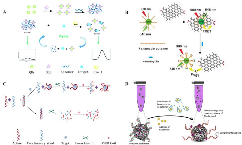
Antibiotics 2020,9,9,787
Antibiotics2020, x FOR PEER REVIEW 23 of
of18
18
method for development of sensors that use different detection methods. Therefore, aptamers are
After accumulation in the food chain, these residual antibiotics eventually enter the human body
ideal for molecular recognition with high specificity and affinity [56,57]. Additionally, aptamers can
and can be very damaging to human health. Antibiotic abuse will accelerate the spread of antibiotic
be used to introduce components for signal amplification, such as nanomaterials and fluorescent
resistance and lead to decreased human immunity [19,20]. To date, China, the European Union,
compounds, which greatly improves the detection sensitivity, shortens the detection time, and
Japan, and the United States have established maximum residue limits (Table 1) for gentamicin,
broadens the application prospects. Due to the unique optical, electrochemical, and mechanical
kanamycin, neomycin, streptomycin, and dihydrostreptomycin [21,22]. Therefore, a rapid, sensitive,
properties of nanomaterials, such as metal nanoparticles, carbon nanotubes, nanocomposite, and
and inexpensive method should be established for the detection of AG residues in food and the
nanostructuredmaterials, they have been widely used in aptasensing strategies. These nanomaterials
environment. The reported AGs assay methods include high performance liquid chromatography
are applied as catalytic tools, immobilization platforms, or as optical or electroactive labels in
(LC) [23–26], LC-mass spectrometry (MS) [27–34], capillary electrophoresis-MS [35,36], microbial
biosensor schemes in order to improve their performance [58]. Some AG aptamers are shown in Table
assays [37], and enzyme-linked immunoassays [38–41].
2.
Figure1.1.Chemical
Figure Chemicalstructure
structureof
ofaminoglycoside
aminoglycosideantibiotics.
antibiotics.
Table 1. Maximum residue limits (MRLs) of some aminoglycosides in various countries.
Table 1. Maximum residue limits (MRLs) of some aminoglycosides in various countries.
MRL (µg kg−1 )
Species or Detection MRL (μg kg−1)
Drug Name The European The United
Product
Species or Object
Detection China The The Japan
Drug Name Union States
Product Object China European United Japan
Muscle, Fat 100 50
Union 100/400
States 100
Liver 2000 200 300 2000
Pig, Cattle Muscle, Fat 100 50 100/400 100
Gentamicin Kidney 5000 750 400 5000
Liver
Milk 2000
200 200
100 300 - 2000 200
Pig, Cattle
Gentamicin Chicken Kidney
Tissue 5000
100 750
- 400 100 5000 -
Milk Fat
Muscle, 200- 100 - - 200 40
Chicken Liver
Tissue 100- 600
- 100 - - 40
Cattle
Kidney
Muscle, Fat -- 2500
100 - - 40 40
Milk - 150 - 400
Kanamycin Liver - 600 - 40
Cattle 100/50
Muscle
Kidney -- 100
2500 - - 40
(chicken)
Pig, Milk -- 150 - - 400 100
Fat 100
Chicken
100/50
100/50
Liver
Muscle -- 600
100 - - (chicken)
(chicken)
Kanamycin 100/500
Fat
Kidney -- 100
2500 - - 100
(chicken)
Pig, Chicken 100/50
Chicken Egg
Liver -- -
600 - - 500
Muscle, Fat, (chicken)
500 500 1200/-/3600 100/500500
Liver
Cattle, Pig, Kidney - 2500 -
Neomycin Kidney 10,000 5000 7200 (chicken)
10,000
Chicken
Chicken Milk
Egg 500
- 1500
- - 150 500 500
Egg
Muscle, 500 500 1200/-- 500
Muscle, Fat, 500 500 500
Cattle, Fat, Liver 600 500 /3600500 600
Streptomycin/ Cattle, Pig, Liver
Neomycin Sheep, Pig Kidney 10,000 5000 7200 10,000
Dihydrostreptomycin Chicken Kidney 1000 1000 2000 1000
Cattle Milk
Milk 500
200 1500
200 150 - 500 200
Egg 500 500 - 500
Cattle, Muscle,
600 500 500 600
Sheep, Pig Fat, Liver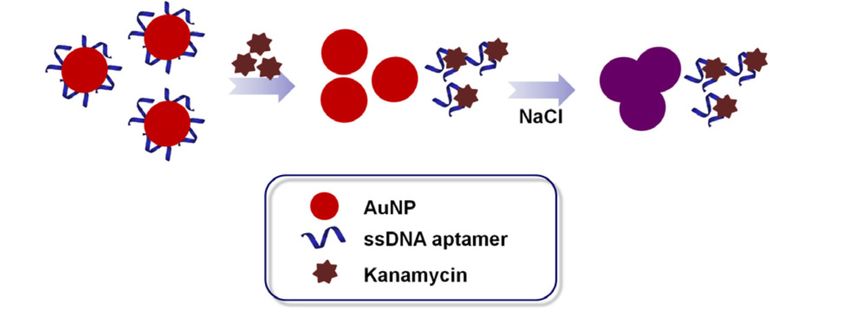
Antibiotics 2020, 9, 787                                                                            3 of 18

      Because AGs are highly polar, they are poorly preserved in conventional reversed-phase LC,
and they cannot be detected using ultraviolet or fluorescence detectors unless pre- or post-column
derivatization is performed because they do not contain chromophores or fluorophores [42–45].
However, derivatization makes the instrument conditions more complex, and the additional processing
steps required lead to loss of the analytical substance. Furthermore, excess reagents and derivatives
can interfere with the results. LC-MS has been widely used for the analysis of AGs. However,
different AGs are structurally similar and produces are similar ions after fragmentation, so quantitative
determination of AGs is challenging. Additionally, the use of LC-MS for AG detection requires the
use of volatile mobile phase additives, which is not beneficial for the long-term condition of the
instrument. Consequently, pretreatment is needed when the sample is analyzed by an instrumental
method. Because AGs are strongly polar and exist as polyanions in aqueous solutions, they are difficult
to extract. Generally, solid-phase extraction is used to extract and purify AGs, but this method has a
low recovery rate and the pretreatment steps are complex. Instrumental analysis has high sensitivity
and accuracy, but it is expensive, requires trained operators, and is difficult to use for rapid on-site
detection [46,47]. Microbial methods and enzyme-linked immunoassays are relatively simple but have
long detection times and large experimental errors, which can result in interference and false positive
for antibiotics with similar structures [48].
      With recent developments in biotechnology, sensors based on specific biometric elements have
been widely used for antibiotic residue detection [49]. Biosensors have strong specificities and high
sensitivities, and are simple, small, and portable. The disadvantages of antibodies and enzymes
as classical biosensors are that they have poor thermal stabilities, are difficult to modify, and have
high production costs [50]. Antibody preparation usually requires immunization of animals and
hybridoma techniques. AGs are small-molecule haptens and cannot stimulate the body to produce
corresponding antibodies on their own, so it is difficult to obtain highly sensitive antibodies [21].
Aptamers as “chemical antibodies” are single-stranded deoxyribonucleic acid (ssDNA) or ribonucleic
acid (RNA) sequences of approximately 10–100 bases that can specifically bind to a target [51]. They can
be obtained from the nucleic acid molecular library through in vitro screening via the systematic
evolution of ligands by exponential enrichment (SELEX) technique. As molecular recognition elements,
aptamers can identify a wide range of target molecules, including metal ions, small drug molecules,
proteins, viruses, animal cells, and tissues [52,53]. Although aptamers have many similar properties
to antibodies, they have a number of advantages over traditional antibodies [54,55]. First, they have
stronger specificities. Their affinities to target molecule are higher than those of antibodies, and their
dissociation constants (Kd ) are far lower than those of antibodies and can be as low as nanomole to
picomole per liter levels. Second, they can be used for a wider range of target molecules. Screening
and synthesis studies have shown an aptamer can theoretically be selected for any target substance,
whereas antibodies cannot. Third, aptamers are simpler to prepare than antibodies, and can be rapidly
synthesized in large quantities in vitro. By contrast, antibodies need to be produced in an animal or
cells, and the preparation conditions are relatively complex. Fourth, aptamers have good stabilities and
are easy to store. Whereas antibodies are unstable and prone to irreversible degradation, aptamers are
more stable and can recover their active conformations. Fifth, aptamers have low molecular weights
and are easily chemically modified. Consequently, they can be coupled with various molecules by
simple chemical reactions, which provides a convenient and flexible method for development of sensors
that use different detection methods. Therefore, aptamers are ideal for molecular recognition with high
specificity and affinity [56,57]. Additionally, aptamers can be used to introduce components for signal
amplification, such as nanomaterials and fluorescent compounds, which greatly improves the detection
sensitivity, shortens the detection time, and broadens the application prospects. Due to the unique
optical, electrochemical, and mechanical properties of nanomaterials, such as metal nanoparticles,
carbon nanotubes, nanocomposite, and nanostructuredmaterials, they have been widely used in
aptasensing strategies. These nanomaterials are applied as catalytic tools, immobilization platforms,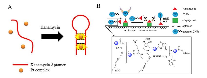
Antibiotics 2020, 9, 787                                                                                      4 of 18

or as optical or electroactive labels in biosensor schemes in order to improve their performance [58].
Some AG aptamers are shown in Table 2.

                                 Table 2. Specific aptamer sequences of several AGs.

                  AGs                   Aptamer Sequence (50 –30 )             Kd (µmol L−1 )        Ref.
              Kanamycin        TGGGGGTTGAGGCTAAGCCGA                                0.079            [59]
              Neomycin B       GGCCUGGGCGAGAAGUUUAGGCC                               1.24            [60]
                               TAGGGAATTCGTCGACGGATCCGGGG
             Streptomycin      TCTGGTGTTCTGCTTTGTTCTGTCGGGT                         0.199            [61]
                               CGTCTGCAGGTCGACGCATGCGCCG
              Tobramycin       GACTAGGCACTAGTC                                      0.042            [62]

2. Application of Aptamer Biosensors in the Detection of AGs
     As a recognition element, aptamers do not directly generate detection signals. Therefore,
researchers usually combine aptamers with nanoscale gold, quantum dots, or another material
that can generate a photoelectric signal to form a composite probe. Aptamer biosensors are developed
in combination with electrochemical and optical detection systems to realize the detection of target
compounds. The operating modes for aptamer-based biosensors have been described in the reported
review paper [63]. The differentially designed principles are necessary for different targets, as the
differences in aptamer sequences and target characteristics. Previous reviews have focused on the
classification of the perspective of compounds rather than the design of biosensors especially for
AGs [63,64]. This review focuses on aptamer sensors (aptasensors) using colorimetric, fluorescent,
chemiluminescent, surface plasma resonance (SPR), and electrochemical techniques for the detection
of AGs. Table 3 summarizes different aptasensor methods and their detection performances.

                     Table 3. Summary of aptasensor types, detection methods, and performances.

      Sensor Type             Method                  Strategy               Analytes             LOD*       Ref.*
                                               High-salt induce AuNPs*
                                                                            Kanamycin           25 nmol/L    [59]
                           NaCl-AuNPs       aggregation from red to blue
      Colorimetric
                                               the catalytic chromogenic
                                              reaction of AuNPs mimics      Streptomycin        86 nmol/L    [65]
                                                          enzymes
                                              based on analyte-protected
                           NaCl-AgNPs             and aptamer-selective     Kanamycin           2.6 ng/mL    [66]
                                                        mechanism
                                             labeled with Exo III, AuNPs
                                                                            Kanamycin           321 pmol/L   [67]
                                                         and FAM*.
                              Labeled       Poly A and FAM modified at
                                                                            Neomycin B      0.01 µmol/L      [68]
       Fluorescent                              the two ends of aptamer
                                               A fluorescent “signal-on”
                                             switch aptasensor based on     Streptomycin        0.03 ng/mL   [69]
                                                         QDs-SSB*
                                              The fluorescence resonant
                                                energy transfer based on
                                                                            Kanamycin            9 pmol/L    [70]
                                            UCNPs* and graphene oxide
                                                            (GO)
                                            digestion of dsDNA by ExoIII
                                            and the ability of SYBR Gold    Streptomycin    54.5 nmol/L      [71]
                            Non-labeled
                                                   as a fluorescent dye
                                            dsDNA-capped mesoporous
                                                 silica nanoparticles and   Kanamycin           7.5 nmol/L   [72]
                                                       Rhodamine B
Antibiotics 2020, 9, 787 5 of 18
Table 3. Cont.
Sensor Type Method Strategy Analytes LOD* Ref.*
Cold light The Pt complex performs
Kanamycin 143 nmol/L [73]
Chemiluminescence probe signal transduction
FALIA* assay CNP*-aptasensor probe Kanamycin 5× 10⁻⁸ ng/mL [74]
Surface plasmon Competitive RNA aptamer modified by
Neomycin B 5 nmol/L [60]
resonance effect methyl groups
GR* and AuNPs modified on
Differential Kanamycin 0.03 pmol/L
glassy carbon electrode [75]
pulse Streptomycin 0.3 pmol/L
surface as adaptor carriers
voltammetry conductive polymer/gold 9.4 ± 0.4
(DPV) Kanamycin [76]
Electrochemical self-assembled nanocomposite nmol/L
Square wave The background signal
Kanamycin 1 pmol/L [77]
voltammetry compressed by exonuclease
(SWV) nanoscale metal organic
Kanamycin 0.16 pmol/L [78]
framework
Alternating
current Target induces signal probe
Kanamycin 3.3 pmol/L [79]
voltammetry transfer
(ACV)
Electrochemical screen printing carbon
Kanamycin 0.11 ng/mL [80]
impedance electrode
spectroscopy protein-oriented carbon
nanoparticles embedded with
(EIS) Tobramycin 6.7 pg mL−1 [81]
SnOx and TiO2
nanocrystalline
* Limit of detection(LOD), references (Ref.), Gold nanoparticles (AuNPs), fluorescein amidite(FAM), a single stranded
DNA binding protein on quantum dots (QDs-SSB), upconversion nanoparticles (UCNPs), fluorescence-based
aptamer-linked immunosorbent assay(FALIA), carbon nanoparticles(CNP), Graphene(GR).
3. Colorimetric Aptamer Sensors
The biggest advantage of the colorimetric method is that preliminary experimental results
can be obtained easily by direct observation of color changes using a cell phone chromatism or
spectrophotometer without the need for complex equipment [82]. Consequently, the colorimetric
method is inexpensive, simple, and rapid [83–85]. Gold nanoparticles (AuNPs) are an excellent material
for colorimetric aptasensors because they have unique optical properties, good biocompatibility,
large surface areas, and high absorption efficiencies. Furthermore, AuNPs are also able to enhance
other optical signals like fluorescence and light scattering [86–89]. AuNP-based aptamer colorimetric
analysis has been widely used in kanamycin detection. Song [59] screened a kanamycin ssDNA
aptamer, Ky2 (TGGGGGTTGAGGCTAAGCCGA), using the SELEX method, and established a
AuNP-colorimetric method using this aptamer (Figure 2). In this method, an aqueous sodium chloride
solution of dispersed AuNPs was wine red. In the presence of aptamers, the AuNPs coordinated
with the aptamers via van der Waals attractions to maintain dispersion of the AuNPs in the solution.
When kanamycin was added to the system, the aptamers bound to it specifically and more strongly
than to the AuNPs. This resulted in dissociation of the aptamers from the AuNPs surface, aggregation
of the AuNPs in the salt solution, a change in the solution color changed from red to blue or even purple,
and a change in the absorbance. The detection limit of this method for kanamycin was 25 nmol/L.
Compared with ssDNA, double stranded DNA does not protect AuNPs from salt-induced aggregation
because of its rigid structure. Zhang [90] designed a colorimetric sensor using AuNPs and dsDNA
to detect kanamycin. In the absence of kanamycin, the aptamer formed a stable DNA double strand
with the complementary DNA strand, which resulted in salt aggregation of AuNPs. In the presence of
kanamycin, the aptamer was released to bind to its target, and complementary DNA was adsorbed on
the surfaces of the AuNPs to protect them from salt-induced aggregation. The absorbance ratio was
linearly correlated to the concentration of kanamycin in the range of 0.02–0.3 mol/L, and the detection
limit was 8 nmol/L. To extend on the classic AuNP colorimetric aptasensors, Chen et al. [91] introduced
fluorescence labeling to develop a sensor with a dual signals for kanamycin A. The detection limitformed a stable DNA double strand with the complementary DNA strand, which resulted in salt
aggregation of AuNPs. In the presence of kanamycin, the aptamer was released to bind to its target,
and complementary DNA was adsorbed on the surfaces of the AuNPs to protect them from salt-
induced aggregation. The absorbance ratio was linearly correlated to the concentration of kanamycin
in the range
Antibiotics 2020, 9, of
7870.02–0.3 mol/L, and the detection limit was 8 nmol/L. To extend on the classic6AuNP
of 18
colorimetric aptasensors, Chen et al. [91] introduced fluorescence labeling to develop a sensor with a
dual signals for kanamycin A. The detection limit of this method reached 0.3 nmol/L, and it was
of this method reached 0.3 nmol/L, and it was successfully applied to the analysis of milk samples.
successfully applied to the analysis of milk samples. Another colorimetric aptasensor has been
Another colorimetric aptasensor has been developed using the catalyzed chromogenic reactions of
developed using the catalyzed chromogenic reactions of various enzymes or mimic enzymes. Zhao
various enzymes or mimic enzymes. Zhao [65] established a new method for streptomycin colorimetric
[65] established a new method for streptomycin colorimetric detection using the simulated enzyme
detection using the simulated enzyme catalytic activity of AuNPs. When there was no streptomycin,
catalytic activity of AuNPs. When there was no streptomycin, the aptamer bound to the AuNPs,
the aptamer bound to the AuNPs, which reduced the activity of the AuNPs enzyme. In the presence of
which reduced the activity of the AuNPs enzyme. In the presence of streptomycin, the aptamer could
streptomycin, the aptamer could not bind to the AuNPs, and the enzyme activity of the AuNPs was
not bind to the AuNPs, and the enzyme activity of the AuNPs was observed. The detection limit of
observed. The detection limit of this method for streptomycin was 86 nmol/L and the linear range
this method for streptomycin was 86 nmol/L and the linear range was 0.1–0.5 mol/L. Silver
was 0.1–0.5 mol/L. Silver nanoparticles (AgNPs) have similar optical properties to AuNPs and also
nanoparticles (AgNPs) have similar optical properties to AuNPs and also aggregate in the presence
aggregate in the presence of a salt. Because the amino group in kanamycin can bind to AgNPs via a
of a salt. Because the amino group in kanamycin can bind to AgNPs via a Ag-N bond, it can adsorb
Ag-N bond, it can adsorb on the surface of unmodified AgNPs and prevent salt-induced aggregation.
on the surface of unmodified AgNPs and prevent salt-induced aggregation. This protective
This protective mechanism will be weakened after kanamycin binds to an aptamer. According to the
mechanism will be weakened after kanamycin binds to an aptamer. According to the selection
selection mechanism of the aptamer and AgNPs protection by the analyzed target, Xu [66] designed
mechanism of the aptamer and AgNPs protection by the analyzed target, Xu [66] designed a new
a new aptasensor. This method could detect kanamycin in milk samples at 0.05–0.6 µg/mL levels
aptasensor. This method could detect kanamycin in milk samples at 0.05–0.6 μg/mL levels within 20
within 20 min with a detection limit of 2.6 ng/mL. Aptamer biosensors using AuNPs or AgNPs for
min with a detection limit of 2.6 ng/mL. Aptamer biosensors using AuNPs or AgNPs for colorimetric
colorimetric detection have strong specificities and high sensitivities, are simple to make and easy to
detection have strong specificities and high sensitivities, are simple to make and easy to use, and have
use, and have been widely used in field and for label free detection. However, AuNP-based aptamer
been widely used in field and for label free detection. However, AuNP-based aptamer colorimetric
colorimetric analysis usually need large amount of aptamer, as the dispersion of the AuNPs and the
analysis usually need large amount of aptamer, as the dispersion of the AuNPs and the binding of
binding of target all need certain amount of aptamer, so the sensitivity of colorimetric aptasensors
target all need certain amount of aptamer, so the sensitivity of colorimetric aptasensors should be
should be more improved through powerful signal amplification methods.
more improved through powerful signal amplification methods.
Figure Schematic
2. 2.
Figure illustration
Schematic ofof
illustration the AuNP-based
the colorimetric
AuNP-based assay
colorimetric forfor
assay detection of of
detection kanamycin [59].
kanamycin [59].
4. Fluorescent Aptamer Sensors
4. Fluorescent Aptamer Sensors
Fluorescence is a highly sensitive optical property. The effectiveness of aptasensors using
Fluorescence is a highly sensitive optical property. The effectiveness of aptasensors using
fluorescent-labeled probes has been confirmed in many experimental studies. Sharma constructed an
fluorescent-labeled probes has been confirmed in many experimental studies. Sharma constructed an
aptaswitch sensor complex using a combination of fluorophore and quenching labeled oligonucleotides
aptaswitch sensor complex using a combination of fluorophore and quenching labeled
and an aptamer that recognizes chloramphenicol [92,93]. Aptamer fluorescence detection uses the
oligonucleotides and an aptamer that recognizes chloramphenicol [92,93]. Aptamer fluorescence
specific recognition of an aptamer and target antibiotics to regulate the energy transfer efficiency
detection uses the specific recognition of an aptamer and target antibiotics to regulate the energy
between a fluorescent donor and a recipient, to achieve quantitative detection of target antibiotics
transfer efficiency between a fluorescent donor and a recipient, to achieve quantitative detection of
through changes in the fluorescence intensity or polarization [94,95]. According to the different modes
target antibiotics through changes in the fluorescence intensity or polarization [94,95]. According to
of action of the fluorophore and aptamer, the sensors can be classed as labeled and unlabeled fluorescent
the different modes of action of the fluorophore and aptamer, the sensors can be classed as labeled
aptasensors. To construct labeled fluorescent aptasensors, organic small molecule fluorescent dyes
or fluorescent nanomaterials are usually used to label the sensor probes. Ramezani [67] designed a
fluorescent aptasensor using a kanamycin aptamer complementary sequence labeled with exonuclease
III, AuNPs, and carboxyfluoresce in FAM. This sensor was suitable for the detection of kanamycin
residues in food with a detection limit as low as 321 pmol/L. Ling [68] divided the RNA aptamer of
neomycin B into two segments, one of which was absorbed on the surfaces of AuNPs by polyadenylate,
and the other labeled with the FAM fluorophore. When neomycin B was present in the samples,
the target material and the two nucleic acid fragments were rapidly assembled into a compact H-shapedAntibiotics 2020, 9, 787 7 of 18 structure on the AuNPs surface, leading to quenching of the FAM fluorophore. The concentration of neomycin B in the solution was inversely proportional to the fluorescence intensity, and the detection limit for neomycin B was 0.01 mol/L. Because of the shortcomings of fluorescent dyes, such as poor photobleaching resistance and vulnerability of the fluorescence performance to external factors, some fluorescent nanomaterials with better performance have been applied to the construction of fluorescent aptasensors for AGs. Quantum dots (QDs) have attracted much attention because of their unique optical and electronic properties, including high luminescence, strong light stability, good resistance to light bleaching, wide absorption bands, and adjustable sizes [96,97]. Wu [69] designed a novel fluorescent switch sensor using QDs labeled with ssDNA binding protein (SSB) and exonuclease I for assisted target recovery, and applied it to streptomycin detection (Figure 3A). The fluorescent probes were synthesized by labeling QDs with SSB, which could bind to the aptamer specifically. When an aptamer was added as a bridge ligand, it hybridized with SSB. At the same time, the QDs scattered in the solution aggregated, which resulted in self-quenching, and the sensor state changed from “on” to “off”. In the presence of streptomycin and exonuclease I, the aptamer preferentially bound to the target. Exonuclease I then digested the aptamer target into a single nucleotide, and the released target could participate in the reaction cycle and produce a strong fluorescence signal. At this point, the distance between the QDs increased and the fluorescence intensity recovered. Thus, the switch changed from the “off” state to the “on” state. Under the optimum conditions, there was a good linear relationship between the fluorescence intensity and streptomycin concentration in the range of 0.1–100 ng/mL and the detection limit of this method was about 0.03 ng/mL. Li [70] used upconversion with nanoparticles and graphene oxide to develop an aptasensor using fluorescence resonant energy transfer technology for detection of kanamycin (Figure 3B). Under the optimized conditions, the method had a wide linear detection range (0.01–3 nmol/L), low detection limit (9 pmol/L), and showed good performance on application to real samples. Compared with a labeled fluorescent aptasensor, time-consuming probe labeling and purification steps were not required for construction of this unlabeled fluorescent aptasensor, which saved time and reduced inter-batch differences in the sensor preparation. Taqhdis [71] used exonuclease III, a fluorescent dye (SYBR Gold), and an aptamer complementary strand to establish an unlabeled fluorescence analysis method for detection of streptomycin in milk and blood samples (Figure 3C). Without streptomycin, the fluorescence intensity was weak. After adding streptomycin, the aptamer combined with the target, leading to release of the aptamer complementary strand, which protected against exonuclease III activity. With addition of SYBR Gold, a strong fluorescence signal was observed. The sensor had high selectivity for streptomycin, and the detection limit reached 54.5 nmol/L. Dehghani [72] constructed a double-stranded “molecular gate” closed mesoporous silicon probe by efficiently loading mesoporous silicon nanoparticles (MSNs) on small molecule fluorescent dyes, and developed a fluorescent aptasensor for detection of kanamycin without the need for a signal amplifier (Figure 3D). The amine-modified complementary chain was covalently fixed on the MSNs surface and the unlabeled aptamer was also fixed on the MSNs surface through pairing with the complementary chain. In the presence of kanamycin, the aptamer specifically bound to it and was separated from its complementary chain and the double-stranded “molecular gate” was destroyed, leading to release of rhodamine B. The fluorescence intensity of the solution increased with leakage of rhodamine B. Kanamycin was detected by measuring the fluorescence intensity. The linear range for measurements using the relative fluorescence intensity was 24.75–137.15 nmol/L and the detection limit was 7.5 nmol/L. Fluorometric sensing is more promising methodology to analyze and measure AGs quantitatively and sensitively compared to colorimetric assay. However, most of the fluorescent aptamer sensors could only detect one target in an assay, if more sensors could be designed using multicolor quantum dots, it would be helpful to achieve the efficient detection of multi-targets.
Antibiotics 2020, 9, x FOR PEER REVIEW 8 of 18
Fluorometric sensing is more promising methodology to analyze and measure AGs quantitatively
and sensitively compared to colorimetric assay. However, most of the fluorescent aptamer sensors
could only
Antibiotics detect
2020, 9, 787 one target in an assay, if more sensors could be designed using multicolor quantum
8 of 18
dots, it would be helpful to achieve the efficient detection of multi-targets.
Figure 3.
Figure 3. Schematic
Schematic illustration
illustration of of aa fluorescent
fluorescent aptasensor.
aptasensor. (A)
(A) AA fluorescent “on” switch
fluorescent “on” switch aptasensor
aptasensor
containing QDs-SSB [69]. (B) Fluorescence resonance energy transfer between
containing QDs-SSB [69]. (B) Fluorescence resonance energy transfer between kanamycin aptamerkanamycin aptamer
UCNPs and
UCNPs andgraphene
graphene [70].[70]. (C) Unlabeled
(C) Unlabeled fluorescence
fluorescence analysis
analysis [71]. [71]. (D) dsDNA-modified
(D) dsDNA-modified mesoporous
mesoporous silicon nanoparticles (MSNs) loaded
silicon nanoparticles (MSNs) loaded with rhodamine B [72]. with rhodamine B [72].
5. Chemiluminescent Aptamer Sensors
Chemiluminescence, also known as cold light, refers to light radiation produced by chemical chemical
reactions in the absence of any light, heat, or electric field field excitation
excitation [98].
[98]. Because there is no need for
an external excitation light source, interference from background or stray light is avoided, the level
of noise is reduced, and the signal-to-noise ratio is greatly improved. Because Because of of its
its high
high sensitivity,
sensitivity,
wide linear range, fast analysis speed, easy operation, and miniaturization, the
linear range, fast analysis speed, easy operation, and miniaturization, the chemiluminescence chemiluminescence
method has been widely used in biology, pharmacy, chemistry, chemistry, environmental
environmental science,
science, and clinical
medicine
medicine [98–100].
[98–100]. Ma et al. [73][73] developed
developed a chemiluminescent
chemiluminescent probe using an aptamer for detection
of kanamycin
kanamycinatat trace levels
trace in aquatic
levels products
in aquatic (Figure(Figure
products 4A). They
4A).used
Theytheused
chemiluminescent transition
the chemiluminescent
metal platinum
transition metal(Pt) rather than
platinum (Pt) arather
traditional
than organic dye because
a traditional organicthe optical
dye physical
because properties
the optical of Pt
physical
were more sensitive
properties of Pt were to microenvironment
more sensitive changes, and it had a longer
to microenvironment phosphorescence
changes, and it had half-life and
a longer
aphosphorescence
larger Stoke shiftvalues. The chemiluminescent probe used a Pt complex for
half-life and a larger Stoke shiftvalues. The chemiluminescent probe used a Pt signal transduction.
Normally,
complex for thesignal
chemiluminescence emitted bythe
transduction. Normally, thischemiluminescence
complex in water was very by
emitted weak,
thisbut when in
complex inserted
water
into
was very weak, but when inserted into the double helix DNA structure, the strength ofgave
the double helix DNA structure, the strength of the chemiluminescence increased, which the
excellent signal transduction.
chemiluminescence increased, When
which only the excellent
gave Pt complex and aptamer
signal were present
transduction. When in water,
only the the aptamer
Pt complex
was
and in a free folded
aptamer state. When
were present kanamycin
in water, was added
the aptamer was in to the system,
a free folded thestate.
aptamer
When specifically
kanamycin bound
was
to it and its conformation changed to a hairpin structure. This double-helix hairpin
added to the system, the aptamer specifically bound to it and its conformation changed to a hairpin structure promoted
insertion
structure.of thedouble-helix
This Pt complex into the aptamer
hairpin structurefragment
promotedand enhanced
insertion thePtchemiluminescence
of the complex into the aptamer signal.
The detection
fragment and limit in an the
enhanced aqueous solution was 143
chemiluminescence nmol/L
signal. Theand the linear
detection range
limit in anwas 0.2–150
aqueous mol/L.
solution
Lin [74] developed a simple, rapid, and highly sensitive method for kanamycin
was 143 nmol/L and the linear range was 0.2–150 mol/L. Lin [74] developed a simple, rapid, and analysis using carbon
nanoparticles
highly sensitive (CNPs)
method andfor
an kanamycin
aptamer. Inanalysis
this method,
usingluminescent CNPs with(CNPs)
carbon nanoparticles high waterand stability and
an aptamer.
excellent luminescence were synthesized by a microwave-assisted method
In this method, luminescent CNPs with high water stability and excellent luminescence were (Figure 4B). Amine-modified
kanamycin aptamer was fixed on the surface of the CNPs with carboxyl groups, which gave the CNPs
aptasensor excellent selectivity and stability. Kanamycin was analyzed using the developed CNPsAntibiotics 2020, 9, x FOR PEER REVIEW 9 of 18
synthesized by a microwave-assisted method (Figure 4B). Amine-modified kanamycin aptamer was
Antibiotics 2020, 9, 787 9 of 18
fixed on the surface of the CNPs with carboxyl groups, which gave the CNPs aptasensor excellent
selectivity and stability. Kanamycin was analyzed using the developed CNPs aptasensor on the basis
of competitive
aptasensor inhibition
on the basis of mechanism.
competitive The content
inhibition of kanamycin
mechanism. Thein milk was
content analyzed successfully
of kanamycin in milk was
with a detection
analyzed limit as
successfully low
with as 5 × 10⁻⁸limit
a detection as low as 5 × 10⁻⁸ ng/mL.
ng/mL.
Figure 4.4.Schematic
Figure illustration
Schematic of a of
illustration chemiluminescent aptasensor.
a chemiluminescent (A) A luminescent
aptasensor. probe containing
(A) A luminescent probe
an aptamer and Pt(II) [73]. (B) A carbon nanoparticles aptasensor [74].
containing an aptamer and Pt(II) [73]. (B) A carbon nanoparticles aptasensor [74].
6. SPR Aptamer Sensors
6. SPR Aptamer Sensors
With the SPR technique, compounds are detected using refractive index changes or chemical
With the SPR technique, compounds are detected using refractive index changes or chemical
changes resulting from optical coupling of metal films (gold or silver). SPR is a highly sensitive optical
changes resulting from optical coupling of metal films (gold or silver). SPR is a highly sensitive optical
sensing technology relying on the interactions of light with the free electrons in a semi-transparent
sensing technology relying on the interactions of light with the free electrons in a semi-transparent
noble metallic layer or chip and can realize the real-time monitoring of small changes in the effective
noble metallic layer or chip and can realize the real-time monitoring of small changes in the effective
refractive index of a metal-dielectric interface [101–103]. SPR sensors have the advantages of high
refractive index of a metal-dielectric interface [101–103]. SPR sensors have the advantages of high
throughput, no need for labeling, simple operation, and provide results faster than other methods.
throughput, no need for labeling, simple operation, and provide results faster than other methods.
With the rapid development of aptamer technology, aptamer SPR biosensors have attracted increasing
With the rapid development of aptamer technology, aptamer SPR biosensors have attracted
attention. An aptamer selected by the SELEX method can be fixed on a SPR chip surface. When the
increasing attention. An aptamer selected by the SELEX method can be fixed on a SPR chip surface.
target object passes through the sensor chip, the aptamer connected to the SPR substrate will specifically
When the target object passes through the sensor chip, the aptamer connected to the SPR substrate
recognize the target object, leading to changes in the resonance conditions, the reflectivity, and the SPR
will specifically recognize the target object, leading to changes in the resonance conditions, the
output signal for detection [104]. De-los-Santos-Alvarez [60] constructed a competitive aptasensor
reflectivity, and the SPR output signal for detection [104]. De-los-Santos-Alvarez [60] constructed a
using methylated RNA aptamer-binding SPR technology and applied it to detection of neomycin B.
competitive aptasensor using methylated RNA aptamer-binding SPR technology and applied it to
SPR sensors provide sensitive and detailed information on the affinities and dynamics in biomolecular
detection of neomycin B. SPR sensors provide sensitive and detailed information on the affinities and
interactions. Real time binding curves can be obtained by monitoring the change in the angle of
dynamics in biomolecular interactions. Real time binding curves can be obtained by monitoring the
laser light on a gilded film. Both the concentration of antibiotics in the substance to be measured
change in the angle of laser light on a gilded film. Both the concentration of antibiotics in the
and the dissociation constant and stoichiometric value of neomycin B binding to its aptamer can be
substance to be measured and the dissociation constant and stoichiometric value of neomycin B
measured using the response of the SPR sensor. The linear range for detection of neomycin B in milk
binding to its aptamer can be measured using the response of the SPR sensor. The linear range for
was 10 nmol/L–100 µmol/L and the detection limit was 5 nmol/L. Although SPR aptamer sensors have
detection of neomycin B in milk was 10 nmol/L–100 μmol/L and the detection limit was 5 nmol/L.
great potential in AGs testing due to the advantage of the high through analysis, the high cost of
Although SPR aptamer sensors have great potential in AGs testing due to the advantage of the high
supporting equipment and chips limits its application in field testing.
through analysis, the high cost of supporting equipment and chips limits its application in field
testing.
7. Electrochemical Aptasensors
Electrochemical
7. Electrochemical aptasensors are modified antibiotic aptamers that use a substrate material with
Aptasensors
good conductivity. These types of sensors have become a focus in the field of antibiotic detection because
they Electrochemical aptasensors
have good specificities, largeare modified
linear ranges,antibiotic aptamers
low detection that
limits, usedetection
low a substrate material
costs, with
are simple
good conductivity. These types of sensors have become a focus in the field of antibiotic
and fast to operate, are easy to miniaturize, and can be used for on-line monitoring. Nowadays, detection
because electrochemical
various they have good specificities, large linear
aptasensor designs haveranges,
been low detectionand
established limits, low detection
extensively costs, are
employed for
simple and fast to operate, are easy to miniaturize, and can be used for on-line
applications related to clinical di-agnostics, biomedical research, environmental monitoring, and foodmonitoring.
Nowadays,
analysis variousAelectrochemical
[105–107]. aptasensor
typical electrochemical designs
aptamer have
sensor been of
consists established
an electrode andcovered
extensively
with
employed for applications related to clinical di-agnostics, biomedical research,
an aptamer which, upon binding the analyte, undergoes a conformational switch affecting current environmental
flow through the system [57]. According to their output parameters, such as the impedance, current,Antibiotics 2020, 9, 787 10 of 18
and potential, electrochemical aptasensors can generally be divided into the following three types:
impedance sensors, current sensors, and potential sensors [108].
Electrochemical impedance spectroscopy (EIS) displays impedance signals by monitoring changes
in electron transfer resistance, whereas square wave voltammetry (SWV), differential pulse voltammetry,
and alternating current voltammetry all show changes in current. In recent years, nanomaterials,
such as carbon nanotubes, graphene, conducting polymers, and metal nanoparticles have been widely
used to construct aptasensors because they have high specific surface areas, good biocompatibilities,
high conductivities, unique physical and chemical properties, and excellent performance for improving
the efficiency of electron transfer on an electrode surface [107]. Feng [75] prepared two kinds of
electrochemical aptasensors by modifying the surfaces of glassy carbon electrodes with graphene and
AuNPs by electrodeposition to use as carriers. The prepared aptasensors were applied to quantitative
detection of kanamycin and streptomycin. Using differential pulse voltammetry, the detection limits
for kanamycin and streptomycin were 0.03 pmol/L and 0.3 pmol/L, respectively. The sensor had high
sensitivity, good selectivity, good reproducibility, and good stability. Zhu et al. [76] constructed an
electrochemical aptasensor using a conductive polymer/gold self-assembled nanocomposite and applied
it to detection of kanamycin with high sensitivity. They covalently immobilized a kanamycin aptamer
onto a AuNP conductive polymer composed of poly-(2,5-di-(2-thienyl)-1H-pyrrole-1-(p-benzoic acid))
as a sensor probe (Figure 5A). The concentration of kanamycin was determined by voltammetry.
The detection limit of the sensor was 9.4 ± 0.4 nmol/L. Xu et al. [77] used SWV with exonuclease
in electrochemical sensors. Because of the cyclic shearing effect, the background signal was greatly
compressed, which increased the absolute value of the change in the response signal and improved
the sensitivity. Detection of kanamycin was realized with a detection limit as low as 1 pmol/L.
Chen [78] proposed a new electrochemical biocode containing a nanoscale metal organic framework
(NMOF) for simultaneous detection of multiple antibiotics, including kanamycin. In this study,
an amine-functionalized NMOF was used as a substrate to carry different metal ions. The metal
NMOFs were labeled with complementary strands of aptamers toward different targets. After specific
binding of the aptamers to the targets, the corresponding metal NMOFs were released into the
supernatant after magnetic separation and detected by SWV (Figure 5B). The method had high
sensitivity and the detection limit was 0.16 pmol/L. Li et al. [79] constructed an electrochemical
aptasensor using a target-induced signal probe transfer mechanism, and applied it to detection
of kanamycin residues in milk, water, and serum samples using alternating current voltammetry
(Figure 5C). This system gave an ultra-low background signal because of dissociation of the signal probe,
which improved the sensitivity. The detection limit for kanamycin was as low as 3.3 pmol/L, and the
detection was rapid. Sharma [80] developed an impedance aptasensor using a functionalized aptamer
complementary sequence for a silk-screen-printed carbon electrode for the detection of kanamycin
in milk using EIS. The signal mechanism of the sensor involved enhancement of the impedance.
Before addition of the target compound, the electron transfer efficiency was high and the impedance
was low. After addition of the target, the aptamer specifically captured the target and formed a
complex covering the electrode surface. This obstructed the electron transfer channel and increased
the impedance. The concentration of the target could be measured using the change in impedance.
Under the optimized analysis conditions, the detection limit of the sensor was 0.11 ng/mL and the linear
range was 1.2–75 ng/mL, which was far less than the residue limit for kanamycin in milk (150 ng/mL).
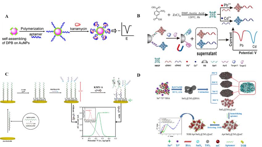
Wang [81] constructed an electrochemical aptasensor using protein-oriented CNPs embedded with
nanocrystalline SnOx and TiO2 to detect tobramycin with good sensitivity. A series of mesoporous
carbon nanospheres embedded with SnOx and TiO2 nanocrystals were obtained by pyrolysis of a
SnOx @TiO2 @bovine serum albumin nanocomposite at different temperatures using titanium butyrate
and sodium stannate trihydrate as precursors and bovine serum albumin as the template (Figure 5D).
SnOx @TiO2 @mC900 exhibited good electrochemical activity and high biological affinity among a
series of SnOx @TiO2 @mC nanocomposites. According to the electrochemical impedance spectroscopy
results, the LOD was 6.7 pg/mL. The fabricated SnOx @TiO2 @mC900 nanocomposite aptasensor had anAntibiotics 2020, 9, x FOR PEER REVIEW 11 of 18
Antibiotics 2020, 9, 787 11 of 18
series of SnOx@TiO2@mC nanocomposites. According to the electrochemical impedance spectroscopy
results, the LOD was 6.7 pg/mL. The fabricated SnOx@TiO2@mC900 nanocomposite aptasensor had
an ultra-low
ultra-low detection
detection limit forlimit for tobramycin.
tobramycin. A numberAof number of electrochemical
electrochemical aptasensors
aptasensors were introduced were
in
introduced
the past years into
the past years
improve to improve
performance performance
and simplify theand
AGssimplify the AGs
detection detection
on site, but theon site, but of
sensitivity the
sensitivity
these of these
aptasensors wasaptasensors was
not sufficient not sufficient
to apply them in to apply
real themasinthe
samples, real samples,
problem as the problem
of interface effect onof
interfacesurface.
electrode effect on electrode surface.
Figure5.5. Schematic
Figure Schematic illustration
illustration ofofananelectrochemical
electrochemicalaptasensor.
aptasensor.(A)(A)Conductive
Conductivepolymer/gold self-
polymer/gold
assembled nanocomposite [76]. (B) Aptamer–metal ion nanoscale metal organic
self-assembled nanocomposite [76]. (B) Aptamer–metal ion nanoscale metal organic framework (MOF)framework (MOF)
electrochemicalbiocodes
electrochemical biocodesfor
fordetection
detectionofofmultiple
multipleantibiotics
antibiotics[78].
[78].(C)
(C)The
Thetarget-induced
target-inducedsignal
signalprobe
probe
transfer mechanism [79]. (D) Preparation of BSA-directed SnO x@TiO2@mC nanocomposites and
transfer mechanism [79]. (D) Preparation of BSA-directed SnOx @TiO2 @mC nanocomposites and
applicationtototobramycin
application tobramycindetection
detection[81].
[81].
8.8.Other
OtherAptasensors
Aptasensors
InInsummary,
summary, many
many aptamer
aptamer biosensors
biosensorshave been
have developed
been developed forfor
thethe
detection
detectionof AGs,
of AGs, but but
most of
most
these are targeted
of these are targetedto single compounds.
to single compounds. Sensors thatthat
Sensors can can
detect groups
detect of compounds
groups of compounds are preferred for
are preferred
rapid on-site
for rapid detection.
on-site Caglayan
detection. [109] designed
Caglayan an aptasensor-based
[109] designed elliptical
an aptasensor-based polarized
elliptical light sensor
polarized light
for the determination
sensor of AGs inofdairy
for the determination AGsproducts. Kanamycin
in dairy products. and neomycin
Kanamycin were successfully
and neomycin detected
were successfully
with good with
detected sensitivity
good and minimum
sensitivity and detection
minimumlimits of 0.22limits
detection ng/mL of and
0.220.048
ng/mL ng/mL, respectively.
and 0.048 ng/mL,
Tang and his team [110] designed an evanescent wave sensor (EWA) using
respectively. Tang and his team [110] designed an evanescent wave sensor (EWA) using target target binding to promote
fluorescence quenching
binding to promote for group-specific
fluorescence quenchingdetection of AGs in a fully
for group-specific onlineofmode.
detection AGs in In afluorescence
fully online
quenching with an EWA,
mode. In fluorescence a fluorophore-labeled
quenching with an EWA, DNA aptamer with selectivity
a fluorophore-labeled DNA aptamer for kanamycin was
with selectivity
used for both recognition of the target in solution and signal transduction to
for kanamycin was used for both recognition of the target in solution and signal transduction to thethe EWA optical fibers.
The
EWA number
opticalof the aptamers
fibers. The number form multiple-strand
of the aptamers form complex (M-APT)complex
multiple-strand on the fibers
(M-APT) wasoninversely
the fibers
proportional
was inversely toproportional
the AG concentration. The minimum
to the AG concentration. detection
The minimum limit of this limit
detection method formethod
of this AGs was for
26AGs
nmol/L. The sensor responded specifically to all AGs detected, but not to other
was 26 nmol/L. The sensor responded specifically to all AGs detected, but not to other types of types of antibiotics.
With the development
antibiotics. of digital technology,
With the development intelligent platforms
of digital technology, intelligentare frequently
platforms used in scientific
are frequently used in
research. Using a digital fluorescence detector as readout device, Wang [111] developed
scientific research. Using a digital fluorescence detector as readout device, Wang [111] developed an an intelligent
platform
intelligentfor detection
platform offor multiple AGs of
detection using a ratiometric
multiple AGs paper-based device. Quantitative
using a ratiometric paper-based detection
device.
was realized according
Quantitative detection to wastherealized
relationship between
according to thethe change in the
relationship digital
between fluorescence
the change in the detector
digital
signal and the target concentration. Sensitive analysis of streptomycin, tobramycin,
fluorescence detector signal and the target concentration. Sensitive analysis of streptomycin, and kanamycin
could be realized
tobramycin, andsimultaneously
kanamycin could using this platform.
be realized simultaneously using this platform.
9. ConclusionsAntibiotics 2020, 9, 787 12 of 18
9. Conclusions
With improvements in production and living standards, environmental protection and food safety
issues have aroused widespread concern. In recent years, the extensive use and even abuse of antibiotics
have posed a serious threat to the environment and food safety. Consequently, detection of antibiotic
residues in food and the environment has attracted increasing attention. With the rapid development
of aptamer screening technologies, biosensors containing aptamers have provided a new method for
rapid detection of AG residues in the environment and food. At the same time, various nanoscale
and composite materials combined with electrochemical, optical, and photoelectrochemical detection
technologies have been used to develop aptasensors with different signal amplification and output
modes. Although there has been progress in research on biosensors developed from AG aptamers,
most sensors are in theoretical and laboratory-research stages. Therefore, practical application of these
sensors on a large scale remains distant. Consequently, it is important to develop rapid, high-quality,
inexpensive, digital, and intelligent biosensor technologies. Future work should focus on the following
aspects. First, there are few types of aptamers that can be used for the detection of AG residues.
Screening for more types of antibiotics or class-specific aptamers that can recognize common groups
will be important for detection of AG residues. Second, development of multi-functional nanomaterials
and strengthening of the application compatibility between nanomaterials, molecular recognition
elements, and conversion elements is required to improve aptasensors and meet the need for portable,
inexpensive, and simple sensors that can be used for on-site testing. Third, environmental and food
sample matrices are complex and high-throughput and specific sample purification and enrichment
methods aptasensor are required to reduce the impact of matrix effects on aptasensors and improve the
accuracy. With the rapid development of aptamers, problems restricting the development of sensors
will eventually be overcome. The development of a fast, sensitive, and portable aptamer biosensor will
broaden the application range and commercial prospects for rapid detection of AG residues in the field.
Author Contributions: Original draft preparation, Y.L.; formal analysis, N.W.; data curation, C.L.; project
administration, X.G.; writing—review and revise, A.L. All authors have read and agreed to the published version
of the manuscript.
Funding: The authors are very grateful for the powerful suggestions from the anonymous referees. We also
thank the support from the Beijing Agricultural Forestry Academy Youth Foundation (QNJJ201903), Research
Foundation of Beijing Academy of Agriculture and Forestry Sciences (KJCX20180407), Beijing Natural Science
Foundation (8182021) and China Agriculture Research System of Peach (CARS-30-1-21).
Conflicts of Interest: The authors declare that they have no conflict of interest.
References
1. Jospe-Kaufman, M.; Siomin, L.; Fridman, M. The relationship between the structure and toxicity of
aminoglycoside antibiotics. Bioorg. Med. Chem. Lett. 2020, 30. [CrossRef]
2. Glinka, M.; Wojnowski, W.; Wasik, A. Determination of aminoglycoside antibiotics: Current status and
future trends. TRAC Trends Anal. Chem. 2020, 131, 116034. [CrossRef]
3. Schilling, K.; Krmar, J.; Maljuric, N.; Pawellek, R.; Protic, A.; Holzgrabe, U. Quantitative structure-property
relationship modeling of polar analytes lacking UV chromophores to charged aerosol detector response.
Anal. Bioanal. Chem. 2019, 411, 2945–2959. [CrossRef] [PubMed]
4. Toro, I.J.; Rodriguez, C.A.; Zuluaga, A.F. Effectiveness of the antibiotic combinations for enterococcal
infections treatment: A critical review. Rev. Chil. Infectol. 2019, 36, 556–564.
5. Khan, F.; Lee, J.W.; Javaid, A.; Park, S.K.; Kim, Y.M. Inhibition of biofilm and virulence properties of
Pseudomonas aeruginosa by sub-inhibitory concentrations of aminoglycosides. Microb. Pathog. 2020, 146.
[CrossRef]
6. Hussein, M.; Han, M.L.; Zhu, Y.; Zhou, Q.; Lin, Y.W.; Hancock, R.E.W.; Hoyer, D.; Creek, D.J.; Li, J.; Velkov, T.
Metabolomics Study of the Synergistic Killing of Polymyxin B in Combination with Amikacin against
Polymyxin-Susceptible and -Resistant Pseudomonas aeruginosa. Antimicrob. Agents Chemother. 2020, 64.
[CrossRef] [PubMed]You can also read