On-Target CRISPR/Cas9 Activity Can Cause Undesigned Large Deletion in Mouse Zygotes - MDPI
←
→
Page content transcription
If your browser does not render page correctly, please read the page content below
International Journal of
            Molecular Sciences

Article
On-Target CRISPR/Cas9 Activity Can Cause
Undesigned Large Deletion in Mouse Zygotes
Alexey Korablev 1 , Varvara Lukyanchikova 1,2 , Irina Serova 1 and Nariman Battulin 1,2, *
 1    Institute of Cytology and Genetics SB RAS, Novosibirsk 630090, Russia; korablevalexeyn@gmail.com (A.K.);
      taksa_91@mail.ru (V.L.); irina_serova2004@mail.ru (I.S.)
 2    Laboratory of structural and functional genome organization, Novosibirsk State University,
      Novosibirsk 630090, Russia
 *    Correspondence: battulin@gmail.com

 Received: 20 April 2020; Accepted: 19 May 2020; Published: 20 May 2020

 Abstract: Genome engineering has been tremendously affected by the appearance of the
 clustered regularly interspaced short palindromic repeats (CRISPR)/CRISPR-associated protein
 9 (CRISPR/Cas9)-based approach. Initially discovered as an adaptive immune system for prokaryotes,
 the method has rapidly evolved over the last decade, overtaking multiple technical challenges and
 scientific tasks and becoming one of the most effective, reliable, and easy-to-use technologies for
 precise genomic manipulations. Despite its undoubtable advantages, CRISPR/Cas9 technology
 cannot ensure absolute accuracy and predictability of genomic editing results. One of the major
 concerns, especially for clinical applications, is mutations resulting from error-prone repairs of
 CRISPR/Cas9-induced double-strand DNA breaks. In some cases, such error-prone repairs can cause
 unpredicted and unplanned large genomic modifications within the CRISPR/Cas9 on-target site.
 Here we describe the largest, to the best of our knowledge, undesigned on-target deletion with a
 size of ~293 kb that occurred after the cytoplasmic injection of CRISPR/Cas9 system components into
 mouse zygotes and speculate about its origin. We suppose that deletion occurred as a result of the
 truncation of one of the ends of a double-strand break during the repair.

 Keywords: CRISPR/Cas9; on-target deletions; large deletion; truncation; cytoplasmic microinjections;
 zygotic microinjections; Kit knockout mice

1. Introduction
      Nowadays,       the     clustered   regularly     interspaced      short   palindromic     repeats
(CRISPR)/CRISPR-associated protein 9 (CRISPR/Cas9) system, known as the most powerful
tool for gene editing, is intensively used in a wide range of biological studies, for conducting various
screenings on cell cultures [1–4] and creating unique genome-modified animal models [5–8]. To date,
two major strategies have been successfully developed and used to create genome-modified animals
by using the CRISPR/Cas9 system. The first method, chimera production, consists of combining a
recipient morula or blastocyst stage embryo with ES cells with modified genetic background [8–10].
The chimeric embryo represents features of both origins, and feather crossings are required to
obtain the animal with desired phenotype. The second method, microinjection into mouse zygotes,
allows us to modify genomes in different ways, generating knockouts [11], point mutations [12],
knock-ins [13–15], allele humanization [13], and large chromosome rearrangements [6,16–18], as
well as producing genetically modified animals, in one step. Both methods have advantages and
disadvantages, but microinjection into zygotes is a less-costly and less-time-consuming technique, with
a high-efficiency outcome. Additionally, CRISPR/Cas9 technology has given rise to high expectations
as a new approach for developing treatment strategies against a spectrum of genetic diseases [19,20]

Int. J. Mol. Sci. 2020, 21, 3604; doi:10.3390/ijms21103604                          www.mdpi.com/journal/ijms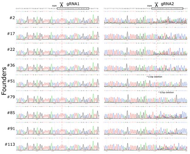
Int. J. Mol. Sci. 2020, 21, 3604                                                                      2 of 14

and effective clinical use in vivo and ex vivo. Such a tremendous involvement of the current technique
in genomic editing can be explained by the easy application, high efficiency, and remarkable flexibility
of this system. However, excessive efficacy of CRISPR/Cas9 system can become a critical point for
many genomic editing applications, if an undesigned genome locus will be edited (off-target activity).
      The Cas9 nuclease has an ability to create unspecific cleavages in a non-target locus, specifically if
off-target sites are similar in sequence to the desired target sites. An off-target activity may depend on
the chromatin state and target site accessibility [21], varies from cell to cell, and needs to be adjusted in
a case-by-case manner. To date, multiple off-target prediction tools have been proposed [22–25], and
most of them utilize algorithms based on sequence similarity. Furthermore, numerous molecular biology
strategies were described to significantly reduce off-target genome activities: alternative editing enzymes,
such as dimeric fusions with FokI [26,27], CpfI [28,29], small Cas9 orthologs [30], CasX [31], and modified
spCas9 enzymes [32,33]; improved delivery strategies, such as adeno-associated viral vectors packaging
the CRISPR-Cas9 system [34,35], inorganic and lipid nanoparticles [36–39], and preassembled CRISPR/Cas
RNPs [40]; protocol modifications with an intracellular concentration of the nuclease and duration of a
nuclease activity [41,42]; truncated gRNAs [43]; and double-nicking strategy [44].
      Another type of an incorrect editing outcome is associated with the occurrence of inaccurate
genome editing at the target locus [45–48]. After DNA cleavage and introducing a double-strand break
(DSB), cellular systems recognize and repair it. If the DNA repair occurs, using a non-homologous
end-joining (NHEJ) path, an error-prone mechanism of the double-strand break repair may happen and
result in the appearance of small insertions or deletions (INDELs) near the site of the initial cleavage.
Consequently, INDELs can cause frameshift mutations. By applying this molecular mechanism, the
production of gene knockouts has been successfully developed [49,50].
      At times, unpredicted large genome modifications can take place within the CRISPR/Cas9
editing process. Recently, unplanned deletions at on-target sites were shown to be more than several
kilobases [51]. In addition, creating mice with chromosomal rearrangements can be accompanied
by hidden complexities, such as a spectrum of structural rearrangements occurring in the same
animal [16,52] or rearrangements larger than originally designed [48]. The frequency of these events
is extremely low, but the occurrence of them may affect adjacent regulatory elements and cause a
serious impact on the activity pattern of surrounding genes. Unanticipated and unpredicted on-target
mutations can be considered an even bigger problem of the CRISPR/Cas9 technology than off-target
activity, as they do not depend on Cas9 nuclease activities but are associated with the functioning of
DNA repair mechanisms in cells. Therefore, to control the phenomenon, it is necessary not to change
the CRISPR/Cas9 activity mechanism but to somehow influence the functioning of DSB repair systems.
      A set of various high-throughput methods to predict and detect off-target mutations have been
developed [53–55]. Nonetheless, no method has been proposed for quantifying the entire spectrum of
possible mutations that can occur at on-target sites. Large undesigned on-target mutations resulting
from the repair of DSB induced by Cas9 are known from a small number of individual examples of
such rearrangements [48,51,56]. However, despite the difficulty in detection, this phenomenon should
be accurately studied because it can cause dangerous obstacles, especially for clinical applications.
      In the current work, we describe the largest, to the best of our knowledge, undesigned on-target
deletion that occurred after the injection of CRISPR/Cas9 system components into mouse zygotes.

2. Results

2.1. Mouse Genome Editing
     To examine the role of CCCTC-binding factor (CTCF) protein in maintaining a three-dimensional
organization of the genome, we planned to obtain mice through a deletion of CTCF binding sites clustered
in the intergenic region between the Kit and Kdr genes. Based on available data, the particular CTCF
cluster of four binding sites, the particular CTCF cluster determines the boundary between two topological
domains (Figure 1). To generate the desired genetic modification, we designed two gRNAs targeted
Int. J. Mol. Sci. 2020, 21, 3604 3 of 14
Int. J. Mol. Sci. 2020, 21, 3604 3 of 15
upstream and downstream
we designed two gRNAs from the selected
targeted upstreamCTCF sites. The components
and downstream of CRISPR/Cas9
from the selected systems,
CTCF sites. The a mix
ofcomponents
two gRNAsofand Cas9 mRNA,
CRISPR/Cas9 were adelivered
systems, into
mix of two the zygotes
gRNAs and Cas9through
mRNA, cytoplasmic microinjections.
were delivered into the In
zygotes
total, through600
we injected cytoplasmic microinjections.
zygotes, where In total,
347 zygotes were we injected 600
transplanted zygotes,
into where 347
CD-1 female zygotes
mice. After all the
were transplanted
manipulations, into female
14 CD-1 CD-1 female mice.produced
recipients After all 113
the live
manipulations, 14 CD-1
and three dead female recipients
pups.
produced 113 live and three dead pups.
Figure1.1.Three-dimensional
Figure organization(Hi-C
Three-dimensional organization (Hi-Cmap)
map)and
and localization
localization of of CTCF
CTCF binding
binding sitessites (ChIP-seq)
(ChIP-
seq)
for for Kit/Kdr
Kit/Kdr genomic
genomic locuslocus in mouse
in mouse embryonic
embryonic stemstem cells,
cells, according
according to to [57].
[57]. TwoTwo gRNAswere
gRNAs wereselected
toselected to delete
delete four CTCF four CTCF binding
binding sites in sites in the intergenic
the intergenic regionregion between
between the and
the Kit Kit and
KdrKdr genes.
genes.
2.2.
2.2.Large
LargeUnexpected Deletionininthe
Unexpected Deletion theTarget
TargetRegion
Region
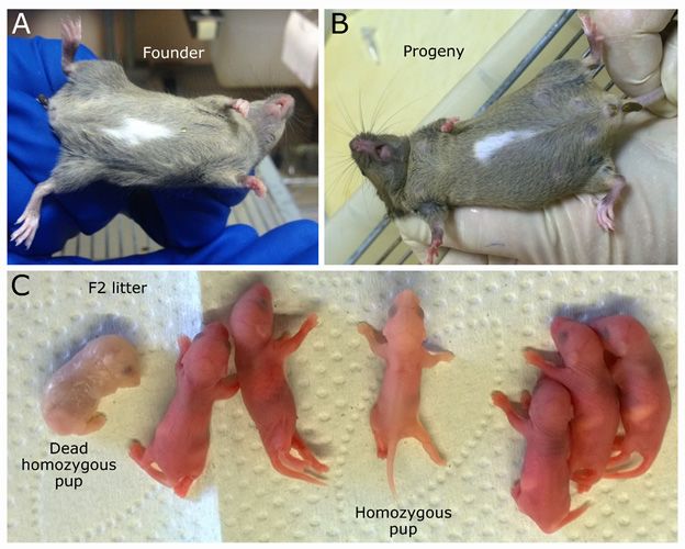
Amongthe
Among the113
113 mice
mice thatthat were
were generated,
generated, wewe detected
detected oneone female
female puppup withwith an unusual
an unusual phenotype.
phenotype. A large white spot was identified on the front part of the body (Figure 2A).
A large white spot was identified on the front part of the body (Figure 2A). Genotyping by polymerase Genotyping
by polymerase
chain chain did
reaction (PCR) reaction (PCR) did
not reveal thenot reveal the
presence of apresence
planned ofgenomic
a plannedrearrangement
genomic rearrangement
in the targeted
in the targeted genomic region in this mouse, as we saw only bands expected for a wild-type allele.
genomic region in this mouse, as we saw only bands expected for a wild-type allele. Surprisingly, we
Surprisingly, we also did not observe any deletions, inversions, or duplications in the targeted locus.
also did not observe any deletions, inversions, or duplications in the targeted locus. To exclude any
To exclude any accidental failure in the development of the founder, we conducted a crossing of the
accidental failure in the development of the founder, we conducted a crossing of the founder female
founder female with a wild-type male. Remarkably, the white spot on the belly was transmitted in
with
accordance withmale.
a wild-type Remarkably,
the Mendelian the white
inheritance spotofondominant
pattern the bellyalleles
was transmitted
(Figure 2B), in accordance
even though thewith the
Mendelian inheritance pattern of dominant alleles (Figure 2B), even though the
shape was slightly different. Moreover, we obtained seven F2 offspring from the crossing between shape was slightly
different. Moreover, we
two F1 heterozygotes obtained
with seven F2phenotype.
a white-spotting offspring fromAmong thethe
crossing betweenwe
seven animals, two F1 heterozygotes
observed two
with
pupsa with
white-spotting phenotype.
typical phenotypes Among
of anemia the skin
and pale seven animals,
color. we observed
Moreover, two pups
the PCR analysis with typical
confirmed
their homozygosity
phenotypes of anemia state.
andHence, the color.
pale skin current mutation the
Moreover, is recessive lethal, confirmed
PCR analysis as homozygous
their animals
homozygosity
were rarely born and usually died in the perinatal period (Figure 2C).
state. Hence, the current mutation is recessive lethal, as homozygous animals were rarely born and
usually died in the perinatal period (Figure 2C).
Several research groups have demonstrated that the white or white-spotting phenotype represents
genetic mutations in the locus near and including the Kit gene (transmembrane tyrosine kinase receptor)
in various organisms, such as alpacas [58], camels [59,60], cats [61], cows [62,63], dogs [64], donkeys [65],
goats [66], horses [67], mice [68–70], pigs [71], rabbits [72], rats [73], yaks [74], humans [75,76], and even
zebrafish [77]. The molecular mechanism behind this phenotype is linked to melanocyte migration
and survival maintained by tyrosine-protein kinase KIT receptor. Due to the lack of melanocytes and,
as a consequence, of melanin production, these regions remain hypopigmented and represent white
spots on the fur.
To establish the molecular nature of the unusual phenotype in our case, the whole genome
sequencing of a heterozygous specimen was performed. Remarkably, within the large region
surrounding the Kit gene (~293 kb), the read coverage was approximately two times lower than that
for the rest of the genome (Figure 3), which is the expected evidence of the deletion in one of the alleles,
i.e., heterozygous deletion. Based on genomic coordinates determined by next-generation sequencingInt. J. Mol. Sci. 2020, 21, 3604 4 of 14
(NGS) data, we designed primers for deletion borders, amplified those regions, and confirmed the
exact coordinates of the deletion, using Sanger sequencing (mm10 chr5:75,588,218-75,881,214; Figure 3).
The right border of the deletion occurred at the expected cutting region for gRNA2, three nucleotides
away from the PAM sequence, whereas the left border moved ~293 kb toward the Kit gene and took
place in the first intron of the Kit gene. Thus, the observed deletion removed 20 out of the 21 exons of
the Kit gene, except for the first one. As the first exon encodes only the 50 -UTR sequence and 22 out of
the 979 amino acids (MRGARGAWDLLCVLLVLLRGQT) of the signal peptide, the generated deletion
Int.referred
can be J. Mol. Sci. to
2020,
as21,
the3604
Kit gene knockout. 4 of 15
Figure 2. Phenotypic
Figure 2. Phenotypicconsequence of an
consequence of anon-target
on-target deletion.
deletion. (A) Founder
(A) Founder with spot
with a white a white spot
on the on the
belly.
belly.(B)(B)F1F1progeny
progenywith withwhite-spotting
white-spotting phenotype.
phenotype. (C) (C) Offspring
Offspring fromfrom
the the crossing
crossing between
between two two
Int. J. Mol. Sci. 2020, 21,with
heterozygotes
heterozygotes with3604 a white
a white spot.
spot. 5 of 15
Several research groups have demonstrated that the white or white-spotting phenotype
represents genetic mutations in the locus near and including the Kit gene (transmembrane tyrosine
kinase receptor) in various organisms, such as alpacas [58], camels [59,60], cats [61], cows [62,63],
dogs [64], donkeys [65], goats [66], horses [67], mice [68–70], pigs [71], rabbits [72], rats [73], yaks [74],
humans [75,76], and even zebrafish [77]. The molecular mechanism behind this phenotype is linked
to melanocyte migration and survival maintained by tyrosine-protein kinase KIT receptor. Due to the
lack of melanocytes and, as a consequence, of melanin production, these regions remain
hypopigmented and represent white spots on the fur.
To establish the molecular nature of the unusual phenotype in our case, the whole genome
sequencing of a heterozygous specimen was performed. Remarkably, within the large region
surrounding the Kit gene (~293 kb), the read coverage was approximately two times lower than that
Figure
for the
Figure Significant
3.rest3.ofSignificantdrop
the genome in(Figure
drop the
in theNGS3),read
NGS read coverage
which visualizes~293
is the expected
coverage visualizes ~293kb kb
evidence deletion
of in heterozygote.
the heterozygote.
theindeletion
deletion the in one of the
Confirmation
alleles,
Confirmation of the
i.e., heterozygous border
of the deletion
deletionwas
deletion.
border wasperformed
Based on genomic
performed viaSanger
via Sanger sequencing.
coordinates determined by next-generation
sequencing.
sequencing (NGS) data, we designed primers for deletion borders, amplified those regions, and
2.3. Off-Target
2.3. or Truncation
Off-Target or Truncation
confirmed the exact coordinates of the deletion, using Sanger sequencing (mm10
chr5:75,588,218-75,881,214;
One of the possible
One of the possible Figure
reasons
reasons that3). The
may
that right
maygive border
giverise
riseto of
tosuchthean
such deletion
an occurred
unplanned
unplanned at thecan
deletion
deletion expected
canbebe cutting
thethe result of
result
region
the mistaken for gRNA2,
off-target
of the mistaken three nucleotides
Cas9 activity
off-target away from
withinwithin
Cas9 activity the left the PAM
theborder sequence,
region.
left border whereas
To test
region. To this the left border
test hypothesis,
this hypothesis, moved
we checked
we
~293 kb the
checked toward
list ofthe Kit geneoff-target
potential and tooksites
place
forinboth
the gRNAs
first intron
usedofinthetheKit gene. Thus,
experiment. the observed
However, none
deletion
of removed
the predicted 20 out of
off-target thewas
sites 21 located
exons ofwithin
the Kitthegene, except
deletion for the first
coordinates one.S1).
(Table AsTherefore,
the first exon
we
encodes only the 5′-UTR sequence
believe that this scenario could unlikely have happened. and 22 out of the 979 amino acids
(MRGARGAWDLLCVLLVLLRGQT)
Furthermore, the regular PCR analysis of thedid
signal
not peptide,
reveal any thespecimen
generated deletion
with can be referred
the deletion, inversion, to
as the Kit gene knockout.
or either duplication of the genomic region flanked by the gRNA recognition sites A,B). ToInt. J. Mol. Sci. 2020, 21, 3604 5 of 14
the list of potential off-target sites for both gRNAs used in the experiment. However, none of the
predicted off-target sites was located within the deletion coordinates (Table S1). Therefore, we believe
that this scenario could unlikely have happened.
Furthermore, the regular PCR analysis did not reveal any specimen with the deletion, inversion,
or either duplication of the genomic region flanked by the gRNA recognition sites A,B). To understand
the molecular basis of the on-targeted deletion occurrence, we characterized Cas9 activities in our
particular CRISPR/Cas9 experiment. Because double-stranded DNA breaks typically get repaired
by NHEJ in eukaryotic cells, CRISPR/Cas9-induced DNA cleavages usually contain small mutations,
such as INDELs near the recognition site. In this way, the presence of INDELs can serve as a positive
indicator of the Cas9 activity. To verify the Cas9 activity in the experiment, we amplified and sequenced
gRNA recognition sites from nine randomly selected founders, after the CRISPR/Cas9 experiment.
Although we found mutations near the gRNA2 recognition site in each founder tested, we did not
observe any INDELs in the gRNA1 locus (Figure 4). More likely, due to the gRNA degradation during
the purification step, gRNA1 was not involved in the CRISPR/Cas9-mediated DNA cleavage. As a
result, we did not detect any animal with anticipated structural rearrangements in the CRISPR/Cas9
experiment. However, the CRISPR/Cas9 complex directed by gRNA2 worked effectively, so all founders
that were analyzed had INDELs in this area. Thus, we assume that the unplanned large deletion in the
white-spotted founder was, more likely, the product of the DSB repair that occurred in the gRNA2
Int. J. Mol. Sci. site.
recognition 2020, 21, 3604 6 of 15
Figure 4.4. Sanger
Figure Sanger sequencing
sequencing for
for gRNA1
gRNA1 andand gRNA2
gRNA2 recognition
recognition sites
sites in
in nine
nine randomly
randomly selected
selected
founders.
founders. Overlapping peaks indicate the presence of heterozygous INDELs, resulting from therepair
Overlapping peaks indicate the presence of heterozygous INDELs, resulting from the repair
of DSB.
of DSB.
3. Discussion
Genome editing directed by the CRISPR/Cas9 system is in widespread use in molecular biology
and in therapeutic and clinical trials, making the editing process relatively easy and allowing for the
introduction of various genomic modifications into the genome, with high accuracy and precision.
On the basis of the CRISPR/Cas9 strategy, many editing tools have been developed, such as nucleases,
nickases, and base editors [32,44,78,79]. However, the spCas9 nuclease remains in the highestInt. J. Mol. Sci. 2020, 21, 3604 6 of 14
3. Discussion
Genome editing directed by the CRISPR/Cas9 system is in widespread use in molecular biology
and in therapeutic and clinical trials, making the editing process relatively easy and allowing for the
introduction of various genomic modifications into the genome, with high accuracy and precision. On
the basis of the CRISPR/Cas9 strategy, many editing tools have been developed, such as nucleases,
nickases, and base editors [32,44,78,79]. However, the spCas9 nuclease remains in the highest demand
and serves as the most commonly used tool for introducing double-stranded breaks at the desired
location in the genome. Using the CRISPR/Cas9 system, researchers can produce the DSB with high
efficiency in the target position, but the process of DSB repair is maintained by internal cellular
repair systems independent of scientists. Thus, the repair mechanism can occur in different ways and
sometimes leads to unpredictable consequences. In this work, we describe the case of an extremely
large deletion that happened at the target site after the injection of CRISPR/Cas9 system components
into mouse zygotes. To the best of our knowledge, this is the largest deletion that has occurred after
the repair of CRISPR/Cas9 introduced DSB in zygotes.
At the present time, there is no technology for an unbiased, high-throughput search for large-scale
rearrangements arising from the repair of a DSB. Usually, either PCR or Southern blot is used to detect
genomic editing events. PCR serves as a very convenient detection method, but any mutation that
destroys a primer annealing site leads to false-negative results [80]. This problem can be partially solved
if the primers are moved away from the DSB site as far as possible [81]. However, PCR amplification
of the fragments larger than 2 kb is inconvenient for routine use. Therefore, any rearrangement of
more than a few kilobases cannot be detected by PCR. In our case, we were not able to detect the
presence of deletion in heterozygote animals: Due to removal of a primer annealing site in one homolog,
heterozygote was undistinguishable from a wild-type animal (Figure S1C). However, when genotyping
a homozygous animal, deletions like the one we described here, can be easily detected by a complete
lack of amplification due to the loss of primer annealing sites in the both alleles. PCR analysis will
not reveal rearrangements with unknown boundaries if genome editing does not involve changing a
sequence copy number (such as inversions or translocations). Southern blots can, more likely, detect
almost any rearrangement, but this method is time-consuming and inefficient. Moreover, in most cases,
one cannot reconstruct the exact nucleotide structure of the rearrangement from the Southern blot
picture, which makes this technology not informative enough.
Apparently, third-generation sequencing methods providing long reads may become the most
promising approach for unanticipated structural variation detection. Oxford nanopore sequencing
technology was successfully used to characterize a complex mutation that occurred during the editing
of a mouse zygote genome. The recent work described in Miyamoto et al. (2019) has demonstrated the
appearance of an inversion (approximately 5 kb) and deletion of a fragment between two introduced
DSBs, using the Oxford Nanopore method [82].
The lack of detection methods makes it almost impossible to accurately assess the occurrence
frequency of such large rearrangements. For instance, in the work of Adikusuma et al. (2018), 57 out
of 127 animals that were tested had deletions in the target site of more than 100 bp, and the largest
deletion detected in the experiment was 2.3 kb in size [81]. At the same time, another group reported
that they did not find large deletions within 55 tested embryos after injecting the components of
CRISPR/Cas9 into a zygote [83]. The particular features of the experiment design that can cause the
occurrence of large unplanned deletions are still unknown. However, it has been shown in experiments
on HEK293T cells that the use of a single Cas9D10A nickase for genome editing prevents on- and
off-target indels and chromosomal truncations, whereas the Cas9 nuclease approach sometimes leads
to unintended rearrangements. Therefore, it can be assumed that DNA double-strand break is the
main cause of large on-target deletions. In addition, p53 knockout cells experience significantly more
chromosomal truncations at the target site, compared with the control cell population, suggesting a
strong involvement of p53 in chromosomal instability induced by CRISPR/Cas9 genome editing [56].
For further progress in this area, the mechanisms of the appearance of large deletions should be furtherInt. J. Mol. Sci. 2020, 21, 3604 7 of 14
examined. There are still no reliable data on the molecular mechanism of the appearance of large
deletions. This is especially important for genome editing in zygotes because several recent studies,
including ours, show that major DSB repair pathways may have unusual high activity in early embryos
that lead to the formation of concatemers from injected DNA molecules [84,85]. It is possible that such
peculiar properties of DSB repair in zygotes will have a role in the formation of large on-target deletion
during genome editing.
4. Materials and Methods
4.1. gRNA Design and gRNA and Cas9 mRNA Preparation
To select gRNAs for the region that determines formation of the TAD boundary in Kit/Kdr locus,
we analyzed CTCF Chip-seq data and Hi-C data from Bonev et al. (2017) [57], in HiGlass visualization
tool (Figure 1) [86]. We found that a cluster of four CTCF sites forms Kit/Kdr TAD border. Based on
their genomic location, we selected regions for CRISPR/Cas9 editing. Two gRNAs were designed
by using Benchling software (https://benchling.com). Then, DNA templates for in vitro transcription
were amplificated with three oligoDNA-primers (Table 1). The reaction was performed in a total
volume of 100 µL containing 1 U of Q5-HF polymerase, 20 µL of ×5 Q5 Reaction Buffer (NEB, M0491),
200 µM of each deoxynucleotide (dATP, dCTP, dGTP, and dTTP), 1000 pmol of T7-gRNA1-FWD or
T7-gRNA2-FWD, 100 pmol of gRNA-REV, and 1000 pmol of REV. The reaction was conducted under
the following conditions: 11 cycles (20 s at 95 ◦ C, 20 s at 58 ◦ C, and 20 sec at 72 ◦ C) and a final incubation
at 72 ◦ C for 2 min. The reaction products were verified on agarose gel and then purified with cleaning
columns (Zymo Research, DNA Clean & Concentrator-5). Purified DNA template was measured with
NanoDrop 2000 and then proceeded with in vitro transcription.
Table 1. DNA oligos for gRNA production and PCR genotyping.
Name Sequence
GTTAATACGACTCACTATAGCGCCGTAAGTGCTGAAAAGGT
T7-gRNA1-FWD
TTTAGAGCTAGAAATAGCAAGTTAA
GTTAATACGACTCACTATAGAGGTAAGAGCAATCCGGTAGT
T7-gRNA2-FWD
TTTAGAGCTAGAAATAGCAAGTTAA
AAAAGCACCGACTCGGTGCCACTTTTTCAAGTTGATAACG
gRNA-REV
GACTAGCCTTATTTTAACTTGCTATTTCTAGCTCTA
REV AAAAGCACCGACTCGGTGCCACTTTTTCAAG
86 TCTTTGTCCTGTTACCGCCC
87 TTGACATGACTCCATGCCCC
88 CCTACGAGCCTTCACGTTGT
89 TGAGGACCGCTGATAGGGAA
90 AAGGCTGTTGTACTGCGTGA
91 ATGATCTCGTGGCGTCATCC
93 GTGCTATGGGAGCCGAAAGA
94 CATAGCCTCTTGCCTTCCGT
179 ATGTTTGGCTGACGCTGAGA
The gRNA recognition sequence is underlined.
The in vitro transcription of gRNA oligos was performed with a HiScribe™ T7 High Yield RNA
Synthesis Kit (NEB, E2040S, Ipswitch, MA, USA), in accordance with the manufacturer’s protocol. The
in vitro transcription of Cas9 mRNA was performed with a HiScribe™ T7 ARCA mRNA Kit (with
tailing) (NEB, E2060S, Ipswitch, MA, USA), in accordance with the manufacturer’s protocol. RNAInt. J. Mol. Sci. 2020, 21, 3604 8 of 14
Clean & Concentrator-25 (Zymo Research, R1017, Irvine, CA, USA) was used for gRNAs and Cas9
mRNA purification, in accordance with the manufacturer’s protocol.
4.2. Preparation of gRNA and Cas9 mRNA
The in vitro transcription of gRNA oligos was performed with a HiScribe™ T7 High Yield RNA
Synthesis Kit (NEB, E2040S, Ipswitch, MA, USA), in accordance with the manufacturer’s protocol. The
in vitro transcription of Cas9 mRNA was performed with a HiScribe™ T7 ARCA mRNA Kit (with
tailing) (NEB, E2060S, Ipswitch, MA, USA), in accordance with the manufacturer’s protocol. RNA
Clean & Concentrator-25 (Zymo Research, R1017, Irvine, CA, USA) was used for gRNAs and Cas9
mRNA purification, in accordance with the manufacturer’s protocol.
4.3. Animals
Six-week-old F1 hybrid CBA/J x C57BL/6J female mice were superovulated with 7.5 ME of PMSG
(Folligon, Intervet, Boxmeer, Holland) on the first day 1 (4:00 p.m.) and 7.5 ME of hCG (Chorulon,
Intervet, Boxmeer, Holland) on the third day (1:00 p.m). Females were crossed with C57BL/6J males,
and on the next morning (fourth day), the females were checked for copulation plugs. The individuals
with plugs were euthanized by cervical dislocation, then oviducts were excised, and zygote–cumulus
mass complexes were flushed out into an M2 medium (M7167, Sigma-Aldrich) by dissecting the
ampulla. Cumulus cells were removed by a hyaluronidase (H3506, Sigma-Aldrich) treatment, and
the zygotes were rinsed into a fresh M2 medium. Then, the zygotes were transferred into an M16
medium (M7292, Sigma-Aldrich) and cultivated in the medium drops covered with mineral oil (M8410,
Sigma-Aldrich) in a cell culture incubator with 5% CO2 in the air.
4.4. Microinjection
After the incubation, two pronuclear zygotes were placed into a drop of the M2 medium covered
with mineral oil and then microinjected into the cytoplasm by mixing two gRNAs (10 ng/uL each) and
mRNA Cas9 (50 ng/uL) diluted in nuclease-free water. Next, injected embryos were cultured for a
short time (1–2 h), in drops of the M16 medium covered with mineral oil at 37 ◦ C and an atmosphere
of 5% CO2 . Finally, the embryos survived after microinjection were transplanted into the oviducts of
the pseudopregnant CD-1 females (0.5 d.p.c.).
The mice were maintained on a 12-hour light/dark cycle, with ad libitum food and water, in a
conventional animal facility. All experiments were conducted at the Department of Experimental
Animal Genetic Resources, at the Institute of Cytology and Genetics, SB RAS (RFMEFI61914X0005
and FMEFI61914X0010). All the procedures and technical manipulations with animals were in
compliance with the European Communities Council Directive of 24 November 1986 (86/609/EEC) and
approved by the Bioethical Committee at the Institute of Cytology and Genetics (Permission N45 from
16 November 2018).
4.5. Genotyping
Genomic DNA was isolated from murine tails by placing them in 500 µL of tail lysis buffer
containing 100 mM NaCl, 10 mM Tris pH 8.0, 25 mM EDTA, 0.5% sodium dodecyl sulfate, and
0.2 µg/µL proteinase K and then incubating them at 56 ◦ C, until the tissue completely dissolved.
Residual fur and bones were removed by centrifugation, and lysates were transferred to the fresh tubes
and treated by a standard phenol–chloroform extraction method, followed by ethanol precipitation.
Then, ~20 ng (2 µL) of genomic DNA was used for PCR genotyping. The reaction was performed in a
total volume of 25 µL containing 1× Taq AS buffer (67 mM Tris-HCl, pH 8.8; 16.6 mM (NH4)2SO4,
0.01% Tween-20) with 1.5 mM MgCl2, 0.2 mM of each deoxynucleotide (dATP, dCTP, dGTP, and dTTP),
0.4 mM of forward and reverse primers (Table 1 and tab1D), and 1 U of Taq polymerase. The reaction
was conducted under the following conditions: initial denaturation for 3 min at 95 ◦ C, 35 cycles (30 s
at 95 ◦ C, 30 s at 63 ◦ C, and 2 min at 72 ◦ C), and a final incubation at 72 ◦ C for 3 min. PCR productsInt. J. Mol. Sci. 2020, 21, 3604 9 of 14
were analyzed by electrophoresis with 2% agarose gels in a Tris–EDTA–acetate buffer and sequenced,
using the BigDye® Terminator v3.1 kit (Thermo Fisher Scientific, Waltham, MA, USA), on an Applied
Biosystems 3500 Genetic Analyzer, in accordance with the manufacturer’s recommendations.
4.6. Whole Genome Sequencing
Genomic DNA was extracted from the tail tissue of a heterozygous F1 mouse with white spot
and subjected to paired-end 2× 150 bp Illumina sequencing, generating approximately 400 million
reads. Raw reads were analyzed by using FastQC software, to ensure high data quality, and mapped
to the mouse genome (mm10) with Bowtie 2 with default parameters. To perform data visualization,
alignments were converted to BAM format, sorted and indexed, using SAMtools, and loaded as the
IGV browser track.
4.7. Data Availability
All sequencing data will be publicly available upon publication, via NCBI Sequencing Read
Archive (SRA) under SRX8102546.
5. Conclusions
Here we describe the largest, to the best of our knowledge, undesigned on-target deletion with a
size of ~293 kb that occurred after the cytoplasmic injection of CRISPR/Cas9 system components into
mouse zygotes. We propose that deletion occurred as a result of the truncation of one of the ends of a
double-strand break during the repair.
Supplementary Materials: Supplementary materials can be found at http://www.mdpi.com/1422-0067/21/10/
3604/s1. Figure S1. PCR detection of the rearrangements in founders. Table S1. Potential of target sites.
Author Contributions: Conceptualization, N.B.; Methodology, A.K. and I.S.; Investigation, A.K. and V.L.; Writing
Original Draft Preparation, A.K., V.L. and N.B.;Writing Review & Editing, V.L. and N.B.; Project Administration,
N.B.; Funding Acquisition, N.B. All authors have read and agreed to the published version of the manuscript.
Funding: The reported study was funded by RFBR, with research project no. 18-29-07022. WGS was performed
with support from the Ministry of Education and Science of Russian Federation, grant #2019-0546 (FSUS-2020-0040).
NGS data analysis was performed with support from the computational cluster of the Novosibirsk State University
and computational nodes of the Institute of Cytology and Genetics (budget project no. 0324-2019-0041-C-01).
Acknowledgments: We thank Veniamin Fish man and Oleg Serov for fruitful discussions, and critical comments.
Also, we thank Boris Skryabin, Leonid Gubar, and Helen Kaiser for protocols and suggestions for manipulation
with mouse embryos and transgenic mouse production.
Conflicts of Interest: The authors declare no conflict of interest.
References
1. Crowther, M.D.; Dolton, G.; Legut, M.; Caillaud, M.E.; Lloyd, A.; Attaf, M.; Galloway, S.A.E.; Rius, C.;
Farrell, C.P.; Szomolay, B.; et al. Genome-wide CRISPR–Cas9 screening reveals ubiquitous T cell cancer
targeting via the monomorphic MHC class I-related protein MR1. Nat. Immunol. 2020, 21, 178–185. [CrossRef]
[PubMed]
2. Hart, T.; Chandrashekhar, M.; Aregger, M.; Steinhart, Z.; Brown, K.R.; MacLeod, G.; Mis, M.; Zimmermann, M.;
Fradet-Turcotte, A.; Sun, S.; et al. High-Resolution CRISPR Screens Reveal Fitness Genes and
Genotype-Specific Cancer Liabilities. Cell 2015, 163, 1515–1526. [CrossRef]
3. Picco, G.; Chen, E.D.; Alonso, L.G.; Behan, F.M.; Gonçalves, E.; Bignell, G.; Matchan, A.; Fu, B.; Banerjee, R.;
Anderson, E.; et al. Functional linkage of gene fusions to cancer cell fitness assessed by pharmacological and
CRISPR-Cas9 screening. Nat. Commun. 2019, 10, 2198. [CrossRef] [PubMed]
4. Shalem, O.; Sanjana, N.E.; Hartenian, E.; Shi, X.; Scott, D.A.; Mikkelsen, T.S.; Heckl, D.; Ebert, B.L.; Root, D.E.;
Doench, J.G.; et al. Genome-Scale CRISPR-Cas9 Knockout Screening in Human Cells. Science 2014, 343,
84–87. [CrossRef] [PubMed]Int. J. Mol. Sci. 2020, 21, 3604 10 of 14
5. Carroll, K.J.; Makarewich, C.A.; McAnally, J.; Anderson, D.M.; Zentilin, L.; Liu, N.; Giacca, M.; Bassel-Duby, R.;
Olson, E.N. A mouse model for adult cardiac-specific gene deletion with CRISPR/Cas9. Proc. Natl. Acad. Sci.
USA 2016, 113, 338–343. [CrossRef] [PubMed]
6. Korablev, A.N.; Serova, I.A.; Serov, O.L. Generation of megabase-scale deletions, inversions and duplications
involving the Contactin-6 gene in mice by CRISPR/Cas9 technology. BMC Genet. 2017, 18. [CrossRef]
[PubMed]
7. Pristyazhnyuk, I.E.; Minina, J.; Korablev, A.; Serova, I.; Fishman, V.; Gridina, M.; Rozhdestvensky, T.S.;
Gubar, L.; Skryabin, B.V.; Serov, O.L. Time origin and structural analysis of the induced CRISPR/cas9
megabase-sized deletions and duplications involving the Cntn6 gene in mice. Sci. Rep. 2019, 9, 14161.
[CrossRef] [PubMed]
8. Kraft, K.; Geuer, S.; Will, A.J.; Chan, W.; Paliou, C.; Borschiwer, M.; Harabula, I.; Wittler, L.; Franke, M.;
Ibrahim, D.M.; et al. Deletions, inversions, duplications: Engineering of structural variants using CRISPR/Cas
in mice. Cell Rep. 2015, 10, 833–839. [CrossRef]
9. Artus, J.; Hadjantonakis, A.-K. Generation of Chimeras by Aggregation of Embryonic Stem Cells with
Diploid or Tetraploid Mouse Embryos. Methods Mol. Biol. 2011, 693, 37–56.
10. Lupiáñez, D.G.; Kraft, K.; Heinrich, V.; Krawitz, P.; Brancati, F.; Klopocki, E.; Horn, D.; Kayserili, H.;
Opitz, J.M.; Laxova, R.; et al. Disruptions of topological chromatin domains cause pathogenic rewiring of
gene-enhancer interactions. Cell 2015, 161, 1012–1025. [CrossRef]
11. Yang, H.; Wang, H.; Shivalila, C.S.; Cheng, A.W.; Shi, L.; Jaenisch, R. XOne-step generation of mice carrying
reporter and conditional alleles by CRISPR/cas-mediated genome engineering. Cell 2013, 154, 1370–1379.
[CrossRef] [PubMed]
12. Jin, Y.; Lee, A.; Oh, J.H.; Lee, H.W.; Ha, S.J. The R229Q mutation of Rag2 does not characterize severe
immunodeficiency in mice. Sci. Rep. 2019, 9, 4415. [CrossRef] [PubMed]
13. Yoshimi, K.; Kunihiro, Y.; Kaneko, T.; Nagahora, H.; Voigt, B.; Mashimo, T. ssODN-mediated knock-in with
CRISPR-Cas for large genomic regions in zygotes. Nat. Commun. 2016, 7, 10431. [CrossRef] [PubMed]
14. Chu, V.T.; Weber, T.; Graf, R.; Sommermann, T.; Petsch, K.; Sack, U.; Volchkov, P.; Rajewsky, K.; Kühn, R.
Efficient generation of Rosa26 knock-in mice using CRISPR/Cas9 in C57BL/6 zygotes. BMC Biotechnol. 2016,
16, 4. [CrossRef]
15. Kvon, E.Z.; Zhu, Y.; Kelman, G.; Novak, C.S.; Plajzer-Frick, I.; Kato, M.; Garvin, T.H.; Pham, Q.;
Harrington, A.N.; Hunter, R.D.; et al. Comprehensive In Vivo Interrogation Reveals Phenotypic Impact of
Human Enhancer Variants. Cell 2020, 180, 1262–1271.e15. [CrossRef]
16. Boroviak, K.; Doe, B.; Banerjee, R.; Yang, F.; Bradley, A. Chromosome engineering in zygotes with CRISPR/Cas9.
Genesis 2016, 54, 78–85. [CrossRef]
17. Saito, R.; Koebis, M.; Nagai, T.; Shimizu, K.; Liao, J.; Wulaer, B.; Sugaya, Y.; Nagahama, K.; Uesaka, N.;
Kushima, I.; et al. Comprehensive analysis of a novel mouse model of the 22q11.2 deletion syndrome: A
model with the most common 3.0-Mb deletion at the human 22q11.2 locus. Transl. Psychiatry 2020, 10, 35.
[CrossRef]
18. Kato, T.; Hara, S.; Goto, Y.; Ogawa, Y.; Okayasu, H.; Kubota, S.; Tamano, M.; Terao, M.; Takada, S. Creation
of mutant mice with megabase-sized deletions containing custom-designed breakpoints by means of the
CRISPR/Cas9 system. Sci. Rep. 2017, 7, 59. [CrossRef]
19. German, D.M.; Mitalipov, S.; Mishra, A.; Kaul, S. Therapeutic Genome Editing in Cardiovascular Diseases.
JACC Basic Transl. Sci. 2019, 4, 122–131. [CrossRef]
20. Porteus, M.H. Towards a new era in medicine: Therapeutic genome editing. Genome Biol. 2015, 16, 286.
[CrossRef]
21. Kim, D.; Kim, J.-S. DIG-seq: A genome-wide CRISPR off-target profiling method using chromatin DNA.
Genome Res. 2018, 28, 1894–1900. [CrossRef] [PubMed]
22. Bae, S.; Park, J.; Kim, J.-S. Cas-OFFinder: A fast and versatile algorithm that searches for potential off-target
sites of Cas9 RNA-guided endonucleases. Bioinformatics 2014, 30, 1473–1475. [CrossRef] [PubMed]
23. Cradick, T.J.; Qiu, P.; Lee, C.M.; Fine, E.J.; Bao, G. COSMID: A Web-based Tool for Identifying and Validating
CRISPR/Cas Off-target Sites. Mol. Ther. Nucleic Acids 2014, 3, e214. [CrossRef] [PubMed]
24. Hsu, P.D.; Scott, D.A.; Weinstein, J.A.; Ran, F.A.; Konermann, S.; Agarwala, V.; Li, Y.; Fine, E.J.; Wu, X.;
Shalem, O.; et al. DNA targeting specificity of RNA-guided Cas9 nucleases. Nat. Biotechnol. 2013, 31, 827–832.
[CrossRef]Int. J. Mol. Sci. 2020, 21, 3604 11 of 14
25. Labuhn, M.; Adams, F.F.; Ng, M.; Knoess, S.; Schambach, A.; Charpentier, E.M.; Schwarzer, A.; Mateo, J.L.;
Klusmann, J.-H.; Heckl, D. Refined sgRNA efficacy prediction improves large- and small-scale CRISPR–Cas9
applications. Nucleic Acids Res. 2018, 46, 1375–1385. [CrossRef]
26. Havlicek, S.; Shen, Y.; Alpagu, Y.; Bruntraeger, M.B.; Zufir, N.B.M.; Phuah, Z.Y.; Fu, Z.; Dunn, N.R.;
Stanton, L.W. Re-engineered RNA-Guided FokI-Nucleases for Improved Genome Editing in Human Cells.
Mol. Ther. 2017, 25, 342–355. [CrossRef]
27. Tsai, S.Q.; Wyvekens, N.; Khayter, C.; Foden, J.A.; Thapar, V.; Reyon, D.; Goodwin, M.J.; Aryee, M.J.;
Joung, J.K. Dimeric CRISPR RNA-guided FokI nucleases for highly specific genome editing. Nat. Biotechnol.
2014, 32, 569–576. [CrossRef]
28. Fagerlund, R.D.; Staals, R.H.J.; Fineran, P.C. The Cpf1 CRISPR-Cas protein expands genome-editing tools.
Genome Biol. 2015, 16, 251. [CrossRef]
29. Zetsche, B.; Gootenberg, J.S.; Abudayyeh, O.O.; Slaymaker, I.M.; Makarova, K.S.; Essletzbichler, P.; Volz, S.E.;
Joung, J.; Van Der Oost, J.; Regev, A.; et al. Cpf1 Is a Single RNA-Guided Endonuclease of a Class 2
CRISPR-Cas System. Cell 2015, 163, 759–771. [CrossRef]
30. Kim, E.; Koo, T.; Park, S.W.; Kim, D.; Kim, K.; Cho, H.-Y.; Song, D.W.; Lee, K.J.; Jung, M.H.; Kim, S.; et al.
In vivo genome editing with a small Cas9 orthologue derived from Campylobacter jejuni. Nat. Commun.
2017, 8, 14500. [CrossRef]
31. Liu, J.-J.; Orlova, N.; Oakes, B.L.; Ma, E.; Spinner, H.B.; Baney, K.L.M.; Chuck, J.; Tan, D.; Knott, G.J.;
Harrington, L.B.; et al. CasX enzymes comprise a distinct family of RNA-guided genome editors. Nature
2019, 566, 218–223. [CrossRef] [PubMed]
32. Kleinstiver, B.P.; Prew, M.S.; Tsai, S.Q.; Topkar, V.V.; Nguyen, N.T.; Zheng, Z.; Gonzales, A.P.W.; Li, Z.;
Peterson, R.T.; Yeh, J.-R.J.; et al. Engineered CRISPR-Cas9 nucleases with altered PAM specificities. Nature
2015, 523, 481–485. [CrossRef] [PubMed]
33. Kleinstiver, B.P.; Pattanayak, V.; Prew, M.S.; Tsai, S.Q.; Nguyen, N.T.; Zheng, Z.; Keith Joung, J. High-fidelity
CRISPR–Cas9 nucleases with no detectable genome-wide off-target effects. Nature 2016. [CrossRef] [PubMed]
34. Lau, C.-H.; Suh, Y. In vivo genome editing in animals using AAV-CRISPR system: Applications to translational
research of human disease. F1000Research 2017, 6, 2153. [CrossRef]
35. Mendell, J.R.; Al-Zaidy, S.; Shell, R.; Arnold, W.D.; Rodino-Klapac, L.R.; Prior, T.W.; Lowes, L.; Alfano, L.;
Berry, K.; Church, K.; et al. Single-Dose Gene-Replacement Therapy for Spinal Muscular Atrophy. N. Engl. J.
Med. 2017, 377, 1713–1722. [CrossRef]
36. Ding, Y.; Jiang, Z.; Saha, K.; Kim, C.S.; Kim, S.T.; Landis, R.F.; Rotello, V.M. Gold Nanoparticles for Nucleic
Acid Delivery. Mol. Ther. 2014, 22, 1075–1083. [CrossRef]
37. Glass, Z.; Li, Y.; Xu, Q. Nanoparticles for CRISPR–Cas9 delivery. Nat. Biomed. Eng. 2017, 1, 854–855.
[CrossRef]
38. Lee, K.; Conboy, M.; Park, H.M.; Jiang, F.; Kim, H.J.; Dewitt, M.A.; Mackley, V.A.; Chang, K.; Rao, A.;
Skinner, C.; et al. Nanoparticle delivery of Cas9 ribonucleoprotein and donor DNA in vivo induces
homology-directed DNA repair. Nat. Biomed. Eng. 2017, 1, 889–901. [CrossRef]
39. Sago, C.D.; Lokugamage, M.P.; Paunovska, K.; Vanover, D.A.; Monaco, C.M.; Shah, N.N.; Gamboa Castro, M.;
Anderson, S.E.; Rudoltz, T.G.; Lando, G.N.; et al. High-throughput in vivo screen of functional mRNA
delivery identifies nanoparticles for endothelial cell gene editing. Proc. Natl. Acad. Sci. USA 2018, 115,
E9944–E9952. [CrossRef]
40. Staahl, B.T.; Benekareddy, M.; Coulon-Bainier, C.; Banfal, A.A.; Floor, S.N.; Sabo, J.K.; Urnes, C.; Munares, G.A.;
Ghosh, A.; Doudna, J.A. Efficient genome editing in the mouse brain by local delivery of engineered Cas9
ribonucleoprotein complexes. Nat. Biotechnol. 2017, 35, 431–434. [CrossRef]
41. Cameron, P.; Fuller, C.K.; Donohoue, P.D.; Jones, B.N.; Thompson, M.S.; Carter, M.M.; Gradia, S.; Vidal, B.;
Garner, E.; Slorach, E.M.; et al. Mapping the genomic landscape of CRISPR–Cas9 cleavage. Nat. Methods
2017, 14, 600–606. [CrossRef] [PubMed]
42. Ramakrishna, S.; Kwaku Dad, A.-B.; Beloor, J.; Gopalappa, R.; Lee, S.-K.; Kim, H. Gene disruption by
cell-penetrating peptide-mediated delivery of Cas9 protein and guide RNA. Genome Res. 2014, 24, 1020–1027.
[CrossRef] [PubMed]
43. Fu, Y.; Sander, J.D.; Reyon, D.; Cascio, V.M.; Joung, J.K. Improving CRISPR-Cas nuclease specificity using
truncated guide RNAs. Nat. Biotechnol. 2014, 32, 279–284. [CrossRef] [PubMed]Int. J. Mol. Sci. 2020, 21, 3604 12 of 14
44. Ran, F.A.; Hsu, P.D.; Lin, C.-Y.; Gootenberg, J.S.; Konermann, S.; Trevino, A.E.; Scott, D.A.; Inoue, A.;
Matoba, S.; Zhang, Y.; et al. Double nicking by RNA-guided CRISPR Cas9 for enhanced genome editing
specificity. Cell 2013, 154, 1380–1389. [CrossRef]
45. van Overbeek, M.; Capurso, D.; Carter, M.M.; Thompson, M.S.; Frias, E.; Russ, C.; Reece-Hoyes, J.S.; Nye, C.;
Gradia, S.; Vidal, B.; et al. DNA Repair Profiling Reveals Nonrandom Outcomes at Cas9-Mediated Breaks.
Mol. Cell 2016. [CrossRef]
46. Ghezraoui, H.; Piganeau, M.; Renouf, B.; Renaud, J.-B.; Sallmyr, A.; Ruis, B.; Oh, S.; Tomkinson, A.E.;
Hendrickson, E.A.; Giovannangeli, C.; et al. Chromosomal Translocations in Human Cells Are Generated by
Canonical Nonhomologous End-Joining. Mol. Cell 2014, 55, 829–842. [CrossRef]
47. Tan, E.-P.; Li, Y.; Del Castillo Velasco-Herrera, M.; Yusa, K.; Bradley, A. Off-target assessment of CRISPR-Cas9
guiding RNAs in human iPS and mouse ES cells. Genesis 2015, 53, 225–236. [CrossRef]
48. Birling, M.-C.; Schaeffer, L.; André, P.; Lindner, L.; Maréchal, D.; Ayadi, A.; Sorg, T.; Pavlovic, G.; Hérault, Y.
Efficient and rapid generation of large genomic variants in rats and mice using CRISMERE. Sci. Rep. 2017, 7,
43331. [CrossRef]
49. Li, D.; Qiu, Z.; Shao, Y.; Chen, Y.; Guan, Y.; Liu, M.; Li, Y.; Gao, N.; Wang, L.; Lu, X.; et al. Heritable gene
targeting in the mouse and rat using a CRISPR-Cas system. Nat. Biotechnol. 2013, 31, 681–683. [CrossRef]
50. Wang, H.; Yang, H.; Shivalila, C.S.; Dawlaty, M.M.; Cheng, A.W.; Zhang, F.; Jaenisch, R. One-step generation
of mice carrying mutations in multiple genes by CRISPR/cas-mediated genome engineering. Cell 2013, 153,
910–918. [CrossRef]
51. Kosicki, M.; Tomberg, K.; Bradley, A. Repair of double-strand breaks induced by CRISPR–Cas9 leads to large
deletions and complex rearrangements. Nat. Biotechnol. 2018, 36, 765–771. [CrossRef] [PubMed]
52. Boroviak, K.; Fu, B.; Yang, F.; Doe, B.; Bradley, A. Revealing hidden complexities of genomic rearrangements
generated with Cas9. Sci. Rep. 2017, 7, 12867. [CrossRef] [PubMed]
53. Pattanayak, V.; Lin, S.; Guilinger, J.P.; Ma, E.; Doudna, J.A.; Liu, D.R. High-throughput profiling of off-target
DNA cleavage reveals RNA-programmed Cas9 nuclease specificity. Nat. Biotechnol. 2013, 31, 839–843.
[CrossRef] [PubMed]
54. Frock, R.L.; Hu, J.; Meyers, R.M.; Ho, Y.-J.; Kii, E.; Alt, F.W. Genome-wide detection of DNA double-stranded
breaks induced by engineered nucleases. Nat. Biotechnol. 2015, 33, 179–186. [CrossRef]
55. Tsai, S.Q.; Zheng, Z.; Nguyen, N.T.; Liebers, M.; Topkar, V.V.; Thapar, V.; Wyvekens, N.; Khayter, C.;
Iafrate, A.J.; Le, L.P.; et al. GUIDE-seq enables genome-wide profiling of off-target cleavage by CRISPR-Cas
nucleases. Nat. Biotechnol. 2015, 33, 187–197. [CrossRef]
56. Cullot, G.; Boutin, J.; Toutain, J.; Prat, F.; Pennamen, P.; Rooryck, C.; Teichmann, M.; Rousseau, E.;
Lamrissi-Garcia, I.; Guyonnet-Duperat, V.; et al. CRISPR-Cas9 genome editing induces megabase-scale
chromosomal truncations. Nat. Commun. 2019. [CrossRef]
57. Bonev, B.; Mendelson Cohen, N.; Szabo, Q.; Fritsch, L.; Papadopoulos, G.L.; Lubling, Y.; Xu, X.; Lv, X.;
Hugnot, J.P.; Tanay, A.; et al. Multiscale 3D Genome Rewiring during Mouse Neural Development. Cell 2017,
171, 557–572.e24. [CrossRef]
58. Jackling, F.C.; Johnson, W.E.; Appleton, B.R. The Genetic Inheritance of the Blue-eyed White Phenotype in
Alpacas (Vicugna pacos). J. Hered. 2014, 105, 941–951. [CrossRef]
59. Holl, H.; Isaza, R.; Mohamoud, Y.; Ahmed, A.; Almathen, F.; Youcef, C.; Gaouar, S.; Antczak, D.; Brooks, S. A
Frameshift Mutation in KIT is Associated with White Spotting in the Arabian Camel. Genes 2017, 8, 102.
[CrossRef]
60. Volpato, G.; Dioli, M.; Di Nardo, A. Piebald Camels. Pastoralism 2017, 7, 3. [CrossRef]
61. David, V.A.; Menotti-Raymond, M.; Wallace, A.C.; Roelke, M.; Kehler, J.; Leighty, R.; Eizirik, E.; Hannah, S.S.;
Nelson, G.; Schäffer, A.A.; et al. Endogenous Retrovirus Insertion in the KIT Oncogene Determines White
and White spotting in Domestic Cats. G3 Genes Genomes Genet. 2014, 4, 1881–1891. [CrossRef]
62. Fontanesi, L.; Tazzoli, M.; Russo, V.; Beever, J. Genetic heterogeneity at the bovine KIT gene in cattle breeds
carrying different putative alleles at the spotting locus. Anim. Genet. 2010, 41, 295–303. [CrossRef] [PubMed]
63. Häfliger, I.M.; Hirter, N.; Paris, J.M.; Wolf Hofstetter, S.; Seefried, F.R.; Drögemüller, C. A de novo germline
mutation of KIT in a white-spotted Brown Swiss cow. Anim. Genet. 2020, 51, 449–452. [CrossRef] [PubMed]
64. Wong, A.K.; Ruhe, A.L.; Robertson, K.R.; Loew, E.R.; Williams, D.C.; Neff, M.W. A de novo mutation in
KIT causes white spotting in a subpopulation of German Shepherd dogs. Anim. Genet. 2013, 44, 305–310.
[CrossRef] [PubMed]Int. J. Mol. Sci. 2020, 21, 3604 13 of 14
65. Haase, B.; Rieder, S.; Leeb, T. Two variants in the KIT gene as candidate causative mutations for a dominant
white and a white spotting phenotype in the donkey. Anim. Genet. 2015, 46, 321–324. [CrossRef]
66. Nazari-Ghadikolaei, A.; Mehrabani-Yeganeh, H.; Miarei-Aashtiani, S.R.; Staiger, E.A.; Rashidi, A.; Huson, H.J.
Genome-Wide Association Studies Identify Candidate Genes for Coat Color and Mohair Traits in the Iranian
Markhoz Goat. Front. Genet. 2018, 9. [CrossRef]
67. Hauswirth, R.; Jude, R.; Haase, B.; Bellone, R.R.; Archer, S.; Holl, H.; Brooks, S.A.; Tozaki, T.; Penedo, M.C.T.;
Rieder, S.; et al. Novel variants in the KIT and PAX3 genes in horses with white-spotted coat colour
phenotypes. Anim. Genet. 2013, 44, 763–765. [CrossRef]
68. Chabot, B.; Stephenson, D.A.; Chapman, V.M.; Besmer, P.; Bernstein, A. The proto-oncogene c-kit encoding a
transmembrane tyrosine kinase receptor maps to the mouse W locus. Nature 1988, 335, 88–89. [CrossRef]
69. Geissler, E.N.; Ryan, M.A.; Housman, D.E. The dominant-white spotting (W) locus of the mouse encodes the
c-kit proto-oncogene. Cell 1988, 55, 185–192. [CrossRef]
70. Nocka, K.; Majumder, S.; Chabot, B.; Ray, P.; Cervone, M.; Bernstein, A.; Besmer, P. Expression of c-kit gene
products in known cellular targets of W mutations in normal and W mutant mice–evidence for an impaired
c-kit kinase in mutant mice. Genes Dev. 1989, 3, 816–826. [CrossRef]
71. Lim, H.T.; Zhong, T.; Cho, I.C.; Seo, B.Y.; Kim, J.H.; Lee, S.S.; Ko, M.S.; Park, H.B.; Kim, B.W.; Lee, J.H.; et al.
Novel alternative splicing by exon skipping in KIT associated with whole-body roan in an intercrossed
population of Landrace and Korean Native pigs. Anim. Genet. 2011, 42, 451–455. [CrossRef] [PubMed]
72. Fontanesi, L.; Vargiolu, M.; Scotti, E.; Latorre, R.; Faussone Pellegrini, M.S.; Mazzoni, M.; Asti, M.;
Chiocchetti, R.; Romeo, G.; Clavenzani, P.; et al. The KIT Gene Is Associated with the English Spotting Coat
Color Locus and Congenital Megacolon in Checkered Giant Rabbits (Oryctolagus cuniculus). PLoS ONE
2014, 9, e93750. [CrossRef] [PubMed]
73. Tsujimura, T.; Hirota, S.; Nomura, S.; Niwa, Y.; Yamazaki, M.; Tono, T.; Morii, E.; Kim, H.; Kondo, K.;
Nishimune, Y. Characterization of Ws mutant allele of rats: A 12-base deletion in tyrosine kinase domain of
c-kit gene. Blood 1991, 78, 1942–1946. [CrossRef] [PubMed]
74. Zhang, M.Q.; Xu, X.; Luo, S.J. The genetics of brown coat color and white spotting in domestic yaks (Bos
grunniens). Anim. Genet. 2014. [CrossRef]
75. Ezoe, K.; Holmes, S.A.; Ho, L.; Bennett, C.P.; Bolognia, J.L.; Brueton, L.; Burn, J.; Falabella, R.; Gatto, E.M.;
Ishii, N. Novel mutations and deletions of the KIT (steel factor receptor) gene in human piebaldism. Am. J.
Hum. Genet. 1995, 56, 58–66.
76. Giebel, L.B.; Spritz, R.A. Mutation of the KIT (mast/stem cell growth factor receptor) protooncogene in
human piebaldism. Proc. Natl. Acad. Sci. USA 1991, 88, 8696–8699. [CrossRef]
77. Parichy, D.M.; Mellgren, E.M.; Rawls, J.F.; Lopes, S.S.; Kelsh, R.N.; Johnson, S.L. Mutational Analysis of
Endothelin Receptor b1 (rose) during Neural Crest and Pigment Pattern Development in the Zebrafish Danio
rerio. Dev. Biol. 2000, 227, 294–306. [CrossRef]
78. Anzalone, A.V.; Randolph, P.B.; Davis, J.R.; Sousa, A.A.; Koblan, L.W.; Levy, J.M.; Chen, P.J.; Wilson, C.;
Newby, G.A.; Raguram, A.; et al. Search-and-replace genome editing without double-strand breaks or donor
DNA. Nature 2019, 576, 149–157. [CrossRef]
79. Rees, H.A.; Liu, D.R. Base editing: Precision chemistry on the genome and transcriptome of living cells. Nat.
Rev. Genet. 2018, 19, 770–788. [CrossRef]
80. Shin, H.Y.; Wang, C.; Lee, H.K.; Yoo, K.H.; Zeng, X.; Kuhns, T.; Yang, C.M.; Mohr, T.; Liu, C.; Hennighausen, L.
CRISPR/Cas9 targeting events cause complex deletions and insertions at 17 sites in the mouse genome. Nat.
Commun. 2017. [CrossRef]
81. Adikusuma, F.; Piltz, S.; Corbett, M.A.; Turvey, M.; McColl, S.R.; Helbig, K.J.; Beard, M.R.; Hughes, J.;
Pomerantz, R.T.; Thomas, P.Q. Large deletions induced by Cas9 cleavage. Nature 2018. [CrossRef] [PubMed]
82. Miyamoto, S.; Aoto, K.; Hiraide, T.; Nakashima, M.; Takabayashi, S.; Saitsu, H. Nanopore sequencing reveals
a structural alteration of mirror-image duplicated genes in a genome-editing mouse line. Congenit. Anom.
2019. [CrossRef] [PubMed]
83. Ma, H.; Marti-Gutierrez, N.; Park, S.-W.; Wu, J.; Hayama, T.; Darby, H.; Van Dyken, C.; Li, Y.; Koski, A.;
Liang, D.; et al. Ma et al. reply. Nature 2018, 560, E10–E23. [CrossRef] [PubMed]
84. Smirnov, A.; Fishman, V.; Yunusova, A.; Korablev, A.; Serova, I.; Skryabin, B.V.; Rozhdestvensky, T.S.;
Battulin, N. DNA barcoding reveals that injected transgenes are predominantly processed by homologous
recombination in mouse zygote. Nucleic Acids Res. 2019. [CrossRef] [PubMed]Int. J. Mol. Sci. 2020, 21, 3604 14 of 14
85. Skryabin, B.V.; Kummerfeld, D.-M.; Gubar, L.; Seeger, B.; Kaiser, H.; Stegemann, A.; Roth, J.; Meuth, S.G.;
Pavenstädt, H.; Sherwood, J.; et al. Pervasive head-to-tail insertions of DNA templates mask desired
CRISPR-Cas9–mediated genome editing events. Sci. Adv. 2020, 6, eaax2941. [CrossRef] [PubMed]
86. Kerpedjiev, P.; Abdennur, N.; Lekschas, F.; McCallum, C.; Dinkla, K.; Strobelt, H.; Luber, J.M.; Ouellette, S.B.;
Azhir, A.; Kumar, N.; et al. HiGlass: Web-based visual exploration and analysis of genome interaction maps.
Genome Biol. 2018, 19, 125. [CrossRef] [PubMed]
© 2020 by the authors. Licensee MDPI, Basel, Switzerland. This article is an open access
article distributed under the terms and conditions of the Creative Commons Attribution
(CC BY) license (http://creativecommons.org/licenses/by/4.0/).You can also read