The Role of Extracellular Vesicles in Cutaneous Remodeling and Hair Follicle Dynamics - MDPI
←
→
Page content transcription
If your browser does not render page correctly, please read the page content below
International Journal of
            Molecular Sciences

Review
The Role of Extracellular Vesicles in Cutaneous
Remodeling and Hair Follicle Dynamics
Elisa Carrasco 1,2,3, * , Gonzalo Soto-Heredero 1,2,3        and María Mittelbrunn 1,2,3
 1    Immunometabolism and Inflammation Laboratory, Department of Cell Biology and Immunology, Centro de
      Biología Molecular Severo Ochoa (CBMSO) Consejo Superior de Investigaciones Científicas-Universidad
      Autónoma, 28049 Madrid, Spain; gonzalo.soto@cbm.csic.es (G.S.-H.); mmittelbrunn.imas12@h12o.es (M.M.)
 2    Department of Molecular Biology, Faculty of Sciences, Universidad Autónoma de Madrid (UAM), 28049
      Madrid, Spain
 3    Regulation of Cellular Homeostasis Laboratory, Area of Rare and Genetically Based Diseases, Instituto de
      Investigación Hospital 12 de Octubre, 28041 Madrid, Spain
 *    Correspondence: elisa.carrasco@cbm.csic.es

 Received: 30 April 2019; Accepted: 2 June 2019; Published: 5 June 2019

 Abstract: Extracellular vesicles (EVs), including exosomes, microvesicles, and apoptotic bodies, are
 cell-derived membranous structures that were originally catalogued as a way of releasing cellular
 waste products. Since the discovery of their function in intercellular communication as carriers of
 proteins, lipids, and DNA and RNA molecules, numerous therapeutic approaches have focused on the
 use of EVs, in part because of their minimized risk compared to cell-based therapies. The skin is the
 organ with the largest surface in the body. Besides the importance of its body barrier function, much
 attention has been paid to the skin in regenerative medicine because of its cosmetic aspect, which
 is closely related to disorders affecting pigmentation and the presence or absence of hair follicles.
 The use of exosomes in therapeutic approaches for cutaneous wound healing has been reported and
 is briefly reviewed here. However, less attention has been paid to emerging interest in the potential
 capacity of EVs as modulators of hair follicle dynamics. Hair follicles are skin appendices that mainly
 comprise an epidermal and a mesenchymal component, with the former including a major reservoir
 of epithelial stem cells but also melanocytes and other cell types. Hair follicles continuously cycle,
 undergoing consecutive phases of resting, growing, and regression. Many biomolecules carried
 by EVs have been involved in the control of the hair follicle cycle and stem cell function. Thus,
 investigating the role of either naturally produced or therapeutically delivered EVs as signaling
 vehicles potentially involved in skin homeostasis and hair cycling may be an important step in the
 attempt to design future strategies towards the efficient treatment of several skin disorders.

 Keywords: extracellular vesicles; exosomes; apoptotic bodies; skin; hair follicles; hair cycle; stem
 cells; immune cells

1. Introduction
      The skin is the largest organ in the body in terms of extension (1.5–2 m2 in humans) and comprises
around 16% of total bodyweight [1]. As the major body barrier, it plays an essential role in both
stimuli sensing and defense against environmental insults and pathogens. The outer skin layer is the
epidermis, a stratified epithelium of ectodermal origin with a high turnover rate. It is mainly built by
keratinocytes but also contains other cell types, such as melanocytes, Merkel cells, and Langerhans
cells, which are involved in pigmentation, sensing, and immune response, respectively. Resting below
the epidermal basal membrane is the dermis, a mesenchymal layer of connective tissue containing
collagen fibers and fibroblasts as well as blood vessels and nerve endings. Finally, the composition

Int. J. Mol. Sci. 2019, 20, 2758; doi:10.3390/ijms20112758                          www.mdpi.com/journal/ijms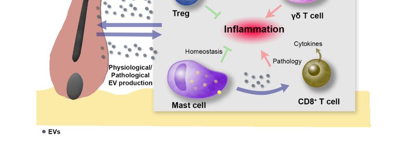
Int. J. Mol. Sci. 2019, 20, 2758 2 of 15
Int. J. Mol. Sci. 2019, 20, x FOR PEER REVIEW 2 of 15
of the subjacent hypodermal tissue is dependent on the part of the body (e.g., fat, connective tissue,
composition of the subjacent hypodermal tissue is dependent on the part of the body (e.g., fat,
bone). Skin appendices include hair follicles (HFs) and glands. HFs are important for warmth function
connective tissue, bone). Skin appendices include hair follicles (HFs) and glands. HFs are important
in mammals but also account for social and cosmetic features in humans. The HF cycle continuously
for warmth function in mammals but also account for social and cosmetic features in humans. The
occurs over the life of the organism, involving successive phases of rest (telogen), growth (anagen),
HF cycle continuously occurs over the life of the organism, involving successive phases of rest
and regression (catagen) [2] (Figure 1). Directly below the sebaceous gland and at the level of insertion
(telogen), growth (anagen), and regression (catagen) [2] (Figure 1). Directly below the sebaceous
of the piloerector muscle, the HF bulge, which hosts a major reservoir of skin stem cells (SCs), is
gland and at the level of insertion of the piloerector muscle, the HF bulge, which hosts a major
located [3]. Due to ease of accessibility and knowledge collected about the location and function of skin
reservoir of skin stem cells (SCs), is located [3]. Due to ease of accessibility and knowledge collected
SCs, the skin in general, and the HF in particular, have drawn much attention as targets for regenerative
about the location and function of skin SCs, the skin in general, and the HF in particular, have drawn
treatments. Among these, the use of extracellular vesicles as a safer alternative in comparison with
much attention as targets for regenerative treatments. Among these, the use of extracellular vesicles
cell-based approaches is gaining importance.
as a safer alternative in comparison with cell-based approaches is gaining importance.
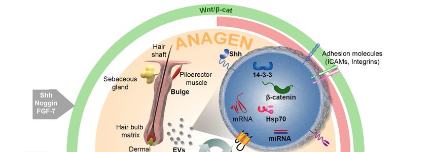
Figure 1. Regulation of the hair follicle cycle. Hair follicles continuously cycle, undergoing consecutive
Figure
phases 1. Regulation
of growing (anagen),of the hair follicle
regression cycle.
(catagen), andHair follicles
resting continuously
(telogen). cycle, undergoing
The fine regulation of hair
consecutive phases of growing (anagen), regression (catagen), and
follicle dynamics globally depends on the coordinated alternation between Wnt/β-catenin (green) resting (telogen). Theandfine
regulation(red)
BMP/Smad of hair folliclewhich
signals, dynamics
mainly globally
emanate depends
from theon the coordinated
dermal papilla alternation
and controlbetween Wnt/β-
the behavior
catenin (green) and BMP/Smad (red) signals, which mainly emanate from the
of follicular epithelial cells. Several factors and molecules have been found to affect different steps dermal papilla and
of the cycle, including Sonic hedgehog (Shh), Noggin, FGF-7, and reactive oxygen species (ROS), to
control the behavior of follicular epithelial cells. Several factors and molecules have been found
affectwork
which different steps of
as inductors of the cycle,TGFβ-1,
growth; including Sonic
which hedgehog
is involved in (Shh),
catagenNoggin, FGF-7,
onset; and and which
FGF-18, reactive
oxygen telogen.
regulates species (ROS),
The role which work as inductors
of extracellular vesiclesof(EVs)
growth; TGFβ-1,
in skin whichassessed
has been is involved in catagen
to date with
onset; and FGF-18, which regulates telogen. The role of extracellular vesicles
a major focus on their effects on the wound healing process but suggesting substantial effects on (EVs) in skin has been
assessed to date with a major focus on their effects on the wound healing
hair cycling. For instance, EVs containing Wnt3a, Wnt11, Wnt4, β-catenin, and 14-3-3 proteins may process but suggesting
substantial
contribute effects
to hair on hair
growth cycling. ForWnt
by enhancing instance, EVs HSP70
signaling; containing Wnt3a,exosomes
containing Wnt11, Wnt4, β-catenin,
have been and
related
to 14-3-3 proteins
pigmentation may and
defects; contribute to hair
the routes growth
involving by enhancing
Pi3K/AKT, MAPK/ERK, Wnt STAT3,
signaling;
andHSP70 containing
IGF1 have been
exosomes
connected with have been related effects
exosome-mediated to pigmentation defects;
in skin, therefore and potential
becoming the routes involving
targets Pi3K/AKT,
for EV-mediated
MAPK/ERK,
therapeutic STAT3, and IGF1 have been connected with exosome-mediated effects in skin, therefore
approaches.
becoming potential targets for EV-mediated therapeutic approaches.
Extracellular vesicles (EVs) comprise a heterogeneous variety of membrane-enclosed structures
Extracellular
in terms vesicles (EVs)
of size, mechanisms of comprise a heterogeneous
biogenesis, composition, variety
cargo, of
andmembrane-enclosed
functions. They structures
contain
in terms of size, mechanisms of biogenesis, composition, cargo, and functions. They contain
transmembrane proteins and enclose components derived from the donor cell that may include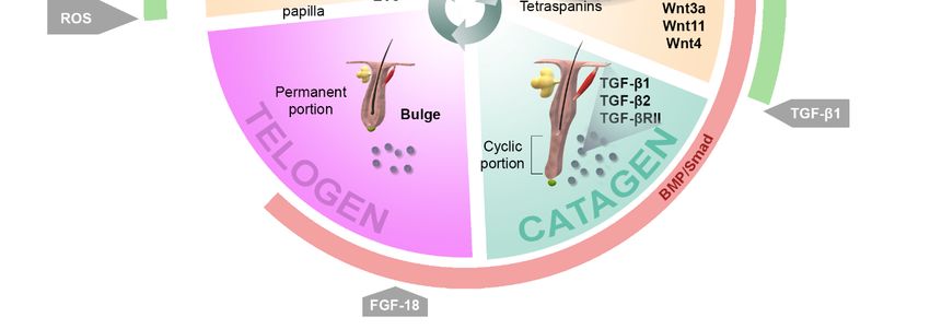
Int. J. Mol. Sci. 2019, 20, 2758                                                                     3 of 15

transmembrane proteins and enclose components derived from the donor cell that may include
proteins, lipids, and DNA and RNA molecules [4–6]. Most types of cells can secrete EVs, from bacterial
to animal—including human—to plant cells, which suggests that such an evolutionary conserved
mechanism [7] could be playing an essential role in cellular function. EVs can be isolated from
body fluids or cell culture supernatants and are nowadays becoming central signaling vehicles and
critical players in cell-cell communication, thus being implicated in physiological and pathological
processes [8].
      According to their route of biogenesis, EVs are generally classified into three subtypes, namely,
exosomes, microvesicles, and apoptotic bodies, with the former having been more frequently connected
to skin regenerative processes. Exosomes are 30–150 nm vesicles which originate via the endocytic
pathway. Normally, inward budding of the plasma membrane or fusion of vesicles gives rise to early
endosomes, which in turn can undergo inward budding, forming so-called multivesicular bodies
(MVBs). The intraluminal vesicles created by this process can either be directed to degradation in late
endosomes, involving lysosomes, or can be secreted to the extracellular space by exocytosis, which is
mediated by the fusion of MVBs with the plasma membrane [9,10]. Since the first characterization of
exosomes in the 1980s [11,12] and in light of later studies proving their role beyond the clearance of
waste products as immune players and even as anti-tumor vaccines [13,14], much research has been
directed at understanding their mechanisms of biogenesis. It is well established that Rab GTPase
family members play a key role in the modulation of exosome secretion, mainly along the pathway that
involves Endosomal Sorting Complexes Required for Transport (ESCRT), though the process can also
occur in an ESCRT-independent manner, in which tretraspanins (CD81, CD9, and CD63) and lipids play
a crucial role [7,10]. Microvesicles are larger lipid bilayer membrane-enclosed structures (0.1–1 µm)
which originate via outward budding of the cell membrane. Although their mechanism of formation
has not yet been uncovered in detail, it is known to involve a rearrangement of plasma membrane
phospoholipids, enzymatic processes, and cytoskeletal-mediated contraction, which eventually allows
for the fission of the membrane protrusions. Finally, relatively large (up to several microns) apoptotic
bodies are formed by outward blebbing of the plasma membrane of apoptotic cells, meaning they
usually contain cellular fragments [15]. Despite the increasing consensus about EV classification, it
is hard to distinguish them once they reach the extracellular space. Their interaction with recipient
cells can occur in different ways: ligand-receptor interaction; internalization by clathrin-dependent
endocytosis, caveolin-mediated uptake, macropinocytosis, or phagocytosis mediated by specific
receptors; and direct fusion with the plasmatic membrane of the recipient cell, thereby involving the
release of EV content in the cytoplasm of the recipient cell [16]. These processes can affect a number
of key cellular signaling pathways that modulate essential cellular processes, such as proliferation,
differentiation, migration, and cell death.

2. Extracellular Vesicles in Cutaneous Regenerative Medicine

2.1. Use of Extracellular Vesicles to Boost Skin Regeneration
      The skin wound healing process comprises four stages: (1) hemostatic, (2) inflammatory,
(3) proliferative, and (4) remodeling [17,18]. During this process, the skin is sequentially: (1) activated
to recruit repairing cell types; (2) cleaned of pathogens by the immune system; (3) stimulated to
provoke the proliferation of fibroblasts and the production of the extracellular matrix; and (4) closed by
the structural adjustment of the newly produced extracellular matrix. EVs obtained from a plethora of
cell types, mainly including mesenchymal stem cells (MSCs) of different origin, but also dermal papilla
(DP) cells, amniotic epithelial cells, keratinocytes, and endothelial progenitors, among others, have
been tested within different models of skin injury, such as skin wounds in healthy and diabetic rodents
or severe burns in rats (as reviewed in [17,19,20]). Key steps of the healing process have been proven to
benefit from the action of EVs, including cell proliferation, migration [21–25], angiogenesis [22,26–29]
and collagen deposition [22,25,28–30], and are mainly mediated by enhanced AKT/ERK and Wnt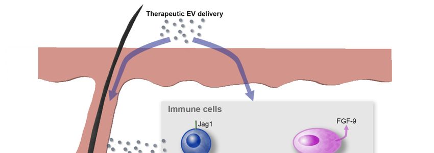
Int. J. Mol. Sci. 2019, 20, 2758                                                                    4 of 15

signaling. Overall, the effects of EVs can be summarized in accelerated wound healing and reduced
scar formation.
     Additionally, it has been suggested that multipotent neural SCs obtained from HFs can serve
as exosome producers, based on the beneficial effects of a conditioned medium for the treatment of
ischemia-reperfusion-induced lung injury in a rat model [31]. Hence, HFs can also be postulated as a
source of exosome-producing SCs.
     However, robust conclusions and defined clinical protocols are still hard to define because of the
lack of consensus regarding the source of EVs used in different studies, the models chosen to test EV
treatments, and the methods used for EV delivery. Additionally, the technical procedures used to isolate,
purify, and quantify exosomes and microvesicles, which have been fairly extensively reviewed [32],
are indeed a crucial point to consider in order to allow data standardization. For these reasons, the
molecular mechanisms of the action of EVs in wound healing need to be further characterized, with
special attention paid to simultaneous effects on hair growth.

2.2. Regulation of the Pigmentation Process by Exosomes
      The cell population of melanocytes, which are resident in the skin but originate at the neural crest
during embryonic development [33], accounts for skin and hair pigmentation. It has been suggested
that cytosolic proteins, such as the heat shock 70 kDa protein (HSP70) chaperone, can be recruited and
sequestered in exosomes as a mere consequence of their physical interaction with other proteins [7].
The role of HSP70 in progressive depigmentation has been confirmed in vivo using a mouse model of
autoimmune vitiligo [34]. Vitiligo is an autoimmune disorder that involves progressive depigmentation
mediated by a T-cell response to melanocytes. The amount of HSP70 is increased in the supernatant of
vitiligo versus the control melanocytes, and, when secreted into the extracellular space by melanocytes,
HSP70 interacts with antigen-presenting dendritic cells, enhancing their uptake and processing of
antigens. This leads to the activation of T-cells, which are ultimately responsible for the loss of
HSP70-producing melanocytes. The mouse model used by Denman and colleagues was based on the
introduction by gene gun vaccination of eukaryotic plasmids encoding melanocyte differentiation
antigens. When combined with the vaccination protocol, the introduction of inducible HSP70 resulted
in significantly accelerated depigmentation. In addition, this protein is found in exosomes derived
from most cell types and it is therefore likely to occur in exosomes from melanocytes. In light of
these observations, it is tempting to speculate that the release of HSP70-containing vesicles to the
extracellular milieu by melanocytes may contribute to the disease.
      Importantly, not only can the exosomes potentially secreted by melanocytes affect the pigmentation
process, but also signaling molecules carried by exosomes originating in other cell types resident in
the skin could affect melanocytes. In this regard, one sophisticated study has compared the effects of
exosomes obtained from human keratinocytes of different skin phototypes to stimulate melanocyte
function, as well as the potential of ultraviolet B (UVB) light to modulate this capacity. Interestingly,
the expression of key proteins participating in the pigmentation process, such as the enzyme tyrosinase,
the melanocyte isoform of microphthalmia-associated transcription factor (MITF), which is the master
transcriptional regulator of melanogenesis, and the Rab27a protein, involved in the mobilization of
melanosomes, were proven to be increased in melanocytes in the presence of exosomes either obtained
from Caucasian donors and treated with UVB, or obtained from black donors [35]. This groundbreaking
work demonstrated for the first time that miRNAs contained in exosomes secreted by keratinocytes
have the ability to modulate pigmentation. On the other hand, another study has reported the ability of
keratinocyte exosome-derived miR-675 to decrease MITF levels in melanocytes [36]. Altogether, these
observations may prompt new strategies for modulating skin pigmentation and hair pigmentation
through the potential effects driven by exosomes on HF bulge resident melanocyte precursors.
Int. J. Mol. Sci. 2019, 20, 2758 5 of 15
3. Role of Extracellular Vesicles in Hair Follicle Function
One previous study focused on rodent incisors found evidence of exosomes mediating
epithelium-mesenchyme molecular crosstalk [37]. Since the HF is considered one of the prototypical
systems for epithelial-mesenchymal crosstalk, it can be postulated as a top candidate to benefit from
EV-based clinical approaches. Moreover, regenerative hair waves continuously occur over mice
lifetimes and it has been demonstrated that the cycling rhythm declines with age, although it is possible
to rescue the cycling capability by transplanting aged skin into a younger donor [38]. In addition,
transplanted DP cells are able to induce hair growth [39,40]. In sum, these observations suggest that
hair growth induction is rather dependent on secreted factors than a cell-autonomous process.
3.1. Effects of Extracellular Vesicles on Hair Follicle Dynamics
3.1.1. Exosomes as Signaling Mediators with the Potential to Modulate Hair Cycling
Bone morphogenetic proteins (BMP), which emanate from dermal cells and adipocytes, maintain
SC quiescence during telogen [3]. Interestingly, DP cells undergo oscillating and out of phase
expression of BMP factors and of those inducing Wnt signaling, which allows for the fine regulation
of HF dynamics [41] (Figure 1). Significant findings involving a link between skin and hair follicle
regeneration and EVs have been compiled in Table 1, with emphasis on the signaling pathways that
mediate these effects. It is well established that Wnt factors are master regulators of HF morphogenesis
and hair growth [42]. In fact, epidermal Wnt ligands play a central role in wound-induced de novo hair
formation in adult skin. Hence, they have been pointed out as potential targets for the treatment of
hair-related syndromes like alopecia [43]. On the other hand, active Wnt factors have been identified as
exosome-secreted molecules which can be contained in the interior compartment of these vesicles [44]
as well as transported exteriorly [45]. Several studies have demonstrated that Wnt signaling in recipient
cells can be mediated by horizontal transfer of the proteomic contents of EVs [46]. For instance, EVs
from breast cancer cells have been shown to induce Wnt5a in macrophages, which in turn increases
macrophage-induced invasiveness of the MCF-7 cell line [47]. In addition, Wnt signaling has been
found to be activated in target cells by EVs containing both β-catenin, the major effector protein of
the canonical Wnt pathway, and 14-3-3 proteins [48]. The latter can bind Dvl-2 and GSK-3β, which
in turn mitigates the interaction between GSK-3β and β-catenin, therefore enhancing Wnt signaling.
In fact, both Wnt4 and Wnt11 secreted in exosomes derived from human umbilical cord MSCs have
been proven to benefit cutaneous regeneration in a rat skin burn model. Wnt4 has been found to
enhance Wnt/β-catenin signaling and angiogenesis in the skin [25,49], while the treatment of MSCs
with 3,30 -diindolylmethane has been shown to induce Wnt11 expression in exosomes in an autocrine
fashion, also favoring wound healing through the activation of Wnt/β-catenin signaling [50]. It would
be interesting to further investigate the strong effects of exosome-secreted Wnt11 on hair growth which
have been suggested by the results of this study. In addition, fibroblast-derived exosomes appear to
mobilize Wnt11-mediated autocrine signaling in breast cancer cells, promoting protrusive activity
and motility through the Wnt-planar cell polarity signaling pathway [51]. Interestingly, differential
subsets of Wnt-containing vesicles can be secreted in a distinctive manner in polarized epithelial cells,
a mechanism that has been demonstrated for the release of Wnt3a and Wnt11 in MDCK cells [52] and
which could be of interest in the well-organized architecture of the interfollicular epidermis and HF.
In agreement with these observations, an upregulation in the expression of Wnt3a and Wnt5a, consistent
with hair growth induction, has been found in mouse skin treated with intradermically-injected EVs
obtained from MSCs [53]. Accordingly, exosomes obtained from human DP cells have been found
to extend the anagen phase of the hair cycle by inducing the expression of β-catenin and Sonic
hedgehog (Shh) when injected into mouse skin [54]. Interestingly, in addition to the important role
that Shh signaling plays in hair morphogenesis, its homologous Hh has been shown to be present in
exosome-like vesicles in Drosophila [55].Int. J. Mol. Sci. 2019, 20, 2758 6 of 15
MicroRNAs (miRNAs) are small noncoding RNA molecules which are capable of altering gene
expression post transcriptionally and are typically transported in EVs [56,57]. These molecules
have been implicated in the control of skin and HF development through the modulation of Wnt
signaling [58]. In a step forward, miR-181c contained in human umbilical cord MSC-exosomes was
found to be a central player in attenuating burn-induced inflammation in a rat model [59]. Additionally,
exosomes obtained from synovium-MSCs that overexpress miR-126-3p have been found to promote
increased expression of P-AKT and ERK1/2 in HMEC-1 endothelial cells and contribute to skin wound
healing in diabetic rats [27].
Several important signaling pathways involved in key cellular processes such as cell migration,
proliferation, and survival are activated by epidermal growth factor (EGF) ligands binding their
receptors on the plasma membrane. Among these, the routes involving Pi3K/AKT, MAPK/ERK,
STAT3, and IGF1 have been connected with exosome-mediated effects on skin wound healing
or hair growth [21,25,27,28,53]. For instance, the JAK/STAT pathway is implicated in hair
growth [60]. Transforming Growth Factor (TGF)-α belongs to the EGF family and is upstream
of the Grb2/Sos-Ras-Raf-MEK1,2-ERK1,2 signaling cascade, which is widely accepted as a promoter of
cell proliferation [23]. Since mice with TGF-α deficiency display skin and hair abnormalities [61,62],
TGF-α has been implicated in the control of HF shape [42]. TGF-α selectively stimulates hsp90α
exosome-secretion in human keratinocytes, but not in dermal cells [23]. Interestingly, hsp90α
promotes migration of dermal cells even in the presence of the strong inhibitor TGF-β, which is
abundant in the skin wound environment. Thus, hsp90α exosome-secretion by keratinocytes in
response to TGF-α may constitute a major factor stimulating cell motility in the wound bed [23].
In particular, since inhibitory signals mediated by TGF-β family factors are involved in the control
of HF regression (catagen) in vivo [63,64], it would be interesting to investigate whether TGF-α
stimuli can trigger exosome-mediated secretion of hsp90α by HF epithelial cells and affect hair cycle
progression. More work is also needed to determine to what extent the induction in expression of the
apoptosis suppressor BCL-2 in response to MSC-exosomes [53] could participate in the extension of
the anagen phase.
Table 1. The role of extracellular vesicles in signaling pathways with the potential to modulate
hair cycling.
Signaling Molecules Model Used to Test the
Source of EVs Highlights of the Study Ref.
Pathway Transported via EVs Effects
β-catenin and 14-3-3 HEK293T, EV-mediated activation of Wnt In vitro: HEK293T,
[48]
proteins SW480 signaling in recipient cells COS7, SW480
In vitro: HaCaT,
HuUC-MSC exosomes facilitated
Ea.hy926, rat dermal
wound re-epithelization and cell
Wnt4 HuUC-MSCs fibroblasts [25,49]
proliferation through the
In vivo: Rat skin 2nd
activation of Wnt signaling
degree burn injury
Exosomal Wnt11 autocrine
In vitro: HaCaT, rat
signaling in response to
dermal fibroblasts
Canonical Wnt Wnt11 HuUC-MSCs 3-30 -diindolylmethane increased [50]
In vivo: Rat skin 2nd
markers of stemness in MSCs and
degree burn injury
favored wound healing
MDCK, Different populations of exosomes
HEK293, carrying Wnt factors secreted by
Wnt3a, Wnt11 [52]
fibroblast L epithelial cells depending on the
cells cell polarity and cell type
EVs contributed to hair growth in
Mouse
Wnt3a, Wnt5a mice by promoting telogen to In vivo: Mouse skin [53]
BM-MSCs
anagen conversion of HFsInt. J. Mol. Sci. 2019, 20, 2758 7 of 15
Table 1. Cont.
Signaling Molecules Model Used to Test the
Source of EVs Highlights of the Study Ref.
Pathway Transported via EVs Effects
Mouse fibroblast-derived
Mouse exosomes mobilized
Wnt-planar cell In vitro: MDA-MB-231
Wnt11 fibroblast L Wnt11-mediated autocrine [51]
polarity In vivo: SCID mice
cells signaling, promoting protrusive
activity and motility
Exosomes extended the anagen
Canonical Wnt; phase of the hair cycle in mice by
Not characterized HuDPCs In vivo: Mouse skin [54]
Shh inducing the expression of
β-catenin and Shh
Hh transport via exosomes along
Hh Hh Drosophila In vitro: Cl8 [55]
cytonems
Exosomes overexpressing
In vivo: Rat
miR-181c reduced burn
TLR4 miR-181c HuUC-MSCs full-thickness burn [59]
inflammation by downregulating
injury
the TLR4 signaling pathway
Improvement in the healing
capacity of wound dressings by In vitro: Human dermal
incorporating exosomes derived fibroblast, HMEC-1
mi-126-3p HuS-MSCs from miR126-overexpressing In vivo: Full-thickness [27]
HuS-MSCs, which led to the excisional skin wound in
activation of AKT and ERK1/2 diabetic rats
through phosphorylation
Key pathways for wound healing In vitro: Diabetic versus
EGF/EGFR ERK1/2 BM-MSCs including Akt, ERK, and STAT3, normal wound patient [21]
activated by MSC-exosomes fibroblasts
ERK1/2-mediated improved In vitro: HMEC-1
angiogenesis in response to In vivo: Full-thickness
ERK1/2 HuEPCs [28]
exosomes with beneficial effects excisional skin wound in
on wound healing diabetic rats
Stimulation of the secretion of
In vitro: Primary
hsp90α in exosomes by
TGF-α HKCs neonatal HKCs, dermal [23]
HuK-promoted migration of both
cells
epidermal and dermal cells
The table compiles significant findings involving a link between skin and hair follicle regeneration and EVs, with
emphasis on the pathways and the specific signaling molecules mediating these effects. Legend: BM-MSCs, bone
marrow-derived mesenchymal stem cells; EGF, Epidermal Growth Factor; EGFR, Epidermal Growth Factor Receptor;
EV, extracellular vesicles; Hh, Hedgehog; HKCs, human keratinocytes; HuDPCs, human dermal papilla cells;
HuEPCs, human endothelial progenitor cells; HuS-MSCs, human synovium mesenchymal stem cells; HuUC-MSCs,
human umbilical cord mesenchymal stem cells; Shh, Sonic hedgehog; TGF, Transforming Growth Factor.
3.1.2. Use of Extracellular Vesicles to Stimulate Hair Growth: Evidence and Clues
Although only a few studies have focused on the use of exosomes to stimulate hair growth, the
findings in this field are promising. A patented study has provided the first evidence of exosomes
obtained from MSCs being a central component of a pharmaceutical composition directed at enhancing
hair growth [65]. The induction of hair growth was accompanied by an improvement in wound healing,
which is actually not surprising, since both processes have previously been linked by a number of
studies, and different mechanisms involved in this link have been pointed out [66–68]. In agreement
with these observations, subcutaneous administration of conditioned media from human amniotic
fluid-derived MSCs in a full thickness wound model in rats has been found to promote hair regrowth,
together with the acceleration of wound healing [69]. However, only a small number of studies with
several limitations have so far been focused on the characterization of the mechanisms involved in the
beneficial effects of exosomes on hair growth. Importantly, intradermically injected MSC-EVs have
been shown to favor telogen to anagen transition in vivo in a mouse model [44]. An increase in the
expression of proliferation, survival, and migration markers in DP cells treated in vitro with MSC-EVs
was pointed out by the authors as a sign of stimulation, including induced expression of PCNA, P-AKT,
P-ERK, and growth factors such as VEGF and IGF-1. However, since those traits are not typically found
in DP cells over the hair cycle [65], determining the signaling molecules that mediate the induction
of hair growth in response to treatment with EVs in vivo will be key to unequivocally unveiling theInt. J. Mol. Sci. 2019, 20, 2758 8 of 15
molecular mechanisms mediating this effect. In a recent study, human DP exosomes were injected into
mouse skin to promote hair growth [54], which can mimic the physiological paracrine effects that DP
cells exert on epithelial cells. An induction in β-catenin and Shh levels was detected in treated skin,
as well as in epithelial hair follicle outer root sheath cells isolated from human scalps and cultured
with DP exosomes. These results consistently revealed the participation of Wnt/β-catenin and Shh
pathways in the molecular mechanisms driving hair growth in response to exosomes. Interestingly,
anagen extension was due not only to a premature anagen onset but also to a delay of the catagen phase
in mice. This suggests that additional molecular mechanisms responsible for hindering the transition
to the catagen stage could be involved in enhanced hair growth. Finally, exosomes containing hTert
have been proven to be secreted by cancer cells and incorporated by telomerase negative fibroblasts,
turning them into nonmalignant cells with telomerase activity [70]. Since HF SC proliferation occurs as
a consequence of conditional telomerase induction [71], hTert-containing exosomes could contribute to
triggering the proliferation of slow-cycling SCs in the HF bulge and favor the transition from telogen
to anagen, ultimately promoting hair growth.
Overall, these observations suggest that since EV cargo has the potential to target a wide range of
molecular processes and recipient cells, EVs emerge as both natural mediators potentially participating
in the control of the hair cycle and promising delivery vehicles for the improvement of skin and hair
regeneration. More work needs to be done in order to determine both the physiological contribution of
exosomes to the HF cycle in vivo and the therapeutic potential of the use of exosomes in the clinics in
order to modulate hair growth.
3.2. Immune System Cells and Hair Follicles
Different types of EVs have been implicated by a myriad of studies in interactions that involve
and affect immune cells [4,72]. Concurrently, important molecules that are released via EVs and
participate in immunomodulation are also recognized as essential factors involved in wound repair [18].
For instance, TGF-β1 belongs to the secretome of mesenchymal stromal cells and is released via
exosomes [73]. Additionally, the extracellular functions carried out by exosome-secreted hsp proteins
include stimulation of immunological cytokine production, activation of antigen-presenting cells,
and anticancer functions [74]. In light of this knowledge, immune cells are indeed key candidates
which participate in the regulation of tissue and hair regeneration as exosome producer or recipient
cells (Figure 2). Hence, therapeutic strategies for hair-related syndromes should also target immune
cell populations. In this sense, several pieces of evidence indicate that important tissue regeneration
processes are mediated by an intimate molecular dialog established between immune cells and other
skin and HF resident cells. For instance, Fgf-9 secreted by γδ-T cells modulates HF neogenesis after
skin wounding in adult mice through the activation of Wnt expression and Wnt signaling in skin
fibroblasts [75]. In addition, regulatory T-cells have also been identified as promoters of proliferation
and differentiation of HF SCs [76]. On the contrary, signals that inhibit hair growth have been
conferred to molecules secreted by immune cells, such as prostaglandin D2 (PGD2), which blocks HF
regeneration through the Gpr44 receptor. This opens up the possibility of therapeutically inhibiting
PGD2 production or Gpr44 signaling to promote skin regeneration [77]. The opposed effects of different
molecules transported via EVs are expected to be controlled depending on the status of the recipient
cells (e.g., the surface receptors being expressed), as well as the tissue microenvironment. In the case
of EVs potentially delivered with therapeutic purposes, the specific molecules loaded as cargo can
be chosen.Int. J. Mol. Sci. 2019, 20, 2758 9 of 15
Int. J. Mol. Sci. 2019, 20, x FOR PEER REVIEW 9 of 15
Figure 2. Extracellular-vesicle-mediated crosstalk between immune cells and hair follicles. EVs can be
Figure 2. Extracellular-vesicle-mediated crosstalk between immune cells and hair follicles. EVs can be
naturally produced under physiological/pathological conditions or, alternatively, can be therapeutically
naturally produced under physiological/pathological conditions or, alternatively, can be
delivered. EVs can contribute to the modulation of hair follicle stem cell function by acting either
therapeutically delivered. EVs can contribute to the modulation of hair follicle stem cell function by
directly or indirectly through their effect on immune cells. In this sense, different types of immune
acting either directly or indirectly through their effect on immune cells. In this sense, different types
cells are involved in the control of hair follicle dynamics. Skin-resident regulatory T (Treg) cells that
of immune
express highcells are of
levels involved
the Notchin the control
ligand of hair (Jag1)
Jagged-1 folliclefacilitate
dynamics. hairSkin-resident
follicle stemregulatory T (Treg)
cell function and
cells that express high levels of the Notch ligand Jagged-1 (Jag1) facilitate hair
contribute to hair follicle regeneration. Skin resident mast cells contribute to hair follicle immune follicle stem cell
function and contribute to hair follicle regeneration. Skin resident mast cells contribute
privilege under physiological conditions but are known to become proinflammatory in alopecia areata, to hair follicle
immune
in which mastprivilege under less
cells contain physiological
TGF-β1 and conditions but are known
produce exosomes to become
that induce proinflammatory
T lymphocytes in
to proliferate
alopecia
and secreteareata, in which
cytokines. mastcell
The γδ-T cells contain less
population TGF-β1
in mouse skinand produce
secretes FGF-9,exosomes that induce
which modulates hairT
lymphocytes to proliferate and secrete cytokines. The γδ-T cell population in mouse
follicle neogenesis after skin wounding. In addition, epithelial and dermal hair follicle cells can secrete skin secretes
FGF-9,
EVs thatwhich modulates
potentially hair hair
target other follicle neogenesis
follicle after skin
cell populations wounding.
or skin residentIn addition,
immune cells,epithelial and
contributing
dermal hair follicle cells can secrete EVs that potentially target other hair follicle
to the modulation of local inflammation. Purple arrows indicate the flux of EVs; red arrows indicate cell populations or
skin resident immune
proinflammatory stimuli;cells, contributing
green to the
T bars indicate modulation of local
anti-inflammatory inflammation. Purple arrows
stimuli.
indicate the flux of EVs; red arrows indicate proinflammatory stimuli; green T bars indicate anti-
The importance
inflammatory of the crosstalk between immune cells and skin and HF cells has also been
stimuli.
highlighted by the classification of alopecia areata as an autoimmune disorder involving a switch of
In this
mast cells context,
towards the use of exosomes
a proinflammatory as carriers
phenotype of Mast
[78]. danger-associated molecular
cells in alopecia patterns
areata display (DAMPs)
lower levels
and
of molecules
TGF-β that regulate
and secrete exosomesT-cell functionthe
that mediate caninduction
exert either a direct
of T-cells effect on HF
to proliferate andSCs or ancytokine
increase indirect
effect through
production the activation
(Figure of immune
2). This could cellsthe
motivate involved
use ofin the inflammatory
exosomes response.ofThis
for the treatment can mimic
autoimmune
molecular
diseases signals
that affectthat
hairparticipate in the
loss, serving activation
as delivery of skinfor
vehicles SCssignaling
in response to skin damage,
molecules although
that contribute to
it remains
the immune unknown
privilegewhether exosomes
of HFs, for example arebyinvolved in this
facilitating the type of mechanism
suppression of majorduring physiological
histocompatibility
skin homeostasis
complex and repair.
class I molecules or by Strikingly,
inducing theexosomes
expression enriched
of immune in telomeric repeat-containing
privilege guardians RNA
like TGF-β1/2.
(TERRA),
In thiswhich arethe
context, non-coding RNAs that
use of exosomes contribute
as carriers to telomere function,
of danger-associated have been
molecular identified
patterns (DAMPs)[79].
These TERRA-enriched exosomes have been found to stimulate inflammatory
and molecules that regulate T-cell function can exert either a direct effect on HF SCs or an indirect cytokines from
immune
effect cellsthe
through and thus have
activation been postulated
of immune as telomere-specific
cells involved in the inflammatory alarmins. TheThis
response. authors have
can mimic
therefore proposed
molecular signals thatthat TERRA molecules
participate transported
in the activation of skinby
SCsthese exosomes
in response towork as telomere-specific
skin damage, although it
DAMPs which account for telomeric dysfunction and trigger an inflammatory
remains unknown whether exosomes are involved in this type of mechanism during physiological response in the
recipient cells. Given that the HF bulge SC niche is known to be enriched in cells
skin homeostasis and repair. Strikingly, exosomes enriched in telomeric repeat-containing RNA with relatively longer
telomereswhich
(TERRA), [80], this mechanismRNAs
are non-coding couldthat
be relevant
contributeastoatelomere
potentialfunction,
way to have
signalbeen
telomere attrition
identified [79].
affecting the HF SC compartment, revealing stem dysfunction and aging, which are linked to hair-
related syndromes. Another scenario where DAMPs could gain importance is in the involution phaseInt. J. Mol. Sci. 2019, 20, 2758 10 of 15
These TERRA-enriched exosomes have been found to stimulate inflammatory cytokines from immune
cells and thus have been postulated as telomere-specific alarmins. The authors have therefore proposed
that TERRA molecules transported by these exosomes work as telomere-specific DAMPs which account
for telomeric dysfunction and trigger an inflammatory response in the recipient cells. Given that the HF
bulge SC niche is known to be enriched in cells with relatively longer telomeres [80], this mechanism
could be relevant as a potential way to signal telomere attrition affecting the HF SC compartment,
revealing stem dysfunction and aging, which are linked to hair-related syndromes. Another scenario
where DAMPs could gain importance is in the involution phase of the hair cycle (catagen). This stage is
characterized by synchronic keratinocyte apoptosis in the regressing proximal hair bulb and constitutes
a unique model of dramatic but physiologically programmed epithelial cell death [81]. Since the
release of apoptotic bodies by follicular epithelial cells has been reported [82–84] but their signaling
capacities generally neglected, we propose that new insights into this process could help to understand
the molecular mechanisms that orchestrate hair cycling.
4. Concluding Remarks and Future Directions
The emergent role of EVs in HF dynamics is likely to become a high-impact tool in cosmetic
and skin regenerative biomedicine. In line with recent work revisiting the isolation and purification
methods of different types of EVs [5,45,85], the unification of procedures used to obtain and administer
EVs is needed in order to generate more reliable and comparable data, as well as to implement
novel techniques for the in vivo characterization of EV-driven mechanisms related to HF biology.
In this regard, witty approaches are needed to more deeply explore the physiological and pathological
occurrence of EV crosstalk among different HF subpopulations. Good examples of such strategies are
the two photon approach [86] and the combined intravital microscopy with genetic lineage tracing [87],
which was conceived by Greco’s lab in an attempt to observe the release of exosomes by mutant cells
within the upper portion of the HF and epidermis and their subsequent clearing by both epithelial
and immune cells. In summary, the ease of accessibility of skin may be strongly advantaged with
regard to both the possibility of implementing novel treatments and the potential to serve as a source
of exosome-secreting cells.
Author Contributions: All authors substantially contributed to this work. Conceptualization, E.C. and M.M.;
writing—original draft preparation, E.C. and G.S.-H.; review and editing, E.C., G.S.-H., and M.M.
Funding: This research was funded by the Fondo de Investigación Sanitaria, Instituto de Salud Carlos III
(CP 14/00219), Fondo Europeo de Desarrollo Regional (FEDER), H2020-EU.1.1.—European Research Council
(ERC-2016-StG 715322-EndoMitTalk), and Instituto de Salud Carlos III (FIS16/188). E.C. was supported by the
Atracción de Talento Investigador grant 2017-T2/BMD-5766 (Comunidad de Madrid and Universidad Autónoma
de Madrid). G.S.-H. was funded by an FPI grant (Universidad Autónoma de Madrid). M.M. was supported by
the Miguel Servet program (Instituto de Investigación del Hospital 12 de Octubre).
Acknowledgments: We thank Manuel Guerrero for assistance with the preparation of the figures.
Conflicts of Interest: The authors declare no conflict of interest.
Abbreviations
BMP Bone Morphogenetic Protein
DAMPS Danger-Associated Molecular Patterns
DP Dermal Papilla
EGF Epidermal Growth Factor
ESCRT Endosomal Sorting Complexes Required for Transport
EV Extracellular Vesicles
HF Hair Follicle
HSP70 Heat shock 70 kDa protein
miRNA MicroRNA
MITF Microphthalmia-associated transcription factor
MSC Mesenchymal Stem CellInt. J. Mol. Sci. 2019, 20, 2758 11 of 15
MVB Multivesicular Body
PGD2 Prostaglandin D2
SC Stem Cell
Shh Sonic Hedgehog
TERRA Telomeric repeats-containing RNA
TGF Transforming Growth Factor
UVB Ultraviolet B
References
1. Clark, R.A.F.; Ghosh, K.; Tonnesen, M.G. Tissue Engineering for Cutaneous Wounds. J. Invest. Dermatol.
2007, 127, 1018–1029. [CrossRef] [PubMed]
2. Hardy, M.H. The Secret Life of the Hair Follicle. Trends Genet. 1992, 8, 55–61. [CrossRef]
3. Solanas, G.; Benitah, S.A. Regenerating the Skin: A Task for the Heterogeneous Stem Cell Pool and
Surrounding Niche. Nat. Rev. Mol. Cell Biol. 2013, 14, 737–748. [CrossRef] [PubMed]
4. Théry, C.; Ostrowski, M.; Segura, E. Membrane Vesicles as Conveyors of Immune Responses. Nat. Rev.
Immunol. 2009, 9, 581–593. [CrossRef] [PubMed]
5. Jeppesen, D.K.; Fenix, A.M.; Franklin, J.L.; Higginbotham, J.N.; Zhang, Q.; Zimmerman, L.J.; Liebler, D.C.;
Ping, J.; Liu, Q.; Evans, R.; et al. Reassessment of Exosome Composition. Cell 2019, 177, 428–445. [CrossRef]
[PubMed]
6. Jimenez, L.; Yu, H.; McKenzie, A.J.; Franklin, J.L.; Patton, J.G.; Liu, Q.; Weaver, A.M. Quantitative Proteomic
Analysis of Small and Large Extracellular Vesicles (EVs) Reveals Enrichment of Adhesion Proteins in Small
EVs. J. Proteome Res. 2019, 18, 947–959. [CrossRef]
7. Van Niel, G.; D’Angelo, G.; Raposo, G. Shedding Light on the Cell Biology of Extracellular Vesicles. Nat. Rev.
Mol. Cell Biol. 2018, 19, 213–228. [CrossRef]
8. Mittelbrunn, M.; Sánchez-Madrid, F. Intercellular Communication: Diverse Structures for Exchange of
Genetic Information. Nat. Rev. Mol. Cell Biol. 2012, 13, 328–335. [CrossRef]
9. Tkach, M.; Théry, C. Communication by Extracellular Vesicles: Where We Are and Where We Need to Go.
Cell 2016, 164, 1226–1232. [CrossRef]
10. Villarroya-Beltri, C.; Baixauli, F.; Gutiérrez-Vázquez, C.; Sánchez-Madrid, F.; Mittelbrunn, M. Sorting It out:
Regulation of Exosome Loading. Semin. Cancer Biol. 2014, 28, 3–13. [CrossRef]
11. Harding, C.; Heuser, J.; Stahl, P. Receptor-Mediated Endocytosis of Transferrin and of the Transferrin Receptor
in Rat Reticulocytes Recycling. J. Cell. Biol. 1983, 97, 329–339. [CrossRef] [PubMed]
12. Pan, B.T.; Teng, K.; Wu, C.; Adam, M.; Johnstone, R.M. Electron Microscopic Evidence for Xternalization of
the Transferrin Receptor in Vesicular Form in Sheep Reticulocytes. J. Cell Biol. 1985, 101, 942–948. [CrossRef]
[PubMed]
13. Raposo, G.H.W.; Nijman, W.; Stoorvogel, R.; Liejendekker, C.V.; Harding, C.J.; Melief, J.G.B. Lymphocytes
Secrete Antigen-PresentingVesicles. J. Exp. Med. 1996, 183, 1161–1172. [CrossRef] [PubMed]
14. Zitvogel, L.; Regnault, A.; Lozier, A.; Wolfers, J.; Flament, C.; Tenza, D.; Ricciardi-Castagnoli, P.; Raposo, G.;
Zitvogel, L.; Regnault, A.; et al. Eradication of Established Murine Tumors Using a Novel Cell-Free Vaccine:
Dendritic Cell-Derived Exosomes. Nat. Med. 1998, 4, 594–600. [CrossRef] [PubMed]
15. Yáñez-Mó, M.; Siljander, P.R.M.; Andreu, Z.; Zavec, A.B.; Borràs, F.E.; Buzas, E.I.; Buzas, K.; Casal, E.;
Cappello, F.; Carvalho, J.; et al. Biological Properties of Extracellular Vesicles and Their Physiological
Functions. J. Extracell. Vesicles 2015, 4, 1–60. [CrossRef] [PubMed]
16. Colombo, M.; Raposo, G.; Théry, C. Biogenesis, Secretion, and Intercellular Interactions of Exosomes and
Other Extracellular Vesicles. Annu. Rev. Cell Dev. Biol. 2014, 30, 255–289. [CrossRef]
17. Cabral, J.; Ryan, A.E.; Griffin, M.D.; Ritter, T. Extracellular Vesicles as Modulators of Wound Healing.
Adv. Drug Deliv. Rev. 2018, 129, 394–406. [CrossRef] [PubMed]
18. Shaw, T.J.; Martin, P. Wound Repair at a Glance. J. Cell Sci. 2009, 122, 3209–3213. [CrossRef] [PubMed]
19. Riazifar, M.; Pone, E.J.; Lötvall, J.; Zhao, W. Stem Cell Extracellular Vesicles: Extended Messages of
Regeneration. Annu. Rev. Pharmacol. Toxicol. 2017, 57, 125–154. [CrossRef] [PubMed]
20. Than, U.T.T.; Guanzon, D.; Leavesley, D.; Parker, T. Association of Extracellular Membrane Vesicles with
Cutaneous Wound Healing. Int. J. Mol. Sci. 2017, 18, 956. [CrossRef] [PubMed]Int. J. Mol. Sci. 2019, 20, 2758 12 of 15
21. Shabbir, A.; Cox, A.; Rodriguez-Menocal, L.; Salgado, M.; Van Badiavas, E. Mesenchymal Stem Cell Exosomes
Induce Proliferation and Migration of Normal and Chronic Wound Fibroblasts, and Enhance Angiogenesis
In Vitro. Stem Cells Dev. 2015, 24, 1635–1647. [CrossRef] [PubMed]
22. Zhang, J.; Guan, J.; Niu, X.; Hu, G.; Guo, S.; Li, Q.; Xie, Z.; Zhang, C.; Wang, Y. Exosomes Released from
Human Induced Pluripotent Stem Cells-Derived MSCs Facilitate Cutaneous Wound Healing by Promoting
Collagen Synthesis and Angiogenesis. J. Transl. Med. 2015, 13, 49. [CrossRef] [PubMed]
23. Cheng, C.-F.; Fan, J.; Fedesco, M.; Guan, S.; Li, Y.; Bandyopadhyay, B.; Bright, A.M.; Yerushalmi, D.; Liang, M.;
Chen, M.; et al. Transforming Growth Factor (TGF )-Stimulated Secretion of HSP90: Using the Receptor
LRP-1/CD91 To Promote Human Skin Cell Migration against a TGF -Rich Environment during Wound
Healing. Mol. Cell. Biol. 2008, 28, 3344–3358. [CrossRef] [PubMed]
24. Guo, S.C.; Tao, S.C.; Yin, W.J.; Qi, X.; Yuan, T.; Zhang, C.Q. Exosomes Derived from Platelet-Rich Plasma
Promote the Re-Epithelization of Chronic Cutaneous Wounds via Activation of YAP in a Diabetic Rat Model.
Theranostics 2017, 7, 81–96. [CrossRef] [PubMed]
25. Zhang, B.; Wang, M.; Gong, A.; Zhang, X.; Wu, X.; Zhu, Y.; Shi, H.; Wu, L.; Zhu, W.; Qian, H.; et al.
HucMSc-Exosome Mediated-Wnt4 Signaling Is Required for Cutaneous Wound Healing. Stem Cells 2015,
33, 2158–2168. [CrossRef] [PubMed]
26. Li, X.; Jiang, C.; Zhao, J. Human Endothelial Progenitor Cells-Derived Exosomes Accelerate Cutaneous
Wound Healing in Diabetic Rats by Promoting Endothelial Function. J. Diabetes Complicat. 2016, 30, 986–992.
[CrossRef] [PubMed]
27. Tao, S.-C.; Guo, S.-C.; Li, M.; Ke, Q.-F.; Guo, Y.-P.; Zhang, C.-Q. Chitosan Wound Dressings Incorporating
Exosomes Derived from MicroRNA-126-Overexpressing Synovium Mesenchymal Stem Cells Provide
Sustained Release of Exosomes and Heal Full-Thickness Skin Defects in a Diabetic Rat Model. Stem Cells
Transl. Med. 2017, 6, 736–747. [CrossRef] [PubMed]
28. Zhang, J.; Chen, C.; Hu, B.; Niu, X.; Liu, X.; Zhang, G.; Zhang, C.; Li, Q.; Wang, Y. Exosomes Derived from
Human Endothelial Progenitor Cells Accelerate Cutaneous Wound Healing by Promoting Angiogenesis
through Erk1/2 Signaling. Int. J. Biol. Sci. 2016, 12, 1472–1487. [CrossRef] [PubMed]
29. Geiger, A.; Walker, A.; Nissen, E. Human Fibrocyte-Derived Exosomes Accelerate Wound Healing in
Genetically Diabetic Mice. Biochem. Biophys. Res. Commun. 2015, 467, 303–309. [CrossRef] [PubMed]
30. Hu, L.; Wang, J.; Zhou, X.; Xiong, Z.; Zhao, J.; Yu, R.; Huang, F.; Zhang, H.; Chen, L. Exosomes Derived
from Human Adipose Mensenchymal Stem Cells Accelerates Cutaneous Wound Healing via Optimizing the
Characteristics of Fibroblasts. Sci. Rep. 2016, 6, 1–11. [CrossRef] [PubMed]
31. Peng, C.-K.; Wu, S.-Y.; Tang, S.-E.; Li, M.-H.; Lin, S.-S.; Chu, S.-J.; Huang, K.-L. Protective Effects of Neural
Crest-Derived Stem Cell-Conditioned Media against Ischemia-Reperfusion-Induced Lung Injury in Rats.
Inflammation 2017, 40, 1532–1542. [CrossRef] [PubMed]
32. McBride, J.D.; Rodriguez-Menocal, L.; Badiavas, E.V. Extracellular Vesicles as Biomarkers and Therapeutics
in Dermatology: A Focus on Exosomes. J. Investig. Dermatol. 2017, 137, 1622–1629. [CrossRef] [PubMed]
33. Lin, J.Y.; Fisher, D.E. Melanocyte Biology and Skin Pigmentation. Nature 2007, 445, 843–850. [CrossRef]
[PubMed]
34. Denman, C.J.; McCracken, J.; Hariharan, V.; Klarquist, J.; Oyarbide-Valencia, K.; Guevara-Patĩo, J.A.; Caroline
Le Poole, I. HSP70i Accelerates Depigmentation in a Mouse Model of Autoimmune Vitiligo. J. Investig.
Dermatol. 2008, 128, 2041–2048. [CrossRef] [PubMed]
35. Lo Cicero, A.; Delevoye, C.; Gilles-Marsens, F.; Loew, D.; Dingli, F.; Guéré, C.; André, N.; Vié, K.; Van Niel, G.;
Raposo, G. Exosomes Released by Keratinocytes Modulate Melanocyte Pigmentation. Nat. Commun. 2015, 6.
[CrossRef] [PubMed]
36. Kim, N.H.; Choi, S.H.; Kim, C.H.; Lee, C.H.; Lee, T.R.; Lee, A.Y. Reduced MiR-675 in Exosome in H19
RNA-Related Melanogenesis via MITF as a Direct Target. J. Investig. Dermatol. 2014, 134, 1075–1082.
[CrossRef]
37. Jiang, N.; Xiang, L.; He, L.; Yang, G.; Zheng, J.; Wang, C.; Zhang, Y.; Wang, S.; Zhou, Y.; Sheu, T.J.; et al.
Exosomes Mediate Epithelium-Mesenchyme Crosstalk in Organ Development. ACS Nano 2017, 11, 7736–7746.
[CrossRef]
38. Chen, C.C.; Murray, P.J.; Jiang, T.X.; Plikus, M.V.; Chang, Y.T.; Lee, O.K.; Widelitz, R.B.; Chuong, C.M.
Regenerative Hair Waves in Aging Mice and Extra-Follicular Modulators Follistatin, Dkk1, and Sfrp4.
J. Investig. Dermatol. 2014, 134, 2086–2096. [CrossRef]Int. J. Mol. Sci. 2019, 20, 2758 13 of 15
39. Jahoda, C.A.B.; Horne, K.A.; Oliver, R.F. Induction of Hair Growth by Implantation of Cultured Dermal
Papilla Cells. Nature 1984, 311, 560–562. [CrossRef]
40. Reynolds, A.J.; Jahoda, C.A.B. Cultured Dermal Papilla Cells Induce Follicle Formation and Hair Growth by
Transdifferentiation of an Adult Epidermis. Development 1992, 115, 587–593.
41. Plikus, M.V.; Mayer, J.A.; De La Cruz, D.; Baker, R.E.; Maini, P.K.; Maxson, R.; Chuong, C.M. Cyclic Dermal
BMP Signalling Regulates Stem Cell Activation during Hair Regeneration. Nature 2008, 451, 340–344.
[CrossRef] [PubMed]
42. Millar, S.E. Molecular Mechanisms Regulating Hair Follicle Development. J. Investig. Dermatol. 2002,
118, 216–225. [CrossRef] [PubMed]
43. Myung, P.S.; Takeo, M.; Ito, M.; Atit, R.P. Epithelial Wnt Ligand Secretion Is Required for Adult Hair Follicle
Growth and Regeneration. J. Investig. Dermatol. 2013, 133, 31–41. [CrossRef] [PubMed]
44. Gross, J.C.; Chaudhary, V.; Bartscherer, K.; Boutros, M. Active Wnt Proteins Are Secreted on Exosomes. Nat.
Cell Biol. 2012, 14, 1036–1045. [CrossRef] [PubMed]
45. McBride, J.D.; Rodriguez-Menocal, L.; Guzman, W.; Candanedo, A.; Garcia-Contreras, M.; Badiavas, E.V.
Bone Marrow Mesenchymal Stem Cell-Derived CD63 + Exosomes Transport Wnt3a Exteriorly and Enhance
Dermal Fibroblast Proliferation, Migration, and Angiogenesis In Vitro. Stem Cells Dev. 2017, 26, 1384–1398.
[CrossRef] [PubMed]
46. Gangoda, L.; Boukouris, S.; Liem, M.; Kalra, H.; Mathivanan, S. Extracellular Vesicles Including Exosomes
Are Mediators of Signal Transduction: Are They Protective or Pathogenic? Proteomics 2015, 15, 260–271.
[CrossRef]
47. Menck, K.; Klemm, F.; Gross, J.C.; Pukrop, T.; Wenzel, D.; Binder, C. Induction and Transport of Wnt 5a during
Macrophage-Induced Malignant Invasion Is Mediated by Two Types of Extracellular Vesicles. Oncotarget
2015, 4, 2057. [CrossRef] [PubMed]
48. Dovrat, S.; Caspi, M.; Zilberberg, A.; Lahav, L.; Firsow, A.; Gur, H.; Rosin-Arbesfeld, R. 14-3-3 and β-Catenin
Are Secreted on Extracellular Vesicles to Activate the Oncogenic Wnt Pathway. Mol. Oncol. 2014, 8, 894–911.
[CrossRef] [PubMed]
49. Zhang, B.; Wu, X.; Zhang, X.; Sun, Y.; Yan, Y.; Shi, H.; Zhu, Y.; Wu, L.; Pan, Z.; Zhu, W.; et al. Human
Umbilical Cord Mesenchymal Stem Cell Exosomes Enhance Angiogenesis Through the Wnt4/β-Catenin
Pathway. Stem Cells Transl. Med. 2015, 4, 513–522. [CrossRef]
50. Shi, H.; Xu, X.; Zhang, B.; Xu, J.; Pan, Z.; Gong, A.; Zhang, X.; Li, R.; Sun, Y.; Yan, Y.; et al. 3,30 -Diindolylmethane
Stimulates Exosomal Wnt11 Autocrine Signaling in Human Umbilical Cord Mesenchymal Stem Cells to
Enhance Wound Healing. Theranostics 2017, 7, 1674–1688. [CrossRef]
51. Luga, V.; Zhang, L.; Viloria-Petit, A.M.; Ogunjimi, A.A.; Inanlou, M.R.; Chiu, E.; Buchanan, M.; Hosein, A.N.;
Basik, M.; Wrana, J.L. Exosomes Mediate Stromal Mobilization of Autocrine Wnt-PCP Signaling in Breast
Cancer Cell Migration. Cell 2012, 151, 1542–1556. [CrossRef] [PubMed]
52. Chen, Q.; Takada, R.; Noda, C.; Kobayashi, S.; Takada, S. Different Populations of Wnt-Containing Vesicles
Are Individually Released from Polarized Epithelial Cells. Sci. Rep. 2016, 6, 35562. [CrossRef] [PubMed]
53. Rajendran, R.L.; Gangadaran, P.; Bak, S.S.; Oh, J.M.; Kalimuthu, S.; Lee, H.W.; Baek, S.H.; Zhu, L.; Sung, Y.K.;
Jeong, S.Y.; et al. Extracellular Vesicles Derived from MSCs Activates Dermal Papilla Cell in Vitro and
Promotes Hair Follicle Conversion from Telogen to Anagen in Mice. Sci. Rep. 2017, 7, 15560. [CrossRef]
[PubMed]
54. Zhou, L.; Wang, H.; Jing, J.; Yu, L.; Wu, X.; Lu, Z. Regulation of Hair Follicle Development by Exosomes
Derived from Dermal Papilla Cells. Biochem. Biophys. Res. Commun. 2018, 500, 325–332. [CrossRef] [PubMed]
55. Gradilla, A.C.; González, E.; Seijo, I.; Andrés, G.; Bischoff, M.; González-Mendez, L.; Sánchez, V.; Callejo, A.;
Ibáñez, C.; Guerra, M.; et al. Exosomes as Hedgehog Carriers in Cytoneme-Mediated Transport and Secretion.
Nat. Commun. 2014, 5. [CrossRef] [PubMed]
56. Miller, K.J.; Brown, D.A.; Ibrahim, M.M.; Ramchal, T.D.; Levinson, H. MicroRNAs in Skin Tissue Engineering.
Adv. Drug Deliv. Rev. 2015, 88, 16–36. [CrossRef] [PubMed]
57. Ning, M.S.; Andl, T. Control by a Hair’s Breadth: The Role of MicroRNAs in the Skin. Cell. Mol. Life Sci.
2013, 70, 1149–1169. [CrossRef]
58. Ahmed, M.I.; Alam, M.; Emelianov, V.U.; Poterlowicz, K.; Patel, A.; Sharov, A.A.; Mardaryev, A.N.;
Botchkareva, N.V. MicroRNA-214 Controls Skin and Hair Follicle Development by Modulating the Activity
of the Wnt Pathway. J. Cell Biol. 2014, 207, 549–567. [CrossRef]You can also read


