Assessment of Prolonged Dengue Virus Infection in Dermal Fibroblasts and Hair-Follicle Dermal Papilla Cells - MDPI
←
→
Page content transcription
If your browser does not render page correctly, please read the page content below
Article
Assessment of Prolonged Dengue Virus Infection in
Dermal Fibroblasts and Hair-Follicle Dermal
Papilla Cells
Kai-Che Wei 1,2,3,†, Wan-Ju Wei 1,†, Yi-Shan Liu 4,5, Li-Chen Yen 6 and Tsung-Hsien Chang 6,*
 1 Department of Dermatology, Kaohsiung Veterans General Hospital, Kaohsiung 81362, Taiwan;
   kcwei@vghks.gov.tw (K.-C.W.), wjwei@vghks.gov.tw (W.-J.W.)
 2 Faculty of Yuh-Ing Junior College of Health Care and Management, Kaohsiung 80776, Taiwan

 3 National Yang Ming University, Taipei 11211, Taiwan

 4 Department of Dermatology, E-Da Hospital, I-Shou University, Kaohsiung 84001, Taiwan;

   ed104456@edah.org.tw
 5 Graduate Institute of Science Education and Environmental Education, National Kaohsiung Normal

   University, Kaohsiung 82446, Taiwan
 6 Department and Graduate Institute of Microbiology and Immunology, National Defense Medical Center,

   Taipei 11490, Taiwan; yenlichen@mail.ndmctsgh.edu.tw (L.-C.Y.), changth@mail.ndmctsgh.edu.tw (T.-
   H.C.)
 * Correspondence: changth@mail.ndmctsgh.edu.tw
 † Both authors contribute equally to this study.

 Received: 21 November 2019; Accepted: 26 February 2020; Published: 28 February 2020

 Abstract: Dengue virus (DENV)-mediated hair loss is one of the post-dengue fatigue syndromes
 and its pathophysiology remains unknown. Whether long-term or persistent infection with DENV
 in the scalp results in hair loss is unclear. In this study, we cultured human dermal fibroblasts (WS1
 cells) and primary human hair-follicle dermal papilla cells (HFDPCs) in the long term with DENV-
 2 infection. The production of virion, the expression of inflammatory and anti-virus genes, and their
 signaling transduction activity in the infected cells were analyzed. DENV-2 NS3 protein and DENV-
 2 5′ UTR RNA were detected in fibroblasts and HFDPCs that were subjected to long-term infection
 with DENV-2 for 33 days. A significant amount of DENV-2 virion was produced by both WS1 cells
 and HFDPCs in the first two days of acute infection. The virion was also detected in WS1 cells that
 were infected in the long term, but HFDPCs failed to produce DENV-2 after long-term culture. Type
 I and type III interferons, and inflammatory cytokines were highly expressed in the acute phase of
 DENV infection in HFPDC and WS1 cells. However, in the long-term cultured cells, modest levels
 of anti-viral protein genes were expressed and we observed reduced signaling activity, which was
 correlated with the level of virus production changes. Long-term infection of DENV-2
 downregulated the expression of hair growth regulatory factors, such as Rip1, Wnt1, and Wnt4. This
 in vitro study shows that the long-term infection with DENV-2 in dermal fibroblasts and dermal
 papilla cells may be involved with the prolonged-DENV-infection-mediated hair loss of post-
 dengue fatigue syndrome. However, direct evidence for viral replication in the human hair of a
 dengue victim or animal infection model is required.

 Keywords: dengue virus; hair loss; hair-follicle dermal papilla cells; dermal fibroblasts;
 inflammation

1. Introduction
     Dengue fever is an acute infectious disease caused by the dengue virus (DENV), which is
transmitted by mosquitoes to humans. About 400 million people are infected per year worldwide.

Viruses 2020, 12, 267; doi:10.3390/v12030267                                  www.mdpi.com/journal/viruses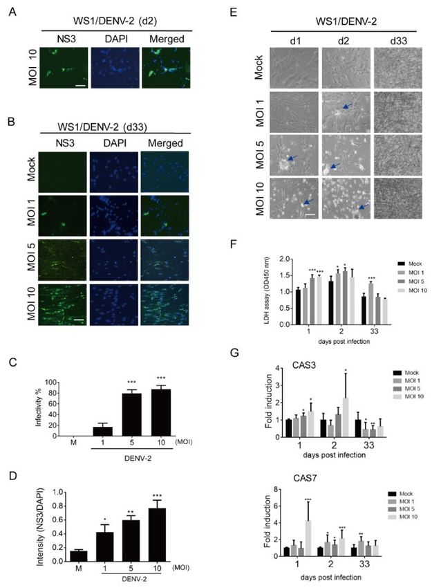
Viruses 2020, 12, 267                                                                               2 of 18

Due to global warming, more than 3 billion people are now residing in areas threatened by DENV
infection [1]. There are four different serotypes of the virus (DENV types 1–4), and each type can
cause disease. The levels of DENV infection vary from mild to severe, ranging from self-limited
febrile dengue fever, skin rash, drowsiness, agitation, liver enlargement, and dengue hemorrhagic
fever (DHF), to even death. A second DENV infection may result in life-threatening dengue shock
syndrome (DSS). Currently, no specific therapy is available for the infection other than supportive
treatments [2,3].
      Although most victims of DENV survive, more than half of patients experience post-dengue
fatigue syndrome (PDFS) for months after recovery [4,5]. The presentations of PDFS include
prolonged chronic fatigue, headache, fever, hair loss, arthralgia, major depression, memory loss, and
reasoning problems, and PDFS usually lasts for three to six months but can last for up to two years
[4–7]. The persistent symptoms are frequently associated with older age, dengue with warning signs
(at least one dengue fever symptom presented), hospitalization, thrombocytopenia, respiratory
distress, and comorbidities [5,8]. It is not clear which DENV serotype infection causes PDFS most
frequently. PDFS has a seriously negative impact on patient quality of life in terms of physical and
psychological limits, and can cause severe economic and social damage [8,9].
      Most studies focused on the acute manifestation of dengue illness. If persistent symptoms affect
a non-negligible proportion of the population, researchers have likely underestimated the problem
of PDFS. Knowledge of this long-term sequela of dengue is essential for understanding its
contribution to the morbidity of DENV infection.
      The underlying mechanisms for post-dengue syndrome remain unclear. One hypothesis is that
DENV infection induces a change in the immune system, which results in an imbalance of underlying
immune status because autoimmune-related cytokines and markers in post-dengue victims are
increased [4]. However, there are immune privilege areas in the human body, which include hair
follicles, neurologic tissue, the eyes, and the reproductive system. Thus, another possible mechanism
is that the virus resides within some immune privilege tissues for a period of time after convalescence,
so infection is persistent and the immune system fails to expel them.
      Flaviviruses are not considered viruses, which normally establish persistent infection in vivo.
However, the emerging Zika virus, which is a flavivirus genetically very similar to DENV, is
challenging this preconception [10]. Similar to PDFS, a chronic and potentially incapacitating clinical
entity, post-infectious syndrome has been noted in later phases following an initial recovery from
infection in victims of Zika virus [11]. The most probable mechanism for post-infectious syndrome
due to the Zika virus is that active viral replication continues in immune-privileged organs [12,13].
      In theory, DENV can also cause persistent infection. In vitro evidence shows that a number of
cell lines are persistently infected with DENV, including monocytes [14] and some immortalized cell
lines [15]. In vivo, the most definitive evidence for viral persistence is the isolation of viable virus or
the demonstration of viral antigens or RNA long after acute illness. However, only a few reports
support this concept. Persistent shedding of DENV-RNA has been demonstrated in vaginal
secretions up to 18 days from the onset of symptoms [16]. DENV RNA was detected in the semen of
a man returning from Thailand to Italy at day 37 after the onset of symptoms [17]. Indirect serological
evidence shows that anti-DENV immunoglobulin M (IgM) persists much longer than previously
thought. Anti-DENV IgM was detectable in 70.5% and 46.2% of 44 subjects at 6 and 12 months using
enzyme-linked immunosorbent assay (ELISA), respectively [18]. Persistent DENV infection was
diagnosed in an immunosuppressed patient, which showed that CD8+ T lymphocytes-mediated
cellular immune response is critical to DENV clearance [19]. All together, we think determining the
persistence of the DENV virus and its impact is important.
      A serious dengue outbreak occurred in Taiwan during 2014 and 2015, with 59,516 cases of
dengue fever confirmed, resulting in a total of 252 deaths. An abnormally high proportion of patients
who experienced early diffuse hair loss was observed, which was not explained by telogen effluvium
[20]. Reports of hair loss due to DENV infection are sporadic globally [21–26]. The clinical
observations agree with these reports: hair loss is not associated with severity of the infection [24].
Viruses 2020, 12, 267 3 of 18
Hair loss occurs in many victims as early as the initial two weeks post-infection. More than two-thirds
of cases occur within the first two months post-infection [21–24,26].
The hair growth cycle is well-orchestrated by complex interactions between different types of
cells and many signaling pathways, in which hair-follicle stem cells, dermal papilla cells, and
adipocytes are involved [27]. Dermal papilla plays an important role in epithelial–mesenchymal
interactions [28,29] and regulates hair growth by secreting factors, including wingless-related
integration site (Wnt), transforming growth factor beta (TGF-β), Fibroblast growth factor (Fgf) 7/10,
Noggin, and ectodysplasin A (Eda) to adjacent responding cells at the base of the hair follicle [30–32].
c-Myc also plays a role in the proliferation and differentiation of hair follicles, and stimulation
of the expression of Myc can interfere with the regulation of the mouse hair growth cycle [33–35].
Progranulin, a key regulator of inflammation, is able to regulate the hair cycle [36]. Receptor-
interacting serine/threonine protein kinase 1 (RIPK1 or RIP1) may affect hair growth since RIP1 can
increase both intracellular and extracellular progranulin protein levels by promoting the translation
rate of progranulin without affecting mRNA levels [37]. In addition, dermal fibroblasts produce
fibroblast growth factor 20 (FGF20) and FGF10 to control the condensation of human hair-follicle
dermal papilla cells (HFDPCs) and the differentiation of keratinocytes [38,39].
We previously reported that DENV can directly infect HFDPCs, resulting in cytokine change,
which could lead to a clinically distinct hair loss following DENV infection [20]. High susceptibility
to DENV infection by dermal fibroblasts was reported [40,41]. Because the host immune response
provides defenses against virus invasion and replication, the virus must avoid elimination by the
host’s immune response to establish a pathogenic or long-term infection [42]. The hair follicle is an
immune-privileged site. As such, we examined whether this kind of infection can persist for a long
time and cause a subsequent inflammatory cytokine or hair growth factors change. Therefore, we
conducted this follow up investigation to determine whether DENV can establish a long-term
infection in dermal fibroblasts and papilla cells.
2. Materials and Methods
2.1. Cell Culture
WS1 cells, which are normal human skin fibroblast cells (Bioresource Collection and Research
Center, BCRC:60300, Hsinchu, Taiwan), were cultured in minimum essential media (MEM) with 10%
heat-inactivated fetal bovine serum (FBS). Primary human hair-follicle dermal papilla cells (HFDPCs)
were isolated from the hair papilla of normal human scalp hair follicles (Cat. 602t-05a, Cell
Applications, Inc. San Diego, CA, USA). HFDPCs were cultured in collagen-coated flasks using
papilla cell growth medium (Cell Applications, Inc., San Diego, CA, USA), which was supplemented
with 12% FBS at 37 °C and 5% CO2.
2.2. Dengue Virus
The strain of DENV-2 (PL046) (GenBank accession no. AJ968413.1) was isolated from patients
with dengue fever. The virus was propagated in a C6/36 mosquito host cell line (CRL-1660, ATCC,
Manassas, VA, USA) that was grown in RPMI 1640 medium containing 5% FBS at 28 °C. The culture
media of DENV-2 infected C6/36 cells were titrated using a plaque-forming assay on the baby
hamster kidney (BHK21) cell line (BCRC: 60041, Hsinchu, Taiwan).
2.3. Virus Plaque Assay
BHK21 cells (2 × 10⁵ cells/well) were infected with different dilutions of the supernatants. Ten-
fold serial dilutions of virus supernatants were added to BHK21 cells in six-well plates and were
inoculated at 37 °C for 2 h. After 2 h of adsorption, the cells were overlaid with 1% agarose (SeaPlaque;
Lonza Rockland, Inc. Rockland, ME, USA) containing Dulbecco’s Modified Eagle Medium (DMEM)
medium with 2% FBS. After 7 days of incubation, cells were fixed with 10% formaldehyde and stained
with 0.5% crystal violet. Plaques (DENV-infected foci) were counted and the virus titration was
calculated in log10 plaque-forming units per milliliter (PFU/mL).Viruses 2020, 12, 267 4 of 18
2.4. Viral Infection
HFDPCs (4 × 10⁴ cells per well) were seeded in 12-well plates and incubated overnight. The cells
were replaced with serum-free medium, then infected or not infected (mock control) with DENV-2
at different multiplicities of infection (MOIs) of 1, 5, and 10. After 4 h of adsorption, virus
supernatants were removed and the cells grown in papilla cell growth medium supplemented with
2% FBS. WS1 cells (4 × 10⁴ cells per well) were also seeded in 12-well plates and followed by DENV-
2 infection at different MOIs (1, 5, and 10). After 2 h of adsorption, the virus supernatants were
removed and the cells were incubated with MEM supplemented with 10% FBS. During the long
period of culture, the medium was renewed every 3 days without subculture. The cell morphological
changes were captured at different time points at 1, 2 and 33 days post-infection using phase contrast
light microscopy. The cell total RNA and lysates underwent gene and protein expression analysis.
The culture medium of infected cells was harvested for the plaque-forming assay.
2.5. Immunofluorescence Assay
DENV2 infected-WS1 cells and HFDPCs were fixed with 4% paraformaldehyde for 30 min, and
then permeabilized with 0.5% Triton X-100 in phosphate buffered saline (PBS) for 10 min. Cells were
blocked with 10% skim milk in PBS. The cells were stained with primary anti-DENV NS3 (#YH0034,
1:500, Yao-Hong Biotechnology, New Taipei city, Taiwan) at 4 °C overnight. After PBS washes, the
cells were stained with goat anti-mouse immunoglobulin G (IgG)-Alexa Fluor 488-conjugated
secondary antibody (#A11001, Invitrogen, ThermoFisher Scientific, Waltham, MA, USA) for 2 h at
room temperature. Nuclei were stained with 4′,6′-diamidino-2-phenylindole (DAPI). The
fluorescence signals were observed and captured by a fluorescence microscope (objective 100×; Axio
Observer A1, Zeiss, Oberkochen, Germany). The fluorescence intensity was quantified using
National Institutes of Health (NIH) ImageJ software (imagej.nih.gov) To score the mean fluorescence
intensity, the exact same outline was used on an adjacent area of the image to provide a background
value. The intensity ratio of NS3 (Alexa Fluor 488)/DAPI levels was calculated. An unpaired t-test
using parametric distribution was used to measure differences between DENV-2 infection and non-
infection and were considered significant when p < 0.05.
2.6. Lactate Dehydrogenase (LDH) Cell Cytotoxicity Assay
WS1 and HFDPCs (4 × 10⁴ cells per well) were seeded in 12-well plates and incubated overnight.
The cells were then infected by DENV-2 (MOI 1, 5, and 10). The culture supernatants were harvested
at days 1, 2, and 33 post-infection and stored at −80 °C before use. Cell activity in cell supernatants
was assessed using an LDH-Cytotoxicity Assay Kit II (#ab65393; Abcam, Cambridge, MA, USA)
according to the manufacturer’s instructions. Cell cytotoxicity was quantified by measuring the
absorbance of solution at 450 nm wavelength using a EPOCHTM 2 microplate reader (BioTek,
Winooski, VT, USA). All experiments were performed in triplicate.
2.7. RNA Extraction and Quantitative Real-Time Polymerase Chain Reaction (qRT-PCR) Analysis
Total RNA was extracted from mock or DENV-infected cells by adding 500 μL Trizol reagent
(Invitrogen, Thermo Fisher Scientific) according to the manufacturer’s instructions. The RNA pellet
was resuspended in 30 μL of RNase-free distilled water and stored at −80 °C. For cDNA synthesis, 5
μg of total RNA was used for reverse transcription using SuperScriptTM III reverse transcriptase kit
(#18080093, Invitrogen, ThermoFisher Scientific, Waltham, MA, USA) according to the
manufacturer’s instructions. Real-time polymerase chain reaction (PCR) was performed using 3 μL
cDNA, 3 μM specific primers targeting the genes of interest, and 1× (final concentration) SYBR green
PCR Master mix (#4312704, Applied Biosystems, Waltham, MA) in a final reaction volume of 10 μL.
Amplification in an Applied Biosystems StepOnePlusTM real-time PCR system involved activation at
95 °C for 20 min followed by 40 amplification cycles at 95 °C for 3 s, 60 °C for 1 s. Real-time data were
analyzed using StepOnePlusTM software (Applied Biosystems, Waltham, MA). mRNA expression
(fold induction) was quantified by calculating the 2-ΔΔCt value, with glyceraldehyde-3-phosphateViruses 2020, 12, 267 5 of 18
dehydrogenase (GAPDH) mRNA as the endogenous control. The primer sequences are shown in
Table S1.
2.8. Immunoblotting Assay
Mock or DENV-infected HFDPCs and WS1 cells were cultured for 1, 2, and 33 days. The whole
cell extracts were prepared with protein lysis buffer (2% sodium dodecyl sulfate (SDS), 50 mM Tris-
HCl, pH 7.5) containing protease inhibitor and phosphatase inhibitor cocktail (Roche, Basel,
Switzerland). Protein concentration was determined using a Bradford assay kit (#5000116, BioRad,
Hercules, CA, USA). We separated 50 µg protein lysates in 10% acrylamide sodium dodecyl sulfate
polyacrylamide gel electrophoresis (SDS-PAGE) gel and transferred to polyvinylidene difluoride
(PVDF) membranes. Membranes were blocked with 5% milk in Tris-buffered saline, 0.05% Tween
X100 (TBST) for 1 h at room temperature, and then incubated with primary antibody overnight at 4
°C. After washing with TBST buffer, the membranes were incubated with horseradish peroxidase-
conjugated secondary antibody for 2 h at room temperature and then revealed using enhanced
chemiluminescent (ECL) reagent (Advansta, San Jose, CA, USA). Image and emission signal density
measurements were captured and quantified using a BioSpectrum Image System (UVP, Upland, CA).
Primary antibodies include anti-DENV-NS3 (GTX124252, GeneTex, Hsinchu, Taiwan), anti-retinoic
acid-inducible gene-I (RIG-I; D14G6; #3743, Cell Signaling, Danvers, MA), anti-MAVS (#3993, Cell
Signaling), anti-phospho-NF-κB p65 (Ser536) (#3033, Cell Signaling), anti-NF-κB p65 (C-20) (#sc-372,
Santa Cruz, CA, USA), and anti-GAPDH (#60004-1-Ig, Proteintech, Rosemont, IL, USA).
2.9. Statistical Analysis
Data are presented as mean ± standard deviation (SD) of at least 3 independent experiments.
Significant differences or correction between groups were analyzed using two tailed Student’s t-test
or Pearson’s correction test (GraphPad Prism software, La Jolla, CA, USA). A p-value less than 0.05
was considered to indicate statistical significance.
3. Results
3.1. Prolonged Infection of Dengue Virus (DENV-2) in WS1 (Human Dermal Fibroblast) Cells
To determine whether long-term or persistent infection with DENV in the scalp results in hair
loss in patients experiencing post-dengue syndromes, two cell types supporting hair growth, dermal
fibroblasts and HFDPCs, were used to determine the prolonged DENV-2 infectivity. First, DENV-2
NS3 was detected using an immunofluorescence assay in dermal fibroblasts (WS1 cells) at day 2 of
the acute infection phase. The results showed that WS1 cells are susceptible to DENV-2, as expected
(Figure 1A). At day 33 of long-term culture after infection, DENV NS3 was still detected in WS1 cells
(Figure 1B). The DENV-2 infectivity was counted using an immunofluorescence assay, which showed
that approximately 80% cells with MOI 5 and 10 presented NS3 (Figure 1C); the increased
immunofluorescence intensity of NS3 was determined in a MOI-dependent manner (Figure 1D). In
the bright field images, the DENV-induced cytopathic effect (CPE) was seen at days 1 and 2 in WS1
cells with a high MOI (5 and 10) for DENV-2 infection, but was not evident on day 33 (Figure 1E).
DENV-2-induced cell death was also confirmed by a LDH cell cytotoxicity assay; compared to the
acute infection phase, long-term culture WS1 cells showed a lower level of LDH release (Figure 1F).
The cell death genes caspase 3 (CAS3) and caspase 7 (CAS7) were also highly induced in the acute
infection phase, but downregulated on day 33 (Figure 1G).Viruses 2020, 12, 267 6 of 18
Figure 1. Long-term infection of dengue virus (DENV)-2 in WS1 (human dermal fibroblast) cells. (A)
4 × 10⁴ WS1 cells were infected with mock or DENV-2 multiplicities of infection (MOI) 10 for 2 days,
the DENV-2 infected cells were detected by immunofluorescence assay with anti-NS3 antibody
(green). The images merged with 4′,6′-diamidino-2-phenylindole (DAPI) staining of cell nuclei (blue)
are also shown, scale bar: 100 μm. (B) The immunofluorescence assay was conducted with DENV-2
(MOI 1, 5, and 10) infected WS1 cells at day 33 post infection. (C) According to the
immunofluorescence data of prolonged infection, the DENV-2 infectivity was cultured with cells
expressed NS3. (D) The immunofluorescence intensity of NS3 vs. DAPI on day 33 post-infected cells.
(E) The morphologies of DENV-2 infected WS1 cells were observed and captured by phase contrast
light microscopy at days 1, 2, and 33 post-infection. The arrows indicate the spots of cytopathic effect
(CPE). (F) Lactate dehydrogenase (LDH) cell cytotoxicity assay was conducted with culture medium
harvested at days 1, 2, and 33 post-infection. (G) Quantitative reverse-transcription polymerase chain
reaction (qRT-PCR) analysis of mRNA expression of caspase 3 (CAS3, upper panel) and caspase 7
(CAS7, lower panel) in WS1 cells (4 × 10⁴) infected with DENV-2 (MOI 1, 5, and 10) for 1, 2, and 33
days. Relative mRNA expression normalized to that of glyceraldehyde-3-phosphate dehydrogenase
(GAPDH), and the fold induction to mock control. Data are shown as means ± standard deviation
(SD, n = 3). Student’s t-test *, p < 0.05; **, p < 0.01; ***, p < 0.001 compared with mock infection groups.
3.2. DENV-2 Replication in Long-Term Cultured WS1 Cells
DENV-2 NS3 protein was detected in long-term infected WS1 cells, the level of viral RNA (5′-
untranslated region, 5′-UTR) expression and virion production during the period of infection was
further measured. The replication of DENV-2 5′-UTR RNA was observed in WS1 cells after days 1
and 2 post-infection. The viral RNA was also detected after 33 days in cells that were infected with
DENV-2 but the level of viral RNA was lower than that for the acute infection phase (Figure 2A).
WS1 produced high amounts of DENV-2 virion on days 1 and 2 post-infection; on day 33, a
certain amount of DENV virion was also detected in WS1 cells (Figures 2B, C). These results show
that the complete life cycle or virion assembling of DENV-2 occurred in WS1 cells. To understand
whether DENV-2 virulence changed after long-term infection in WS1 cells, the plaque size wasViruses 2020, 12, 267 7 of 18
measured. Compared to day 1, virus from day 33 showed a slight reduction in plaque size, which
implicated that DENV-2 virulence does not change after long-term infection in WS1 cells (Figure 2D).
Figure 2. DENV-2 RNA replication and virion production in WS1 cells after long-term culture. (A) 4
× 104 WS1 cells were infected with DENV-2 MOI 1, 5, and 10; the DENV-2 RNA (5′-UTR) replication
was detected by qRT-PCR on days 1, 2, and 33 post-infection. The DENV-2 RNA expression was
normalized to the Gapdh gene and the fold induction to mock control. Data are presented mean ± SD
from three independent tests. (B) The culture medium of DENV-2-infected WS1 cells were harvested
and diluted (dilution factor: 10–1 to 10–6) for plaque assay. (C) The virus yield by WS1 cells and
quantified in plaque assay are presented in log10 plaque-forming units per milliliter (PFU/mL) from
three independent assays. (D) The diameter of the plaque size in DENV-2 plaque assay was measured
using Image J software (imagej.nih.gov). Data are presented mean ± SD, * p < 0.05, *** p < 0.001, *** p
< 0.005 vs. mock control.
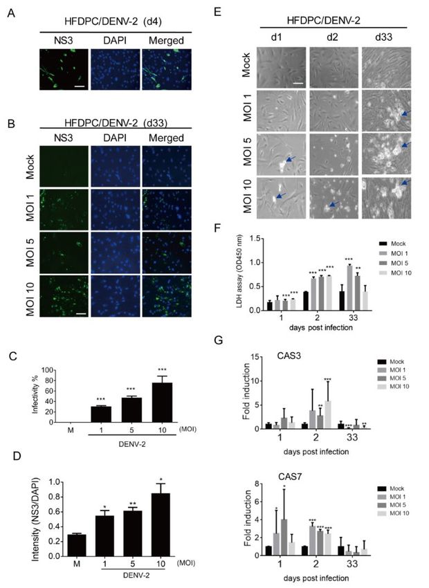
3.3. Prolonged Infection of DENV-2 in Human Hair-Follicle Dermal Papilla Cells (HFDPCs)
The DENV-2-infected HFDPCs were investigated using an immunoflourescence assay, in which
DENV-2 NS3 was detected in the acute inifection phase (day 4) and prolonged infection phase (day
33). In addition, a dots staining pattern of NS3 was noted (Figures 3A,B). The DENV-2 infectivity and
immunofluorescence level of NS3 increased in HFDPCs in a MOI-dependent manner on day 33 post-
infection (Figures 3C,D). In the bright field images, the DENV-2-induced cytopathic effect (CPE) was
observed on days 1, 2, and 33 in long-term cultured HFDPCs (Figure 3E). The LDH assay revealed
that DENV-2 caused severe cell damage on day 33 (Figure 3F). These data suggested that the dots
staining pattern for NS3 immunofluorescence assay, shown in Figure 3B, might be the cell death
debris after DENV-2 long-term infection. In addition, CAS3 and CAS7 expression were induced by
DENV-2 on days 1 and 2 post-infection; compared to the acute infection phase, the prolonged-
infected HFDPCs expressed a relatively lower RNA level of CAS3 and CAS7, which might due to the
severe CPE (Figure 1G).Viruses 2020, 12, 267 8 of 18
Figure 3. Analysis of the prolonged infection of DENV-2 in hair-follicle dermal papilla cells
(HFDPCs). (A) HFDPCs (4 × 10⁴) were infected with mock or DENV-2 MOI 10 for four days; an
immunofluorescence assay was conducted to detect DENV-2 NS3 (green). The images merged with
DAPI staining of cell nuclei (blue) are also shown, scale bar: 100 μm. (B) DENV-2-infected cells were
detected by an immunofluorescence assay in DENV-2 (MOI 1, 5, and 10)-infected HFDPCs cells after
prolonged infection for 33 days. (C) DENV-2 infectivity at day 33 post-infection was calculated. (D)
The intensity shows the fluorescence ratio of NS3 vs. DAPI in post-infected cells on day 33. (E) The
morphologies of DENV-2 (MOI 1, 5, and 10)-infected HFDPCs were obtained by phase contrast light
microscopy at days 1, 2, and 33 post-infection. The arrows indicate the spots of CPE. (F) The culture
medium was harvested on day 1, 2, and 33 post-infection for the LDH cell cytotoxicity assay. (G) qRT-
PCR analysis of mRNA expression of caspase 3 (CAS3, upper panel) and caspase 7 (CAS7, lower panel)
in HFDPCs with DENV-2 (MOI 1, 5 and 10) infection. Relative mRNA expression normalized to that
of Gapdh and the fold induction to mock control. Data are presented as means ± SD (n = 3). Student’s
t-test *, p < 0.05; **, p < 0.01; ***, p < 0.001 compared with mock infection groups.
3.4. Detection of DENV-2 in Long-Term Cultured HFDPCs
In HFDPCs, the high level of DENV-2 RNA was detected on day 1 and 4 post-infection; at 33
days, a lower level of viral RNA was detected than during the acute infection phase (Figure 4A). The
high titer of DENV-2 virion was detected on days 1 and 2 post-infection in HFDPCs, but the long-
term culture HDFPCs failed to produce DENV2 (Figures 4B,C). In addition, compared to the virion
harvested on day 1 post-infection, a slightly reduction of plaque size was found on day 2 (Figure 4D).Viruses 2020, 12, 267 9 of 18
Figure 4. Evaluation of the DENV-2 replication in HFDPCs after prolonged infection. (A) The DENV-
2 RNA (5′-UTR) replication was detected by qRT-PCR in HFDPCs (4 × 10⁴) with DENV-2 (MOI 1, 5,
and 10) after 1, 2, and 33 days. The expression level of DENV-2 RNA was normalized to Gapdh, and
the fold induction to mock control. Data are presented as mean ± SD from three independent tests.
(B) The plaque assay was conducted with the culture medium of DENV-2-infected HFDPCs (dilution
factor: 10–1 to 10–6). (C) The virus yield by HFDPCs from the plaque assay are presented in log10 plaque-
forming units per milliliter (PFU/mL) from three independent assays. (D) The diameter of the plaque
size in the plaque assay. Data are presented as mean ± SD, * p < 0.05, *** p < 0.001, *** p < 0.005 vs. mock
control.
3.5. Anti-Viral Inflammatory Responses Moderately Activated in Long-Term Cultured WS1 Cells
Innate immunity plays an important role against viral infection. Type I interferons (IFN α/β) and
type III interferons (IFNλ1), which are induced by viral infection, are an important first defense [43].
Inflammation in the upper hair follicle was found to be involved in the pathogenic progress of
alopecia areata [44]. Regulation of host anti-viral inflammation and cell death due to acute infection
with DNEV-2 in HFDPCs could be responsible for hair loss [20].
To evaluate the gene expression of anti-virus and inflammation in long-term DENV-2-infected
cells compared to the acute infection, qRT-PCR was conducted. The expressions of IFNα, IFNβ,
IFNλ1, Rig-I, OAS-1, MxA, IL6, IL8, and TNFα were increased at either day 1 or day 2 in WS1 post-
infection or on both days. These genes’ expressions were decreased on day 33; however, compared
with mock infection, IFNα, Rig-I, OAS-1, IRF7, MxA, and IL6 were also expressed at a statistically
significant higher level. In particular, we observed significant expression of IRF7 on day 33 (Figure
5A).
Because the mRNA of anti-viral inflammatory genes was detected in WS1, the retinoic acid-
inducible gene I (RIG-I)/mitochondrial antiviral-signaling protein (MAVS)/NF-κB p65 signaling axis
[45] for anti-viral inflammation in long-term DENV-infected WS1 cells was examined using
immunoblotting. The immunoblots indicated the expression levels of DENV-NS3, RIG-I, MAVS, and
NF-κB p65 protein, and phosphorylated NF-κB p65 (phospho-NF-κB p65) protein. First, DENV-2 NS3
was detected in the DENV acute and long-term infection phases, which supports the DENV-2 RNA
and virion production shown in Figures 1 and 2. Compared to the mock group, DENV-2 enhanced
the protein expression of RIG-I in WS1 cells on days 1, 2, and 33 post-DENV-infection in WS1 cells.
The enhanced protein level of MAVS was detected on day 1 and day 33 of the infection (Figure 5B).
These data indicated a prolonged activation of antiviral signaling in DENV-2-infected fibroblasts.Viruses 2020, 12, 267 10 of 18
The inflammation signaling in WS1 cells with DENV-2 infection was evaluated by immunoblotting
of the protein level of phospho-NF-κB p65 and total NF-κB p65. The data showed that phospho-NF-
κB p65 and total NF-κB p65 were down-regulated with high MOIs of 5 and 10 in the first two days
after infection, but an increased level of phospho-NF-κB p65 was noted at 33 days of cultivation
(Figure 5B).
Figure 5. Prolonged DENV-2 induces anti-viral inflammation in WS1 cells. (A) qRT-PCR of the
indicated antiviral inflammation genes’ mRNA expression in WS1 cells infected with DENV-2 at MOI
1, 5, and 10 for 1, 2, and 33 days. The gene expression was normalized to internal control gene, Gapdh.
Data are presented as mean ± SD from three independent tests, * p < 0.05, ** p < 0.01, *** p < 0.005 vs.
mock control. (B) WS1 cells were infected by DENV at MOI 1, 5, and 10 for 1, 2, and 33 days. The cell
lysates were subjected to acrylamide sodium dodecyl sulfate polyacrylamide gel electrophoresis
(SDS-PAGE) and Western blot analysis with antibody against DENV NS3, retinoic acid-inducible
gene I (RIG-I), mitochondrial antiviral-signaling protein (MAVS), NF-κB p65. GAPDH expression was
the internal loading control. Data are presented from three independent tests. Quantification of each
protein level with GAPDH was normalized to the mock control.
3.6. Anti-Viral Inflammatory Responses in HFDPCs with Prolonged DENV-2 Infection
HFDPCs exhibited similar expression in the antiviral inflammation genes with WS1 cells. IFNα,
IFNβ, IFNλ1, Rig-I, IRF3, IRF7, OAS-1, MxA, IL6, IL8, and TNFα expressions were increased at either
day 1 or day 2 in HFDPCs post-infection or on both days. Compared to mock samples, significant
levels of IFNλ1, Rig-I, MAVS, IRF7, OAS-1, MxA, and IL6 were induced in the long-term infected cells,
although in lower amounts than in acute infection (Figure 6A). The change in the expression level of
the host genes might be associated with the virus replication level during the period of infection; forViruses 2020, 12, 267 11 of 18
example, IFNλ and IL6 expression were correlated with the level of DENV-2 virion titer in HFDPC as
shown in Figure 4 (Table S2).
Immunoblotting of the RIG-I/MAVS/NF-κB signaling pathway in HFDPCs showed that RIG-I
protein was increased by DENV-2 at days 1, 2, and 33 post-infection. In the high-MOI infected cells,
a downregulated RIG-I was noted at MOI 10 infection at day 2, but which increased again at day 33
(Figure 6B). We observed a variation in the protein level of MAVS and NF-κB p65 during the period
of infection; MAVS was induced at day 1 of the acute infection phase, and then reduced to a modest
protein level (Figure 4B). Similar to MAVS, a transient protein induction pattern of phospho-NF-κB
p65 and total NF-κB p65 was detected in HFDPCs.
Figure 6. Analysis of the anti-viral inflammation in HFDPCs with acute or long-term DENV-2
infections. (A) qRT-PCR of the indicated mRNA expression in WS1 cells infected with DENV-2 at
MOI 1, 5, and 10 for 1, 2, and 33 days. The gene expression was normalized to Gapdh. Data are
presented as mean ± SD from three independent tests, * p < 0.05, ** p < 0.01, *** p < 0.005 vs. mock
control. (B) The DENV-2-infected HFDPCs lysates were subjected to SDS-PAGE and Western blot
analysis. Representative data were obtained from three independent tests. Quantification of each
protein level at days 1, 2, and 33 days with GAPDH was normalized to individual mock control.
3.7. Downregulation of Hair Growth Genes by DENV-2 Long-Term Infection
The acute and prolonged DENV-2 infection caused subsequent changes of anti-viral
inflammatory signaling and cytokine expression. We wanted to understand whether the hair growth
regulatory factors were also affected by DENV-2 at different infection periods. qRT-PCR in WS1
showed that Rip1, Wnt1, Wnt4, and cMyc expressions were inducted by DENV-2 (MOI 1, 5, and 10)
after one and/or two days of infection, but decreased at day 33 post-infection, particularly in the MOI
5 and 10 infected WS1 cells (Figure 7A). In HFDPCs, the increased levels of Rip1, Wnt1, and Wnt4
were only detected at day 1 after DENV-2 infection, but decreased at days 2 and 33 in comparison to
the mock infection (Figure 7B). A different pattern of cMyc expression was noted in HFDPCs, where
cMyc expression remained at a substantial level after the long-term culture (Figure 7B).Viruses 2020, 12, 267 12 of 18
Figure 7. The expression of hair growth cycle genes in cells with DENV-2 infection for indicated days.
(A, B) qRT-PCR of Rip-1, Wnt1, Wnt4 and cMyc was conducted in WS1 and HFDPCs with DENV-2
(MOI 1, 5, and 10) for 1, 2, and 33 days. The mRNA expression level was normalized to Gapdh, and
the fold induction to the mock control. Data are presented as mean ± SD from three independent tests.
* p < 0.05, ** p < 0.01, *** p < 0.005 vs. mock control.
4. Discussion
In this study, we used a long-term DENV-2 infection model for human dermal fibroblasts (WS1
cells) and primary human follicle dermal papilla cells (HFDPC) to determine the possible mechanism
for DENV-mediated hair loss of post-dengue fatigue syndromes in vitro. We found that DENV-2
RNA existed in both long-term infected cell types. DENV-2 virion was produced in long-term
infected WS1 cells, but the HFDPCs failed to produce DENV-2 after a long-term culture. This reveals
that although DENV-2-infected HFDPCs expressed RNA and protein, the complete life cycle or virion
assembling of DENV-2 was defective. Antiviral inflammation genes, related to signaling activity and
hair growth regulatory factors, were detected in both cell types, which showed that DENV-2 virus is
a critical factor for inflammation in cells after long-term infection. The data for detectable long-term
infection with DENV-2 in dermal fibroblasts and dermal papilla showed a pathogenic mechanism,
which may be involved in DENV-mediated hair loss during post-dengue fatigue syndrome (Figure
S1).
Skin cells, such as dermal fibroblasts, HPDPCs, epidermal keratinocytes, and dendritic cells,
were previously found to be permissive to flaviviruses, DENV, West Nile virus (WNV), and Zika
virus (ZIKV) infection [20,46–48]. Here, we revealed that dermal fibroblasts are also a competent cell
type in which a prolonged infection of over one month with DENV can be established without CPE
(Figures 1 and 2). DENV-2 can establish a persistent infection in Raji cells (Burkitt’s lymphoma), but
the infection period examined was only four days [15].Viruses 2020, 12, 267 13 of 18
The role of HFDPCs in hair loss caused by acute infection with DENV-2 was previously
speculated [20]. We further reveal that DENV viral protein and RNA are persistently expressed in
HFDPCs after long-term infection. The DENV-2-mediated CPE that is present in HFDPCs inhibits
virion production (Figures 3 and 4). The results showed that persistent infection with DENV-2 and
virion production in the scalp depends on the specific cell types. Therefore, a future study should
determine whether dermal fibroblasts, HFDPCs, and other cell types, such as bulge stem cells or
adipocytes, are involved in cell growth that causes DENV infection-mediated hair loss. Dendritic cells
were found to play a critical role in the establishment of viral persistent infection [49]. Thus,
understanding whether DENV can persistently infect skin dendritic cells (Langerhans cells) is
important, which are the targets of DENV [50]. Whether delayed DENV-2 clearance prolongs
replication of the virus should also be determined.
Inflammatory and anti-viral genes were significantly induced in HFDPC and WS1 cells at day 1
and 2 after DENV infection, but only low levels of these genes were detected at day 33 after DENV
infection. The DENV-2 acute infection-mediated anti-viral inflammation response in human dermal
fibroblasts is supported by other flaviviruses studies on a clinical isolate DENV-2, WNV, or ZIKV
[41,47,51]. The change in the expression of host genes in HFDPCs was corelated with the DENV-2
RNA expression, which demonstrates that inflammatory and anti-viral genes are induced in DENV-
infected cells in a viral load-dependent manner (Table S2). This result is consistent with the results of
a previous study of A549 cells (lung carcinoma cells) that are infected with DENV [52].
Viral RNA is a potent pathogen-associated molecular pattern that activates numerous receptors
and induces strong immune responses [53]. In terms of DENV infection, the viral RNA is recognized
by the host RNA sensors, which are mainly RIG-I-like receptors (RLRs) and toll-like receptors [4,52].
RIG-I/MDA5 senses genomic RNA or double-stranded (ds) which is the product of an intermediate
step of DENV replication. This activates intracellular pathways and leads to the production of anti-
viral effectors, including interferon and pro-inflammatory cytokines [54]. In this study, we observed
that RIG-I gene and protein are induced in the late persistent infection phase, possibly due to the
persistent expression of viral RNA, which activates anti-viral inflammatory signaling (Figures 5 and
6) [55,56]. However, the dynamic changes of singling protein might be due to the DENV-2-mediated
HFDPC death or downregulation of RIG-I/MAVS signaling, as shown in previous reports [57,58]. We
hypothesized that the changes in the level of expression of signaling protein in both cell types is
correlated with the expression of antiviral inflammation genes. The early pathological processes of
alopecia areata may involve inflammatory cell activity in the upper hair follicle; potentially, the cycles
for hair growth may be altered by inflammation [44].
IFN-λ is a crucial defense mechanism for barrier tissues such as mucosal epithelial cells against
viral infection [59,60]. IFN-λ-mediated signaling is also required to control Yellow fever virus
infection [61]. The pathophysiological role of IFN-λ in cutaneous lupus erythematosus, psoriasis, and
atopic dermatitis has been reported [59,62]. The data for this study showed that in addition to type I
IFN, type III IFN (IFNλ1) was significantly induced by DENV-2 in dermal fibroblast and HFDPCs in
the acute infection phase (Figures 5 and 6). This may regulate an antiviral response in skin; therefore,
the biological role of IFN-λ in skin tissue should be determined.
IRF7 is a transcription factor induced by IFN to regulate IFN activity. It is also involved in the
viral infection that causes lethal immune disease [63,64]. We determined a significant induction of
IRF7 in WS1 cells and HPDPCs at day 33 after DENV infection (Figures 5 and 6). This result may
suggest a pathogenic activity for IRF7 in long-term infected cells. We think that the long-term effect
of IRF7 in viral-infection-mediated disease is worthy of future study.
Wnt/β-catenin pathway is the core component for regulating hair growth and telogen-to-anagen
phase transformation. Altered expression of Wnt ligands or Wnt antagonists can induce
dysregulation of the murine hair-follicle cycle and causes alopecia. The Wnt signaling pathway can
interfere with BMP, Eda, and Notch pathways, and this disturbance subsequently causes hair loss
[65,66]. In the literature, many kinds of Wnt factors were found to be involved in regulating hair-
growth cycles, including Wnt 1a, -3, -4, -5, -7, -10, and -11 [67–71]. However, fundamental
discrepancies exist in the regulatory modulators of the Wnt pathway in human and murine modelsViruses 2020, 12, 267 14 of 18
[72]. Thus, studying and identifying which single factor of the Wnt family is mainly responsible for
initiating hair loss are difficult. The disturbed expression of any signaling pathway can lead to hair
growth alteration [72]. However, we found that Wnt1 and Wnt4 gene levels were downregulated after
prolonged DENV-2 infection in dermal fibroblasts and dermal papilla cells (Figure 7), which indicates
a linkage between DENV-2-mediated hair loss and the Wnt pathway.
During DENV virus infection, inflammation and apoptosis play important roles in
pathophysiology. Serine-threonine kinase is a key regulator of both cell death and cell survival and
can regulate downstream apoptotic-associated factors. In the regulation of the normal hair cycle and
hair development, apoptosis is an essential step since the presence of apoptotic cells in hair follicles
is a decisive indicator for catagen [73]. This effect supports our data, which showed that RIP1
expression was reduced in the long-term DENV-2-infected cells (Figure 7), which may be involved
in the virus-mediated hair loss.
The findings suggest that the cycles for hair growth are disturbed because DENV causes
persistent infection in hair-follicle dermal papilla cells. This persistent infection may cause chronic
prolonged hair loss in post-dengue victims. However, we did not directly determine whether
persistent infection causes long-term alteration of hair cycles. Direct evidence for viral replication in
the human hair of dengue victims or in animal infection models is lacking. Different hair-associated
cells must be co-cultured and the in vivo shedding of hair should be analyzed to verify delayed
prolonged hair loss in post-dengue syndrome cases with alopecia.
Supplementary Materials: The following are available online at www.mdpi.com/xxx/s1, Figure S1. Schematic
diagram of the hypothesis, Table S1. The primer sequences used in qPCR analysis, Table S2. The correlation of
virus titer and host gene induction.
Author Contributions: K.-C.W. and T.-H.C. conceived and designed the experiments. W.-J.W. performed the
experiments. K.-C.W. and T.-H.C. analyzed the data. Y.-S. L. and L.-C.Y. contributed reagents and materials. K.-
C.W. and T.-H.C. wrote the manuscript. All authors reviewed the manuscript. All authors have read and agreed
to the published version of the manuscript.
Funding: This work was supported in part by grants from the Ministry of Science and Technology of Taiwan
(MOST 106-2314-B-075B-007-MY2, MOST 108-2314-B-075B-005).
Conflicts of Interest: The authors declare no competing financial interests. The funders had no role in the study
design, data collection and analysis, or preparation of the manuscript or decision to publish.
References
1. Wilder-Smith, A.; Ooi, E.E.; Horstick, O.; Wills, B. Dengue. Lancet 2019, 393, 350–363.
2. Halstead, S.; Wilder-Smith, A. Severe dengue in travellers: Pathogenesis, risk and clinical
management. J Travel Med 2019, 26.
3. Martina, B.E.; Koraka, P.; Osterhaus, A.D. Dengue virus pathogenesis: An integrated view. Clin
Microbiol Rev 2009, 22, 564–581.
4. Garcia, G.; Gonzalez, N.; Perez, A.B.; Sierra, B.; Aguirre, E.; Rizo, D.; Izquierdo, A.; Sanchez, L.; Diaz,
D.; Lezcay, M., et al. Long-term persistence of clinical symptoms in dengue-infected persons and its
association with immunological disorders. Int J Infect Dis 2011, 15, e38-43.
5. Teixeira, L.A.S.; Nogueira, F.; Nascentes, G.A.N. Prospective study of patients with persistent
symptoms of dengue in brazil. Rev Inst Med Trop Sao Paulo 2017, 59, e65.
6. Seet, R.C.; Quek, A.M.; Lim, E.C. Post-infectious fatigue syndrome in dengue infection. J Clin Virol
2007, 38, 1–6.
7. Shin, H.; Choi, S.J.; Cho, A.R.; Kim, D.Y.; Kim, K.H.; Kwon, O. Acute stress-induced changes in
follicular dermal papilla cells and mobilization of mast cells: Implications for hair growth. Ann Derm.
2016, 28, 600–606.
8. Tiga, D.C.; Undurraga, E.A.; Ramos-Castaneda, J.; Martinez-Vega, R.A.; Tschampl, C.A.; Shepard, D.S.Viruses 2020, 12, 267 15 of 18
Persistent symptoms of dengue: Estimates of the incremental disease and economic burden in mexico.
Am J Trop Med Hyg 2016, 94, 1085–1089.
9. Hung, T.M.; Wills, B.; Clapham, H.E.; Yacoub, S.; Turner, H.C. The uncertainty surrounding the
burden of post-acute consequences of dengue infection. Trends Parasitol 2019, 35, 673–676.
10. Paz-Bailey, G.; Rosenberg, E.S.; Doyle, K.; Munoz-Jordan, J.; Santiago, G.A.; Klein, L.; Perez-Padilla, J.;
Medina, F.A.; Waterman, S.H.; Adams, L.E. Persistence of zika virus in body fluids. New Engl. J. Med.
2018, 379, 1234–1243.
11. Chang, C.; Ortiz, K.; Ansari, A.; Gershwin, M.E. The zika outbreak of the 21st century. J Autoimmun
2016, 68, 1–13.
12. Hirsch, A.J.; Smith, J.L.; Haese, N.N.; Broeckel, R.M.; Parkins, C.J.; Kreklywich, C.; DeFilippis, V.R.;
Denton, M.; Smith, P.P.; Messer, W.B., et al. Zika virus infection of rhesus macaques leads to viral
persistence in multiple tissues. Plos Pathog 2017, 13, e1006219.
13. Pierson, T.C.; Diamond, M.S. The emergence of zika virus and its new clinical syndromes. Nature
2018, 560, 573–581.
14. Kurane, I.; Kontny, U.; Janus, J.; Ennis, F.A. Dengue-2 virus infection of human mononuclear cell lines
and establishment of persistent infections. Arch. Virol. 1990, 110, 91–101.
15. Takasaki, T.; Takada, K.; Kurane, I. Electron microscopic study of persistent dengue virus infection:
Analysis using a cell line persistently infected with dengue-2 virus. Intervirology 2001, 44, 48.
16. Iannetta, M.; Lalle, E.; Musso, M.; Carletti, F.; Scorzolini, L.; D’Abramo, A.; Chinello, P.; Castilletti, C.;
Ippolito, G.; Capobianchi, M.R. Persistent detection of dengue virus rna in vaginal secretion of a
woman returning from sri lanka to italy, april 2017. Eurosurveillance 2017, 22.
17. Lalle, E.; Colavita, F.; Iannetta, M.; Teklè, S.G.; Carletti, F.; Scorzolini, L.; Bordi, L.; Vincenti, D.;
Castilletti, C.; Ippolito, G. Prolonged detection of dengue virus rna in the semen of a man returning
from thailand to italy, january 2018. Eurosurveillance 2018, 23.
18. Chien, Y.-W.; Liu, Z.-H.; Tseng, F.-C.; Ho, T.-C.; Guo, H.-R.; Ko, N.-Y.; Ko, W.-C.; Perng, G.C.
Prolonged persistence of igm against dengue virus detected by commonly used commercial assays.
Bmc Infect. Dis. 2018, 18, 156.
19. Ng, K.H.; Zhang, S.L.; Tan, H.C.; Kwek, S.S.; Sessions, O.M.; Chan, C.Y.; Liu, I.D.; Lee, C.K.; Tambyah,
P.A.; Ooi, E.E., et al. Persistent dengue infection in an immunosuppressed patient reveals the roles of
humoral and cellular immune responses in virus clearance. Cell Host Microbe 2019, 26, 601–605 e603.
20. Wei, K.-C.; Huang, M.-S.; Chang, T.-H. Dengue virus infects primary human hair follicle dermal
papilla cells. Front. Cell. Infect. Microbiol. 2018, 8.
21. Hitani, A.; Yamaya, W.; To, M.; Kano, I.; Honda-Hosono, N.; Takasaki, T.; Haruki, K. [a case of dengue
fever and subsequent long-lasting depression accompanied by alopecia in a japanese traveler
returning from bali, indonesia]. Kansenshogaku Zasshi 2015, 89, 279–282.
22. Jensenius, M.; Gundersen, S.G.; Vene, S.; Bruu, A.L. [dengue fever imported to norway. Serologically
confirmed cases 1991-96]. Tidsskr Nor Laegeforen 1997, 117, 4230–4233.
23. Qiu, F.X.; Gubler, D.J.; Liu, J.C.; Chen, Q.Q. Dengue in china: A clinical review. Bull World Health
Organ 1993, 71, 349–359.
24. Tristao-Sa, R.; Kubelka, C.F.; Zandonade, E.; Zagne, S.M.; Rocha Nde, S.; Zagne, L.O.; Araujo, N.F.;
Amin, B.; Fazoli, F.; Souza, L.J., et al. Clinical and hepatic evaluation in adult dengue patients: A
prospective two-month cohort study. Rev Soc Bras Med Trop 2012, 45, 675–681.
25. Harn, M.R. [clinical study on dengue fever during 1987-1988 epidemic at kaohsiung city, southernViruses 2020, 12, 267 16 of 18
taiwan]. Gaoxiong Yi Xue Ke Xue Za Zhi 1989, 5, 58–65.
26. Chu, C.-B.; Yang, C.-C. Dengue-associated telogen effluvium: A report of 14 patients. Dermatol. Sin.
2017, 35, 124–126.
27. Driskell, R.R.; Clavel, C.; Rendl, M.; Watt, F.M. Hair follicle dermal papilla cells at a glance. J Cell Sci
2011, 124, 1179–1182.
28. Millar, S.E. Molecular mechanisms regulating hair follicle development. J Invest Derm. 2002, 118, 216–
225.
29. Alonso, L.; Fuchs, E. The hair cycle. J Cell Sci 2006, 119, 391–393.
30. Oshimori, N.; Fuchs, E. Paracrine tgf-beta signaling counterbalances bmp-mediated repression in hair
follicle stem cell activation. Cell Stem Cell 2012, 10, 63–75.
31. Ramos, R.; Guerrero-Juarez, C.F.; Plikus, M.V. Hair follicle signaling networks: A dermal papilla-
centric approach. J Invest Derm. 2013, 133, 2306–2308.
32. Rezza, A.; Wang, Z.; Sennett, R.; Qiao, W.; Wang, D.; Heitman, N.; Mok, K.W.; Clavel, C.; Yi, R.;
Zandstra, P., et al. Signaling networks among stem cell precursors, transit-amplifying progenitors,
and their niche in developing hair follicles. Cell Rep 2016, 14, 3001–3018.
33. Bull, J.; Pelengaris, S.; Hendrix, S.; Chronnell, C.; Khan, M.; Philpott, M. Ectopic expression of c-myc in
the skin affects the hair growth cycle and causes an enlargement of the sebaceous gland. Br. J.
Dermatol. 2005, 152, 1125–1133.
34. Wang, N.; Yang, T.; Li, J.; Lei, M.; Shi, J.; Qiu, W.; Lian, X. The expression and role of c-myc in mouse
hair follicle morphogenesis and cycling. Acta Histochem. 2012, 114, 199–206.
35. Flores, A.; Schell, J.; Krall, A.S.; Jelinek, D.; Miranda, M.; Grigorian, M.; Braas, D.; White, A.C.; Zhou,
J.L.; Graham, N.A. Lactate dehydrogenase activity drives hair follicle stem cell activation. Nat. Cell
Biol. 2017, 19, 1017–1026.
36. Kato, M.; Hasunuma, N.; Nakayama, R.; Takeda, J.; Itami, S.; Taira, M.; Manabe, M.; Osada, S.-I.
Progranulin, a secreted tumorigenesis and dementia-related factor, regulates mouse hair growth. J.
Dermatol. Sci. 2009, 53, 234–236.
37. Mason, A.R.; Elia, L.P.; Finkbeiner, S. The receptor-interacting serine/threonine protein kinase 1
(ripk1) regulates progranulin levels. J Biol Chem 2017, 292, 3262–3272.
38. Biggs, L.C.; Makela, O.J.; Myllymaki, S.M.; Das Roy, R.; Narhi, K.; Pispa, J.; Mustonen, T.; Mikkola,
M.L. Hair follicle dermal condensation forms via fgf20 primed cell cycle exit, cell motility, and
aggregation. Elife 2018, 7.
39. Kumtornrut, C.; Yamauchi, T.; Koike, S.; Aiba, S.; Yamasaki, K. Androgens modulate keratinocyte
differentiation indirectly through enhancing growth factor production from dermal fibroblasts. J
Derm. Sci 2019, 93, 150–158.
40. Kurane, I.; Janus, J.; Ennis, F.A. Dengue virus infection of human skin fibroblasts in vitro production
of ifn-beta, il-6 and gm-csf. Arch Virol 1992, 124, 21–30.
41. Bustos-Arriaga, J.; Garcia-Machorro, J.; Leon-Juarez, M.; Garcia-Cordero, J.; Santos-Argumedo, L.;
Flores-Romo, L.; Mendez-Cruz, A.R.; Juarez-Delgado, F.J.; Cedillo-Barron, L. Activation of the innate
immune response against denv in normal non-transformed human fibroblasts. Plos Negl Trop Dis
2011, 5, e1420.
42. Randall, R.E.; Griffin, D.E. Within host rna virus persistence: Mechanisms and consequences. Curr
Opin Virol 2017, 23, 35–42.
43. Mesev, E.V.; LeDesma, R.A.; Ploss, A. Decoding type i and iii interferon signalling during viralViruses 2020, 12, 267 17 of 18
infection. Nat Microbiol 2019, 4, 914–924.
44. Zhang, B.; Zhao, Y.; Cai, Z.; Caulloo, S.; McElwee, K.J.; Li, Y.; Chen, X.; Yu, M.; Yang, J.; Chen, W., et
al. Early stage alopecia areata is associated with inflammation in the upper dermis and damage to the
hair follicle infundibulum. Australas J Derm. 2013, 54, 184–191.
45. Ramos, H.J.; Gale, M., Jr. Rig-i like receptors and their signaling crosstalk in the regulation of antiviral
immunity. Curr Opin Virol 2011, 1, 167–176.
46. Garcia, M.; Wehbe, M.; Leveque, N.; Bodet, C. Skin innate immune response to flaviviral infection. Eur
Cytokine Netw 2017, 28, 41–51.
47. Hamel, R.; Dejarnac, O.; Wichit, S.; Ekchariyawat, P.; Neyret, A.; Luplertlop, N.; Perera-Lecoin, M.;
Surasombatpattana, P.; Talignani, L.; Thomas, F., et al. Biology of zika virus infection in human skin
cells. J Virol 2015, 89, 8880–8896.
48. Kim, J.A.; Seong, R.K.; Son, S.W.; Shin, O.S. Insights into zikv-mediated innate immune responses in
human dermal fibroblasts and epidermal keratinocytes. J Invest Derm. 2019, 139, 391–399.
49. Lambotin, M.; Raghuraman, S.; Stoll-Keller, F.; Baumert, T.F.; Barth, H. A look behind closed doors:
Interaction of persistent viruses with dendritic cells. Nat Rev Microbiol 2010, 8, 350–360.
50. Wu, S.J.; Grouard-Vogel, G.; Sun, W.; Mascola, J.R.; Brachtel, E.; Putvatana, R.; Louder, M.K.;
Filgueira, L.; Marovich, M.A.; Wong, H.K., et al. Human skin langerhans cells are targets of dengue
virus infection. Nat Med 2000, 6, 816–820.
51. Arnold, S.J.; Osvath, S.R.; Hall, R.A.; King, N.J.; Sedger, L.M. Regulation of antigen processing and
presentation molecules in west nile virus-infected human skin fibroblasts. Virology 2004, 324, 286–296.
52. Chang, T.H.; Liao, C.L.; Lin, Y.L. Flavivirus induces interferon-beta gene expression through a
pathway involving rig-i-dependent irf-3 and pi3k-dependent nf-kappab activation. Microbes Infect
2006, 8, 157–171.
53. Jensen, S.; Thomsen, A.R. Sensing of rna viruses: A review of innate immune receptors involved in
recognizing rna virus invasion. J Virol 2012, 86, 2900–2910.
54. Urcuqui-Inchima, S.; Cabrera, J.; Haenni, A.L. Interplay between dengue virus and toll-like receptors,
rig-i/mda5 and micrornas: Implications for pathogenesis. Antivir. Res 2017, 147, 47–57.
55. Kawai, T.; Akira, S. Innate immune recognition of viral infection. Nat Immunol 2006, 7, 131–137.
56. Oshiumi, H.; Kouwaki, T.; Seya, T. Accessory factors of cytoplasmic viral rna sensors required for
antiviral innate immune response. Front Immunol 2016, 7, 200.
57. Dalrymple, N.A.; Cimica, V.; Mackow, E.R. Dengue virus ns proteins inhibit rig-i/mavs signaling by
blocking tbk1/irf3 phosphorylation: Dengue virus serotype 1 ns4a is a unique interferon-regulating
virulence determinant. MBio 2015, 6, e00553-00515.
58. He, Z.; Zhu, X.; Wen, W.; Yuan, J.; Hu, Y.; Chen, J.; An, S.; Dong, X.; Lin, C.; Yu, J. Dengue virus
subverts host innate immunity by targeting adaptor protein mavs. J. Virol. 2016, 90, 7219–7230.
59. Lazear, H.M.; Nice, T.J.; Diamond, M.S. Interferon-lambda: Immune functions at barrier surfaces and
beyond. Immunity 2015, 43, 15–28.
60. Wack, A.; Terczynska-Dyla, E.; Hartmann, R. Guarding the frontiers: The biology of type iii
interferons. Nat Immunol 2015, 16, 802–809.
61. Douam, F.; Soto Albrecht, Y.E.; Hrebikova, G.; Sadimin, E.; Davidson, C.; Kotenko, S.V.; Ploss, A. Type
iii interferon-mediated signaling is critical for controlling live attenuated yellow fever virus infection
in vivo. MBio 2017, 8.
62. Zahn, S.; Rehkamper, C.; Kummerer, B.M.; Ferring-Schmidt, S.; Bieber, T.; Tuting, T.; Wenzel, J.Viruses 2020, 12, 267 18 of 18
Evidence for a pathophysiological role of keratinocyte-derived type iii interferon (ifnlambda) in
cutaneous lupus erythematosus. J Invest Derm. 2011, 131, 133–140.
63. Castillo Ramirez, J.A.; Urcuqui-Inchima, S. Dengue virus control of type i ifn responses: A history of
manipulation and control. J Interferon Cytokine Res 2015, 35, 421–430.
64. Noisakran, S.; Onlamoon, N.; Hsiao, H.M.; Clark, K.B.; Villinger, F.; Ansari, A.A.; Perng, G.C.
Infection of bone marrow cells by dengue virus in vivo. Exp Hematol 2012, 40, 250–259 e254.
65. Reinke, J.; Sorg, H. Wound repair and regeneration. Eur. Surg. Res. 2012, 49, 35–43.
66. Muhammad, S.A.; Fatima, N.; Paracha, R.Z.; Ali, A.; Chen, J.Y. A systematic simulation-based meta-
analytical framework for prediction of physiological biomarkers in alopecia. J. Biol. Res. -Thessalon.
2019, 26, 2.
67. Reddy, S.; Andl, T.; Bagasra, A.; Lu, M.M.; Epstein, D.J.; Morrisey, E.E.; Millar, S.E. Characterization
of wnt gene expression in developing and postnatal hair follicles and identification of wnt5a as a
target of sonic hedgehog in hair follicle morphogenesis. Mech. Dev. 2001, 107, 69–82.
68. Plikus, M.V. New activators and inhibitors in the hair cycle clock: Targeting stem cells’ state of
competence. J. Investig. Dermatol. 2012, 132, 1321–1324.
69. Dong, L.; Hao, H.; Xia, L.; Liu, J.; Ti, D.; Tong, C.; Hou, Q.; Han, Q.; Zhao, Y.; Liu, H. Treatment of
mscs with wnt1a-conditioned medium activates dp cells and promotes hair follicle regrowth. Sci. Rep.
2014, 4, 1–9.
70. Dong, L.; Hao, H.; Liu, J.; Tong, C.; Ti, D.; Chen, D.; Chen, L.; Li, M.; Liu, H.; Fu, X. Wnt1a maintains
characteristics of dermal papilla cells that induce mouse hair regeneration in a 3d preculture system.
J. Tissue Eng. Regen. Med. 2017, 11, 1479–1489.
71. Tripurani, S.K.; Wang, Y.; Fan, Y.-X.; Rahimi, M.; Wong, L.; Lee, M.-H.; Starost, M.F.; Rubin, J.S.;
Johnson, G.R. Suppression of wnt/β-catenin signaling by egf receptor is required for hair follicle
development. Mol. Biol. Cell 2018, 29, 2784–2799.
72. Hawkshaw, N.; Hardman, J.; Alam, M.; Jimenez, F.; Paus, R. Deciphering the molecular morphology
of the human hair cycle: Wnt signalling during the telogen–anagen transformation. Br. J. Dermatol.
2019.
73. Magerl, M.; Tobin, D.J.; Müller-Röver, S.; Hagen, E.; Lindner, G.; McKay, I.A.; Paus, R. Patterns of
proliferation and apoptosis during murine hair follicle morphogenesis. J. Investig. Dermatol. 2001, 116,
947–955.
© 2020 by the authors. Licensee MDPI, Basel, Switzerland. This article is an open access
article distributed under the terms and conditions of the Creative Commons Attribution
(CC BY) license (http://creativecommons.org/licenses/by/4.0/).You can also read