Chinchilla and Murine Models of Upper Respiratory Tract Infections with Respiratory Syncytial Virus
←
→
Page content transcription
If your browser does not render page correctly, please read the page content below
JOURNAL OF VIROLOGY, May 2005, p. 6035–6042                                                                                       Vol. 79, No. 10
0022-538X/05/$08.00⫹0 doi:10.1128/JVI.79.10.6035–6042.2005
Copyright © 2005, American Society for Microbiology. All Rights Reserved.

 Chinchilla and Murine Models of Upper Respiratory Tract Infections
                  with Respiratory Syncytial Virus
    Negin Gitiban,2† Joseph A. Jurcisek,1† Randall H. Harris,1‡ Sara E. Mertz,2 Russell K. Durbin,2
                             Lauren O. Bakaletz,1* and Joan E. Durbin2
      Columbus Children’s Research Institute Center for Microbial Pathogenesis1 and Center for Vaccines and Immunity2 and
                          Department of Pediatrics, The Ohio State University College of Medicine and
                                                Public Health, Columbus, Ohio
                                             Received 4 August 2004/Accepted 9 January 2005

                                                                                                                                                     Downloaded from http://jvi.asm.org/ on February 17, 2021 by guest
             Respiratory syncytial virus (RSV) is a major cause of lower respiratory tract infections in infants and the
          elderly. While the primary infection is the most serious, reinfection of the upper airway throughout life is the
          rule. Although relatively little is known about either RSV infection of the upper respiratory tract or host
          mucosal immunity to RSV, recent literature suggests that RSV is the predominant viral pathogen predisposing
          to bacterial otitis media (OM). Herein, we describe mouse and chinchilla models of RSV infection of the
          nasopharynx and Eustachian tube. Both rodent hosts were susceptible to RSV infection of the upper airway
          following intranasal challenge; however, the chinchilla proved to be more permissive than the mouse. The
          chinchilla model will likely be extremely useful to test the role of RSV in bacterial OM and the efficacy of RSV
          vaccine candidates designed to provide mucosal and cytotoxic T-lymphocyte immunity. Ultimately, we hope to
          investigate the relative ability of these candidates to potentially protect against viral predisposal to bacterial
          OM.

   Many factors contribute to the prevalence of middle ear                      Nearly all respiratory tract viruses can predispose to bacte-
infections in children as well as to the chronic or recurrent                rial OM, but different viruses, and even different strains of the
nature of otitis media (OM). These include immunological                     same virus, can differ in their relative ability to do so. The
immaturity, existence of other infections, anatomic positioning              degree to which a particular virus compromises the airway,
of the Eustachian tube (ET), and genetic predisposition (10,                 particularly the ET, has a tremendous influence on the occur-
14, 22). However, whereas the multifactorial nature of OM is                 rence and severity of OM (28, 77). Whereas the cited studies
well known, it has only recently become fully appreciated that               describe intrastrain variability in the ability of influenza virus to
both acute and chronic OM are truly polymicrobial infections,                predispose to OM, there are also ample data to suggest that
involving any of several upper respiratory tract (URT) viruses               other viruses are more commonly associated with OM, partic-
and one or more of three bacterial pathogens.                                ularly respiratory syncytial virus (RSV) (35, 37, 39, 46, 47, 49,
   There is now ample epidemiological evidence and data gen-                 53, 61, 64, 66, 73).
erated by PCR-based assays, immunoassays, and direct culture                    Thus, in addition to causing secondary URT infections
to support the association of URT viruses with acute bacterial               throughout life, RSV is a key viral copathogen of OM. Despite
OM (2, 3, 15, 16, 39, 46–50, 55, 60, 64, 67–69). Moreover, peak              its importance, the specific mechanisms by which RSV predis-
incidence of OM occurs in concert with peak periods of viral                 poses to OM have not been studied, largely due to the absence
isolation (38, 56, 64). Importantly, exposure to URT viruses                 of a relevant animal model. While many animals can be in-
(primarily via day care attendance or association with siblings)             fected with RSV, following direct inoculation of the respiratory
is a significant risk factor and/or predictor for early onset,               tract, only humans and chimpanzees are highly permissive for
frequent, or recurrent OM (51, 58). In a prospective study of                human RSV replication. Several animal models of RSV-in-
596 infants, Daly et al. (20) found that exposure to URT                     duced infection of the lower respiratory tract exist (13, 18, 19,
viruses is indisputably the single most important predictor for              54), but RSV pathogenesis in the uppermost airway has not
early acute OM. While the crucial role of URT viruses in the                 been extensively studied. Very little virus has been detected in
pathogenesis of bacterial OM is now firmly established (24,                  the nasal cavities of mice infected intranasally (i.n.) (30), al-
32–36, 64, 65), we do not yet have a complete understanding of               though RSV infection of nasal mucosa has been shown in the
the mechanisms involved.                                                     more permissive cotton rat (63).
                                                                                Herein we present evidence that murine and chinchilla hosts
                                                                             are susceptible to nasopharyngeal (NP) and ET infection by
  * Corresponding author. Mailing address: Columbus Children’s Re-           RSV following intranasal challenge. In addition, we have ob-
search Institute, Rm. W591, The Ohio State University College of             served that chinchillas, unlike mice, are relatively permissive
Medicine & Public Health, Department of Pediatrics, 700 Children’s           for RSV infection, showing signs of illness in addition to evi-
Drive, Columbus, OH 43205-2696. Phone: (614) 722-2915. Fax: (614)            dence of virus replication and virus-induced inflammation.
722-2818. E-mail: BakaletL@pediatrics.ohio-state.edu.
  † These authors contributed equally to this work.
                                                                             These rodent models will provide useful and relevant in vivo
  ‡ Present address: Claflin University, Department of Biology, Or-          systems to assay both the pathogenesis of and the host immune
angeburg, S.C.                                                               response to RSV infection, as well as provide models in which

                                                                      6035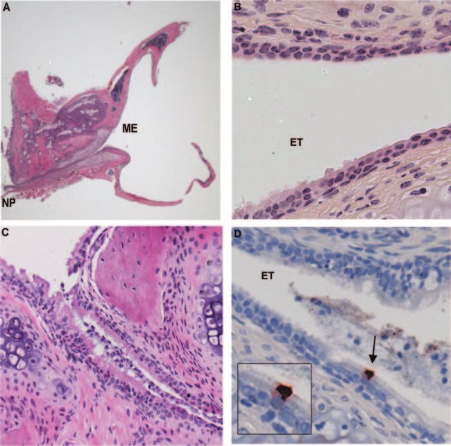
6036 GITIBAN ET AL. J. VIROL.
to assay the relative efficacy of potential RSV vaccine candi- Serum neutralization assay. RSV (500 PFU/ml) was mixed with serial dilu-
dates. tions of chinchilla serum and incubated at room temperature for 30 min. These
mixtures (100 l) were used to inoculate confluent monolayers of mouse
STAT1⫺/⫺ fibroblast cells grown in 24-well plates. After a 1-h incubation at 37°C,
MATERIALS AND METHODS infected wells were washed and refed with growth medium plus 0.5% methyl-
Virus preparation and assay. High-titer stocks of RSV (A2 strain) were cellulose. After 2 days, plates were stained with crystal violet and plaques
prepared on HEp2 cell monolayers by modification of published methods (30). counted. The neutralizing titer was defined as the serum dilution that resulted in
In order to detect low levels of bacterial or mycoplasmal contamination, cultures a 50% reduction in plaque number compared to controls that had no serum.
were maintained in the absence of antibiotics. Virus titers were determined by Histology. Sections through the NP and ET of mice were obtained following
plaque assay on STAT1⫺/⫺ mouse fibroblasts (23). Equivalent numbers of formalin fixation and decalcification. For immunohistochemistry, sections were
plaques were counted on STAT1⫺/⫺ fibroblast and HEp2 monolayers, but they blocked with a Vector Avidin/Biotin blocking kit (Vector Laboratories, Inc.,
appeared much more rapidly on the knockout murine cells: 36 h versus 5 days, Burlingame, CA) and Super Sniper (Biocare Medical, Walnut Creek, CA) before
respectively (data not shown). overnight incubation at 4°C with goat anti-RSV antibodies, 1:500 (Biodesign,
Animals. Nine juvenile chinchillas (Chinchilla lanigera) (mean weight, 399 ⫾ Saco, ME). Staining was visualized using a biotinylated mouse anti-goat second-
74 g) were purchased from Rauscher’s Chinchilla Ranch (LaRue, OH) and used ary (ScyTek, Logan, UT) antibody, streptavidin-horseradish peroxidase (Biocare
for these studies, after resting for 7 to 10 days. Juvenile chinchillas, approxi- Medical), the chromogen AEC (DAKO, Carpinteria, CA), and a hematoxylin
counterstain.
Downloaded from http://jvi.asm.org/ on February 17, 2021 by guest
mately 3 months of age and weighing 300 to 350 g, were used here due to our
previous demonstration of the preferred use of juvenile chinchillas when dem- For indirect fluorescence microscopy, ETs recovered from chinchillas inocu-
onstrating adenovirus predisposition to bacterial invasion of the middle ear (8, lated with 107 PFU of RSV were embedded in low-temperature embedding
45). Male and female BALB/c mice were obtained from Harlan Laboratories medium or cryocompound (Fisher Scientific, Pittsburgh, PA) and cryosectioned.
(Indianapolis, IN) and were infected at 6 to 10 weeks of age. Sections (5 m) were fixed with cold 3.5% paraformaldehyde in PBS (137 mM
Infection. Mice were infected i.n. following methodologies that were devel- NaCl, 2.7 mM KCl, 10 mM Na2HPO4, 2 mM KH2PO4, pH 7.4), washed, blocked
oped to deliver an inoculum specifically to the upper airway using a very small with 0.5% ovalbumin in PBS, and incubated 2 h at room temperature with
volume (43, 76). Briefly, each mouse was lightly anesthetized, placed in a supine fluorescein isothiocyanate (FITC)-conjugated goat anti-RSV (Biodesign Inter-
position, and given 107 PFU of RSV in a 20-l volume. Two microliters per naris national, Saco, Maine). FITC-conjugated goat immunoglobulin (Ig) (Caltag Lab-
were delivered at 0, 2, 7, 9, and 11 min. There were three mice per data point. oratories, Burlingame, CA) was used as a non-RSV specific control antiserum
Chinchillas were also inoculated i.n. with RSV, again delivered specifically to and Evans blue was used on all tissues as a counterstain (this dye will fluoresce
the upper airway (76). The dosages (in total PFU delivered), 103, 105, 106, or 107, red under the conditions used to image FITC labeling, which appears as green
were administered by passive inhalation of droplets of viral suspension delivered fluorescence). Images were captured using a Zeiss LSM 410 confocal microscope
to the nares of chinchillas that were lightly anesthetized with xylazine (2 mg/kg (Carl Zeiss, Thornwood, NY).
of body weight; Fort Dodge Animal Health, Fort Dodge, IA) and ketamine (10
mg/kg; Phoenix Scientific Inc., St. Joseph MO) and were laying in a prone
position. Initially, we used three chinchillas to attempt to identify the dose of RESULTS
RSV that would induce maximal compromise of Eustachian tube function. Thus,
one animal received a dose of 103, 105, or 107 PFU of RSV for this early Murine susceptibility to RSV infection of the NP and ET
assessment. Subsequently, to better define RSV-induced histopathology and following i.n. challenge. BALB/c mice have been widely used
determine the relative reproducibility of RSV-induced upper airway compromise
in this host, six additional juvenile chinchillas were inoculated with either 106 or
to study lower respiratory tract infection by RSV following i.n.
107 PFU RSV (3 chinchillas/dose). No chinchillas were inoculated with either inoculation, but titers from mouse nasal cavities are generally
sterile saline or sterile tissue culture medium in the present study due to the very low, on the order of 100 to 1,000 PFU/nose. Similar results
existing extensive evaluation of the normal histology, Eustachian tube function, have been reported by others (30). Here, we examined the
and microbial flora resident in this host under these control conditions in the
course of NP and ET infection by RSV in BALB/c mice by
literature (6, 7, 9, 17, 26, 27, 44, 52, 59, 72).
Otoscopy and tympanometry. Chinchillas were evaluated daily, with signs of immunohistochemical methods, looking for cells expressing
tympanic membrane inflammation determined by video otoscopy (Video Vet- RSV antigens rather than scoring virus titers by plaque assay.
Scope system; MedRx, Seminole, FL) and rated on a 0 to 4⫹ scale, as described An important departure from previous studies (21) was our
previously (8, 45, 76). Tympanometry (EarScan, South Daytona, FL) was used to method for inoculation (43, 76) using a concentrated virus
monitor changes in middle ear pressure, tympanic width, and tympanic mem-
brane compliance, also as described previously (8, 45).
suspension and delivering the inoculum in multiple small ali-
Lavages and recovery of tissues for quantitative viral culture. RSV-infected quots over a 20-min interval. Unlike typical i.n. protocols that
mice and uninfected controls were sacrificed at various times postinfection by use larger (50 to 100 l) volumes, this method allows the
CO2 inhalation. For virus titration, lungs and nasal mucosa were immediately inoculum to be absorbed by the nasal mucosa rather than being
frozen in liquid nitrogen. Prior to plaque assay, tissues were weighed and ho-
delivered through the nose into the trachea and esophagus.
mogenized in 1 ml phosphate-buffered saline (PBS). Tissue homogenates were
assayed as described above. Histologic examination was performed on sections from the
In chinchillas, middle ear lavages were performed on day 4 post-RSV inocu- NPs and ETs of BALB/c mice at days 4, 7, and 10 after RSV
lation by first venting the superior cephalid bulla with a 25-gauge needle and infection. Transverse sections, or cross sections, through the
depositing 300 l of pyrogen-free sterile saline into the middle ear cavity. Lavage nasal cavity were taken at the levels used routinely in toxicol-
fluids were then recovered from the middle ear by “epitympanic tap,” which
involved withdrawing these fluids from the middle ear cavity via use of a 1.5-in.,
ogy studies. The mucosal lining of the nasal turbinates and
25-gauge hypodermic needle. NP lavage was also performed on days 4 or 8 septum is primarily columnar respiratory epithelium at the
postinoculation of RSV by passive inhalation of 500 l pyrogen-free sterile level of the incisors (level I), transitioning to primarily olfac-
saline. Lavage fluids were collected as they drained from the contralateral naris tory epithelium at the level of the second molar (level III) (74).
and snap-frozen in liquid nitrogen until used for determination of viral titer. As
A transverse section taken at level III from an uninfected
per our usual protocol, in all cases epitympanic taps were performed prior to NP
lavage to limit the possibility of introducing commensal bacteria into the tym- mouse is pictured in Fig. 1A. Superiorly is the bony skull to
panum via refluxing of lavage fluids. which the centrally located nasal septum is attached. At the
Lung and tracheal tissues were recovered on day 4 after RSV inoculation, lower aspect is the palate; just above the palate is the septal
whereas NP and ET mucosae were recovered on days 4 or 8. Middle ear lavage window with two triangular shaped masses of nasal-associated
fluid, lungs, and tracheal mucosa retrieved on day 4 were from a single animal
that received 107 PFU, whereas all other specimens were retrieved from three
lymphoid tissue positioned at its ventrolateral edges. Ciliated
chinchillas per dose of either 106 or 107 PFU RSV. All fluids and tissues were respiratory epithelium lines the ventral portion of the nasal
snap-frozen in liquid nitrogen. cavity and the maxillary sinus. Sections from infected animals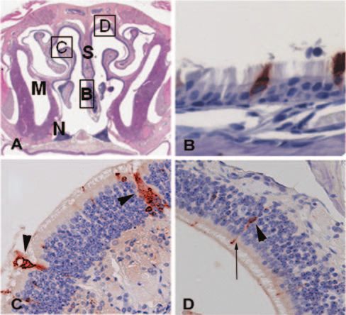
VOL. 79, 2005 URT RSV INFECTION 6037
polar neurons with antibody staining of their axons and den-
drites (Fig. 1C and D). These findings were consistent over the
course of three experiments, and staining of olfactory mucosa
was noted in the chinchilla as well.
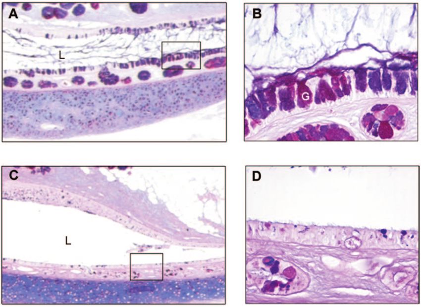
Murine ETs were removed at 4, 7, and 10 days, formalin
fixed, and extensively decalcified. Longitudinal sections of both
infected and uninfected ETs were prepared and examined
(Fig. 2A). Both ends of the murine ET are lined by
pseudostratified, ciliated mucosa intermixed with goblet cells
(Fig. 2B, C, and D). In all mice examined, infected or unin-
fected, a sparse inflammatory infiltrate consisting of lympho-
cytes and eosinophils was present in the lamina propria under-
lying the nasopharyngeal ET opening. The mid-portion of the
ET tube is lined by cuboidal, sparsely ciliated epithelium (Fig.
Downloaded from http://jvi.asm.org/ on February 17, 2021 by guest
2B). Figure 2D shows RSV infection of a single ciliated cell
present in the distal ET at the opening to the middle ear, with
the red-brown immunostaining concentrated at its apical, cili-
ated surface. Only rare, single infected cells were identified
among six animals examined at day 4 postinfection, and no
virus-infected cells were identified after day 4. Pathological
FIG. 1. Panel A is a low-power (15⫻) H&E-stained section show- changes in both the NP and ET were subtle. The most notable
ing a transverse section, or cross section, of the murine nasal cavity change was the accumulation of acute inflammatory cells
taken at the level of the second molar. At the superior aspect of the
specimen is the bony skull to which the nasal septum (S) is attached. (mostly neutrophils and macrophages) and mucus in the ET
The palate is present at the lower aspect, and just above the palate are lumen (Fig. 2C) at day 10. Other inflammatory changes in the
the two triangular masses of nasal-associated lymphoid tissue (N). M, NP and ET were mild, with a small increase in inflammatory
maxillary sinus. Immunostained respiratory mucosa lining the nasal cells within the lamina propria and mild goblet cell hyperpla-
septum (B), olfactory mucosa lining the turbinate (C), and the dorsal
sia. These findings are representative of three animals at each
surface of the nasal cavity (D) are shown in panels B, C, and D,
respectively. All specimens shown are from day 4 postinoculation. In time point in each of two experiments.
panel B, red-brown immunostaining is seen at the apical surface of one Chinchilla permissivity to infection of the upper airway with
ciliated cell and throughout the cytoplasm of a second cell (400⫻). In RSV following i.n. challenge. A dose-effect was clearly detect-
panel C, two ducts from Bowman’s glands are staining strongly with the able in chinchillas inoculated with increasing doses of RSV. At
anti-viral antiserum and are marked with arrowheads (200⫻). In panel
D, cell bodies of olfactory neurons (arrowhead) and their dendritic the lower dosages assayed (1 ⫻ 103 and 1 ⫻ 105 PFU/animal),
processes (arrow) are infected with RSV (200⫻). no signs of illness were noted at any time postchallenge. Con-
versely, animals that received higher dosages (e.g., 1 ⫻ 106 and
1 ⫻ 107 PFU/animal) showed signs of acute respiratory tract
were evaluated by staining for RSV antigen expression by using infection. Ruffling of fur and lethargic behavior were observed
polyclonal whole-virus antisera. Infected cells could be visual- by day 4 after challenge in 2 of 6 (33%) animals that received
ized at day 4 when virus production is near or at peak levels the 1 ⫻ 107 PFU inoculum; however, the hallmark sign of
(21) (Fig. 1). Figure 1B shows RSV infection of ciliated respi- infection was mild retraction of the tympanic membrane as
ratory epithelial cells lining the nasal septum. The red-brown measured by both otoscopy and tympanometry. By otoscopy,
immunostaining appears either apically or extends throughout there was evidence of low-grade inflammation of the tympanic
the cytoplasm. This staining pattern is consistent with the bi- membrane (maximum value, ⬃1.5 on a 0 to 4⫹ scale), typical
ology of RSV that is known to infect through, and bud from, of viral OM, with retraction being the most consistent sign of
the luminal surface of respiratory epithelial cells (18). The RSV infection in this host at a dose of 107 PFU.
images shown are representative of multiple sections from Retraction was noted as early as 1 day after i.n. challenge in
each of three animals from each genotype at this time point. chinchillas inoculated with 1 ⫻ 107 PFU RSV. In animals that
The tropism of RSV for respiratory epithelium is well es- received either 1 ⫻ 106 or 1 ⫻ 107 PFU, retraction was max-
tablished; however, a surprising and intriguing result from imal 3 to 4 days after challenge, occurring in 67 to 100% of ears
these studies was its equal affinity for the olfactory mucosa as noted by otoscopy and persisting until at least 9 days after
(Fig. 1C and D). Olfactory mucosa lines the ethmoid turbi- inoculation, although the degree of retraction was less marked
nates, the dorsal wall of the nasal cavity, and the dorsal portion at day 9 (data not shown) than that noted on days 3 to 4. In
of the nasal septum at level III. The olfactory epithelium con- these animals, evidence of retraction was supported by dem-
sists of the supporting sustentacular cells, bipolar olfactory onstration of an underpressured state (Fig. 3A) occurring in 63
neurons, and the excretory ducts from Bowman’s glands. Sus- to 88% of ears that was also maximal 3 to 4 days after challenge
tentacular cells are tall columnar cells, extending from the (Fig. 3B). Middle ear pressure steadily declined after inocula-
basal lamina to the lumen. They have microvilli on their apical tion of RSV, with a mean maximal underpressured middle ear
surface and their nuclei form a layer occupying the upper state of ⫺45 daPa occurring approximately 4 days after i.n.
one-third of the olfactory mucosal layer (40). Morphological challenge with 107 PFU RSV. This underpressured state per-
analysis clearly shows RSV infection of the ductal epithelium sisted until at least 7 days after inoculation, although both
in Fig. 1C. Less prominent, but clearly seen, are infected bi- degree of underpressure and number of ears affected was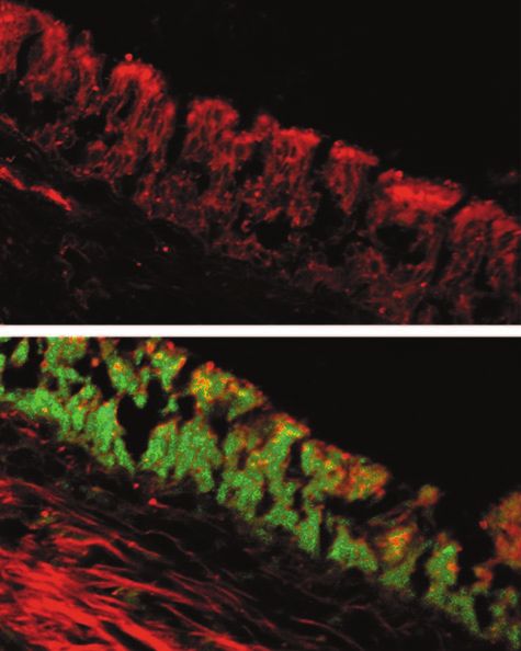
6038     GITIBAN ET AL.                                                                                                                 J. VIROL.

                                                                                                                                                     Downloaded from http://jvi.asm.org/ on February 17, 2021 by guest
  FIG. 2. Panel A is a low-power (15⫻) H&E-stained section showing an entire ET dissected from an uninfected mouse. On the left side of the
image is the NP mucosa at the opening of the ET. The ET opens distally into the middle ear (ME) cavity, which can be seen at the right side of
the section. In panel B (400⫻), ciliated mucosa at the proximal, nasopharyngeal end of the ET gives way to a more flattened epithelial cell type.
A section from the ET of a RSV-infected mouse (panel C) shows the accumulation of mucous and inflammatory cells in the lumen at day 10 after
inoculation (200⫻). Staining with anti-RSV antiserum shows that ciliated ET epithelium can be infected. The cell shown in panel D is at the distal
end of the ET (400⫻), where it opens into the middle ear cavity.

markedly reduced at this time point. Together, these data                  (H&E)-stained sections of NP and ET mucosae recovered
suggested RSV-induced ET compromise. In addition, nasal                    from chinchillas infected i.n. with RSV revealed signs of mild
lavage fluids recovered from several RSV-infected chinchillas              inflammation. There was a sparse mononuclear submucosal
had an abnormal yellowish-green tint and were notably turbid.              infiltrate with eosinophils proximally. Marked goblet cell hy-
This latter observation is consistent with histopathological               perplasia with mucus hypersecretion into the lumen of the ET
evaluations that showed hypersecretion of mucus into the ET                was however a consistent finding in RSV-infected chinchillas,
lumen in tissues recovered from chinchillas inoculated with                as evidenced by periodic acid-Schiff–Alcian Blue staining
RSV (see below).                                                           which is specific for mucopolysaccharides present in these tis-
   Plaque assays conducted with homogenized NP mucosa and                  sues (Fig. 4). Note relative the abundance of mucus in the
NP lavage fluids indicated that the chinchilla was permissive to           Eustachian tube lumen, the number of heavily stained goblet
RSV infection (Table 1). Both NP mucosal tissue homogenates                cells, and the intensity/character of staining of submucosal
and NP lavage fluid specimens were positive on days 4 and 8                glands in an RSV-infected chinchilla versus a noninfected con-
after challenge at viral doses of ⱖ105 PFU. Preliminary evi-               trol animal when comparing panels A and B versus C and D,
dence in support of restriction of viral replication to the up-            respectively, in Fig. 4. By using a biotin-conjugated goat anti-
permost airway following i.n. challenge in the manner de-                  RSV polyclonal antiserum, we showed the presence of RSV
scribed here was supported by the absence of viral plaques                 antigen in ciliated cells lining the chinchilla ET at various or
when tracheal mucosa and lung tissue recovered 4 days after                multiple points along approximately the proximal two-thirds of
challenge from a single chinchilla that had received the highest           its length (similar to that seen in Fig. 2D), thus demonstrating
dose of RSV (107 PFU) were homogenized and assayed for                     the ability of RSV to infect the chinchilla ET following i.n.
RSV via plaque assay. More extensive evaluation of this phe-               inoculation. When frozen sections of whole chinchilla ETs
nomenon is needed.                                                         (including tissues from the pharyngeal to tympanic orifice),
   RSV infection results in goblet cell hyperplasia, hypersecre-           recovered from animals challenged with RSV, were incubated
tion of mucus, and the clear presence of RSV antigen within                with FITC-conjugated goat IgG antibody, there was no fluo-
cells lining the ET. Examination of hematoxylin and eosin                  rescent labeling (Fig. 5A). Conversely, tissues incubated with
VOL. 79, 2005                                                                                                    URT RSV INFECTION           6039

                                                                                   Further evidence that the chinchilla host was permissive to
                                                                                RSV infection was provided by the demonstration of develop-
                                                                                ment of virus-neutralizing antibody in sera recovered from
                                                                                these animals. No plaque-neutralizing antibody was observed
                                                                                in sera recovered from either five chinchillas prior to challenge
                                                                                or in two chinchillas 8 days after i.n. delivery of either 106 or
                                                                                107 PFU RSV. However, 30 days after receiving either of these
                                                                                latter doses of RSV, plaque-neutralizing antibody with a 50%
                                                                                plaque reduction assay of 1/160 was present in the sera of both
                                                                                animals assayed. To the best of our knowledge, this is the first
                                                                                published report of a chinchilla model for RSV infection of the
                                                                                uppermost airway.

                                                                                                                                                     Downloaded from http://jvi.asm.org/ on February 17, 2021 by guest
                                                                                                         DISCUSSION
                                                                                   As most studies have focused on RSV infection of the lower
                                                                                respiratory tract, relatively little is known about RSV disease of
                                                                                the upper airway, including the NP, ET, and middle ear. To
                                                                                better understand URT infection with RSV and predisposition
                                                                                to bacterial OM by RSV, we need useful and relevant animal
                                                                                models in which to assay pathogenesis and the host immune
                                                                                response to viral infection. Ultimately, animal studies will be
                                                                                necessary to assess the relative efficacy of potential vaccine
                                                                                candidates directed against RSV. Whereas there are several
                                                                                established models of lower airway infection (13, 18, 19), RSV
                                                                                infection of the uppermost airway has not been extensively
                                                                                studied. Barely detectable virus titers were noted in the murine
                                                                                nasal cavity accompanying pulmonary infection (30). Cotton
                                                                                rats infected i.n. with RSV were more permissive for nasal
                                                                                infection, with titers on the order of 104 PFU/g (63). Despite
                                                                                their limitations, both mouse and cotton rat models are very
                                                                                useful for the study of possible immunopathology accompany-
   FIG. 3. Panel A, changes in normalized average middle ear pres-
                                                                                ing delivery of any vaccine candidate; however, mechanistic
sure (⫾ standard deviations [sd]) over time in chinchillas infected with
either 106 or 107 PFU RSV strain A2 via intranasal delivery. Middle             studies have been most informative in the mouse due to the
ear pressure (MEP) was assessed by tympanometry. Panel B, relative              abundance of available murine reagents (1, 29, 41, 57, 71).
percentage of ears that demonstrated reduced middle ear pressure                Nevertheless, at present, little is known about viral OM in the
relative to baseline measurements over time in cohorts that received            mouse or cotton rat.
either 106 or 107 PFU RSV. Values on bars indicate the number of ears
demonstrating a reduced middle ear pressure over the total number of               In the studies described here, using BALB/c mice, we have
ears evaluated.                                                                 determined that the respiratory and olfactory mucosae of the
                                                                                nasal cavity and the ciliated epithelium of the ET can be
                                                                                infected by RSV. Nonetheless, only a small number of infected
FITC-conjugated goat anti-RSV showed distinct fluorescent                       cells can be detected, and there is very little pathology accom-
labeling of the epithelial lining for three quarters of the length              panying infection. In response to infection, submucosal lym-
of the ET from the NP to the tympanic orifice (Fig. 5B).                        phocytes and eosinophils are mildly increased in the NP and at
Control tissues from naive animals did not label with FITC-                     the entrance to the ET. Mucous and inflammatory cells were
conjugated goat anti-RSV antibody (data not shown).                             found along the length of the ET between 7 and 10 days.
                                                                                   In contrast to these observations, we have established that
                                                                                the chinchilla, the preferred rodent host for OM research (25,
                                                                                31), is susceptible to RSV infection. Chinchillas infected i.n.
TABLE 1. RSV titers from chinchilla NP mucosa and lavage fluids
                                                                                with RSV showed milder signs of illness than those shown
                                             RSV titera                         when inoculated similarly with either influenza A virus or with
                                                            Log PFU/ml NP       adenovirus (26, 28, 72). Nonetheless, these animals were man-
Viral dose          Log PFU/g NP mucosa
                                                              lavage fluid      ifesting signs of URT infection, and these observed signs of
                     Day 4           Day 8                Day 4        Day 8    illness were dose dependent. RSV-induced compromise of
                                                                                chinchilla ET function was clearly demonstrable. While the
   105                3.4             NDb                 3.15          ND
   106                3.9             2.4                 2.9           2.3
                                                                                underpressured state induced was not marked, it was nonethe-
   107                3.6             3.6                 3.6           2.6     less present and concordant retraction of the tympanic mem-
  a
                                                                                brane was observed. Moreover, the noted goblet cell hyperpla-
    The data presented are mean titer/group. One chinchilla received 105 PFU,
three received 106 PFU, and four animals received 107 PFU.                      sia within the ET mucosa and increased mucus secretion into
  b
    Not done.                                                                   the ET lumen could provide additional means to predispose to
6040 GITIBAN ET AL. J. VIROL.
Downloaded from http://jvi.asm.org/ on February 17, 2021 by guest
FIG. 4. Periodic acid-Schiff–Alcian Blue staining of mucopolysaccharides present within Eustachian tube tissues showing goblet cell hyperplasia
with mucus hypersecretion into the lumen was a consistent finding in RSV-infected chinchillas. Panel A, (⬃5⫻) mid-Eustachian tube of a chinchilla
4 days after inoculation with 1 ⫻ 107 PFU RSV. Note goblet cell (see representative goblet cell labeled with a white letter “G”) hyperplasia relative
to noninfected chinchilla as shown in panel C. Inset in panel A is shown at ⬃40⫻ in panel B; note abundant mucus in ET lumen (labeled “L”)
and prominent labeling of both goblet cells and glandular structures, submucosally. Inset in panel C is shown at ⬃40⫻ in panel D; note relative
paucity of goblet cells and notably diminished staining of lumenal mucus (Eustachian tube lumen is labeled “L”) and submucosal glands in this
ET, recovered from a non-RSV-infected animal, when compared to inset panel B.
bacterial superinfection of the tympanum. These phenomena, microorganisms that cause OM, and the significant parallels
as well as others, have been shown to be associated with both that exist between the course of combined viral-bacterial dis-
adenovirus and influenza A virus predisposition to bacterial ease induced in chinchillas and the course of natural human
invasion of the middle ear in the chinchilla (26, 28, 72) and thus disease (4, 5). The chinchilla model will thus provide the first
parallel the disease course in children, wherein bacterial OM is experimental system where the predisposition to bacterial OM
a highly prevalent complicating sequela of viral URT infection by RSV might perhaps be rigorously studied. Despite the fact
(36). that, when compared to humans and chimpanzees (18), even
While both of these rodent models will likely be extremely the chinchilla is relatively nonpermissive for RSV, rodent mod-
useful for defining RSV pathogenesis and the host’s immune els of RSV infection may still offer the most practical solution
response to infection of the upper airway, each has distinct regarding the need for small and inbred animals to use in
advantages. The availability of a large range of immunological proof-of-concept studies and preclinical vaccine candidate tri-
reagents in the murine system will allow us to characterize als relevant to the prevention of RSV-induced disease.
virus-specific T- and B-cell responses to primary and secondary In summary, the field of RSV research to date has largely
nasopharyngeal RSV infection in naive and vaccinated wild- been focused on strategies to prevent life-threatening RSV-
type animals. Our demonstration that RSV infection of the induced bronchiolitis and pneumonia in the infant, with little
upper airway can be monitored histologically will allow for the attention paid to protection against upper airway infection.
use of BALB/c mice in challenge experiments, despite the low However, it is the upper airway that is continually reinfected,
virus titers found in the murine nasal cavity. and studies have shown that serum IgGs, though able to pre-
On the other hand, the chinchilla is a well-established model vent lower airway infection, do not constitute effective protec-
for OM research due to its larger size, its susceptibility to the tion against upper airway disease (42, 62, 70). ImmunizationVOL. 79, 2005 URT RSV INFECTION 6041
REFERENCES
1. Alwan, W. H., W. J. Kozlowska, and P. J. Openshaw. 1994. Distinct types of
lung disease caused by functional subsets of antiviral T cells. J. Exp. Med.
179:81–89.
2. Arola, M., O. Ruuskanen, T. Ziegler, J. Mertsola, K. Nanto-Salonen, A.
Putto-Laurila, M. K. Viljanen, and P. Halonen. 1990. Clinical role of respi-
ratory virus infection in acute otitis media. Pediatrics 86:848–855.
3. Arola, M., T. Ziegler, H. Puhakka, O. P. Lehtonen, and O. Ruuskanen. 1990.
Rhinovirus in otitis media with effusion. Ann. Otol. Rhinol. Laryngol. 99:
451–453.
4. Bakaletz, L. O. 2002. Otitis media, p. 259–298. In K. A. Brogden and J. M.
Guthmiller (ed.), Polymicrobial diseases. ASM Press, Washington, D.C.
5. Bakaletz, L. O. 1995. Viral potentiation of bacterial superinfection of the
respiratory tract. Trends Microbiol. 3:110–114.
6. Bakaletz, L. O., R. L. Daniels, and D. J. Lim. 1993. Modeling adenovirus
type 1-induced otitis media in the chinchilla: effect on ciliary activity and
fluid transport function of Eustachian tube mucosal epithelium. J. Infect.
Dis. 168:865–872.
Downloaded from http://jvi.asm.org/ on February 17, 2021 by guest
7. Bakaletz, L. O., and K. A. Holmes. 1997. Evidence for transudation of
specific antibody into the middle ears of parenterally immunized chinchillas
after an upper respiratory tract infection with adenovirus. Clin. Diagn. Lab.
Immunol. 4:223–225.
8. Bakaletz, L. O., B. J. Kennedy, L. A. Novotny, G. Duquesne, J. Cohen, and
Y. Lobet. 1999. Protection against development of otitis media induced by
nontypeable Haemophilus influenzae by both active and passive immuniza-
tion in a chinchilla model of virus-bacterium superinfection. Infect. Immun.
67:2746–2762.
9. Bakaletz, L. O., D. M. Murwin, and J. M. Billy. 1995. Adenovirus serotype
1 does not act synergistically with Moraxella (Branhamella) catarrhalis to
induce otitis media in the chinchilla. Infect. Immun. 63:4188–4190.
10. Bluestone, C. D. 1996. Pathogenesis of otitis media: role of Eustachian tube.
Pediatr. Infect. Dis. J. 15:281–291.
11. Bont, L., J. Versteegh, W. T. Swelsen, C. J. Heijnen, A. Kavelaars, F. Brus,
J. M. Draaisma, M. Pekelharing-Berghuis, R. A. van Diemen-Steenvoorde,
and J. L. Kimpen. 2002. Natural reinfection with respiratory syncytial virus
does not boost virus-specific T-cell immunity. Pediatr. Res. 52:363–367.
FIG. 5. Fluorescent microscopy images of frozen sections of whole 12. Reference deleted.
chinchilla ETs recovered from a chinchilla 4 days after intranasal 13. Byrd, L. G., and G. A. Prince. 1997. Animal models of respiratory syncytial
challenge with 107 PFU RSV. There was no fluorescent labeling when virus infection. Clin. Infect. Dis. 25:1363–1368.
these tissues were incubated with the control antiserum, FITC-conju- 14. Casselbrant, M. L., E. M. Mandel, P. A. Fall, H. E. Rockette, M. Kurs-Lasky,
gated goat IgG (top panel) or when non-RSV-infected control tissues C. D. Bluestone, and R. E. Ferrell. 1999. The heritability of otitis media: a
were labeled with FITC-conjugated goat anti-RSV antibody (not twin and triplet study. JAMA 282:2125–2130.
shown). Conversely, distinct green fluorescent labeling of the mucosal 15. Chonmaitree, T., M. J. Owen, J. A. Patel, D. Hedgpeth, D. Horlick, and V. M.
epithelium lining this tubal organ was seen when frozen ET sections Howie. 1992. Effect of viral respiratory tract infection on outcome of acute
otitis media. J. Pediatr. 120:856–862.
were incubated with FITC-conjugated goat anti-RSV antibody (bot- 16. Chonmaitree, T., M. J. Owen, J. A. Patel, D. Hedgpeth, D. Horlick, and V. M.
tom panel). Red color in tissues is due to fluorescence of the Evans Howie. 1992. Presence of cytomegalovirus and herpes simplex virus in middle
blue dye used as a counterstain. ear fluids from children with acute otitis media. Clin. Infect. Dis. 15:650–653.
17. Chung, M. H., S. R. Griffith, K. H. Park, D. J. Lim, and T. F. DeMaria. 1993.
Cytological and histological changes in the middle ear after inoculation of
influenza A virus. Acta Otolaryngol. 113:81–87.
that induces strong cellular immunity will be particularly im- 18. Collins, P. L., K. McIntosh, and R. M. Chanock. 1996. Respiratory syncytial
portant in this regard, as it has been shown that natural rein- virus, p. 1313–1351. In D. M. Knipe, B. N. Fields, and P. M. Howley (ed.),
fection with RSV does not boost virus-specific T-cell immunity Fields virology, 3rd ed. Lippincott-Raven Publishers, Philadelphia, Pa.
19. Connors, M., A. B. Kulkarni, C. Y. Firestone, K. L. Holmes, H. C. Morse III,
(11). This feature of RSV biology may underlie its ability to A. V. Sotnikov, and B. R. Murphy. 1992. Pulmonary histopathology induced
reinfect throughout life. Importantly, recurrent URT infection by respiratory syncytial virus (RSV) challenge of formalin-inactivated RSV-
with RSV is the predominant risk factor for bacterial OM. In immunized BALB/c mice is abrogated by depletion of CD4⫹ T cells. J. Vi-
rol. 66:7444–7451.
the studies presented here, we have developed two rodent 20. Daly, K. A., J. E. Brown, B. R. Lindgren, M. H. Meland, C. T. Le, and G. S.
models of URT infection by RSV to aid in furthering under- Giebink. 1999. Epidemiology of otitis media onset by six months of age.
Pediatrics 103:1158–1166.
standing of RSV-induced upper airway disease as well as per- 21. Durbin, J. E., T. R. Johnson, R. K. Durbin, S. E. Mertz, R. A. Morotti, R. S.
haps future utility in defining the pathogenic mechanisms of Peebles, and B. S. Graham. 2002. The role of IFN in respiratory syncytial
RSV predisposition to bacterial OM. Ultimately, both models virus pathogenesis. J. Immunol. 168:2944–2952.
22. Fireman, P. 1990. The role of antihistamines in otitis. J. Allergy Clin. Im-
will be useful to support endeavors to develop a vaccine can- munol. 86:638–641.
didate for the prevention of RSV infection of the upper airway. 23. Garcia-Sastre, A., R. K. Durbin, H. Zheng, P. Palese, R. Gertner, D. E. Levy,
and J. E. Durbin. 1998. The role of interferon in influenza virus tissue
ACKNOWLEDGMENTS tropism. J. Virol. 72:8550–8558.
24. Giebink, G. S. 1989. The microbiology of otitis media. Pediatr. Infect. Dis. J.
Studies conducted in the chinchilla host were supported by discre- 8:S18–20.
tionary funds to L.O.B. and, in part, by a grant from the NIH/NIDCD, 25. Giebink, G. S. 1999. Otitis media: the chinchilla model. Microb. Drug Resist.
R01 DC05847. Mouse studies were supported by a grant from the 5:57–72.
NIH, AI47226, to J.E.D. 26. Giebink, G. S., I. K. Berzins, S. C. Marker, and G. Schiffman. 1980. Exper-
imental otitis media after nasal inoculation of Streptococcus pneumoniae and
We thank Jennifer Neelans for manuscript preparation and G. Scott influenza A virus in chinchillas. Infect. Immun. 30:445–450.
Giebink (in memoriam) for many helpful discussions and encourage- 27. Giebink, G. S., M. L. Ripley, and P. F. Wright. 1987. Eustachian tube
ment. The Mouse Phenotyping Service of The Ohio State University histopathology during experimental influenza A virus infection in the chin-
Department of Veterinary Biosciences provided assistance with immu- chilla. Ann. Otol. Rhinol. Laryngol. 96:199–206.
nostaining and data interpretation. 28. Giebink, G. S., and P. F. Wright. 1983. Different virulence of influenza A6042 GITIBAN ET AL. J. VIROL.
virus strains and susceptibility to pneumococcal otitis media in chinchillas. 55. Okamoto, Y., K. Kudo, K. Ishikawa, E. Ito, K. Togawa, I. Saito, I. Moro, J. A.
Infect. Immun. 41:913–920. Patel, and P. L. Ogra. 1993. Presence of respiratory syncytial virus genomic
29. Graham, B. S., L. A. Bunton, P. F. Wright, and D. T. Karzon. 1991. Role of sequences in middle ear fluid and its relationship to expression of cytokines
T lymphocyte subsets in the pathogenesis of primary infection and rechal- and cell adhesion molecules. J. Infect. Dis. 168:1277–1281.
lenge with respiratory syncytial virus in mice. J. Clin. Investig. 88:1026–1033. 56. Okamoto, Y., K. Kudo, K. Shirotori, M. Nakazawa, E. Ito, K. Togawa, J. A.
30. Graham, B. S., M. D. Perkins, P. F. Wright, and D. T. Karzon. 1988. Primary Patel, and P. L. Ogra. 1992. Detection of genomic sequences of respiratory
respiratory syncytial virus infection in mice. J. Med. Virol. 26:153–162. syncytial virus in otitis media with effusion in children. Ann. Otol. Rhinol.
31. Green, B. A., W. J. Doyle, and J. L. Cowell. 1994. Chinchilla model of Laryngol. Suppl. 157:7–10.
experimental otitis media for study of nontypable Haemophilus influenzae 57. Openshaw, P. J., F. J. Culley, and W. Olszewska. 2001. Immunopathogenesis
vaccine efficacy. Methods Enzymol. 235:59–68. of vaccine-enhanced RSV disease. Vaccine 20(Suppl. 1):S27–S31.
32. Hament, J. M., J. L. Kimpen, A. Fleer, and T. F. Wolfs. 1999. Respiratory 58. Paradise, J. L., H. E. Rockette, D. K. Colborn, B. S. Bernard, C. G. Smith,
viral infection predisposing for bacterial disease: a concise review. FEMS M. Kurs-Lasky, and J. E. Janosky. 1997. Otitis media in 2253 Pittsburgh-
Immunol. Med. Microbiol. 26:189–195. area infants: prevalence and risk factors during the first two years of life.
33. Hayden, F. G. 2000. Influenza virus and rhinovirus-related otitis media: Pediatrics 99:318–333.
potential for antiviral intervention. Vaccine 19(Suppl. 1):S66–S70. 59. Park, K., L. O. Bakaletz, J. M. Coticchia, and D. J. Lim. 1993. Effect of
34. Heikkinen, T. 2000. The role of respiratory viruses in otitis media. Vaccine influenza A virus on ciliary activity and dye transport function in the chin-
19(Suppl. 1):S51–S55. chilla Eustachian tube. Ann. Otol. Rhinol. Laryngol. 102:551–558.
35. Heikkinen, T. 2000. Role of viruses in the pathogenesis of acute otitis media.
60. Pitkaranta, A., J. Jero, E. Arruda, A. Virolainen, and F. G. Hayden. 1998.
Pediatr. Infect. Dis. J. 19:S17–S23.
Downloaded from http://jvi.asm.org/ on February 17, 2021 by guest
Polymerase chain reaction-based detection of rhinovirus, respiratory syncy-
36. Heikkinen, T., and T. Chonmaitree. 2000. Viral-bacterial synergy in otitis
tial virus, and coronavirus in otitis media with effusion. J. Pediatr. 133:390–
media: implications for management. Curr. Infect. Dis. Rep. 2:154–159.
394.
37. Heikkinen, T., M. Thint, and T. Chonmaitree. 1999. Prevalence of various
61. Pitkaranta, A., A. Virolainen, J. Jero, E. Arruda, and F. G. Hayden. 1998.
respiratory viruses in the middle ear during acute otitis media. N. Engl.
J. Med. 340:260–264. Detection of rhinovirus, respiratory syncytial virus, and coronavirus infec-
38. Henderson, F. W., A. M. Collier, W. A. Clyde, Jr., and F. W. Denny. 1979. tions in acute otitis media by reverse transcriptase polymerase chain reac-
Respiratory-syncytial-virus infections, reinfections and immunity. A prospec- tion. Pediatrics 102:291–295.
tive, longitudinal study in young children. N. Engl. J. Med. 300:530–534. 62. Plotnicky-Gilquin, H., A. Robert, L. Chevalet, J. F. Haeuw, A. Beck, J. Y.
39. Henderson, F. W., A. M. Collier, M. A. Sanyal, J. M. Watkins, D. L. Fair- Bonnefoy, C. Brandt, C. A. Siegrist, T. N. Nguyen, and U. F. Power. 2000.
clough, W. A. Clyde, Jr., and F. W. Denny. 1982. A longitudinal study of CD4⫹ T-cell-mediated antiviral protection of the upper respiratory tract in
respiratory viruses and bacteria in the etiology of acute otitis media with BALB/c mice following parenteral immunization with a recombinant respi-
effusion. N. Engl. J. Med. 306:1377–1383. ratory syncytial virus G protein fragment. J. Virol. 74:3455–3463.
40. Herbert, R. A., and J. R. Leininger. 1999. Nose, larynx, and trachea, p. 63. Prince, G. A., A. B. Jenson, R. L. Horswood, E. Camargo, and R. M.
259–292. In R. R. Maronpot (ed.), Pathology of the mouse. Cache River Chanock. 1978. The pathogenesis of respiratory syncytial virus infection in
Press, Research Triangle Park, Vienna, Ill. cotton rats. Am. J. Pathol. 93:771–791.
41. Hussell, T., C. J. Baldwin, A. O’Garra, and P. J. Openshaw. 1997. CD8⫹ T 64. Ruuskanen, O., M. Arola, A. Putto-Laurila, J. Mertsola, O. Meurman, M. K.
cells control Th2-driven pathology during pulmonary respiratory syncytial Viljanen, and P. Halonen. 1989. Acute otitis media and respiratory virus
virus infection. Eur. J. Immunol. 27:3341–3349. infections. Pediatr. Infect. Dis. J. 8:94–99.
42. The IMpact-RSV Study Group. 1998. Palivizumab, a humanized respiratory 65. Ruuskanen, O., and T. Heikkinen. 1994. Viral-bacterial interaction in acute
syncytial virus monoclonal antibody, reduces hospitalization from respiratory otitis media. Pediatr. Infect. Dis. J. 13:1047–1049.
syncytial virus infection in high-risk infants. Pediatrics 102:531–537. 66. Sarkkinen, H., O. Ruuskanen, O. Meurman, H. Puhakka, E. Virolainen, and
43. Johnson, S. A., M. G. Ottolini, M. E. Darnell, D. D. Porter, and G. A. Prince. J. Eskola. 1985. Identification of respiratory virus antigens in middle ear
1996. Unilateral nasal infection of cotton rats with respiratory syncytial virus fluids of children with acute otitis media. J. Infect. Dis. 151:444–448.
allows assessment of local and systemic immunity. J. Gen. Virol. 77:101–108. 67. Sarkkinen, H. K., P. E. Halonen, P. P. Arstila, and A. A. Salmi. 1981.
44. Jurcisek, J. A., J. E. Durbin, D. F. Kusewitt, and L. O. Bakaletz. 2003. Detection of respiratory syncytial, parainfluenza type 2, and adenovirus
Anatomy of the nasal cavity in the chinchilla. Cells Tissues Organs 174:136– antigens by radioimmunoassay and enzyme immunoassay on nasopharyngeal
152. specimens from children with acute respiratory disease. J. Clin. Microbiol.
45. Kennedy, B. J., L. A. Novotny, J. A. Jurcisek, Y. Lobet, and L. O. Bakaletz. 13:258–265.
2000. Passive transfer of antiserum specific for immunogens derived from a 68. Sarkkinen, H. K., O. Meurman, T. T. Salmi, H. Puhakka, and E. Virolainen.
nontypeable Haemophilus influenzae adhesin and lipoprotein D prevents 1983. Demonstration of viral antigens in middle ear secretions of children
otitis media after heterologous challenge. Infect. Immun. 68:2756–2765. with acute otitis media. Acta Paediatr. Scand. 72:137–138.
46. Klein, B. S., F. R. Dollete, and R. H. Yolken. 1982. The role of respiratory 69. Shaw, C. B., N. Obermyer, S. J. Wetmore, G. A. Spirou, and R. W. Farr. 1995.
syncytial virus and other viral pathogens in acute otitis media. J. Pediatr. Incidence of adenovirus and respiratory syncytial virus in chronic otitis me-
101:16–20. dia with effusion using the polymerase chain reaction. Otolaryngol. Head
47. Klein, J. O., and D. W. Teele. 1976. Isolation of viruses and mycoplasmas Neck Surg. 113:234–241.
from middle ear effusions: a review. Ann. Otol. Rhinol. Laryngol. 85:140– 70. Simoes, E. A., J. R. Groothuis, D. A. Tristram, K. Allessi, M. V. Lehr, G. R.
144. Siber, and R. C. Welliver. 1996. Respiratory syncytial virus-enriched globulin
48. Koivunen, P., T. Kontiokari, M. Niemela, T. Pokka, and M. Uhari. 1999. for the prevention of acute otitis media in high risk children. J. Pediatr.
Time to development of acute otitis media during an upper respiratory tract 129:214–219.
infection in children. Pediatr. Infect. Dis. J. 18:303–305.
71. Srikiatkhachorn, A., W. Chang, and T. J. Braciale. 1999. Induction of Th-1
49. Korppi, M., M. Leinonen, M. Koskela, P. H. Makela, and K. Launiala. 1989.
and Th-2 responses by respiratory syncytial virus attachment glycoprotein is
Bacterial coinfection in children hospitalized with respiratory syncytial virus
epitope and major histocompatibility complex independent. J. Virol. 73:
infections. Pediatr. Infect. Dis. J. 8:687–692.
6590–6597.
50. Korppi, M., M. Leinonen, P. H. Makela, and K. Launiala. 1991. Mixed
infection is common in children with respiratory adenovirus infection. Acta 72. Suzuki, K., and L. O. Bakaletz. 1994. Synergistic effect of adenovirus type 1
Paediatr. Scand. 80:413–417. and nontypeable Haemophilus influenzae in a chinchilla model of experimen-
51. Kvaerner, K. J., P. Nafstad, J. Hagen, I. W. Mair, and J. J. Jaakkola. 1997. tal otitis media. Infect. Immun. 62:1710–1718.
Early acute otitis media: determined by exposure to respiratory pathogens. 73. Uhari, M., J. Hietala, and H. Tuokko. 1995. Risk of acute otitis media in
Acta Otolaryngol. Suppl. 529:14–18. relation to the viral etiology of infections in children. Clin. Infect. Dis.
52. Miyamoto, N., and L. O. Bakaletz. 1997. Kinetics of the ascension of NTHi 20:521–524.
from the nasopharynx to the middle ear coincident with adenovirus-induced 74. Uraih, L. C., and R. R. Maronpot. 1990. Normal histology of the nasal cavity
compromise in the chinchilla. Microb. Pathog. 23:119–126. and application of special techniques. Environ. Health Perspect. 85:187–208.
53. Monobe, H., T. Ishibashi, Y. Nomura, M. Shinogami, J. Yano, and K. Kaga. 75. Reference deleted.
2002. Presented at the 25th Annual Midwinter Research Meeting, Associa- 76. Visweswaraiah, A., L. A. Novotny, E. J. Hjemdahl-Monsen, L. O. Bakaletz,
tion for Research in Otolaryngology, St. Petersburg Beach, Fla., 27 to 31 and Y. Thanavala. 2002. Tracking the tissue distribution of marker dye
January 2002. following intranasal delivery in mice and chinchillas: a multifactorial analysis
54. Niewiesk, S., and G. Prince. 2002. Diversifying animal models: the use of of parameters affecting nasal retention. Vaccine 20:3209–3220.
hispid cotton rats (Sigmodon hispidus) in infectious diseases. Lab. Anim. 77. Wright, P. F., J. Thompson, and D. T. Karzon. 1980. Differing virulence of
36:357–372. H1N1 and H3N2 influenza strains. Am. J. Epidemiol. 112:814–819.You can also read