JBC Papers in Press. Published on May 1, 2020 as Manuscript RA119.011748 The latest version is
←
→
Page content transcription
If your browser does not render page correctly, please read the page content below
JBC Papers in Press. Published on May 1, 2020 as Manuscript RA119.011748
       The latest version is at https://www.jbc.org/cgi/doi/10.1074/jbc.RA119.011748

Co-targeting of CXCR4 and hedgehog pathways disrupts tumor-stromal crosstalk and improves
chemotherapeutic efficacy in pancreatic cancer

Mohammad Aslam Khan1,2, Sanjeev Kumar Srivastava1,2, Haseeb Zubair1,2, Girijesh Kumar Patel2, Sumit
Arora2, Moh’d Khushman3, James Elliot Carter1, Gregory Stephen Gorman4, Seema Singh1,2,5, Ajay Pratap
Singh1,2,5*
1
  Department of Pathology, College of Medicine, University of South Alabama, Mobile, AL 36617;
2
  Department of Oncologic Sciences, Mitchell Cancer Institute, University of South Alabama, Mobile, AL
36604; 3Department of Medical Oncology, Mitchell Cancer Institute, University of South Alabama, Mobile,
AL 36604; 4Pharmaceutical Sciences Research Institute, Samford University, Birmingham, AL 35229;
5
  Department of Biochemistry and Molecular Biology, College of Medicine, University of South Alabama,
Mobile, AL 36688

              Running title: CXCR4 and Hh pathways in pancreatic cancer chemoresistance

*Corresponding Author

                                                                                                              Downloaded from http://www.jbc.org/ by guest on October 24, 2020
Ajay Pratap Singh, PhD
Department of Pathology, College of Medicine
Cancer Biology Program, Mitchell Cancer Institute
University of South Alabama
1660 Springhill Avenue, Mobile, AL 36604
Tel: +1 251-445-9843, Fax: +1 251-460-6994
Email: asingh@health.southalabama.edu

Keywords: pancreatic cancer, C-X-C motif chemokine receptor 4 (CXCR4), chemoresistance, sonic
hedgehog, stromal cells, paracrine signaling, chemotherapy, pancreatic stellate cell (PSC), cancer stemness

ABSTRACT                                               and sphere formation in vehicle- or gemcitabine-
                                                       treated mono- and co-cultured PC cells. Treatment
Pancreatic cancer (PC) remains a therapeutic           of orthotopic pancreatic tumor-bearing mice with
challenge because of its intrinsic and extrinsic       gemcitabine alone or in combination with a CXCR4
chemoresistance mechanisms. Here, we report that       antagonist (AMD3100) or hedgehog inhibitor
C-X-C motif chemokine receptor 4 (CXCR4) and           (GDC-0449) display reduced tumor growth.
hedgehog       pathways     cooperate      in   PC     Notably, we show that the triple combination
chemoresistance via bi-directional tumor-stromal       treatment is the most effective resulting in nearly
crosstalk. We show that when PC cells are co-          complete suppression of tumor growth. Immuno-
cultured with pancreatic stellate cells (PSCs) they    histochemical analysis of Ki67 and cleaved
are significantly more resistant to gemcitabine        caspase-3 confirm these findings from in vivo
toxicity than those grown in monoculture. We also      imaging and tumor measurements. Our findings
demonstrate      that   this     co-culture-induced    provide preclinical and mechanistic evidence that a
chemoresistance is abrogated by inhibition of the      combination of gemcitabine treatment with targeted
CXCR4 and hedgehog pathways. Similarly, the co-        inhibition of both the CXCR4 and hedgehog
culture-induced altered expression of genes in PC      pathways improves outcomes in a PC mouse model.
cells associated with gemcitabine metabolism,
antioxidant defense, and cancer stemness is also       Pancreatic cancer is the third leading cause of
reversed upon CXCR4 and hedgehog inhibition.           cancer-related death in the United States with an
We have confirmed the functional impact of these       expected 57,600 new cases and 47,050 deaths in
genetic alterations by measuring gemcitabine           2020 (1). It has the lowest 5-year survival rate
metabolites, reactive oxygen species production,       among all cancers, which has stayed in single digit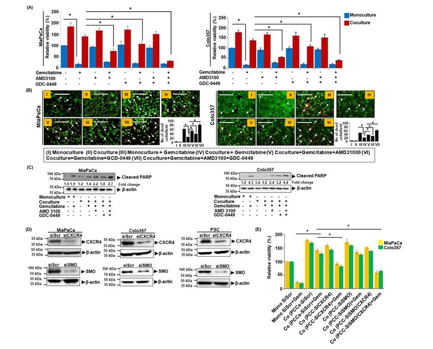
CXCR4 and Hh pathways in pancreatic cancer chemoresistance

since past several decades (2). Asymptomatic               cells induced CXCR4 upregulation providing
progression leading to late diagnosis as well as the       further support for its role as a counter-defense
lack of effective therapies are considered important       mechanism (10). CXCR4 is aberrantly expressed in
factors for such a grim prognosis of PC (3). Despite       PC cells (27,28) and activated by stromal-derived
recent advancements in therapeutic regimens,               chemokine, CXCL12, which is abundantly present
outcomes continue to be disappointing (4-6).               in pancreatic TME as well as at the sites of
Therefore, considering the continuous increase in          metastases (29). In additional findings, we
incidence and mortality of PC, it is extremely             demonstrated that CXCL12/CXCR4 signaling
important that we develop novel mechanism-                 engaged in forming a bi-directional tumor-stromal
based therapies for effective management of this           signaling loop by inducing the expression of sonic
devastating malignancy.                                    hedgehog (SHH), a ligand for hedgehog signaling
                                                           pathway (21). SHH is shown to promote pancreatic
Chemoresistance in PC appears to be a highly               tumor desmoplasia by inducing proliferation and
complicated phenomenon involving both intrinsic            differentiation of pancreatic stellate cells (PSCs)
and extrinsic mechanisms (7-11). Intrinsic                 into myofibroblasts and thus impacts therapeutic
resistance is attributed to the highly genetically         outcome indirectly (23,30).
advanced nature of pancreatic tumors that

                                                                                                                 Downloaded from http://www.jbc.org/ by guest on October 24, 2020
culminate into multiple overlapping and                    Here we examined the therapeutic significance of
compensatory signaling pathways promoting cell             co-targeting of CXCR4 and hedgehog pathways
survival, stemness and drug metabolism (8,12). On          by hypothesizing their cooperative roles in tumor-
the other hand, extrinsic resistance is associated         stromal interaction-driven PC chemoresistance.
with unique histopathological characteristics of           Our findings in co-culture and orthotopic
pancreatic tumors as well as paracrine signaling           pancreatic tumor mouse models demonstrate that
that is operative through bi-directional tumor-            co-targeting of CXCR4 and hedgehog provide
stromal crosstalk (13-17). Studies suggest that            significant improvement in therapeutic efficacy of
pancreatic     tumor      cell-      derived   SHH         gemcitabine. Mechanistically, we demonstrate
predominantly acts on PSCs and induces                     that the crosstalk of PC cells with PSCs leads to
desmoplasia (18), whereas CXCL12 is mostly                 tumor supportive changes in gemcitabine
derived from activated fibroblasts and promote             metabolism, anti-oxidant and stemness properties
growth, aggressiveness and chemoresistance of              via altered gene expression, which could be
PC cells(19-21). Pancreatic stroma is highly               reversed by targeting of CXCR4 and/or hedgehog
dense and fibrotic, and considered to be an                pathways. Together, these findings provide strong
attractive therapeutic target (16,22). However,            preclinical evidence in support of a novel
stromal targeting has resulted in mixed                    combination therapy against PC.
therapeutic responses likely due to stromal
heterogeneity and its dual impact on pancreatic            RESULTS
tumor pathobiology. Earlier it was shown that              CXCR4 and hedgehog pathways mediate co-
desmoplastic tumor microenvironment (TME)
                                                           culture-induced chemoresistance of pancreatic
acted as a barrier for drug delivery and its               cancer cells
targeting in preclinical model led to improved             Studies from our lab and elsewhere have suggested
therapeutic outcome (23). This approach;                   important roles of CXCR4 and hedgehog (Hh)
however, failed in clinical setting (24). Subsequent       signaling in PC pathobiology and chemoresistance
studies suggested that dense stroma keeps the tumor        via different mechanisms(10,20,21,31). Here we
cells confined at the primary site and its depletion       explored their       role in     mediating the
promoted tumor cell dissemination and                      chemoresistance of PC cells when co-cultured with
immunosuppression (25,26) thus necessitating               pancreatic stellate cells (PSCs). We treated the
deeper mechanistic investigations.                         monocultures and co-cultures with gemcitabine in
We previously demonstrated an important role of            the presence or absence of CXCR4 antagonist
CXCR4 signaling in PC chemoresistance (20). We             (AMD3100) and/or hedgehog inhibitor (GDC-
also showed that the gemcitabine treatment of PC           0449) and measured the viability of PCCs.

                                                       2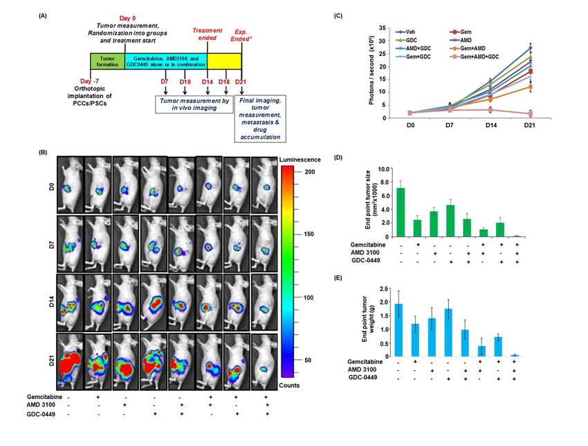
CXCR4 and Hh pathways in pancreatic cancer chemoresistance

Inhibition of either CXCR4 or Hh sensitized the            droplets (Figure 2B). We also monitored the effect
PCCs to gemcitabine toxicity in co-culture (Figure         of CXCR4 and Hh inhibition on the viability of
1A, Figure S1). Notably, combined inhibition of            PSCs in monoculture and coculture with PCCs. Our
CXCR4 and Hh pathways led to a nearly complete             data demonstrate that the inhibition of Hh either by
abolition of co-culture-induced chemoresistance.           GDC-0449 or SMO silencing significantly reduces
Importantly, these effects of combination therapy          PSCs viability in coculture. However, inhibition of
were synergistic in nature when compared with the          CXCR4 had a marginal effect only on PSCs
single drug treatments (Table S1). No significant          viability (Figure 2C and D). To determine the
effect of CXCR4 and/or Hh inhibition; however,             contribution of CXCR4 and Hh signaling inhibition
was recorded on gemcitabine toxicity of PCCs in            in PSCs on the chemoresistance of PC cells, we co-
monocultures. Moreover, treatment of AMD3100               cultured PCCs with CXCR4- and/or SMO-silenced
and GDC-0449 alone or in combination had no                PSCs and treated them with gemcitabine. The data
significant effect on the viability of Colo357 cells       demonstrate that the silencing of SMO in PSCs
in mono- or co-cultures without gemcitabine                effectively      abolished       co-culture-induced
treatment (Figure 1A). These findings were further         chemoresistance (Figure 2E). CXCR4 silencing
confirmed by performing live/dead cell assay using         alone n PSCs; however, did not have a significant
ethidium homodimer-1 (EthD-1) and Calcein-AM               effect.

                                                                                                                   Downloaded from http://www.jbc.org/ by guest on October 24, 2020
staining and PARP cleavage. Reduced tumor cell             CXCR4 and hedgehog pathways mediate co-
death (lesser EthD-1 red fluorescence positivity)          culture-induced expression of chemoresistance-
was observed in co-culture treated with                    associated genes
gemcitabine than those grown in monoculture, and           To elucidate the underlying mechanisms of co-
as expected, co-treatment with AMD3100 and/or              culture-induced chemoresistance in PCCs, we
GDC-0449 promoted cell killing by gemcitabine in           carried out pathway-focused gene expression
co-cultured PCCs (Figure 1B). Similarly, an                analyses. We observed that in PCCs co-cultured
increased signal of cleaved PARP was also reported         with PSCs, expression of a number of genes
in PCCs co-treated with gemcitabine and                    associated    with     gemcitabine       metabolism,
AMD3100 and/or GDC-0449, compared to those                 antioxidant and stemness altered (Figure 3A).
treated with gemcitabine alone (Figure 1C). To             Specifically, MiaPaCa cells in co-culture had an
further confirm the role of CXCR4 and Hh                   upregulation of NT5C1A encoding for cytosolic 5-
signaling in chemoresistance, we silenced CXCR4            nucleotidase     I    A,    an     enzyme      which
and SMO (important mediator of hedgehog                    dephosphorylate gemcitabine monophosphate into
signaling) expression by RNA interference both in          gemcitabine. On the other hand, Colo357 cells in
PCCs (MiaPaCa and Colo357) and PSCs (Figure                co-culture exhibited an increase in the expression of
1D). CXCR4 and/or SMO-silenced PCCs, co-                   CDA, which encodes for cytidine deaminase, an
cultured with PSCs, were treated with gemcitabine,         enzyme that converts gemcitabine into 2′,2′-
and their cell viability was determined. We                difluorodeoxyuridine        (dFdU).       Moreover,
observed that the silencing of CXCR4 alone or in           expression of dCK, encoding deoxycytidine kinase
combination with SMO significantly reduced their           required for conversion of gemcitabine into active
viability when treated with gemcitabine compared           metabolite, decreased in both the cell lines when
to control scrambled siRNA -treated cells (Figure          co-cultured with PSCs. Furthermore, significant
1E). To further support a cooperative role of PSCs         upregulation of genes encoding for antioxidant
in co-culture induced chemoresistance, we used an          enzymes such as glutathione peroxidases GPXs,
additional PSCs line. The resulting data (Figure           and nuclear factor E2-related factor 2 (NRF-2) was
S2) is consistent with findings presented here. We         observed in both the cell lines. In addition,
next determined the effect of PSCs co-culture with         stemness-associated genes, SOX2 and OCT4, were
PCCs on their activation status by examining the           upregulated in MiaPaCa cells co-cultured with
expression of α-SMA, a myofibroblast marker, and           PSCs, whereas Colo357 cells exhibited altered
presence of lipid droplets. Our results in two PSCs        expression of SOX2 and KLF-4 as compared to
lines show that co-culture with PCCs activates             those grown in monoculture (Figure 3A). To
PSCs as evident by increased expression of α-SMA           examine the role of CXCR4 and Hh pathways in
(Figure 2A) and reduced accumulation of lipid              mediating these gene expression changes, we

                                                       3
CXCR4 and Hh pathways in pancreatic cancer chemoresistance

performed qRT-PCR on RNAs isolated from PCCs              expression further suggesting its indirect role in
in co-cultures following treatment with AMD3100           chemoresistance through activation of PSCs.
and GDC-0449, alone or in combination. We
observed that the inhibition of CXCR4 and Hh              Inhibition of CXCR4 and hedgehog pathways
pathways cooperatively led to the abrogation of co-       alters gemcitabine metabolism, ROS levels and
culture-induced upregulation and downregulation           stemness
of NT5C1A and dCK, respectively, in MiaPaCa               To confirm if altered gene expression translates
cells. On the other hand, inhibition of either            into changes in gemcitabine metabolism and ROS
CXCR4 or Hh alone did not alter the co-culture-           generation, we measured their levels in PCCs by
induced downregulation of dCK, but their                  LC-tandem mass spectrometry and H2DCFDA
combined inhibition led to regained expression of         staining, respectively. We observed an enhanced
dCK in Colo357 cells. Interestingly, inhibition of        accumulation of dFdCTP in monocultured PCCs
CXCR4 either alone or in combination with Hh              as compared to those co-cultured with PSCs.
abrogated co-culture-induced CDA upregulation in          Remarkably, inhibition of CXCR4 or/and
Colo357      cells,    while    co-culture-induced        hedgehog pathways significantly enhanced
upregulation of NRF-2 and GPX-1 was                       dFdCTP accumulation in co-cultured PCCs
significantly inhibited upon blocking of CXCR4            (Figure 4A). In contrast, accumulation of

                                                                                                                Downloaded from http://www.jbc.org/ by guest on October 24, 2020
or/and Hh pathways in MiaPaCa cells (Figure 3B).          gemcitabine (dFdC) was higher in co-cultured
Similarly, NRF-2 upregulation was inhibited in the        PCCs, compared to those in monoculture, which
presence of AMD3100 alone or in combination               was significantly reduced upon blocking CXCR4
with GDC-0449 but no significant effect was               or/and hedgehog pathways (Figure 4B). Next, our
observed upon GDC-0449 treatment alone in                 flow cytometry data demonstrated reduction in
Colo357 cells. Additionally, co-culture-induced           basal as well as gemcitabine-induced ROS
GPX-1 upregulation was significantly inhibited in         generation in co-cultured PCCs as compared to
AMD3100 and/or GDC-0449 treated Colo357                   PC cells in monoculture (Figure 4C).
cells. Inhibition of CXCR4 and/or hedgehog                Furthermore, we observed that the blocking
pathways also inhibited SOX2 expression, while            CXCR4 and hedgehog pathway alone or in
OCT4 upregulation was significantly blocked upon          combination abrogated the inhibitory effect of co-
combination treatment of AMD3100 and GDC-                 culture on ROS generation. Next, we examined
0449 in MiaPaCa cells. Interestingly, inhibition of       the role of CXCR4 and hedgehog pathways in co-
CXCR4 alone or in combination with hedgehog               culture-induced stemness of PCCs by monitoring
pathway effectively suppressed co-cultured-               the sphere-forming ability on ultra-low attachment
induced SOX2 and KLF-4 upregulation in Colo357            surface plates. Monoculture and co-culture (with
cells (Figure 3B). To further confirm the                 PSCs) of PCCs were established and treated with
involvement of CXCR4 and SHH signaling                    AMD3100 and/or GDC-0449 in the presence and
pathways in these co-culture-induced changes,             absence of gemcitabine. Thereafter, PCCs were
PCCs were treated with recombinant CXCL12                 plated on ultra-low attachment surface wells and
and/or SHH, and changes in gene expression                inserts containing PCCs or PSCs were placed
analyzed. Data demonstrated that the treatment of         over. Sphere formation was visualized under
CXCL12 caused most of the changes in gene                 microscope after 7 days, which showed greater
expression suggesting an indirect role of Hh              sphere-forming capacity of PCCs in co-culture as
pathway (Figure 3C). In order to further confirm          compared to that in monoculture. Interestingly,
the involvement of CXCR4 and Hh pathway in                gemcitabine-treated PCCs exhibited enhanced
altered gene expression, we examined gene                 stemness in both mon- and co-cultures. Dual
expression changes in CXCR4- or SMO-silenced              inhibition of CXCR4 and hedgehog pathways
PCCs treated with CXCL12 or SHH, respectively.            resulted in reduced formation of spheres, whereas
The data demonstrate that CXCL12- mediated                inhibition of either CXCR4 or hedgehog pathway
downregulation of dCK and upregulation of                 alone did not have any significant effect (Figure 4
NT5C1A, NRF-2 and SOX-2 is significantly                  D).
abrogated in CXCR4-silenced cells (Figure 3D).
SMO-silencing did not have a major effect on gene

                                                      4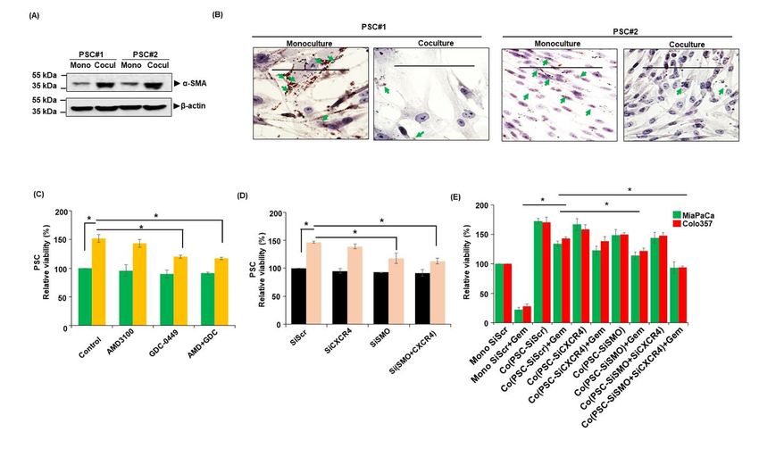
CXCR4 and Hh pathways in pancreatic cancer chemoresistance
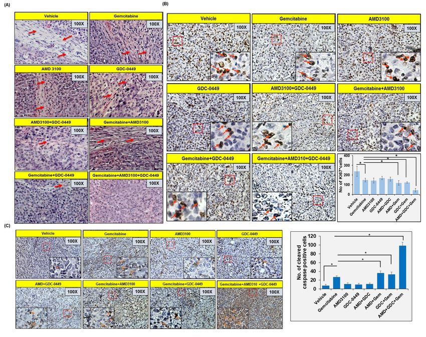
Co-targeting of CXCR4 and hedgehog pathways Ki67 staining (a measure of cell proliferation) was
enhances chemotherapeutic response in vivo reduced in tumor sections from gemcitabine-treated
To obtain the direct evidence in vivo, we evaluated mice, compared to those treated with vehicle only.
the therapeutic efficacy of gemcitabine alone and in In addition, mice treated with AMD3100 and/or
combination with AMD3100 and/or GDC-0449 in GDC-0449 also showed reduction in number of
nude mice. Luciferase-tagged MiaPaCa cells mixed proliferative tumor cells. Combination of
with PSCs were injected into the tail of the pancreas gemcitabine with AMD3100 and/or GDC-0449
of nude mice and tumor formation monitored each showed greater reduction in Ki67 positive cells
alternate day by palpation. After one week, when compared to mice treated with gemcitabine with
tumors became palpable, mice were randomly most remarkable reduction observed in triple
divided into eight treatment groups (Vehicle, combination treated mice (Figure 6B). Tumor
Gemcitabine, AMD3100, GDC-0449, AMD3100 + sections from gemcitabine-treated mice also
GDC-0449, Gemcitabine + AMD3100, showed increased cleaved caspase-3 staining,
Gemcitabine + GDC-0449, Gemcitabine + compared to those from vehicle-treated mice, and
AMD3100 + GDC-0449). Treatments continued the most robust increase in caspase-3 activation was
for two weeks and all mice were sacrificed after a recorded in tumor sections from combined-
week interval (Figure 5A). At day 0 (treatment treatment group (AMD3100, GDC-0449 and
Downloaded from http://www.jbc.org/ by guest on October 24, 2020
initiation) and each subsequent week, tumor growth gemcitabine) mice (Figure 6C).
was monitored by non-invasive in vivo imaging
(Figure 5B and 5C). As expected, the treatment of DISCUSSION
mice with gemcitabine resulted in reduction of
tumor growth, compared to those treated with Pancreatic cancer has remained a therapeutic
vehicle only. Mice treated with AMD3100 alone or challenge despite our improved understanding of
in combination with GDC-0449 (without genetics and molecular pathways involved in its
gemcitabine) showed slower tumor development as etiology and aggressive progression (3,32). Here
compared to those treated with GDC-0449 only. we demonstrated that CXCR4/CXCL12 and
Furthermore, treatment of mice with gemcitabine in hedgehog signaling pathways play an important
combination with either AMD3100 or GDC-0449 role in mediating the chemoresistance of PC cells
had improved therapeutic efficacy, compared to upon co-culturing with PSCs. We identified altered
gemcitabine alone. Combination of gemcitabine gemcitabine metabolism, ROS production and
with AMD3100 had superior effect that that with induction of cancer stemness as important
GDC-0449, which was also confirmed by end-point underlying mechanisms. Combined targeting of
tumor measurement analysis (Figure 5D and 5E). CXCR4 and hedgehog pathways with
Remarkably, triple combination of gemcitabine, chemotherapy almost entirely stalled pancreatic
AMD3100 and GDC-0449 had a tremendous effect tumor growth in an orthotopic mouse model of PC
on the tumor growth compared to any other providing a direct evidence for their therapeutic
treatment group as recorded in non-invasive significance.
imaging and end-point analysis of resected tumor We first focused on investigating the direct roles of
xenografts (Figure 5 B-E). A comparison of the CXCR4 and hedgehog pathways, alone and in
efficacy of different treatments suggested that the concert, in mediating the tumor-stromal
combination therapy provided synergistic interaction-driven chemoresistance of PC. For this,
outcomes (Table S2). Next, we performed we utilized co-culture model to establish bi-
hematoxylin and eosin (H&E) staining and directional crosstalk between PC cells and PSCs.
immunohistochemical analyses (Ki67 and cleaved We found that the treatment of CXCR4 antagonist,
caspase-3) on tumor sections from different AMD3100 and/or hedgehog inhibitor, GDC-0449,
treatment group mice (Figure 6 A-C). Our H&E efficiently reduced co-culture-induced gemcitabine
data suggested extensive desmoplasia in vehicle resistance in PCCs. We also observed that the
treated mice and degree of desmoplasia reduces targeting of CXCR4 was more effective than
upon AMD3100 and/or GDC-0449 treatment and hedgehog inhibition in causing the
complete abolished in mice treated with chemosensitization of PC cells and their co-
gemcitabine along with AMD3100 and GDC-0449.
5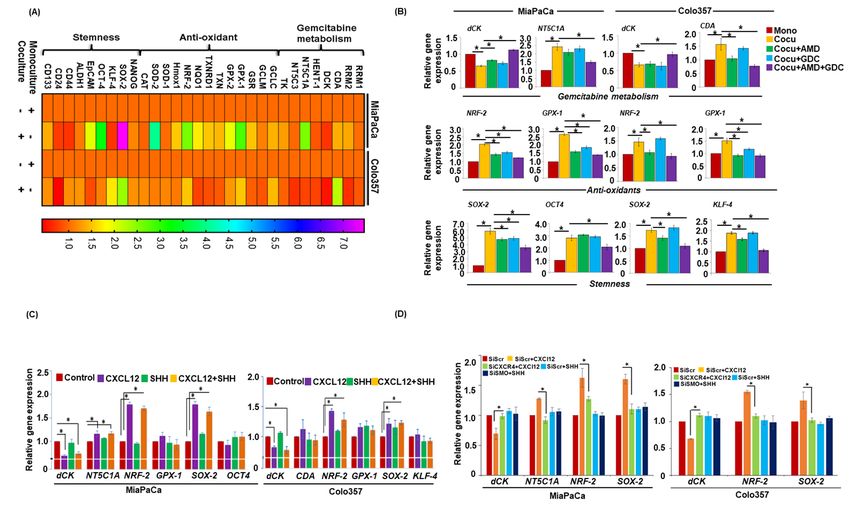
CXCR4 and Hh pathways in pancreatic cancer chemoresistance

targeting nearly abolished co-culture-induced              difluoro-deoxyuridine leading to its reduced
chemoresistance. This is in line with our prior            cellular toxicity(40). In a preclinical study, nab-
observation where treatment with exogenous                 paclitaxel was shown to degrade CDA as a
CXCL12 conferred chemoresistance to PC cells via           synergistic     therapeutic    mechanism       with
activation of survival signaling (20). In addition,        gemcitabine(41). In another study, tumor-
CXCR4 upregulation occurred upon gemcitabine               associated macrophages were shown to upregulate
treatment of PC cells in a counter-defense against         CDA level to induce gemcitabine resistance in PC
chemotherapy(10). Interestingly, our data also             cells(42). Functional impact of the gene expression
supported that PSCs play a significant and direct          changes was confirmed by demonstration of
role in promoting PC chemoresistance besides               reduced intracellular accumulation of gemcitabine
indirectly impacting therapeutic outcome via               triphosphate in co-cultured PC cells, which was
promotion of tumor desmoplasia leading to                  reversed by the inhibition of CXCR4 and/or
restricted drug delivery(16,23). These observations        hedgehog pathways. In contrast, we observed
were further validated in animal studies where we          enhanced gemcitabine accrual in co-cultured PC
established orthotopic pancreatic tumor xenografts         cells, which significantly reduced upon CXCR4
via co-injection of PCCs and PSCs and treated with         and/or hedgehog inhibition. These findings
gemcitabine alone or in combination with                   establish a novel aspect of cooperative function of

                                                                                                                  Downloaded from http://www.jbc.org/ by guest on October 24, 2020
AMD3100 and/or GDC-0449. These inhibitors are              CXCR4 and hedgehog pathways in PC
shown to be effective in mice in previous studies          chemoresistance.
(33,34). Furthermore, orthotopic implantation of
SHH-expressing primary human pancreatic                    Our study also demonstrated that co-culture with
epithelial and cancer cells induce desmoplasia in          PSCs led to reduced intracellular ROS levels in PC
mice(18). Thus, our current findings provide               cells, which were restored upon targeted inhibition
support for a cooperative role of CXCR4 and                of CXCR4 and hedgehog pathways. Increased
hedgehog pathways in establishing a bi-directional         accumulation of ROS following drug treatment is
tumor-stromal crosstalk that has pathobiological           an important mechanism of drug toxicity(43).
significance in growth and chemoresistance of PC           Cancer cells, in general, have heightened levels of
cells.                                                     ROS due to increased metabolic demand of
                                                           proliferating cells for quick energy and cellular
From the mechanistic standpoint, our data                  building blocks(44). Increased ROS likely
demonstrated that the co-culture of PCCs with              contributes to cancer pathogenesis via promotion of
PSCs altered the expression of several genes               genetic instability and it also remodel ECM
associated with gemcitabine metabolism, ROS                components by activating proteolytic enzymes in
generation and cancer stemness. Several of these           cancer cells, which help in tumor cell migration and
genes were indeed found to be regulated through            dissemination (45,46). However, to check the ROS
the cooperative action of CXCR4 and hedgehog               levels beyond a threshold, tumor cells overexpress
signaling pathways. Importantly, the rate limiting         enzymes of the anti-oxidant system(46). Our
enzyme of gemcitabine activation(35), dCK found            findings provide evidence that tumor-stromal
to be reduced in PC cells co-cultured with PSCs.           crosstalk mediated through CXCR4 and hedgehog
dCK expression is positively co-related with overall       pathways control the expression of antioxidant
survival and disease free survival in PC                   genes (NRF-2 and GPX-1). Earlier data suggest that
patients(36). A recent study has also suggested that       NRF-2 is highly expressed in pancreatic tumors and
the forced expression of dCK in PC cells inhibits          patients with low levels of NRF-2 are more
tumor cell proliferation and aggressiveness(37).           sensitive to chemotherapy (47,48). Furthermore,
Other two enzymes that were altered in cell-specific       downregulation of NRF-2 increases gemcitabine
manner were NT5C1A and CDA.                     The        sensitivity of PC cells (47). GPX1, which
dephosphorylating enzyme, NT5C1A, converts                 counteracts oxidative stress, has oncogenic role in
gemcitabine monophosphate into gemcitabine and             several malignancies. However, low GPX-1
is reported to be overexpressed in PC(38,39). On           expression has been correlated with poor survival
the other hand, CDA is a gemcitabine catabolizing          rate of PC patients treated with gemcitabine (49).
enzyme that converts gemcitabine into 2′,2′-               Recent finding suggested GPX-1 as an important

                                                       6
CXCR4 and Hh pathways in pancreatic cancer chemoresistance
regulator of glucose metabolism in PC cells (50). intermittently using established markers either in
This clearly suggests complex and context house and/or by using a commercial service
dependent role of GPX-1 in cancer which needs to provider (Genetica DNA Laboratories, Burlington,
be addressed in great detail. NC and short-tandem repeats genotyping).
Cancer stemness is another important characteristic Reagents and antibodies
associated with the poor response of The following reagents were used in this study:
chemotherapy(51). Stemness potential of the Dulbecco's Modified Eagle Medium (DMEM);
cancer cells is controlled by certain transcription Roswell Park Memorial Institute medium (RPMI-
factors that regulate expression of genes associated 1640); Fetal bovine serum (FBS) (Atlanta
with tumor maintenance, aggressiveness and Biologicals, Lawrenceville, GA); penicillin and
chemoresistance (52,53). We observed increased streptomycin (Invitrogen, Carlsbad, CA); High-
expression of two important stem cell-associated Capacity RNA-to-cDNA™ Kit and SYBR Green
transcription factors, SOX2 and OCT4, in PC cells Master Mix (Applied Biosystems, Carlsbad, CA);
co-cultured with PSCs and targeting of CXCR4 Western blotting SuperSignal West Femto
and/or hedgehog pathways led to their reduced Maximum sensitivity substrate kit (Thermo
expression. SOX2 expression is shown to increase Scientific, Logan, UT); EZ-Dewax (Biogenex,
Fremont, CA); background sniper, polymer and
Downloaded from http://www.jbc.org/ by guest on October 24, 2020
levels of pancreatic CSC markers and increase cell
proliferation and stemness (54). Similarly, probe (Biocare Medical, Concord, CA); Plerixafor
upregulation of OCT4 maintains undifferentiated (AMD3100) and gemcitabine (SelleckChem,
state of induced pluripotent stem cell state, while Houston, TX); Vismodegib (GDC-0449) (LC
the loss of OCT4 induces differentiation of stem Laboratories, MA). LIVE/DEAD™
cells (55). Study suggested that inhibition of OCT4 Viability/Cytotoxicity Kit (Thermo Fisher
and Nanog abolished stemness of pancreatic cancer Scientific). The following antibodies were used: α-
cells (56). In some reports, a role of canonical cleaved PARP (rabbit polyclonal Abcam,
hedgehog signaling in pancreatic CSCs and Cambridge, MA) and rabbit polyclonal cleaved
gemcitabine resistance has also been demonstrated caspas-3 (Cell Signaling Technology, Denver,
(57,58). In agreement, we also observed an MA), mouse monoclonal Ki67 (BD Biosciences,
upregulation of SOX2 in PC cells treated with SHH San Jose, CA), mouse monoclonal biotinylated
although treatment with CXCL12 imparted a anti-β-actin (Sigma-Aldrich, St. Louis, MO), rabbit
greater effect. polyclonal α-SMA (Epitomics, Burlingame, CA),
rabbit polyclonal CXCR4 (Novous Biologicals,
In summary, our findings provide a novel Centennial, CO), mouse monoclonal SMO
mechanistic basis for PC chemoresistance where (Millipore Sigma, Burlington, MA), and
tumor-stromal cross-talk mediated through CXCR4 horseradish peroxidase (HRP) labeled secondary
and hedgehog pathways plays an important role and antibodies (Santa Cruz Biotechnology).
co-targeting of these signaling nodes in a Cell co-culturing, treatments and transfections
combination therapy will lead to superior Pancreatic tumor cells (PCCs; 1.0x10⁵cells/well)
synergistic clinical outcome in PC patients. were seeded in the six-well plates, while pancreatic
stellate cells (PSCs; 1.0 x/105cells/well) were
EXPERIMENTAL PROCEDURES seeded in one-micron pore size insert chambers that
Cell lines and culture conditions were placed over PCCs. For monoculture controls,
Pancreatic cancer cell lines, MiaPaCa and Colo357, we seeded PCCs (1.0x10⁵ cells) in the insert
and pancreatic stellate cells (PSCs) were procured chamber as well. Cells were kept in incubator for
and maintained in culture as previously described 48 h at 37ºC to establish co-culture and thereafter,
(59,60). Human pancreatic stellate cells were media was replaced and cells allowed to grow for
purchased from ScienCell Research Laboratories, additional 24 h and treated with AMD3100 (5
Carlsbad, CA and culture according to their µg/mL) and GD-049 (5 µM) alone or together in
instructions. All the cells were routinely tested for presence and absence of gemcitabine (5 µM) for
mycoplasma contamination and authenticated 24-96h. For studies intended to estimate
gemcitabine metabolites, gemcitabine treatment
7CXCR4 and Hh pathways in pancreatic cancer chemoresistance
was given 4 h prior to cell harvesting. For Briefly, cell lysates were prepared in NP-40 lysis
knockdown of CXCR4 and SMO in pancreatic buffer and protein estimated using DC protein assay
cancer and stellate cells, cells were cultured in 6 kit. Subsequently, 30 µg of protein was resolved on
well plates and transfected with 100 nmol/L of 10-12 % polyacrylamide gels and transferred onto
nontarget or ON-TARGETplus SMARTpool PVDF membranes. Blots were washed, blocked in
CXCR4 and SMO targeting siRNAs (Dharmacon 5% milk and probed with primary antibody
Horizon Discovery, Lafayette, CO) using (1:1000) followed by incubation with secondary
DharmaFECT (Dharmacon Horizon Discovery HRP-antibody (1:2000). β-actin (1:20,000) was
Lafayette, CO) according to the manufacturer's used as an internal control. The signal was detected
instruction. by using Super Signal West Femto maximum
sensitivity substrate kit on the ChemiDoc Imaging
Cell viability and sphere-formation assay
System (Bio-Rad, Hercules, CA).
Pancreatic tumor cell growth was monitored by
counting of number of viable cells on Oil red O staining
haemocytometer following staining with Trypan Pancreatic stellate cells were stained with oil red O
blue. For sphere-formation assay, treated or to determine their activation by visualizing lipid
untreated tumor cells were harvested, counted and droplets using Lipid (Oil Red O) Staining Kit
plated (1000 cells/well) in wells having ultra-low (BioVision, Milpitas, CA). The assay was
Downloaded from http://www.jbc.org/ by guest on October 24, 2020
attachment surface. Culture inserts containing performed according to the manufacturer’s
PCCs or PSCs were placed over plated cells. instruction and images were taken under light
Spheres were visualized under microscope after 7 microscope.
days and photographed. Immunohistochemical and histological analyses
Five micron sections were cut from orthotopic
Measurement of reactive oxygen species
tumor xenograft tissues and processed for H&E
Intracellular reactive oxygen species (ROS) levels
staining and immunohistochemistry as described
were determined as described earlier (11,43).
previously (60). All the antibodies were used at
Briefly, cells from monoculture or co-culture were
1:100 dilutions. Tumor tissue sections were
incubated with DCFDA dye in regular growth
visualized under microscope and photographed.
medium for 30 min at 37 °C, and subsequently
washed with PBS and resuspended in 400µL PBS. Measurement of gemcitabine metabolites
Fluorescence intensity in cells was examined as a Treated cells in monoculture or co-culture were
measure of ROS by flow cytometry on a homogenized 1:10 using a pellet pestle with buffer
FACSCanto IIi (BD Biosciences, San Jose, CA, containing 50:50 Acetonitrile: 10 mM PO4 buffer
USA). pH 9.5 containing 25 µg/mL THU and 2 mg/mL
EDTA. Samples were spiked with internal standard
RNA isolation and quantitative real time-PCR
(10 µL of a spiking solution containing 50 µg/mL
(qRT-PCR)
5-CL-deoxyuridne and 5 µg/mL Cytidine N15
Total RNA was isolated by using TRIzol reagent
triphosphate in DI water) and mixed well. After
and 2 µg of RNA was taken for cDNA synthesis
centrifugation for 5 minutes at 21,000 x g, the
using the High Capacity complementary DNA
supernatant was transferred to autosampler vials
Reverse Transcription Kit following
and analyzed by liquid chromatography-tandem
manufacturer’s instructions. Subsequently, q-PCR
mass spectrometry (LC-MS/MS).
was performed in 96-well plates using SYBR Green
Master Mix, cDNA as a template and specific Orthotopic xenograft study in mice
primer pair sets (Table S3) on an iCycler system All animal experiments were performed under a
(Bio-Rad, Hercules, CA). The thermal conditions protocol approved by the University of South
for real-time PCR assays were as follows: cycle 1: Alabama Institutional Animal Care and Use
95 °C for 10 min, cycle 2 (x 40): 95 °C for 10 sec Committee (IACUC). Immuno-compromised nude
and 58-60 °C for 45 sec. mice (4-6 weeks old; Harlan Laboratories,
Prattville, AL) were anaesthetized with
Preparation of cell lysates and immunoblotting
intraperitoneal (i.p.) injection of ketamine (100
Total protein was isolated, estimated and processed
mg/kg) and xylazine (15 mg/kg). A mixture (1:5) of
for immunoblotting as described earlier (10,61).
8CXCR4 and Hh pathways in pancreatic cancer chemoresistance
luciferase-tagged pancreatic cancer cells (1x10⁶) B2)/2, where A is the larger and B is the smaller of
and PSCs human PSCs (5x10⁶) suspended in 100 the two dimensions.
μL normal saline were injected into the tail region
Statistical analysis
of the pancreas as described previously (62). Once
All the experiments were performed at least three
tumor became palpable (7th day post injection),
times in a biological replicates and data expressed
mice were randomly divided into eight treatment
as mean ± SD. Wherever appropriate, the data were
groups (6 mice/group) and treatment initiated as
also subjected to unpaired two-tailed Student's t test
listed (Table S4). Tumor growth was monitored
and Two-way ANOVA. *p < 0.05 was considered
biweekly by bioluminescence imaging using
as significant.
Xenogen-IVIS–cooled CCD optical system (IVIS
Spectrum) following i.p. injection of D-Luciferin
Data availability
(150 mg/kg). At the end point, final imaging was
The data presented in this manuscript is stored with
performed, and animals sacrificed. Thereafter,
us and is available from corresponding author, for
tumors were resected, weighed and their volumes
sharing upon a reasonable request.
measured using by the following formula: (A ×
Downloaded from http://www.jbc.org/ by guest on October 24, 2020
CONFLICT OF INTEREST
The authors declare no conflict of interest.
9CXCR4 and Hh pathways in pancreatic cancer chemoresistance
REFERENCES
1. Siegel, R. L., Miller, K. D., and Jemal, A. (2020) Cancer statistics, 2020. CA Cancer J Clin
70, 7-30
2. Aier, I., Semwal, R., Sharma, A., and Varadwaj, P. K. (2019) A systematic assessment of
statistics, risk factors, and underlying features involved in pancreatic cancer. Cancer
Epidemiol 58, 104-110
3. Maitra, A., and Hruban, R. H. (2008) Pancreatic cancer. Annu Rev Pathol 3, 157-188
4. Conroy, T., Desseigne, F., Ychou, M., Bouche, O., Guimbaud, R., Becouarn, Y., Adenis,
A., Raoul, J. L., Gourgou-Bourgade, S., de la Fouchardiere, C., Bennouna, J., Bachet, J.
B., Khemissa-Akouz, F., Pere-Verge, D., Delbaldo, C., Assenat, E., Chauffert, B., Michel,
P., Montoto-Grillot, C., Ducreux, M., Groupe Tumeurs Digestives of, U., and Intergroup,
P. (2011) FOLFIRINOX versus gemcitabine for metastatic pancreatic cancer. N Engl J
Med 364, 1817-1825
5. Von Hoff, D. D., Ervin, T., Arena, F. P., Chiorean, E. G., Infante, J., Moore, M., Seay, T.,
Tjulandin, S. A., Ma, W. W., Saleh, M. N., Harris, M., Reni, M., Dowden, S., Laheru, D.,
Downloaded from http://www.jbc.org/ by guest on October 24, 2020
Bahary, N., Ramanathan, R. K., Tabernero, J., Hidalgo, M., Goldstein, D., Van Cutsem,
E., Wei, X., Iglesias, J., and Renschler, M. F. (2013) Increased survival in pancreatic cancer
with nab-paclitaxel plus gemcitabine. N Engl J Med 369, 1691-1703
6. Wang-Gillam, A., Li, C. P., Bodoky, G., Dean, A., Shan, Y. S., Jameson, G., Macarulla,
T., Lee, K. H., Cunningham, D., Blanc, J. F., Hubner, R. A., Chiu, C. F., Schwartsmann,
G., Siveke, J. T., Braiteh, F., Moyo, V., Belanger, B., Dhindsa, N., Bayever, E., Von Hoff,
D. D., Chen, L. T., and Group, N.-S. (2016) Nanoliposomal irinotecan with fluorouracil
and folinic acid in metastatic pancreatic cancer after previous gemcitabine-based therapy
(NAPOLI-1): a global, randomised, open-label, phase 3 trial. Lancet 387, 545-557
7. Fiorini, C., Cordani, M., Gotte, G., Picone, D., and Donadelli, M. (2015) Onconase induces
autophagy sensitizing pancreatic cancer cells to gemcitabine and activates Akt/mTOR
pathway in a ROS-dependent manner. Biochim Biophys Acta 1853, 549-560
8. Fiorini, C., Cordani, M., Padroni, C., Blandino, G., Di Agostino, S., and Donadelli, M.
(2015) Mutant p53 stimulates chemoresistance of pancreatic adenocarcinoma cells to
gemcitabine. Biochim Biophys Acta 1853, 89-100
9. Costantino, C. L., Witkiewicz, A. K., Kuwano, Y., Cozzitorto, J. A., Kennedy, E. P.,
Dasgupta, A., Keen, J. C., Yeo, C. J., Gorospe, M., and Brody, J. R. (2009) The role of
HuR in gemcitabine efficacy in pancreatic cancer: HuR Up-regulates the expression of the
gemcitabine metabolizing enzyme deoxycytidine kinase. Cancer Res 69, 4567-4572
10. Arora, S., Bhardwaj, A., Singh, S., Srivastava, S. K., McClellan, S., Nirodi, C. S., Piazza,
G. A., Grizzle, W. E., Owen, L. B., and Singh, A. P. (2013) An undesired effect of
chemotherapy: gemcitabine promotes pancreatic cancer cell invasiveness through reactive
oxygen species-dependent, nuclear factor kappaB- and hypoxia-inducible factor 1alpha-
mediated up-regulation of CXCR4. J Biol Chem 288, 21197-21207
11. Patel, G. K., Khan, M. A., Bhardwaj, A., Srivastava, S. K., Zubair, H., Patton, M. C., Singh,
S., Khushman, M., and Singh, A. P. (2017) Exosomes confer chemoresistance to pancreatic
cancer cells by promoting ROS detoxification and miR-155-mediated suppression of key
gemcitabine-metabolising enzyme, DCK. Br J Cancer 116, 609-619
12. Ju, H. Q., Gocho, T., Aguilar, M., Wu, M., Zhuang, Z. N., Fu, J., Yanaga, K., Huang, P.,
and Chiao, P. J. (2015) Mechanisms of Overcoming Intrinsic Resistance to Gemcitabine in
10CXCR4 and Hh pathways in pancreatic cancer chemoresistance
Pancreatic Ductal Adenocarcinoma through the Redox Modulation. Mol Cancer Ther 14,
788-798
13. Ansari, D., Ohlsson, H., Althini, C., Bauden, M., Zhou, Q., Hu, D., and Andersson, R.
(2019) The Hippo Signaling Pathway in Pancreatic Cancer. Anticancer Res 39, 3317-3321
14. Chen, F., Long, Q., Fu, D., Zhu, D., Ji, Y., Han, L., Zhang, B., Xu, Q., Liu, B., Li, Y., Wu,
S., Yang, C., Qian, M., Xu, J., Liu, S., Cao, L., Chin, Y. E., Lam, E. W., Coppe, J. P., and
Sun, Y. (2018) Targeting SPINK1 in the damaged tumour microenvironment alleviates
therapeutic resistance. Nat Commun 9, 4315
15. Miyamoto, H., Murakami, T., Tsuchida, K., Sugino, H., Miyake, H., and Tashiro, S. (2004)
Tumor-stroma interaction of human pancreatic cancer: acquired resistance to anticancer
drugs and proliferation regulation is dependent on extracellular matrix proteins. Pancreas
28, 38-44
16. von Ahrens, D., Bhagat, T. D., Nagrath, D., Maitra, A., and Verma, A. (2017) The role of
stromal cancer-associated fibroblasts in pancreatic cancer. J Hematol Oncol 10, 76
17. Thomas, D., and Radhakrishnan, P. (2019) Tumor-stromal crosstalk in pancreatic cancer
and tissue fibrosis. Mol Cancer 18, 14
Downloaded from http://www.jbc.org/ by guest on October 24, 2020
18. Bailey, J. M., Swanson, B. J., Hamada, T., Eggers, J. P., Singh, P. K., Caffery, T., Ouellette,
M. M., and Hollingsworth, M. A. (2008) Sonic hedgehog promotes desmoplasia in
pancreatic cancer. Clin Cancer Res 14, 5995-6004
19. Ene-Obong, A., Clear, A. J., Watt, J., Wang, J., Fatah, R., Riches, J. C., Marshall, J. F.,
Chin-Aleong, J., Chelala, C., Gribben, J. G., Ramsay, A. G., and Kocher, H. M. (2013)
Activated pancreatic stellate cells sequester CD8+ T cells to reduce their infiltration of the
juxtatumoral compartment of pancreatic ductal adenocarcinoma. Gastroenterology 145,
1121-1132
20. Singh, S., Srivastava, S. K., Bhardwaj, A., Owen, L. B., and Singh, A. P. (2010) CXCL12-
CXCR4 signalling axis confers gemcitabine resistance to pancreatic cancer cells: a novel
target for therapy. Br J Cancer 103, 1671-1679
21. Singh, A. P., Arora, S., Bhardwaj, A., Srivastava, S. K., Kadakia, M. P., Wang, B., Grizzle,
W. E., Owen, L. B., and Singh, S. (2012) CXCL12/CXCR4 protein signaling axis induces
sonic hedgehog expression in pancreatic cancer cells via extracellular regulated kinase-
and Akt kinase-mediated activation of nuclear factor kappaB: implications for bidirectional
tumor-stromal interactions. J Biol Chem 287, 39115-39124
22. Neesse, A., Bauer, C. A., Ohlund, D., Lauth, M., Buchholz, M., Michl, P., Tuveson, D. A.,
and Gress, T. M. (2019) Stromal biology and therapy in pancreatic cancer: ready for
clinical translation? Gut 68, 159-171
23. Olive, K. P., Jacobetz, M. A., Davidson, C. J., Gopinathan, A., McIntyre, D., Honess, D.,
Madhu, B., Goldgraben, M. A., Caldwell, M. E., Allard, D., Frese, K. K., Denicola, G.,
Feig, C., Combs, C., Winter, S. P., Ireland-Zecchini, H., Reichelt, S., Howat, W. J., Chang,
A., Dhara, M., Wang, L., Ruckert, F., Grutzmann, R., Pilarsky, C., Izeradjene, K.,
Hingorani, S. R., Huang, P., Davies, S. E., Plunkett, W., Egorin, M., Hruban, R. H.,
Whitebread, N., McGovern, K., Adams, J., Iacobuzio-Donahue, C., Griffiths, J., and
Tuveson, D. A. (2009) Inhibition of Hedgehog signaling enhances delivery of
chemotherapy in a mouse model of pancreatic cancer. Science 324, 1457-1461
24. Allison, M. (2012) Hedgehog hopes lifted by approval... and stung by failure. Nat
Biotechnol 30, 203
11CXCR4 and Hh pathways in pancreatic cancer chemoresistance
25. Rhim, A. D., Oberstein, P. E., Thomas, D. H., Mirek, E. T., Palermo, C. F., Sastra, S. A.,
Dekleva, E. N., Saunders, T., Becerra, C. P., Tattersall, I. W., Westphalen, C. B.,
Kitajewski, J., Fernandez-Barrena, M. G., Fernandez-Zapico, M. E., Iacobuzio-Donahue,
C., Olive, K. P., and Stanger, B. Z. (2014) Stromal elements act to restrain, rather than
support, pancreatic ductal adenocarcinoma. Cancer Cell 25, 735-747
26. Ozdemir, B. C., Pentcheva-Hoang, T., Carstens, J. L., Zheng, X., Wu, C. C., Simpson, T.
R., Laklai, H., Sugimoto, H., Kahlert, C., Novitskiy, S. V., De Jesus-Acosta, A., Sharma,
P., Heidari, P., Mahmood, U., Chin, L., Moses, H. L., Weaver, V. M., Maitra, A., Allison,
J. P., LeBleu, V. S., and Kalluri, R. (2014) Depletion of carcinoma-associated fibroblasts
and fibrosis induces immunosuppression and accelerates pancreas cancer with reduced
survival. Cancer Cell 25, 719-734
27. Saur, D., Seidler, B., Schneider, G., Algul, H., Beck, R., Senekowitsch-Schmidtke, R.,
Schwaiger, M., and Schmid, R. M. (2005) CXCR4 expression increases liver and lung
metastasis in a mouse model of pancreatic cancer. Gastroenterology 129, 1237-1250
28. Wang, Z., Ma, Q., Li, P., Sha, H., Li, X., and Xu, J. (2013) Aberrant expression of CXCR4
and beta-catenin in pancreatic cancer. Anticancer Res 33, 4103-4110
Downloaded from http://www.jbc.org/ by guest on October 24, 2020
29. Zhang, J., Liu, C., Mo, X., Shi, H., and Li, S. (2018) Mechanisms by which
CXCR4/CXCL12 cause metastatic behavior in pancreatic cancer. Oncol Lett 15, 1771-
1776
30. Li, X., Wang, Z., Ma, Q., Xu, Q., Liu, H., Duan, W., Lei, J., Ma, J., Wang, X., Lv, S., Han,
L., Li, W., Guo, J., Guo, K., Zhang, D., Wu, E., and Xie, K. (2014) Sonic hedgehog
paracrine signaling activates stromal cells to promote perineural invasion in pancreatic
cancer. Clin Cancer Res 20, 4326-4338
31. Xiao, G., Wang, X., and Yu, Y. (2017) CXCR4/Let-7a Axis Regulates Metastasis and
Chemoresistance of Pancreatic Cancer Cells Through Targeting HMGA2. Cell Physiol
Biochem 43, 840-851
32. Khan, M. A., Azim, S., Zubair, H., Bhardwaj, A., Patel, G. K., Khushman, M., Singh, S.,
and Singh, A. P. (2017) Molecular Drivers of Pancreatic Cancer Pathogenesis: Looking
Inward to Move Forward. Int J Mol Sci 18
33. Tu, Y., Niu, M., Xie, P., Yue, C., Liu, N., Qi, Z., Gao, S., Liu, H., Shi, Q., Yu, R., and Liu,
X. (2017) Smoothened is a poor prognosis factor and a potential therapeutic target in
glioma. Sci Rep 7, 42630
34. Righi, E., Kashiwagi, S., Yuan, J., Santosuosso, M., Leblanc, P., Ingraham, R., Forbes, B.,
Edelblute, B., Collette, B., Xing, D., Kowalski, M., Mingari, M. C., Vianello, F., Birrer,
M., Orsulic, S., Dranoff, G., and Poznansky, M. C. (2011) CXCL12/CXCR4 blockade
induces multimodal antitumor effects that prolong survival in an immunocompetent mouse
model of ovarian cancer. Cancer Res 71, 5522-5534
35. Saiki, Y., Yoshino, Y., Fujimura, H., Manabe, T., Kudo, Y., Shimada, M., Mano, N.,
Nakano, T., Lee, Y., Shimizu, S., Oba, S., Fujiwara, S., Shimizu, H., Chen, N., Nezhad, Z.
K., Jin, G., Fukushige, S., Sunamura, M., Ishida, M., Motoi, F., Egawa, S., Unno, M., and
Horii, A. (2012) DCK is frequently inactivated in acquired gemcitabine-resistant human
cancer cells. Biochem Biophys Res Commun 421, 98-104
36. Xiong, J., Altaf, K., Ke, N., Wang, Y., Tang, J., Tan, C., Li, A., Zhang, H., He, D., and
Liu, X. (2016) dCK Expression and Gene Polymorphism With Gemcitabine
Chemosensitivity in Patients With Pancreatic Ductal Adenocarcinoma: A Strobe-
Compliant Observational Study. Medicine (Baltimore) 95, e2936
12CXCR4 and Hh pathways in pancreatic cancer chemoresistance
37. Hu, Q., Qin, Y., Xiang, J., Liu, W., Xu, W., Sun, Q., Ji, S., Liu, J., Zhang, Z., Ni, Q., Xu,
J., Yu, X., and Zhang, B. (2018) dCK negatively regulates the NRF2/ARE axis and ROS
production in pancreatic cancer. Cell Prolif 51, e12456
38. Hunsucker, S. A., Spychala, J., and Mitchell, B. S. (2001) Human cytosolic 5'-nucleotidase
I: characterization and role in nucleoside analog resistance. J Biol Chem 276, 10498-10504
39. Patzak, M. S., Kari, V., Patil, S., Hamdan, F. H., Goetze, R. G., Brunner, M., Gaedcke, J.,
Kitz, J., Jodrell, D. I., Richards, F. M., Pilarsky, C., Gruetzmann, R., Rummele, P., Knosel,
T., Hessmann, E., Ellenrieder, V., Johnsen, S. A., and Neesse, A. (2019) Cytosolic 5'-
nucleotidase 1A is overexpressed in pancreatic cancer and mediates gemcitabine resistance
by reducing intracellular gemcitabine metabolites. EBioMedicine 40, 394-405
40. Amrutkar, M., and Gladhaug, I. P. (2017) Pancreatic Cancer Chemoresistance to
Gemcitabine. Cancers (Basel) 9
41. Frese, K. K., Neesse, A., Cook, N., Bapiro, T. E., Lolkema, M. P., Jodrell, D. I., and
Tuveson, D. A. (2012) nab-Paclitaxel potentiates gemcitabine activity by reducing cytidine
deaminase levels in a mouse model of pancreatic cancer. Cancer Discov 2, 260-269
42. Weizman, N., Krelin, Y., Shabtay-Orbach, A., Amit, M., Binenbaum, Y., Wong, R. J., and
Downloaded from http://www.jbc.org/ by guest on October 24, 2020
Gil, Z. (2014) Macrophages mediate gemcitabine resistance of pancreatic adenocarcinoma
by upregulating cytidine deaminase. Oncogene 33, 3812-3819
43. Khan, M. A., Gahlot, S., and Majumdar, S. (2012) Oxidative stress induced by curcumin
promotes the death of cutaneous T-cell lymphoma (HuT-78) by disrupting the function of
several molecular targets. Mol Cancer Ther 11, 1873-1883
44. Kumari, S., Badana, A. K., G, M. M., G, S., and Malla, R. (2018) Reactive Oxygen Species:
A Key Constituent in Cancer Survival. Biomark Insights 13, 1177271918755391
45. Nikitovic, D., Corsini, E., Kouretas, D., Tsatsakis, A., and Tzanakakis, G. (2013) ROS-
major mediators of extracellular matrix remodeling during tumor progression. Food Chem
Toxicol 61, 178-186
46. Schumacker, P. T. (2006) Reactive oxygen species in cancer cells: live by the sword, die
by the sword. Cancer Cell 10, 175-176
47. Lister, A., Nedjadi, T., Kitteringham, N. R., Campbell, F., Costello, E., Lloyd, B., Copple,
I. M., Williams, S., Owen, A., Neoptolemos, J. P., Goldring, C. E., and Park, B. K. (2011)
Nrf2 is overexpressed in pancreatic cancer: implications for cell proliferation and therapy.
Mol Cancer 10, 37
48. Soini, Y., Eskelinen, M., Juvonen, P., Karja, V., Haapasaari, K. M., Saarela, A., and
Karihtala, P. (2014) Nuclear Nrf2 expression is related to a poor survival in pancreatic
adenocarcinoma. Pathol Res Pract 210, 35-39
49. Meng, Q., Shi, S., Liang, C., Liang, D., Hua, J., Zhang, B., Xu, J., and Yu, X. (2018)
Abrogation of glutathione peroxidase-1 drives EMT and chemoresistance in pancreatic
cancer by activating ROS-mediated Akt/GSK3beta/Snail signaling. Oncogene 37, 5843-
5857
50. Meng, Q., Xu, J., Liang, C., Liu, J., Hua, J., Zhang, Y., Ni, Q., Shi, S., and Yu, X. (2018)
GPx1 is involved in the induction of protective autophagy in pancreatic cancer cells in
response to glucose deprivation. Cell Death Dis 9, 1187
51. Nunes, T., Hamdan, D., Leboeuf, C., El Bouchtaoui, M., Gapihan, G., Nguyen, T. T.,
Meles, S., Angeli, E., Ratajczak, P., Lu, H., Di Benedetto, M., Bousquet, G., and Janin, A.
(2018) Targeting Cancer Stem Cells to Overcome Chemoresistance. Int J Mol Sci 19
13CXCR4 and Hh pathways in pancreatic cancer chemoresistance
52. Deshmukh, S. K., Srivastava, S. K., Zubair, H., Bhardwaj, A., Tyagi, N., Al-Ghadhban,
A., Singh, A. P., Dyess, D. L., Carter, J. E., and Singh, S. (2017) Resistin potentiates
chemoresistance and stemness of breast cancer cells: Implications for racially disparate
therapeutic outcomes. Cancer Lett 396, 21-29
53. Zubair, H., Azim, S., Srivastava, S. K., Bhardwaj, A., Marimuthu, S., Patton, M. C., Singh,
S., and Singh, A. P. (2016) Cancer Stem Cells: Concept, Significance, and Management.
in Stem Cells in Toxicology and Medicine (Sahu, S. C. ed.), John Wiley & Sons, Ltd:
Chichester, UK. pp 375-413
54. Herreros-Villanueva, M., Zhang, J. S., Koenig, A., Abel, E. V., Smyrk, T. C., Bamlet, W.
R., de Narvajas, A. A., Gomez, T. S., Simeone, D. M., Bujanda, L., and Billadeau, D. D.
(2013) SOX2 promotes dedifferentiation and imparts stem cell-like features to pancreatic
cancer cells. Oncogenesis 2, e61
55. Herreros-Villanueva, M., Bujanda, L., Billadeau, D. D., and Zhang, J. S. (2014) Embryonic
stem cell factors and pancreatic cancer. World J Gastroenterol 20, 2247-2254
56. Lu, Y., Zhu, H., Shan, H., Lu, J., Chang, X., Li, X., Lu, J., Fan, X., Zhu, S., Wang, Y.,
Guo, Q., Wang, L., Huang, Y., Zhu, M., and Wang, Z. (2013) Knockdown of Oct4 and
Downloaded from http://www.jbc.org/ by guest on October 24, 2020
Nanog expression inhibits the stemness of pancreatic cancer cells. Cancer Lett 340, 113-
123
57. Fu, J., Rodova, M., Roy, S. K., Sharma, J., Singh, K. P., Srivastava, R. K., and Shankar, S.
(2013) GANT-61 inhibits pancreatic cancer stem cell growth in vitro and in
NOD/SCID/IL2R gamma null mice xenograft. Cancer Lett 330, 22-32
58. Singh, B. N., Fu, J., Srivastava, R. K., and Shankar, S. (2011) Hedgehog signaling
antagonist GDC-0449 (Vismodegib) inhibits pancreatic cancer stem cell characteristics:
molecular mechanisms. PLoS One 6, e27306
59. Zubair, H., Azim, S., Srivastava, S. K., Ahmad, A., Bhardwaj, A., Khan, M. A., Patel, G.
K., Arora, S., Carter, J. E., Singh, S., and Singh, A. P. (2016) Glucose Metabolism
Reprogrammed by Overexpression of IKKepsilon Promotes Pancreatic Tumor Growth.
Cancer Res 76, 7254-7264
60. Bhardwaj, A., Srivastava, S. K., Singh, S., Tyagi, N., Arora, S., Carter, J. E., Khushman,
M., and Singh, A. P. (2016) MYB Promotes Desmoplasia in Pancreatic Cancer through
Direct Transcriptional Up-regulation and Cooperative Action of Sonic Hedgehog and
Adrenomedullin. J Biol Chem 291, 16263-16270
61. Khan, M. A., Srivastava, S. K., Bhardwaj, A., Singh, S., Arora, S., Zubair, H., Carter, J.
E., and Singh, A. P. (2015) Gemcitabine triggers angiogenesis-promoting molecular
signals in pancreatic cancer cells: Therapeutic implications. Oncotarget 6, 39140-39150
62. Aiello, N. M., Rhim, A. D., and Stanger, B. Z. (2016) Orthotopic Injection of Pancreatic
Cancer Cells. Cold Spring Harb Protoc 2016, pdb prot078360
14CXCR4 and Hh pathways in pancreatic cancer chemoresistance
FOOTNOTES
ACKNOWLEDGEMENT
Authors would like to acknowledge the funding from NIH/NCI [R01CA175772 and R01CA224306 (to
APS)] and USAMCI. We also thank Dr. Joel Andrews, Manager, Bioimaging Core Facility at USAMCI,
for assistance in microscopy analysis.
ABBREVIATIONS
PC, pancreatic cancer; PSCs, pancreatic stellate cells; ROS, reactive oxygen species; SHH, sonic
hedgehog
Downloaded from http://www.jbc.org/ by guest on October 24, 2020
15CXCR4 and Hh pathways in pancreatic cancer chemoresistance
Downloaded from http://www.jbc.org/ by guest on October 24, 2020
Figure 1. Co culture induces chemoresistance in pancreatic cancer through CXCR4 and hedgehog
pathways. (A) Monoculture of PC cells (MiaPaCa or Colo357) or co-culture with PSCs were treated with
gemcitabine (5 µM) alone, and in combination with AMD3100 (5 µg/ml) and GDC-0449 (5 µM) for 96 h.
Thereafter, viable cells counted and presented as relative percentage viability. Bars represent the average
of triplicates ± S.D. *, p < 0.05. (B) Gemcitabine treated PC cell monoculture and PCC: PSC coculture in
the presence or absence of AMD3100 and/or GDC-0449 and cells were stained with calcein-AM (green
fluorescence) and ethidium homodimer-1 (red fluorescence). Photographs were taken under fluorescence
microscope. Arrows indicate ethidium homodimer-1 positive dead cells. The number of dead cells was
counted in 10 random fields and presented as mean ± SD. *p value < .05. Scale bar is 200 µm. (C) Similarly
as in B, at the end of treatment, total protein was isolated from the harvested PC cells and expression of
cleaved PARP determined by immunoblotting. β-actin was used as an internal control. Fold change shows
the level of cleaved PARP expression after normalizing with β-actin. (D) Pancreatic cancer cells were
transiently transfected with either scrambled (siScr) or CXCR4 (siCXCR4) or SMO (siSMO)‐targeting
siRNA and CXCR4 and SMO silencing was confirmed by immunoblotting. (E) CXCR4 and/or SMO
silenced PCCs were co-cultured with PSCs and treated with gemcitabine (5 µM) for 96 h. Viable cells were
counted and data presented as relative percentage viability. Bars represent the average of triplicates ± S.D.
*, p < 0.05.
16CXCR4 and Hh pathways in pancreatic cancer chemoresistance
Downloaded from http://www.jbc.org/ by guest on October 24, 2020
Figure 2. Activation status of PSCs in mono- and co-culture system and effect of CXCR4 and Hh
inhibition in co-culture on pancreatic stellate cell (PSC) survival and chemoresistance of pancreatic
cancer cells. (A) Two different types of PSCs (PSC#1 and PSC#2) were cocultured with PCCs and
activation of PSCs was determined by checking α-SMA expression through immunoblotting. β-actin was
used as loading control. (B) Lipid droplet staining in monoculture and coculture PSCs was performed using
oil red O staining, and images taken under bright field microscope. Arrows indicate presence of lipid
droplets in PSCs. Scale bar is 100 µm. (C) Monoculture (PSC) or coculture (PSCs with PCCs) was
established and treated with AMD3100 (5 µg/ml) and/or GDC-0449 (5 µM) for 96 h and viable cells were
counted. Data is presented as relative percentage viability. Mean ± S.D., n = 3; *; p < 0.05. (D) CXCR4 or
SMO expression was silenced by RNA interference (data not shown) in PSCs and then mono (PSCs) or
coculture (PSCs with PCCs) was established and cell viability determined. The data is presented as relative
percentage viability. Mean ± S.D., n = 3; *, p < 0.05. (E) CXCR4 and/or SMO silenced PSCs were co-
cultured with PCCs and treated with gemcitabine (5 µM) for 96 h. Viable cells were counted and data
presented as relative percentage viability. Bars represent the average of triplicates ± S.D., *, p < 0.05.
17You can also read